Serum pregnancy-specific
β1
-glycoprotein before embryo
transfer is related to endometrial thickness and to
outcome prognosis in women undergoing in-vitro
fertilization treatment
N.A.Bersinger
1, A.Brandenberger,
E.Berger, C.K.Baumann and
M.H.Birkha¨user
Department of Obstetrics and Gynaecology, University of Berne, Switzerland
1To whom correspondence should be addressed at: Division of Endocrinology, Department of Obstetrics and Gynaecology, Schanzeneckstrasse 1, Berne, CH-3012,
Switzerland
We have previously observed the repeated presence of low but detectable amounts of the trophoblast marker pregnancy-specific β1-glycoprotein (SP1) in the serum of some women undergoing in-vitro fertilization (IVF) treatment around the time of oocyte retrieval. The occur-rence of these signals seemed to be restricted to a defined group of patients which also showed a lower pregnancy success rate in a preliminary study. To test our hypothesis we have analysed 173 consecutive cycles leading to an embryo transfer. Fifty-four cycles (31%) had a serum SP1 level of at least 0.1 ng/ml between days embryo transfer –5 and embryo transfer (group A). Five pregnancies were obtained in this group (pregnancy rate J 9.3%), while in group B, defined by the absence of detectable SP1 before embryo transfer (119 cycles), 36 ongoing pregnancies were achieved (30.3%). Ten of the 41 pregnancies were achieved in 33 first-time non-pregnant patients undergoing further attempts during the study period. Again the pregnancy rate was higher in the first-time group B women (9/23 versus 1/10 for group A). Patients tended to remain in their groups A or B, the latter being associated with a better immediate as well as subsequent chance for pregnancy. Group A cycles had a significantly lower endometrial thickness two days before oocyte retrieval than group B (PJ 0.0011). We postulate that the presence of an unknown, maternal and progester-one- or follicle stimulating hormprogester-one-independent factor in some patients could stimulate tonic ectopic SP1 synthesis and at the same time negatively influence endometrial development.
Key words: endocrinology/IVF-embryo transfer/endomet-rium/SP1
Introduction
There are multiple reasons for failure to achieve pregnancy in infertility treatment by in-vitro fertilization (IVF). Besides the natural (statistical) non-pregnancy cycles and the failures
related to poor embryo quality, the endometrium is responsible for a not insignificant fraction of such unsuccessful treatment cycles. In a previous study (Bersinger et al., 1995a), we have analysed placental and endometrial proteins in serum during the course of IVF cycles of various outcome. One of these proteins, pregnancy-specificβ1-glycoprotein (SP1), is
tropho-blast-derived, but the presence of low levels of this protein in the non-pregnant serum has been published (Ahmed and Klopper, 1985; Mueller and Jones, 1985) although no studies on a possible effect of endometriosis or polycystic ovary syndrome on SP1 have been reported. In the first paper (Ahmed and Klopper, 1985), the incidence of detectable SP1 was higher in the luteal than in the follicular phase; however, SP1 has never been shown to be a progesterone-dependent protein. In pregnancy, SP1 is detected by immunoassay in the serum 1–3 days later than human chorionic gonadotrophin (HCG) (Bersunger et al., 1995a). We have observed that serum SP1 was significantly above background in some IVF cycles on or before embryo transfer. The number of pregnancies obtained in that group of patients was lower than in their counterpart group with no measurable SP1. In this study, we have further investigated this association and discovered a negative correla-tion between the occurrence of such early SP1 signals and the thickness of the endometrium, which is an interesting observation since, at this cycle stage, SP1 is detected before implantation.
Materials and methods Patients and IVF procedure
A total of 173 consecutive cycles (123 women) of conventional IVF were included in this study. Cycles not leading to an embryo transfer were discarded from this sample. Controlled ovarian hyperstimulation was according to a standardized protocol (‘long’ down-regulation) in all cycles, as described earlier (Bersinger et al., 1995a). Follicular size and endometrial thickness were measured 2 days before oocyte retrieval using a Hitachi Sonotron EUB-515 with a vaginal probe at 5 MHz. All measurements were performed by the two medical doctors who subsequently performed the oocyte retrievals and using the same procedural criteria: the entire endometrial thickness was measured in the longitudinal axis at the uterine fundus by placing an electronic calliper at both basalis-myometrial junctions. The reproducibility of this procedure is naturally dependent on the operator and on the physical resolution; under our conditions the error is around 1 mm (10–15%). Oocytes were recovered transvaginally under light sedation under transvaginal ultrasound guidance. Embryo transfers were evaluated by the cumulated embryo score (CES, sum of blastomere number times a numerical measure of morphological assessment ranging from
45 best to 1 5 least) as well as by the quality of the procedure itself (time involved, mechanical difficulties, appearance of mucus or blood, embryos recovered from catheter, 1 5 best, 3 5 least). Serum was obtained at the initiation of the stimulation phase with menopausal gonadotrophins, 3 and/or 2 days before oocyte retrieval, on the day of oocyte retrieval, at embryo transfer (2 days after oocyte retrieval), and 15–17 days after embryo transfer for the diagnosis of pregnancy.
Six volunteers with regular cycles served as non-treated controls. During their cycle, between seven and eight blood samples were obtained at evenly distributed intervals. Ovulation was demonstrated by the observed rise in serum progesterone and its preceding luteinizing hormone (LH) surge which was detected by specific fluoroimmunoassay (LHspec-DELFIA®, Wallac, Turku, Finland). However, these women were nulliparous, i.e. their fertility was not proven; they were not involved in any clinical programme and thus no ultrasound investigations of their endometrium were made.
Hormone determinations
The concentrations of ovarian steroids were determined in all sera by commercially available immunoassays: time-resolved fluoro-immunoassay (DELFIA®, Wallac) for oestradiol and radioimmuno-assay (Coat-a-count®, DPC, Los Angeles, CA, USA) for progesterone. SP1 was determined by a microplate enzyme immunometric assay using polyclonal rabbit antibodies obtained from DAKO (Glostrup, Denmark). According to the manufacturer of the antibody, the preparation was monospecific for SP1 without significant reaction with normal human plasma and no cross-reaction with known placental or endometrial proteins or glycopro-tein hormones. The enzyme-linked immunosorbent assay was developed in our laboratory (Bersinger et al., 1995a) and the international reference preparation 78-610 (WHO, Geneva, Switzerland) was used as a calibrator. The detection limit of the assay was 0.02 to 0.05 ng/ml; in this study, however, a cut-off of 0.1 ng/ml was applied (.5 SD above the background). Intra- and inter-assay coefficients of variance were 4.1 and 8.7%, respectively. Additional specificity studies performed in our laboratory gave a result which was indistinguishable from blank for human recombin-ant follicle stimulating hormone (FSH) (Gonal-F®, Serono Laborat-ories, Aubonne, Switzerland) at 100 IU/ml as well as for highly purified carcinoembryonic antigen (CEA, Sigma, St. Louis, Mo, USA), which belongs to the same family as SP1, at 10 µg/ml.
Analysis, outcome and categorization by SP1
The IVF cycles were divided into ongoing pregnancy (first-time elevated HCG between day embryo transfer115 and embryo transfer117, and subsequent positive fetal heart action; n 5 41) and non-pregnancy (n5 132); three biochemical and five early abortive pregnancies were excluded. For both groups pregnant and non-pregnant cycles, the number of those cycles with at least one positive (above 0.1 ng/ml) serum SP1 reading between 3 days before oocyte retrieval and the day of embryo transfer was recorded; these cycles were attributed to group A. The other cycles had no detectable serum SP1 at any time before embryo transfer18 (group B). The six control (non-IVF) cycles were categorized similarly.
Thirty-three women had more than one IVF–embryo transfer treatment cycle during the study period. The SP1 category (i.e. group A or B) was firstly attributed individually to each cycle (n5 173) as described above. In a second analysis, these categories were compared between the first (always non-pregnant) and the subsequent
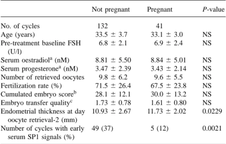
Table I. Clinical parameters in the two outcome groups: pregnant (ongoing) and non-pregnant. All values are means6 SD; probabilities are obtained using the Mann–Whitney non-parametric analysis
Not pregnant Pregnant P-value
No. of cycles 132 41 Age (years) 33.56 3.7 33.16 3.0 NS Pre-treatment baseline FSH 6.86 2.1 6.96 2.4 NS (U/l) Serum oestradiola(nM) 8.816 5.50 8.846 5.01 NS Serum progesteronea(nM) 3.476 2.39 3.436 2.14 NS Number of retrieved oocytes 9.86 6.2 9.66 5.5 NS Fertilization rate (%) 71.56 26.4 67.56 23.8 NS Cumulated embryo scoreb 28.16 12.1 30.06 13.2 NS Embryo transfer qualityc 1.736 0.78 1.616 0.80 NS Endometrial thickness at day 10.936 2.67 11.736 2.02 0.0229
oocyte retrieval-2 (mm)
Number of cycles with early 49 (37) 5 (12) 0.0021 serum SP1 signals (%)
aObtained up to 24 h before human chorionic gonadotrophin administration (‘day HCG’), i.e. 2–3 days before oocyte retrieval.
bA measure of embryo quality involving blastomere number and morphology (see Materials and methods), integrated over the number of transferred embryos.
cA measure of embryo transfer procedure quality involving various aspects (see Materials and methods).
SP15 pregnancy-specificβ1-glycoprotein; FSH5 follicle stimulating hormone; NS5 not significant.
attempt(s), and set in relation to the outcome in the last attempt cycle included in the study period.
Results
In the overall sample of 173 IVF cycles, the two outcome groups non-pregnant (n5 132) and pregnant (n 5 41) did not differ significantly (P. 0.1) in the levels of serum oestradiol and progesterone as determined 2 days before oocyte retrieval, and in the number of retrieved and the fraction of fertilized oocytes (Table I). The CES is also listed and did not differ significantly between the two outcome groups. The quality of the embryo transfer procedure itself was also expressed in our clinic in a semiquantitative way, a measure which was introduced in January 1995 and was thus available for 91/132 non-pregnancy and 31/41 pregnancy cycles of the study population. No significant difference in transfer quality was found between pregnancy and non-pregnancy cycles (Table I). The mean patient age of the pregnant group was 33.1 years (range 26–38 years) and in the non-pregnancy cycles it was 33.5 years (range 23–39 years); again the difference was not significant (P5 0.2676, Mann–Whitney). The same was the case for the baseline FSH levels; the highest observed values were 10.9 in the non-pregnant and 12.4 IU/l in the subsequently pregnant group, respectively. Endometrial thickness, on the other hand, differed significantly between the two groups (see detailed analysis below).
The occurrence of positive SP1 readings on or before embryo transfer (incidence of group A cycles) was different between the two outcome groups: 49 (37%) of the 132 cycles
Figure 1. Serum pregnancy-specificβ1-glycoprotein (SP1) levels in six control cycles obtained from volunteers not subjected to any hormonal treatment and not having intercourse. The dashed line represents the sensitivity of the assay, the dotted line shows the cut-off used in this study for distinguishing the subpopulations A and B (see text and legend to Figure 2).
not leading to pregnancy versus 5 (12%) of the 41 pregnancy cycles were group A cycles. This association was found to be highly significant by Fisher’s test (232 contingency table, P5 0.0021, odds ratio 5 4.251). Such positive SP1 signals were also observed in three of the six control cycles studied; they were evenly distributed over the full cycle (Figure 1).
Thirty-three unsuccessful patients (10 from group A and 23 from group B) underwent a second IVF treatment cycle (of these, 14 had a third and again two of these 14 came for a fourth treatment cycle within the study period). The outcome distributions are presented in Figure 2; of the 23 women with ‘normal’ first-attempt SP1 pattern (group B), eight became pregnant in the second or third attempt (four each). On the other hand, of the sample of the 10 previously group A cycles undergoing a second attempt, six presented the same group A pattern again and three fell into group B. One resulted in a pregnancy on the second attempt (15 months later) and, in another first-attempt group A case, a pregnancy was obtained in the third cycle, but interestingly a group B pattern was recorded in the intermittent second attempt. These findings are presented in Figure 2 in relation to the previous and, where applicable, the following cycle. The SP1 pattern (group A or B attribution) of the second, third, and fourth attempts were not independent from the previous cycle in the same patient, i.e. the individuals tended to remain in their respective groups A or B. This is illustrated by the observation that 37 cycles (76%) showed the same SP1 group pattern as the one in the preceding treatment cycle, while 12 (24%) switched to the other group. When all second/third/fourth attempt cycles were compared with the first attempt for the same patient, 69% presented the same pattern.
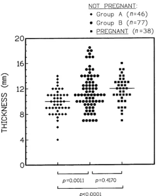
A measurement of the endometrial thickness 2 days before oocyte retrieval was available in 123 non-pregnancy and 38
Figure 2. Outcome and pregnancy-specificβ1-glycoprotein (SP1) group distribution in the first treatment cycle of the 123 patients (top pie) and, for the 33 women who underwent more than one IVF cycle, as a function of the rank of attempt in the subsequent cycles. Patients tended to remain in their respective groups (either in A, far left, or in B, far right), with more pregnancies (P) occurring in the group B population (right column of pies).
pregnancy cycles; the results are shown in relation to the SP1 category (group A or B) and outcome in Figure 3. In the sample of non-pregnancy cycles with abnormally positive SP1 values in the critical observation period (group A, n 5 46) the median thickness was 10 mm (mean 9.86 1.9 mm, SD), which is not a pathological value in itself but nevertheless is significantly below the result not only of the pregnancy cycles (median 12.0 mm, mean 11.76 2.0 mm, n 5 38, P , 0.0001) but also of the non-pregnancy outcome subgroup with no detectable SP1 on or before embryo transfer (group B, median 11.0 mm, mean 11.6 6 2.8 mm, n 5 77; P 5 0.0011 by Mann–Whitney).
Discussion
The usefulness of the determination of endometrial thickness by ultrasound for pregnancy prediction has been confirmed (Noyes et al., 1995) as well as questioned (Coulam et al.,
Figure 3. Endometrial thickness as determined by ultrasound examination 2 days before oocyte retrieval. The horizontal lines are medians. The two-tailed P-values shown have been obtained using the non-parametric Mann–Whitney test in the InStat 2.04 software (GraphPad, San Diego, CA, USA).
1994; Oliveira et al., 1997) in the literature. Our results here favour the application of such measurements in IVF treatment but at the same time clearly indicate that there may be two different subpopulations: one with a slightly reduced thickness, an abnormal SP1 pattern (as group A in this study), and a lower final pregnancy rate; and another one, presenting a thicker endometrium and no detectable mid-cycle SP1. The recent study by Oliveira et al. (1997) involved a similar number of cycles (150) and ovarian stimulation protocol to our study (173 cycles). As a consequence, their values for endometrial thickness, and particularly the difference in thickness between the pregnant and the non-pregnant group (0.6 mm), were comparable to ours (0.8 mm). In spite of using the same method of statistical analysis, significance was not reached in their study (P 5 0.14), while this was the case in our study (P 5 0.02). Nevertheless, we do not think that the results are fundamentally different but that the cut-off applied for significance (P 5 0.05) drew an artificial separation line. Thus the analysis of subgroups (pattern I in the paper by Oliveira et al., absence of ‘abnormal’ SP1 signals in this study) is important. Within our mentioned subgroup without detectable SP1, the ultrasonic determination of endometrial thickness cannot predict the outcome. The quality of the embryo(s) and of the transfer procedure play a major role particularly in this second population, and for analysis one has to make sure that no bias is introduced due to these embryo- or transfer-related parameters. In the set of data
presented here, there were no significant differences in the morphological embryo score or in the transfer quality. Moreover, the outcome groups were unbiased with respect to patient age, hormone levels (pre-HCG oestradiol and progesterone, baseline FSH) and oocyte-related parameters (Table I), thus allowing independent comparison of SP1 and endometrial thickness.
SP1, while found to be only of limited use for pregnancy diagnosis in the HCG-supported luteal phase in IVF treatment (Bersinger et al., 1995a), may eventually be clinically useful before treating a patient with IVF. Our observations in this study indicate that the early occurrence of serum SP1 concentrations above 0.1 ng/ml could be associated with a poor prognosis, certainly for the current treatment cycle (Figure 2), and there is a trend to such poor prognosis also for the subsequent treatment cycles in the same patient, since the incidence of these SP1 signals seemed to be patient-associated. In these group A women, they can occur at any time of the cycle and in the presence as well as in the absence of hormonal treatment, as indicated also by our results obtained from the control volunteer group. For the IVF cycles, we have selected the time-span between days oocyte retrieval-3 and embryo transfer because serum samples are readily available and also because earlier samples (e.g. taken at the beginning of the stimulation phase when oestradiol is at its lowest level) were found to occasionally contain SP1 from the previous cycle. The production of ectopic (non-placental) SP1 must be dependent, at mid-cycle (or before), on maternal mechanisms only.
An early (pre-HCG) elevation of progesterone has been reported to negatively influence the pregnancy rate (Harada et al., 1995; Yovel et al., 1995). In this study, serum progesterone levels immediately (max. 14 h) before HCG administration were available from 153 cycles, and there was no significant difference between the two groups A (‘abnormal’ SP1, mean progesterone 3.31 6 1.36 nM) and B (no detectable SP1, 3.15 6 1.17 nM, mean 6 SD, P 5 0.7698); SP1 indeed does not seem to be dependent of progesterone. The observed correlation between the presence of ectopic, non-pregnancy SP1 (group A) and reduced endometrial thickness could suggest that the role of the endometrium in implantation control may well reach beyond the well-characterized steroid-dependent functions. However, the fact that the correlation is negative makes the concept of a control by SP1 more difficult to place, not only in the frame of established mechanisms but also of earlier hypotheses (e.g. protease inhibition).
A possible endometrial factor, secreted in a cycle-dependent fashion and thus positively related to the ultrasonically determined endometrial thickness, would have to be an inhibitor of SP1 production. It seems more likely to us that the presence of ectopic SP1 as well as the reduced endometrial thickness (or rather the reduced response of the endometrium to oestrogens) would both be parallel consequences of another, unknown cause, e.g. a stimulatory or inhibitory factor of another maternal origin and which is more active in some women than in others. The effect of SP1 production would be independent of the menstrual
cycle since abnormal SP1 could be detected at any time; we have found low levels of SP1 in apparently healthy volunteers in the absence of any hormonal treatment (Figure 1). This observation as well as the use of a standardized stimulation protocol (except FSH dosage) in IVF makes it unlikely that HCG has an effect on SP1. No difference in the incidence of abnormal SP1 signals before or after HCG administration was observed, and sera were generally available before as well as after this treatment for all patients, allowing the pooling of results regarding exposure to HCG. It would also be difficult to explain why, using the same antibody, some patients were positive for SP1 and some were not. In our immunoassay we have found a cross-reactivity level of 0.02% w/w (0.01% molar ratio) for HCG which, however, could be attributed to contaminating SP1 antigen present in the HCG preparation tested in the assay and which is the same as the one administered to the patients where such small amounts of SP1 would be diluted to far below sensitivity (the testing of recombinant HCG could answer this question; however, the answer might not be relevant since the extent of cross-reactivity is low). At this stage it could also be possible that the presence of tonic (constant but low) levels of SP1, as observed in our group A patients, decreases the response of the endometrium (proliferation) during the stimulatory phase of the cycle. This hypothesis could be tested on in-vitro endometrial cultures (see below) but the purity of the currently available SP1 antigen is insufficient for this purpose for the moment. It is too early to suggest an origin and cause of ‘abnormal’ maternal SP1 signals (and concomitantly reduced endometrial thickness), let alone a treatment line for the patients presenting this pattern associated with a reduced chance to obtain a pregnancy. However, it might be useful to measure a mid-cycle serum SP1 concentration and an endometrial thickness in an observation cycle without exogeneous hormonal stimulation prior to running an IVF cycle. These non-invasive investigations may allow providers to identify those women who may have reduced chances of pregnancy success, thereby reducing the cost of treatment. The SP1 test used must have a detection limit of 0.05 ng/ml or less. As mentioned before, abnormal SP1 patterns do not seem to depend on ovarian hyperstimulation and pre-treatment baseline FSH levels did not differ between the two (‘abnormal’, A and normal, B) SP1 groups: the levels were 7.36 6 2.34 IU/l for A and 6.68 6 2.17 IU/l for B, respectively (mean 6 SD, P 5 0.3487). No other medical intervention seemed to influence these early ectopic SP1 signals since we have observed them also in some control volunteer cycles (three out of six) without any hormonal administration such as contraceptive pills, etc. The investi-gation of a possible correlation, in such control cycles, between SP1 and endometrial thickness would afford a large number of volunteers and a more precise dating of the LH surge (i.e. a more frequent sampling) than in this study. As the variations in SP1, in a given individual but between cycles are only small, the ‘non-treated group A’ women may well be the patients presenting with ‘unexplained infertility’ or repeated IVF failures in the future. The
restrictive use of IVF treatment in group A women (a procedure which would probably be unsuccessful anyway) would therefore save costs and increase overall pregnancy rates for a given centre. Unfortunately it is too early to offer help to these women; they are not interested in an overall success rate but in their individual achievement of pregnancy.
In order to achieve such clinical progress, basic investi-gations into the biochemistry of SP1 and its possible role in relation to the endometrium are required. Peri-ovulatory SP1 has been described earlier and before the general availability of IVF treatment; limited biochemical character-ization showed no difference between this and the ‘normal’ (pregnancy-derived or placental) SP1 (Bersinger, 1985). We have also developed and standardized an in-vitro explant culture model (Bersinger et al., 1995b) by means of which the effect of embryo-derived substances (IVF-conditioned media) and other factors on the release of endometrial marker proteins will be studied. The role of ovarian products such as inhibin or oestradiol (Licciardi et al., 1995) will also be studied in the context of a possible correlation with SP1.
Note added at proof
Recombinant HCG was found to shown no cross-reactivity (zero value) in the SP1 assay at 22.5 µg/ml (i.e. it is less than 0.001% w/w).
Acknowledgements
Our thanks go to the nurses in the endocrinology clinic for serum collection, to P.Glanzmann and U.Huber for technical assistance, and to C.B.Smikle for careful reviewing of the manuscript.
References
Ahmed, A.G. and Klopper, A. (1985) Serum concentration of placental proteins in non-pregnant and pregnant subjects. Arch. Gynecol., 237, 41–49.
Bersinger, N.A. (1985) Periovulatory secretion of pregnancy-specificβ1 -glycoprotein (SP1) immunoactivity in humans: comparison with ‘normal’ SP1. Ann. Inst. Pasteur/Immunol., 136D, 37–45. Bersinger, N.A., Brandenberger, A.W. and Birkha¨user, M.H. (1995a)
Endometrial and placental protein markers and ovarian steroids in serum during in-vitro fertilization cycles. Hum. Reprod., 10, 2149– 2154.
Bersinger, N.A., Brandenberger, A.W., Zakher A. et al. (1995b) Production of endometrial placental protein 14 and prolactin by cultured endometrial explants after collagenase and freeze/thaw treatment, and in response to progesterone. Early Preg. Biol. Med., 1, 134–140.
Coulam, C.B., Bustillo, M., Soenksen, D.M. et al. (1994)
Ultrasonographic predictors of implantation after assisted reproduction. Fertil. Steril., 62, 1004–1010.
Harada, T., Yoshida, S., Katagiri, C. et al. (1995) Reduced implantation rate associated with a subtle rise in serum progesterone concentration during the follicular phase of cycles stimulated with a combination of a gonadotrophin-releasing hormone agonist and gonadotrophin. Hum. Reprod., 10, 1060–1064.
Licciardi, F.L., Liu, H.C. and Rosenwaks, Z. (1995) Day 3 estradiol serum concentrations as prognosticators of ovarian stimulation response
and pregnancy outcome in patients undergoing in vitro fertilisation. Fertil. Steril., 64, 991–994.
Mueller, U.W. and Jones, W.R. (1985) Identification of an SP1-like protein in non-pregnancy serum: isolation using a monoclonal antibody. J. Reprod. Immunol., 8, 111–120.
Noyes, N., Liu, H.C., Sultan, K. et al. (1995) Endometrial thickness appears to be a significant factor in embryo implantation in in-vitro fertilisation. Hum. Reprod., 10, 919–922.
Oliveira, J.B., Baruffi, R.L., Mauri, A.L. et al. (1997) Endometrial ultrasonography as a predictor of pregnancy in an in-vitro fertilization programme after ovarian stimulation and gonadotrophin-releasing hormone and gonadotrophins. Hum. Reprod., 12, 2515–2518. Yovel, I., Yaron,Y., Amit, A. et al. (1995) High progesterone levels
adversely affect embryo quality and pregnancy rates in in vitro fertilization and oocyte donation programs. Fertil. Steril., 64, 128–131. Received on May 9, 1997; accepted on March 18, 1998