HAL Id: inserm-00117250
https://www.hal.inserm.fr/inserm-00117250
Submitted on 8 Jan 2007HAL is a multi-disciplinary open access archive for the deposit and dissemination of
sci-L’archive ouverte pluridisciplinaire HAL, est destinée au dépôt et à la diffusion de documents
Endogenous ghrelin regulates episodic growth hormone
(GH) secretion by amplifying GH Pulse amplitude:
evidence from antagonism of the GH secretagogue-R1a
receptor.
Philippe Zizzari, Heather Halem, John Taylor, Jesse Dong, Rakesh Datta,
Michael Culler, Jacques Epelbaum, Marie-Thérèse Bluet-Pajot
To cite this version:
Philippe Zizzari, Heather Halem, John Taylor, Jesse Dong, Rakesh Datta, et al.. Endogenous ghrelin regulates episodic growth hormone (GH) secretion by amplifying GH Pulse amplitude: evidence from antagonism of the GH secretagogue-R1a receptor.. Endocrinology, Endocrine Society, 2005, 146 (9), pp.3836-42. �10.1210/en.2005-0212�. �inserm-00117250�
ENDOGENOUS GHRELIN REGULATES EPISODIC GH SECRETION BY
AMPLIFYING GH PULSE AMPLITUDE: EVIDENCE FROM ANTAGONISM OF THE GHS-R1a RECEPTOR
ZIZZARI P.1, HALEM H.2, TAYLOR J.2, DONG J.Z.2, DATTA R.2, CULLER M.D.2, EPELBAUM J.1, BLUET-PAJOT M.T.1
1UMR. 549 INSERM-Université Paris 5, IFR Broca-Ste Anne, 2ter rue d’Alésia, 75014, Paris France and 2IPSEN 27 Maple St, Millford, MA, USA.
Short title: Effect of a GHS-R1a antagonist on GH secretion and food intake
Key words: GHS receptor, BIM-28163, GH, food intake
Corresponding author: J. EPELBAUM, UMR. 549 INSERM-Paris 5, 2ter rue d’Alésia 75014, Paris France., Tel: (33) 01 40 78 92 32 ; fax : (33) 01 45 80 72 93 ; E-mail : jacques.epelbaum@broca.inserm.fr
Endocrinology. First published May 26, 2005 as doi:10.1210/en.2005-0212
HAL author manuscript inserm-00117250, version 1
HAL author manuscript
Abstract
Ghrelin was purified from rat stomach as an endogenous ligand for the growth hormone secretagogue (GHS) receptor. As a GHS, ghrelin stimulates GH release, but it also has additional activities, including stimulation of appetite and weight gain. Plasma GH and ghrelin secretory patterns appear unrelated whereas many studies have correlated ghrelin variations with food intake episodes. In order to evaluate the role of endogenous ghrelin, GH secretion and food intake were monitored in male rats infused subcutaneously (6 µg/h during 10 h) or intracerebroventrically (5 µg/h during 48 h) with BIM-28163, a full competitive antagonist of the GHS-R1a receptor. Subcutaneous BIM-28163 infusion significantly decreased GH area under the curve during a 6 hour sampling period by 54% and peak amplitude by 46%. Twelve hours after the end of treatment these parameters returned to normal. Central treatment was similarly effective [-37% and 42% for AUC and –44% and 49% for peak amplitude on the first and second days of infusion, respectively]. Neither peripheral nor central BIM-28163 injection modifies GH peak number, GH nadir or IGF-1 levels. In this protocol, food intake is not strongly modified and water intake is unchanged. Subcutaneously infusion of BIM-28163 did not change plasma leptin and insulin levels evaluated at 12:00 h and 16:00 h. On the contrary, central BIM-28163 infusion slightly increased leptin and significantly increased insulin concentrations. Thus, endogenous ghrelin, through GHS-R1a, acts as a strong endogenous amplifier of spontaneous GH peak amplitude. The mechanisms by which ghrelin modifies food intake remain to be defined, and may involve a novel GHS receptor.
Introduction
The secretory pattern of growth hormone (GH) is mainly driven by two counter-regulatory neurohormones, GH-releasing hormone (GHRH) and somatostatin (SRIH). However, the existence of a third factor regulating GH rhythmicity was suggested 20 years ago when met-enkephalin analogues without opiate-like activity were found to exert potent GH-releasing activity (1). Since then, numerous synthetic GH secretagogues (GHS) have been developed and shown to act through a specific G-protein coupled receptor : GHS-R1a (2). GHS-R1a is distinct from the GHRH receptor and it is mainly expressed in the hypothalamus and pituitary gland. In 1999, a 28 amino acid acylated peptide was identified as an endogenous ligand for the GHS-R1a and termed ghrelin (3). It has been demonstrated that the acylation is essential for GHS-R1a binding. Ghrelin is produced mainly by A-like cells in oxyntic glands of the stomach and it is secreted into the bloodstream (4). In addition to the gastro-intestinal tract, ghrelin expression, at either the mRNA or protein level or both, has been identified in a number of tissues, including the hypothalamus (5). In this brain structure, low levels of ghrelin have been measured in the arcuate nucleus (3, 6) which is involved in the control of GH secretion and food intake. As expected ghrelin stimulates GH release (3, 4, 7-9) but also exerts multiple activities, including increased feeding (9) and weight gain (10). The circulating levels and patterns of ghrelin and GH appear weakly related (11) whereas many studies have established strong correlations between ghrelin variations and food intake episodes. Fasting increases circulating ghrelin levels which decrease after food intake (11-13). Ghrelin gene deletion in mice impairs neither growth nor appetite (14, 15) though deleting the GHS-R gene does abolish both ghrelin and synthetic GHS effects on these two functions (16). Thus, the role of endogenous ghrelin in the regulation of pulsatile GH secretion and food intake is not yet clear. The aim of the present study was to define this role by examining the
effects of treating rats with a novel analog of ghrelin, BIM-28163, recently developed as a full competitive antagonist of the GHS-R1a (17).
Materials and Methods 1) In vitro experiments : Binding Assay
The cDNA of the human GHS-R1a was cloned by polymerase chain reaction (PCR) using human brain RNA (Clontech, Palo Alto, CA, USA) as the template. The PCR product was cloned into the pCR2.1 vector using the TA Cloning Kit (Invitrogen, Carlsbad, CA, USA), and subcloned into the mammalian expression vector pcDNA 3.1 (Invitrogen). The plasmid was transfected into the Chinese hamster ovary cell line, CHO-K1 (ATCC, Manassas, VA, USA), by the calcium phosphate method. Single cell clones stably expressing the human GHS-R1a were obtained by selecting transfected cells using 0.8 mg/ml G418 containing selection media (Life Technologies, Carlsbad, CA, USA).
Membranes were prepared for radioligand binding studies by homogenization of CHO-K1 cells expressing the human recombinant GHS-R1a in 20 ml of ice-cold 50 mM Tris-HCl with a Brinkman Polytron (Westbury, NY, USA) (setting 6, 15 sec). The homogenates were washed twice by centrifugation (39,000 g for 10 min), and the final pellets were resuspended in 50 mM Tris-HCl, containing 2.5 mM MgCl2, and 0.1% BSA. For assay, aliquots (0.4 ml) were incubated with 0.05 nM [125I]h-ghrelin (~2000 Ci/mmol), with and without 0.05 ml of unlabeled competing test peptide. After a 60 min incubation (4 °C), the bound [125I]h-ghrelin was separated from the free by rapid filtration through GF/C filters (Brandel, Gaithersburg, MD, USA), which had been previously soaked in 0.5% polyethyleneimine/0.1% BSA. The filters were then washed three times with 5-ml aliquots of ice-cold 50 mM Tris-HCl and 0.1% bovine serum albumin, and the bound radioactivity trapped on the filters was counted by
gamma spectrometry (Wallac LKB, Gaithersburg, MD, USA). Specific binding was defined as the total [125I]ghrelin bound minus that bound in the presence of 1000 nM unlabeled h-ghrelin (Bachem, Torrence, CA, USA).
Intracellular Calcium Mobilization Assay
CHO-K1 cells, expressing the GHS-R1a, were harvested by incubating in a 0.3% EDTA/phosphate buffered saline solution (25 °C), and washed twice by centrifugation. The washed cells were resuspended in Hank's - buffered saline solution (HBSS) for loading of the fluorescent calcium indicator Fura-2AM. Cell suspensions of approximately 106 cells/ml were incubated with 2 mM Fura-2AM for 30 min at 25 °C. Unloaded Fura-2AM was removed by centrifugation twice in HBSS, and the final suspensions were transferred to a spectrofluorometer (Hitachi F-2000) equipped with a magnetic stirring mechanism and a temperature-regulated cuvette holder. After equilibration to 37 °C, the h-ghrelin analogues were added for measurement of intracellular calcium mobilization. The excitation and emission wavelengths were 340 and 510 nm, respectively.
2) In vivo experiments Acute in vivo GH stimulation
Male Sprague-Dawley rats (250g) were anesthetized and fit with a jugular-right atrial cannula 18 h prior to the experiment. To determine basal hormone levels blood samples were withdrawn into heparinized syringes at time -10 and 0. To demonstrate antagonist activity, immediately after the time 0 blood sample, the rats were injected with either BIM-28163 (1000 µg/kg) or vehicle via the indwelling cannula. Ten minutes later, a blood sample was withdrawn and the rats were immediately injected with either h-ghrelin (10 µg/kg) or
vehicle via the cannula. Subsequent blood samples were withdrawn 10, 20 and 40 minutes post-injection of h-ghrelin or vehicle. Plasma was separated and stored until assay for GH.
Chronic in vivo treatments
Four weeks before sampling experiments, adult male Sprague Dawley rats weighing 100–125 g (Charles River Laboratories, Inc., L’arbresle, France) were housed individually in transparent plastic containers placed in a soundproof room with controlled temperature (22-24 °C) and illumination (12 h light, 12 h dark schedule with lights off at 19:00 h). They had free access to food and water and were regularly handled and weighed, to minimize stress effects. Guidelines of the National Institute of Health for the Care and Use of Laboratory
Animals [1996 (7th ed.) Washington, DC: National Academy Press, aka National Research
Council Guide.] were followed.
SC treatment
Six days before the experiment, an osmotic minipump (Alzet Model 2002, flow rate 0.5 µl/h for 14 days; Charles River, France) was connected to a polyethylene catheter and implanted subcutaneously in the back of the animals under ether anaesthesia. The catheter had previously been filled with a combination of saline and BIM or saline. This resulted in delivery of saline during 144 hours followed by BIM-28163 (6 µg/h) or saline during 10 hours. On the day of the experiment (d0) SC treatment started at 9:00 h and rats received IP injection of 300 µg/0.2 ml BIM-28163 or 0.2 ml NaCl at 10:00 h.
ICV treatment
Six days before the experiment, a chronic ICV cannula (Alzet Brain Infusion Kit II, Charles River, France) was inserted into the lateral ventricle of the brain under pentobarbital
anesthesia (60 mg/kg of body weight i.p, Centravet, Plancoet, France). The following coordinates were used: anterior -0.8 mm to the bregma; lateral -1.5 mm to the midline; height -3.8 mm to the bregma, with the incisor bar set at -3.3 mm below the interaural line. The cannula was secured to the skull of the animal with stainless steel and dental cement and connected to an osmotic minipump (Alzet Model 2002; flow rate 0.5 µl/h for 14 days Charles River) implanted under the dorsal skin of the animal. The flow moderator of the pump was connected to the polyethylene catheter containing 72 µl saline, which was administered during the first 6 days in all animals. A small air bubble was inserted to separate the saline solution from an additional 24 µl of BIM-28163 (5 µg/h) or saline to be delivered for an additional 48 h. Treatment started at 8:00 h on d0 until 8:00 h on d2.
Blood sampling schedule
Experiments were performed on freely moving rats. Two days before data collection, an indwelling cannula was inserted into the right atrium under ether anesthesia as previously described (18). Two hours before the sampling and recording period, the distal extremity of the cannula was connected to a polyethylene catheter filled with 25 IU/ml heparinized saline. Blood samples were withdrawn every 20 minutes and centrifuged immediately. Red blood cells were resuspended in saline and reinjected every hour to attenuate hemodynamic modifications. Plasma was stored at -20 °C until assayed for GH.
Feeding behaviour
Experiments on food and water intake were performed on a separate group of animals which underwent the same treatments. Food and water intake were measured continuously with a PC-controlled monitoring system (Feeding/Drinking Monitor, Technical & Scientific
Equipment, Bad Homburg, Germany). Animals were acclimated to this new environment for 1 week before actual monitoring.
3) Hormonal determinations
Plasma GH concentrations were measured by EIA as previously described (19). Values are reported in terms of rGH-RP2, with sensitivity of 5 ng/ml and intra- and interassay coefficients of variation below 7%.
Plasma insulin, total leptin and IGF-1 levels were determined with RIA kits following manufacturer instructions (Linco, Saint Charles, Missouri ; DSL, Webster, Texas, USA, respectively). The sensitivity was 0.2 ng/ml, 0.5 ng/ml and 160 ng/ml respectively. For all assays, the intra- and interassay coefficients of variation were below 10%.
4) Statistical analysis
GH pulse analysis was performed using the Cluster program (20) with the t value set to 2 to maintain false positive rates under 1%. Cluster size was set to two prepeak and two postpeak nadir values. False positive error for peak detection was 7%.
Areas under the GH response curves (AUC) were calculated by mean of trapezoidal analysis.
Values are given as means ± SEM and statistical analysis was performed by ANOVA and paired t test using the JMP IN 5.1 software (SAS Institute Inc, North Carolina, USA).
Results
1) In vitro experiments
BIM-28163 was found to displace labelled h-ghrelin binding to the human GHS-R1a with a IC50 (Ki) of 8.1 + 1.3 nM. Human ghrelin, under the same conditions, had an IC50 of 1.6 + 0.3 nM.
In the calcium mobilization assay, h-ghrelin induced concentration-dependent calcium mobilization in cells stably transfected with the human GHS-R1a between the concentrations of 0.1 and 100nM, with an EC50 of 5.4 + 1.4nM. In contrast, BIM-28163 had no effect on calcium mobilization, even when tested at concentrations as high as 10-6 M (data not shown). When added in combination, BIM-28168 attenuated the ability of h-ghrelin to induce calcium mobilization in a dose-dependent manner, with complete inhibition achieved with a ratio of 100:1 BIM-28163:h-ghrelin (Figure 1), indicating that BIM-28163 acts as a full, competitive antagonist of ghrelin at the GHS-R1a.
Neither [125I]h-ghrelin binding nor ghrelin-induced mobilization of intracellular calcium was observed in the parent CHO-K1 cell line.
2) In vivo experiments Acute in vivo GH stimulation
Ghrelin induced GH stimulation was maximal 10 minutes post injection (data not shown). As illustrated on Figure 2, injection of h-ghrelin (10 µg/kg) alone into freely-moving, unanesthetized rats induced a significant increase in plasma GH, whereas injection of
28163 (1000 µg/kg) was without effect. When BIM-28163 was injected 10 minutes prior to h-ghrelin, the h-ghrelin-induced increase in GH secretion was completely abolished.
Chronic in vivo treatments
BIM-28163 was infused either SC (at the rate of 6 µg/h during 10 h after an IP bolus of 300 µg) or ICV (at the rate of 5 µg/h during 48h) to unanaesthetized, freely-moving rats and blood samples were collected every 20 minutes for 6 hours the first day, one hour after the beginning of infusion, and the following day during the same period. Individual profiles are illustrated on Figure 3. BIM-28163 administration always resulted in a rapid decrease in spontaneous GH release. Throughout BIM-28163 treatment, total GH, calculated as area under the curve, was significantly lower in treated animals compared with their respective controls (table 1). The decrease in total GH corresponded to a similar decrease in GH peak amplitudes. Basal values and GH pulse number were not modified (Table 1). ICV infusion was similarly effective on the first and second day of treatment. Fifteen hours after the end of SC treatment, pulsatile GH secretion was similar to that of controls.
Neither SC nor ICV treatment modified plasma IGF-1 levels, sampled at 15:00 h [SC treatment: BIM-28163 = 779 ± 84 ng/ml (n=5) vs. saline = 855 ± 78 ng/ml; ICV treatment: BIM-28163 = 977 ± 94 ng/ml (n=7) vs. saline = 1105 ± 133 ng/ml (n=7) on the first day, and BIM-28163 = 1199 ± 96 ng/ml (n=7) vs. saline =1162 ± 113 ng/ml (n=7) on the second day]. SC infusion of BIM-28163 changed neither plasma leptin nor insulin levels evaluated at 12:00 h and 16:00 h. In contrast, ICV BIM-28163 infusion slightly increased leptin and significantly increased insulin concentrations (Table 2).
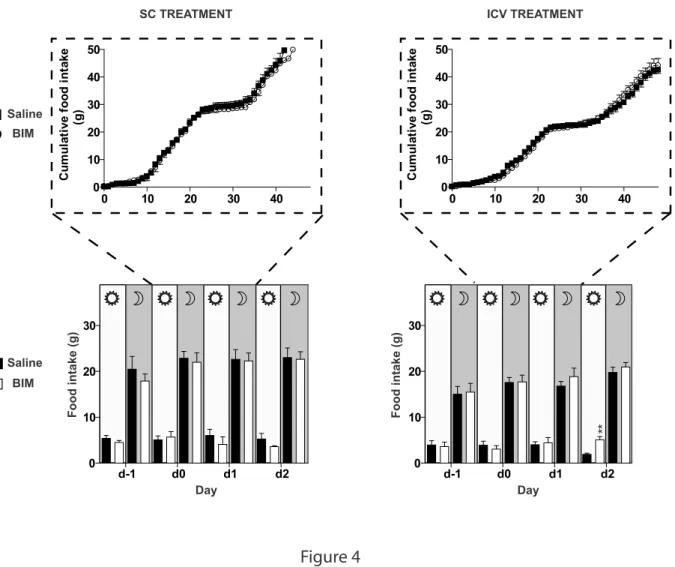
Food intake was modestly increased only during the light-on period on the second day after ICV infusion of BIM-28163 (Figure 4) and water consumption remained unchanged after either peripheral or central BIM-28163 infusion.
Discussion
In this study, either SC or ICV infusion of a GHS-R1a antagonist, BIM-28163, was demonstrated to decrease spontaneous GH secretion without causing major changes in food intake. This h-ghrelin analog is a full competitive antagonist at the GHS-R1a. Indeed, BIM-28163 binds to the h-GHS-R1a receptor and can displace h-ghrelin binding, but BIM-BIM-28163 does not activate GHS-R1a and completely and dose-dependently inhibits the ability of ghrelin to stimulate calcium mobilization in h-GHS-R1a transfected cells and GH secretion in vivo. Morever BIM-28163 blunts the GH releasing effect of ghrelin but not the GHRH induced GH rise (17). Thus, our results indicate that ghrelin, acting through the GHS-R1a, appears to be an endogenous regulator of spontaneous GH secretion but not necessarily of food intake.
Interestingly, antagonism of the GHS-R1a in freely-moving male rats does not impair the pulsatile pattern of GH secretion; however, it significantly lowers pulse amplitude. These results suggest that endogenous ghrelin acts to amplify the basic pulsatile pattern of GH established by the interplay of hypothalamic GHRH and somatostatin (21). Indeed, it has been established that concomitant administration of ghrelin and GHRH produces an increase in GH secretion significantly greater than that induced by either factor alone (22). The observation that antagonism of the GHS-R1a receptor with BIM-28163 lowers spontaneous GH pulse amplitude and total GH secretion without altering the basic pulsatile pattern agrees well with the observation of (23) that treatment of elderly men with MK677, a non-peptide ligand and agonist of the GHS-R1a, results in a significant increase in GH secretion without changing the underlying pulsatile pattern. Previously, repetitive injections of [D-Lys3]- GHRP6, a compound reported to be a GHS-R1a antagonist, did not significantly reduce
plasma GH levels (24); however, a very low dose of [D-Lys3]-GHRP6 was used (750 ng, less than one nanomole) considering the in vitro IC50 (3.2 x 10⁻⁶ M) of this particular analog (25) and its minimum effective dose on in vivo food intake (200 nM) (26). On the other hand, GH secretion was reduced in female (but not in male) transgenic rats expressing an antisense GHS-R mRNA under the control of the tyrosine hydroxylase promoter, thus selectively attenuating GHS-R protein expression in the arcuate nucleus (27). Moreover, in ghsr-null mice serum IGF-1 levels and body weight are reduced compared with wild type littermates suggesting decreased GH secretion (16). Taken together, these data suggest that endogenous ghrelin (or another, yet unidentified, ligand of GHS-R1a) acts as an amplifier of GH pulsatility. At the present time, a strict correlation has not been established between GH and ghrelin secretory episodes (11); however, only total ghrelin was monitored and it cannot be excluded that a stronger correlation occurs between GH and acylated ghrelin which represents only a minor proportion of total plasma ghrelin and has a very short half life (28).
In the subchronic experimental paradigm used herein, total plasma IGF-1 levels were not modified by BIM-28163 infusion. This lack of effect is in keeping with the data obtained on male transgenic rats expressing an antisense GHS-R mRNA in the arcuate nucleus (27) as well as another study in which chronic ICV administration of ghrelin for seven days did not affect IGF-1 levels (29). Moreover, 95% of IGFs circulate bound to IGFBPs and have a half life of 12-15 hours (30, 31); thus, it cannot be excluded that a longer treatment might affect IGF-1 levels.
Interestingly, plasma leptin slightly and insulin concentrations were significantly elevated by ICV BIM-28163 treatment, but not by peripheral administration. The change in plasma leptin concentrations may represent triggering of counter regulatory mechanisms as leptin has been reported to suppress feeding; although the overt effect of ghrelin on feeding in the
present study was minimal. Surprisingly, the effect on insulin secretion was only observed following ICV administration of BIM-28163. Expression of the GHS-R1a has been described in the pancreas, yet direct interaction with BIM-28163 via peripheral administration produced no significant effect on insulin secretion. Results from both GHS-R (16) and ghrelin null (14) mice also suggest that ghrelin exerts only a minimal role in regulating insulin or leptin levels, as these parameters remain largely unchanged in both animal models. Indeed the relationship between ghrelin and insulin secretion is far from clear as, depending on the dose, model and species, ghrelin has been reported to both stimulate and suppress insulin secretion (32). The present results may suggest a central mechanism for stimulation of insulin secretion, as well as provide evidence that this analog does not cross the blood brain barrier; however, further knowledge is clearly needed to fully interpret the present results on leptin and insulin secretion.
The fact that BIM-28163 treatment, either ICV or SC, does not change feeding behaviour is surprising since both GH secretagogues and exogenous ghrelin do stimulate food intake via GHS-R1a (16). Moreover, suppression of feeding has been reported in mice after ICV injections, using two other antagonists: [D-Lys3]-GHRP6, an analog of one of the synthetic GHSs or [D-Arg,D-Phe,D-Trp,Leu]-substance P an analog of substance P (26). Results with these compounds may be difficult to interpret as the first also binds to four of the melanocortin receptors (33) and the second antagonizes many peptide receptors (34, 35). A recent report indicated that the GHS-R1a displays a high constitutive activity and that [D-Arg,D-Phe,D-Trp,Leu]-substance P is a full inverse agonist, rather than a true antagonist (36). One might therefore hypothesize that the GHS-R1a involved in GH regulation and in food intake may have different levels of constitutive activity and, subsequently, different responses to inverse agonists and antagonists. Alternatively, it cannot be excluded that another, as yet unknown, ghrelin receptor may exist. Structural variants of GHS-R have been
found in human, swine, chicken and fish genome (37-39). A truncated version of the GHS receptor, derived from alternative gene splicing, GHS-R 1b, and not yet observed in the rat, cannot be the novel ghrelin receptor since it binds neither ghrelin nor any of the various GHS molecules. However, GHS-R 1b, or another receptor, may interact with GHS-R1a, perhaps through receptor heterodimerization as shown in fish (39), thereby playing a counter regulatory role in controlling either GH secretion or food intake.
Another potential explanation of the discrepant results using GHS-R1a antagonists may rely on the central or peripheral location of the ghrelin systems involved. Central ghrelin probably stimulates food intake and GH secretion through neural mechanisms involving neuronal circuitry (40). An important site for GHS-R1a mediation of GH secretion is the hypothalamic arcuate nucleus (41), where GHS-R mRNA is expressed on both GHRH and NPY-expressing neurons (21). However, peripheral ghrelin can also modulate food intake and GH secretion via vagal afferent fibers (42). Blockade of the vagal afferent, either through vagotomy or through perivagal capsaicin application, totally blocks peripheral ghrelin effects on feeding while it only attenuates the stimulation of GH secretion.
In summary, this study shows that pharmacological dissociation of the effects of endogenous ghrelin on GH secretion and feeding is possible. It will remain to be determined whether other effects of this interesting peptide, such as those on adiposity or cardiac function can also be separately affected. At any rate, the present data support the feasibility of creating ghrelin analogs with selective actions on either GH or feeding behavior.
References
1. Bowers CY, Momany FA, Reynolds GA, Hong A 1984 On the in vitro and in vivo
activity of a new synthetic hexapeptide that acts on the pituitary to specifically release growth hormone. Endocrinology 114:1537-45
2. Howard AD, Feighner SD, Cully DF, Arena JP, Liberator PA, Rosenblum CI,
Hamelin M, Hreniuk DL, Palyha OC, Anderson J, Paress PS, Diaz C, Chou M, Liu KK, McKee KK, Pong SS, Chaung LY, Elbrecht A, Dashkevicz M, Heavens R, Rigby M, Sirinathsinghji DJ, Dean DC, Melillo DG, Van der Ploeg LH, et al. 1996 A receptor in pituitary and hypothalamus that functions in growth hormone release. Science 273:974-7
3. Kojima M, Hosoda H, Date Y, Nakazato M, Matsuo H, Kangawa K 1999 Ghrelin
is a growth-hormone-releasing acylated peptide from stomach. Nature 402:656-60
4. Date Y, Kojima M, Hosoda H, Sawaguchi A, Mondal MS, Suganuma T,
Matsukura S, Kangawa K, Nakazato M 2000 Ghrelin, a novel growth hormone-releasing acylated peptide, is synthesized in a distinct endocrine cell type in the gastrointestinal tracts of rats and humans. Endocrinology 141:4255-61
5. Cowley MA, Smith RG, Diano S, Tschop M, Pronchuk N, Grove KL, Strasburger
CJ, Bidlingmaier M, Esterman M, Heiman ML, Garcia-Segura LM, Nillni EA, Mendez P, Low MJ, Sotonyi P, Friedman JM, Liu H, Pinto S, Colmers WF, Cone RD, Horvath TL 2003 The distribution and mechanism of action of ghrelin in the CNS demonstrates a novel hypothalamic circuit regulating energy homeostasis. Neuron 37:649-61
6. Lu S, Guan JL, Wang QP, Uehara K, Yamada S, Goto N, Date Y, Nakazato M,
Kojima M, Kangawa K, Shioda S 2002 Immunocytochemical observation of ghrelin-containing neurons in the rat arcuate nucleus. Neurosci Lett 321:157-60
7. Tolle V, Zizzari P, Tomasetto C, Rio MC, Epelbaum J, Bluet-Pajot MT 2001 In
vivo and in vitro effects of ghrelin/motilin-related peptide on growth hormone secretion in the rat. Neuroendocrinology 73:54-61
8. Seoane LM, Tovar S, Baldelli R, Arvat E, Ghigo E, Casanueva FF, Dieguez C
2000 Ghrelin elicits a marked stimulatory effect on GH secretion in freely-moving rats. Eur J Endocrinol 143:R7-9
9. Wren AM, Small CJ, Ward HL, Murphy KG, Dakin CL, Taheri S, Kennedy AR,
Roberts GH, Morgan DG, Ghatei MA, Bloom SR 2000 The novel hypothalamic peptide ghrelin stimulates food intake and growth hormone secretion. Endocrinology 141:4325-8
10. Tschop M, Smiley DL, Heiman ML 2000 Ghrelin induces adiposity in rodents. Nature 407:908-13
11. Tolle V, Bassant MH, Zizzari P, Poindessous-Jazat F, Tomasetto C, Epelbaum J,
Bluet-Pajot MT 2002 Ultradian rhythmicity of ghrelin secretion in relation with GH, feeding behavior, and sleep-wake patterns in rats. Endocrinology 143:1353-61
12. Cummings DE, Purnell JQ, Frayo RS, Schmidova K, Wisse BE, Weigle DS 2001
A preprandial rise in plasma ghrelin levels suggests a role in meal initiation in humans. Diabetes 50:1714-9
13. Tschop M, Wawarta R, Riepl RL, Friedrich S, Bidlingmaier M, Landgraf R,
Folwaczny C 2001 Post-prandial decrease of circulating human ghrelin levels. J Endocrinol Invest 24:RC19-21
14. Sun Y, Ahmed S, Smith RG 2003 Deletion of ghrelin impairs neither growth nor
appetite. Mol Cell Biol 23:7973-81
15. Wortley KE, Anderson KD, Garcia K, Murray JD, Malinova L, Liu R,
Moncrieffe M, Thabet K, Cox HJ, Yancopoulos GD, Wiegand SJ, Sleeman MW 2004 Genetic deletion of ghrelin does not decrease food intake but influences metabolic fuel preference. Proc Natl Acad Sci U S A 101:8227-32
16. Sun Y, Wang P, Zheng H, Smith RG 2004 Ghrelin stimulation of growth hormone
release and appetite is mediated through the growth hormone secretagogue receptor. Proc Natl Acad Sci U S A 101:4679-84
17. Halem HA, Taylor JE, Dong JZ, Shen Y, Datta R, Abizaid A, Diano S, Horvath
T, Zizzari P, Bluet-Pajot MT, Epelbaum J, Culler MD 2004 Novel analogs of ghrelin: physiological and clinical implications. Eur J Endocrinol 151 Suppl 2:S071-5
18. Bluet-Pajot MT, Durand D, Drouva SV, Mounier F, Pressac M, Kordon C 1986
Further evidence that thyrotropin-releasing hormone participate in the regulation of growth hormone secretion in the rat. Neuroendocrinology 44:70-5
19. Ezan E, Laplante E, Bluet-Pajot MT, Mounier F, Mamas S, Grouselle D, Grognet
JM, Kordon C 1997 An enzyme immunoassay for rat growth hormone: validation and application to the determination of plasma levels and in vitro release. J Immunoassay 18:335-56
20. Veldhuis JD, Johnson ML 1986 Cluster analysis: a simple, versatile, and robust
algorithm for endocrine pulse detection. Am J Physiol. 250:E486-93
21. Tannenbaum GS, Epelbaum J, Bowers CY 2003 Interrelationship between the
novel peptide ghrelin and somatostatin/growth hormone-releasing hormone in regulation of pulsatile growth hormone secretion. Endocrinology 144:967-74
22. Arvat E, Maccario M, Di Vito L, Broglio F, Benso A, Gottero C, Papotti M,
Muccioli G, Dieguez C, Casanueva FF, Deghenghi R, Camanni F, Ghigo E 2001
humans: comparison and interactions with hexarelin, a nonnatural peptidyl GHS, and GH-releasing hormone. J Clin Endocrinol Metab 86:1169-74
23. Chapman IM, Bach MA, Van Cauter E, Farmer M, Krupa D, Taylor AM,
Schilling LM, Cole KY, Skiles EH, Pezzoli SS, Hartman ML, Veldhuis JD, Gormley GJ, Thorner MO 1996 Stimulation of the growth hormone (GH)-insulin-like growth factor I axis by daily oral administration of a GH secretogogue (MK-677) in healthy elderly subjects. J Clin Endocrinol Metab 81:4249-57
24. Okimura Y, Ukai K, Hosoda H, Murata M, Iguchi G, Iida K, Kaji H, Kojima M,
Kangawa K, Chihara K 2003 The role of circulating ghrelin in growth hormone (GH) secretion in freely moving male rats. Life Sci 72:2517-24
25. Cheng K, Chan WW, Barreto A, Jr., Convey EM, Smith RG 1989 The synergistic
effects of His-D-Trp-Ala-Trp-D-Phe-Lys-NH2 on growth hormone (GH)-releasing factor-stimulated GH release and intracellular adenosine 3',5'-monophosphate accumulation in rat primary pituitary cell culture. Endocrinology 124:2791-8
26. Asakawa A, Inui A, Kaga T, Katsuura G, Fujimiya M, Fujino MA, Kasuga M
2003 Antagonism of ghrelin receptor reduces food intake and body weight gain in mice. Gut 52:947-52
27. Shuto Y, Shibasaki T, Otagiri A, Kuriyama H, Ohata H, Tamura H, Kamegai J,
Sugihara H, Oikawa S, Wakabayashi I 2002 Hypothalamic growth hormone secretagogue receptor regulates growth hormone secretion, feeding, and adiposity. J Clin Invest 109:1429-36
28. Akamizu T, Takaya K, Irako T, Hosoda H, Teramukai S, Matsuyama A, Tada H,
Miura K, Shimizu A, Fukushima M, Yokode M, Tanaka K, Kangawa K 2004 Pharmacokinetics, safety, and endocrine and appetite effects of ghrelin administration in young healthy subjects. Eur J Endocrinol 150:447-55
29. Kim MS, Namkoong C, Kim HS, Jang PG, Kim Pak YM, Katakami H, Park JY,
Lee KU 2004 Chronic central administration of ghrelin reverses the effects of leptin. Int J Obes Relat Metab Disord 28:1264-71
30. Humbel RE 1990 Insulin-like growth factors I and II. Eur J Biochem 190:445-62
31. Guler HP, Zapf J, Schmid C, Froesch ER 1989 Insulin-like growth factors I and II
in healthy man. Estimations of half-lives and production rates. Acta Endocrinol (Copenh) 121:753-8
32. Broglio F, Gottero C, Benso A, Prodam F, Volante M, Destefanis S, Gauna C,
Muccioli G, Papotti M, van der Lely AJ, Ghigo E 2003 Ghrelin and the endocrine pancreas. Endocrine 22:19-24
33. Schioth HB, Muceniece R, Wikberg JE 1997 Characterization of the binding of
MSH-B, HB-228, GHRP-6 and 153N-6 to the human melanocortin receptor subtypes. Neuropeptides 31:565-71
34. Holst JJ, Knuhtsen S, Orskov C, Skak-Nielsen T, Poulsen SS, Nielsen OV 1987 GRP-producing nerves control antral somatostatin and gastrin secretion in pigs. Am J Physiol 253:G767-74
35. Woll PJ, Rozengurt E 1988 [D-Arg1,D-Phe5,D-Trp7,9,Leu11]substance P, a potent
bombesin antagonist in murine Swiss 3T3 cells, inhibits the growth of human small cell lung cancer cells in vitro. Proc Natl Acad Sci U S A 85:1859-63
36. Holst B, Cygankiewicz A, Jensen TH, Ankersen M, Schwartz TW 2003 High
constitutive signaling of the ghrelin receptor--identification of a potent inverse agonist. Mol Endocrinol 17:2201-10
37. McKee KK, Palyha OC, Feighner SD, Hreniuk DL, Tan CP, Phillips MS, Smith
RG, Van der Ploeg LH, Howard AD 1997 Molecular analysis of rat pituitary and hypothalamic growth hormone secretagogue receptors. Mol Endocrinol 11:415-23
38. Geelissen SM, Beck IM, Darras VM, Kuhn ER, Van der Geyten S 2003
Distribution and regulation of chicken growth hormone secretagogue receptor isoforms. Gen Comp Endocrinol 134:167-74
39. Chan CB, Cheng CH 2004 Identification and functional characterization of two
alternatively spliced growth hormone secretagogue receptor transcripts from the pituitary of black seabream Acanthopagrus schlegeli. Mol Cell Endocrinol 214:81-95
40. Nakazato M, Murakami N, Date Y, Kojima M, Matsuo H, Kangawa K,
Matsukura S 2001 A role for ghrelin in the central regulation of feeding. Nature 409:194-8
41. Dickson SL, Luckman SM 1997 Induction of c-fos messenger ribonucleic acid in
neuropeptide Y and growth hormone (GH)-releasing factor neurons in the rat arcuate nucleus following systemic injection of the GH secretagogue, GH-releasing peptide-6. Endocrinology 138:771-7
42. Date Y, Murakami N, Toshinai K, Matsukura S, Niijima A, Matsuo H, Kangawa
K, Nakazato M 2002 The role of the gastric afferent vagal nerve in ghrelin-induced feeding and growth hormone secretion in rats. Gastroenterology 123:1120-8
Figure Legends
Figure 1: Dose-related attenuation of the ability of h-ghrelin to stimulate calcium mobilization in CHO-K1 cells stably transfected with the human GHS-R1a by BIM-28163, a competitive, though non-activating, ligand for GHS-R1a.
Each bar and bracket represents the mean + SEM of 3 replicate experiments. *** : p<0,001 vs ghreline; °°° : p<0,001
Figure 2: Acute blockade by BIM-28163 of h-ghrelin stimulation of GH secretion in vivo Data are presented as peak GH levels observed 10 minutes after the injection of either vehicle, h-ghrelin (10 µg/kg), BIM-28163 (1000 µg/kg), or the combination. *** :p<0.001 vs vehicle
Figure 3: Representative GH secretory patterns during a 6-h sampling period in freely moving male rats that received subchronic SC (left panel) or ICV (right panel) administration of either BIM-28163 (open circles) or saline (closed squares).
In the SC group, rats were infused at d0 with saline or BIM-28163 (6 µg/h) from 9:00 h to 19:00 h and received an IP injection of saline or BIM-28163 (300µg) at 10:00 h. In the ICV group, rats were infused with saline or BIM-28163 (5 µg/h) for 48h from 8:00 h at d0.
Blood samples were collected every 20 min.
Figure 4: Effect of subchronic SC (left panel) or ICV (right panel) administration of BIM-28163 (open symbols) or saline (closed symbols) on food intake.
Upper panel: Cumulative food intake during the 48 h post infusion.
Lower panel: Food intake measured during both the diurnal (7:00 h-19:00 h) and nocturnal period (19:00 h-7:00 h) before, during and after treatment.
Table 1 : Cluster analysis of GH pulsatility parameters in BIM-28163-treated rats.
In the SC group, rats were infused at d0 with saline or BIM-28163 (6 µg/h) from 9:00 h to 19:00 h and received an injection of saline or BIM-28163 (300 µg) at 10:00 h. In the ICV group, rats were infused with saline or BIM-28163 (5 µg/h) for 48 h from 8:00 h at d0. Data are mean ± sem. ** : p<0.01 ; * : p<0.05 vs saline
Table 2 : Effect of subchronic BIM-28163 administration on insulin and leptin plasma levels. SC administration: Hormones were measured in freely moving male rats at 12:00 h and 16:00 h on day 0 (d0: 3 h and 7 h after the start of the treatment) and at the same time on day 1 (d1: 17 h and 21 h after the end of the treatment),
ICV administration: Hormones were measured in freely moving male rats at 12:00 h and 16:00 h on d0 (4 h and 8 h after the start of the treatment) and at the same time on d1 (28 h and 32 h after the start of the treatment),
Data are mean ± sem.** : p<0.01; * : p<0.05 vs saline
Route of mi n istration Sampli n g d ay T rea tm en t A U C N u m b er o f peaks Peak amp litude ng/ml Nadir ng/ml Sa line N = 7 12863 ± 146 7 1.7 ± 0.2 69 ± 11 23 ± 1 d0 BIM-28163 N = 7 5948 ± 1 320 ** 1.7 ± 0.7 37 ± 6 * 24 ± 7 Sa line N = 7 16617 ± 117 4 2.5 ± 0.5 10 1 ± 13 26 ± 4 SC d1 BIM-28163 N =6 13958 ± 211 4 2.6 ± 0.5 81 ± 9 23 ± 4 Sa line N = 7 13085 ± 143 5 2.0 ± 0.4 70 ± 10 23 ± 1 d0 BIM-28163 N = 7 8235 ± 1 163 * 1.6 ± 0.5 38 ± 5 * 21 ± 2 Sa line N = 7 18681 ± 149 7 3.0 ± 0.5 11 4 ± 11 31 ± 5 ICV d1 BIM-28163 N = 5 10926 ± 489 ** 2.2 ± 0.2 59 ± 3 ** 19 ± 1
Table 1
ut e o f n istration Samp lin g day Treatment In su lin ng/ml Leptin ng /m l 12:00 h 16:00 h 12:00 h 16:00 h Sa line N = 5 1.53 ± 0.09 1.77 ± 0.08 4.56 ± 0.27 3.48 ± 0.40 d0 BIM-28163 N = 5 1.78 ± 0.23 1.90 ± 0.24 4.89 ± 0.66 3.78 ± 0.86 Sa line N = 5 1.83 ± 0.13 1.30 ± 0.10 3.72 ± 0.31 2.60 ± 0.25 SC d1 BIM-28163 N = 5 1.65 ± 0.12 1.81 ± 0.08 3.94 ± 0.66 2.88 ± 0.35 Sa line N = 7 1.76 ± 0.22 2.02 ± 0.23 3.74 ± 0.34 3.31 ± 0.24 d0 BIM-28163 N = 7 2.70 ± 0.35 * 3.07 ± 0.32 ** 4.56 ± 0.24 4.10 ± 0.36 Sa line N = 7 1.92 ± 0.26 2.27 ± 0.34 3.56 ± 0.23 2.61 ± 0.16 ICV d1 BIM-28163 N = 5 2.86 ± 0.46 * 3.17 ± 0.70 4.78 ± 0.69 3.60 ± 0.64
Table 2
10 10 100 1000 100 110 120 130 140 150 Ghrelin +BIM-28163
Figure 1
[Ca 2+ ]i mobilization (% Control) *** *** *** °°° nM0 40 80 120 160 200 Treatment Vehicle ***
Ghrelin BIM-28163 BIM-28163 + Ghrelin
Figure 2
GH (ng/ml)
10 11 12 13 14 15 16 0 50 100 150 10 11 12 13 14 15 16 0 50 100 150 10 11 12 13 14 15 16 0 50 100 150 10 11 12 13 14 15 16 0 50 100 150 BIM-28163 Saline 10 11 12 13 14 15 16 0 50 100 150 10 11 12 13 14 15 16 0 50 100 150 10 11 12 13 14 15 16 0 50 100 150 10 11 12 13 14 15 16 0 50 100 150 G H ( ng/ m l)
SC TREATMENT
d0Light on Light off Light on Light off
d1 Sampling Period Saline BIM or S
ICV TREATMENT
d0Light on Light off Light on Light off
d1 BIM or Saline S Time (h) S Sampling Period