HAL Id: hal-01480292
https://hal-amu.archives-ouvertes.fr/hal-01480292
Submitted on 13 Mar 2017HAL is a multi-disciplinary open access archive for the deposit and dissemination of sci-entific research documents, whether they are pub-lished or not. The documents may come from teaching and research institutions in France or abroad, or from public or private research centers.
L’archive ouverte pluridisciplinaire HAL, est destinée au dépôt et à la diffusion de documents scientifiques de niveau recherche, publiés ou non, émanant des établissements d’enseignement et de recherche français ou étrangers, des laboratoires publics ou privés.
predictive of pancreatic adenocarcinoma
Emmanuelle Martinez, Françoise Silvy, Dominique Lombardo, Eric Mas
To cite this version:
Emmanuelle Martinez, Françoise Silvy, Dominique Lombardo, Eric Mas. Single nucleotide polymor-phisms and risk factors predictive of pancreatic adenocarcinoma. Cancer Cell & Microenvironment, 2016, 3, pp.1231 - 1231. �10.14800/ccm.1231�. �hal-01480292�
Page 1 of 12
Single nucleotide polymorphisms and risk factors predictive of
pancreatic adenocarcinoma
Emmanuelle Martinez1,2, Françoise Silvy1,2, Dominique Lombardo1,2, Eric Mas1,2
1Aix-Marseille Université, CRO2, Centre de Recherche en Oncologie biologique et Oncopharmacologie, F-13005, Marseille, France 2INSERM, UMR_S 911, F-13005, Marseille, France
Correspondence: Eric Mas or DominiqueLombardo
E-mail: [email protected] or [email protected] Received: February 25, 2016
Published online: March 28, 2016
Pancreatic ductal adenocarcinoma (PDAC) is a devastating disease progressing asymptomatically until death within months after diagnosis. Defining at-risk populations should promote an early diagnosis and efficient follow-up, therefore avoiding its development. Single nucleotide polymorphisms, or SNPs, constitute the most abundant form of genetic variation in the human genome. SNPs are markers of diverse populations or individuals, yet also can be associated with differences in susceptibility or severity of certain diseases and/or individual responses to drugs. Many SNPs have previously been identified in studies of healthy subjects and patients with different alleles of a given gene. To date, around forty SNPs from the human genome have been correlated with predisposition to PDAC by pan-genomic studies or genome wide association studies (GWAS). However, parts of the human genome located within the GC-rich repeated domain of chromosomes are unsuitable for GWAS. Unfortunately, of those forty SNPs none are currently used in routine clinical protocols as potential biomarkers for PDAC. Exon 11 of the bile salt-dependent lipase gene (BSDL) plays a key role in pancreatic disease and encodes variable number of tandem repeat (VNTR) sequences, therefore Martinez et al. [Oncotarget. 2015; 6: 39855-39864.] hypothesized that a genetic link exists between mutations in VNTR loci and predisposition to pancreatic cancer (PC). Authors reported that the c.1719C>T transition (SNP rs488087) present in BSDL VNTR may be a useful marker for defining a population at risk of developing PC (occurrence: 63.90% in the PC group versus 27.30% in the control group). Notably, the odds ratio (OR) of 4.7 for the T allele was larger than those already determined for other SNPs suspected to be predictive of PC. Further studies on tumor pancreatic tissue suggested that the T allele may favor Kras G12R/G12D somatic mutations which represent negative prognostic factors associated with reduced survival. Furthermore, a robust method using probes for droplet digital (dd)-PCR was designed to specifically discriminate the C/C major from C/T or T/T minor genotypes. Altogether, Martinez et al. propose that detection of the T allele in rs488087 SNP should lead to an in-depth follow-up of these patients, particularly if associated with other potential risk factors of PC.
Keywords: SNP; bile salt-dependent lipase; pancreatic cancer; pancreatic ductal adenocarcinoma
To cite this article: Emmanuelle Martinez, et al. Single nucleotide polymorphisms and risk factors predictive of pancreatic
adenocarcinoma.Can Cell Microenviron 2016; 3: e1231. doi: 10.14800/ccm.1231.
Copyright: © 2016 The Authors. Licensed under a Creative Commons Attribution 4.0 International License which allows users including authors of articles to copy and redistribute the material in any medium or format, in addition to remix, transform, and build upon the material for any purpose, even commercially, as long as the author and original source are properly cited or credited.
Page 2 of 12
Introduction
Pancreatic cancers (PC) represent 10% of all digestive cancers with over 338,000 new cases worldwide in 2012, among which 90% were pancreatic ductal adenocarcinoma
(PDAC), (www.wcrf.org; http://globocan.iarc.fr). The
survival rate is dramatically low with a case-fatality ratio of about 0.9. PDAC could be the second cause of mortality by
cancer by the year 2030 [1]. The 5-year survival rate of PDAC
is less than 4% in western countries [2]. The poor prognosis of
this cancer is mainly due to its lack of response to currently
available therapies [3, 4]. Its curative resection rate is very low
(15% of patients) due to unspecific symptoms, the lack of early biological markers, delayed diagnosis and metastasis formation. Patients diagnosed with advanced or metastatic disease are not elected for surgery (85% of patients) and
show median survival rates ranging from 6 to 9 months [5, 6].
For those patients with inoperable cancer, the main treatment remains standard chemotherapy. Precancerous lesions as well as PDAC occur as a result of alterations affecting genes essential for maintaining cellular functions. These changes can be explained by mutations, deletions or epigenetic modifications, responsible for either a gain or loss of gene function. They can either be inherited (familial cancers, predispositions) or acquired, although in both cases each stage is associated with one or more types of alteration, their evolution and accumulation increasing the invasiveness of lesions.
Single nucleotide polymorphisms (SNPs) are a one-time bi-allelic polymorphism at one base pair on a given locus in a chromosome, constituting the most abundant form of genetic variation in the human genome. Comparing two random human genomes, 99.9% of the DNA sequence is identical. The remaining 0.1% contains sequence variations including SNPs. SNPs are stable, abundant and evenly distributed throughout the genome. In fact, SNPs occur on average, once every 300 nucleotides. This translates to a potential 10 million SNPs in the human genome. Although SNPs are markers of diverse populations or individuals, they also can be associated with differences in susceptibility or severity of certain diseases and/or individual responses to drugs. Many SNPs have previously been identified in studies of healthy subjects and patients with different alleles of a given gene. Currently, pan-genome association studies, or genome wide association studies (GWAS), are one of the most frequently used analytical tools for multifactor diseases such as cancer. GWAS are used to compare the frequency of hundreds of thousands of genetic variants distributed across all chromosomes between a group of cancer patients and a
control group, using high-throughput genotyping
technologies. This is an "agnostic" approach, in the absence of
a hypothesis based on a gene(s) of interest, unlike the genetic type of candidate gene association studies. GWAS aim to identify genetic variants with relevant predisposition to specific diseases by genotyping up to a million SNPs. Although these studies do not require prior knowledge of the functional significance of the variant studied, very large groups of samples are necessary, usually thousands of cases plus paired controls.
Gene wide association studies (GWAS)
In recent studies of PC, particularly PDAC, many pan-genomic studies have highlighted SNPs linked to a predisposition to this dramatic disease (Table 1). In 2009,
Amundadottir and collaborators [7] were the first to show
through GWAS that some chromosome loci could predispose to PDAC. This large-scale study, (about 560,000 SNPs were analyzed), highlighted the presence of SNPs at three loci:, 7q36, 15q14 and 9q34. A study performed on an ethnically restricted population showed that four SNPs (rs6464375, rs7779540, rs6973850, rs1048768) located at the 7q36.2 locus with odds ratios (OR) close to 1.1, are associated with dipeptidyl peptidase 6 (DPP6) and could predispose to PC in
the Japanese population [8]. SNPs rs8028529, rs4130461 and
rs4459505 were discovered on locus 15q14. However these latter SNPs did not impact any gene. The SNP at the 9q34 locus attracted particular attention due to its highly significant
association with PDAC (P-value = 5.37x10⁻⁸). This SNP,
referred to as rs505922, is positioned on the long arm of chromosome 9, the same locus as genes encoding the ABO blood groups. These results confirm those obtained in epidemiological studies from 1950 to 1970, which suggested that group O individuals were less prone to develop PDAC
than individuals with blood groups A, B or AB [9, 10]. More
recently, Wolpin and collaborators [11] tested this hypothesis in
a pan-genomic study and determined which genotype of the ABO system (OO, AO, AA, AB, BO, BB blood groups) is preferentially associated with PDAC. Results revealed that there is a significantly increased risk of developing PC in individuals of A, B or AB blood groups compared to group O individuals, with close relative risk or OR of 1.38, 1.47 and 1.53 respectively. The BB genotype was the most predisposing genotype. In two large and independent populations the age-adjusted incidence rates for PDAC per 100,000 person-years were 27 for participants with blood type O, 36 for blood type A, 41 for blood type AB, and 46
for blood type B [11, 12]. However, the molecular or cellular
connection between the presence of the rs505922 SNP and susceptibility to PC is still debatable. Another group demonstrated that rs505922 is associated with elevated blood levels of tumor necrosis factor alpha (TNF-α), soluble intercellular adhesion molecule 1 (sICAM-1) and alkaline
Page 3 of 12
Table 1. SNPs predictive of pancreatic cancer
Chromosomes Locus Ancestry SNP Nb Nearest gene OR Ref
1 q32.1 European 37908844 NR5A2 0.77 15, 21 p36.13 European 16861827 IGSF21 1.7 21 2 p13.3 European 1486134 ETAA1 1.14 29 3 q29 European 9854771 TP63 0.89 29 5 p13.1 European/Chinese 225280 DAB2 0.81/0.74 30,36 p15.33 European 27336098 CLPTMIL-TERT 0.8/1.19 15 4011681 1.06 15 6 p25.3 Japanese 9502893 FOXQ1 1.29 8 7 q32.3 European 6971499 LINC-PINT 0.79 36 q36.2 European 167020 SHH 1.17/1.38 7 European 172310 SHH 1.17/1.36 7 European 288746 SHH 1.18/1.39 7 Japanese 6464375 DPP6 1.13 8 Japanese 7779540 DPP6 1.14 8 Japanese 6973850 DPP6 1.1 8 Japanese 1048768 DPP6 1.13 8 p13 European 17688601 SUGCT 0.88 29 8 q24.21 European 1561927 PVT1 0.87 36 9 q34 European 505922 ABO locus 1.20/1.44 7
495828 1.19/1.43 7 657152 1.19/1.41 7 630014 488087 CEL 0.85/0.71 4.72 7 53 10 q26.11 European/Chinese 12413624 PRLHR 1.23 30 11 p15.4 European 12362504 SBF2 1.4 21 10500715 SBF2 0.74 21 12 p11.21 Japanese 708224 B1CD1 1.32 8 13 q12.2 European 9581943 PVDX1 1.15 36 q22.1 European/Chinese 9543325 KFL5 1.26 15,31 9564966 KFL12 1.21 14 15 q14 European 8028529 none 1.14/1.31 7 4130461 none 1.10/1.21 7 4459505 none 1.13/1.28 7 16 q23.1 European 7190458 CTRB1/B2 1.46/0.79 36 17 p12-13 European 1042522 TP53 - 52 q25.1 11655237 LINC00673 1.26 29 18 p11.21 European 981621 C18orf1 1.39 21 21 q21.3 European/Chinese 372883 BACH1 0.79/0.69 30 q22.3 European/Chinese 1547374 TFF 0.79/0.79 30 22 q12.1 European 16986825 ZNRF3 1.18 36 q13.32 European/Chinese 5768709 FAM19A5 0.23 30
controls apoptosis of ductal cells and high levels of sICAM-1 is associated with diabetes, a well-known predisposing factor for PDAC. All these elements show that the A and B glyco-antigens may be linked to tissue inflammation susceptibility, therefore increasing the risk of developing a
PDAC [7]. Overall, these studies highlight the ABO blood
group as a potential marker of predisposition to PC.
Other studies have identified several SNPs as markers of
susceptibility to PC. Petersen and colleagues [15] revealed 8
SNPs spread over 3 chromosome loci: 1 - The SNP rs401681 at the 5p15.33 locus which encodes both Cleft lip and palate transmembrane protein 1-like protein (CLPTM1-like protein), also known as cisplatin resistance-related protein 9 (CRR9p), and Telomerase reverse transcriptase (TERT) genes and involved in carcinogenesis. CLPTM1L plays a role in apoptosis and genetic variations are associated with many
cancers including lung cancer and melanoma [16, 17]. A link has
also been described between the overexpression of CLPTM1L
in cancers and resistance to cisplatin treatment [18]. Finally,
genetic variations in CLPTM1L increase tumor growth and
favor aneuploidy in PC [19]. This same locus also appears to be
a potential marker of predisposition to PC in the Han Chinese
population [20]. 2 - The locus 1q32.1 on which the nuclear
receptor subfamily 5, group A member 2 gene (NR5A2) is
located [15, 21] encodes a nuclear receptor (Ftz-F1) which is
mainly expressed in the exocrine pancreas, liver, intestine and ovaries. NR5A2 plays a role in the homeostasis of cholesterol and bile acids as well as in cell proliferation. It is also involved in malignant transformation through interaction with β-catenin, activating a series of target genes, such as cyclin D1, c-myc, PPAR-δ (Peroxisome proliferator-activated receptor-delta) and AF17 (a fusion partner of the
mixed-lineage leukemia gene) [22-26]. 3 - The 13q22.1 locus
with two SNPs, rs9543325 and rs9564966, appears to be associated with susceptibility to PDAC. Indeed, these SNPs are located on a portion of chromosome 13 which encodes
Page 4 of 12
Table 2. SNPs predictive of different cancers and their clinical impact
Disease Gene rs # and SNP location Clinical impact
Smoking-related Cancer (lung and upper aerodigestive tract cancer)
NBS1 (Nijmegen Breakage Syndrome 1) rs709816 : exon 10 rs1061302 : exon 14
Association with smoking status
Colorectal Cancer ERCC1 (Excision Repair Cross-Complementing rodent repair deficiency, complementation group 1)
rs11615 : exon 4 Prediction of clinical outcome to oxaliplatine-based chemotherapy
Chronic Myeloid Leukemia WT1 (Wilms tumor 1 or BCR-ABL fusion gene) rs2229069 : exon 5 rs2227985 : exon 10 rs16754 : exon 7
Correlation with disease characteristics and clinical outcome
Non-Small-Cell Lung Carcinoma
EGFR (Epidermal Growth Factor Receptor) rs2293347 : exon 27 Prediction of clinical outcome in patients treated with gefitinib
Cancers TP53 (Tumor Protein 53) rs111287251 : exon 27 rs1042522
Association with tumor susceptibility to cancer
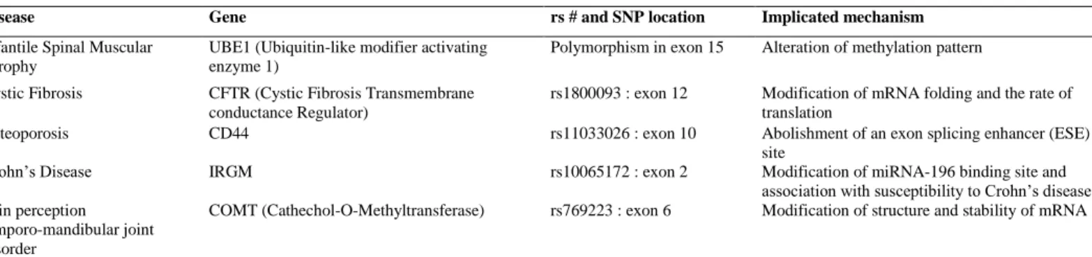
Table 3. SNPs predictive of non-neoplastic diseases and their mechanistic implications
Disease Gene rs # and SNP location Implicated mechanism
Infantile Spinal Muscular Atrophy
UBE1 (Ubiquitin-like modifier activating enzyme 1)
Polymorphism in exon 15 Alteration of methylation pattern
Cystic Fibrosis CFTR (Cystic Fibrosis Transmembrane conductance Regulator)
rs1800093 : exon 12 Modification of mRNA folding and the rate of translation
Osteoporosis CD44 rs11033026 : exon 10 Abolishment of an exon splicing enhancer (ESE) site
Crohn’s Disease IRGM rs10065172 : exon 2 Modification of miRNA-196 binding site and association with susceptibility to Crohn’s disease Pain perception
temporo-mandibular joint disorder
COMT (Cathechol-O-Methyltransferase) rs769223 : exon 6 Modification of structure and stability of mRNA
kruppel-like transcription factor 5 and factor 12 (KLF5 and KLF12, respectively), which are frequently deleted in a large
number of cancers, particularly in PC [27]. This locus is also a
potential marker for susceptibility to breast cancer 1 and 2
(BRCA1/BRCA2) non-mutated breast cancer [28].
Consequently, a particular SNP may not necessarily be specifically associated with a unique cancer type.
Another very recent GWAS study [29] highlighted 4 new
loci that may predispose to PC: 1 -The 17q25.1 locus associated with long intergenic non-coding RNA protein 673 (LINC00673) and the presence of SNP rs11655237. 2 - The locus 7p13 and succinyl-CoA: glutarate CoA transferase (SUGCT) on which the SNP rs17668601 has been noted. 3 - The 3q29 locus and TP63 gene with the presence of the SNP rs9854771. 4 - The locus 2p13.3 associating ewing tumor associated antigen-1 (ETAA1) with the SNP rs1486134. This final locus may predispose to PDAC in the Chinese Han
population [30]. Data presented by Low et al. [8] and Wu et al.
[30],among others [21], seem to suggest that a SNP predisposing
to a given pathology does not necessarily apply to only one ethnicity; other ethnicities should be examined before such a conclusion.
In 2012, Li et al.[31] demonstrated that three genes are
involved in maturity onset diabetes of the young (MODY) which also harbor SNPs predisposing to PC: 1 - The pancreatic and duodenal homeobox 1 gene (PDX-1) involved in MODY-4 diabetes encodes a transcription factor that
regulates the early stage development of the exocrine pancreas. 2 - The hepatocyte nuclear factor-1 alpha or beta genes (HNF1A and HNF1B) are involved in MODY-3 and
MODY-5 diabetes, respectively [32, 33],as well as playing roles
in the growth of β-cells in the islets of Langerhans and control of pancreatic organogenesis. HNF1 and PDX-1 are also
associated with risk of type 2 diabetes and obesity [34]. In this
context, the SNPs rs9554197 and rs9581943 in PDX-1, as well as rs7310409 and rs4794758 in HNF1A and HNF1B, respectively, could contribute to the development of PC in
patients afflicted with obesity or diabetes [31, 35, 36].
SNPs in the clinic
As shown above, a number of SNPs have been experimentally validated as potential predisposition markers for PDAC. However, none of these SNPs is currently used in the clinic, most likely due to their associated low OR (generally close to 1) thus the increase in risk is limited. Tools to detect these SNPs in clinical routines are also lacking. Furthermore, no study has yet shown what functional impact these SNPs could have on cell behavior to explain the onset of PDAC. However, studies in other cancers have shown correlations between the presence of polymorphisms and their potential impact on treatment (sensitivity, resistance) (Table
2). The functional effects of these polymorphisms have been
demonstrated in many other pathology cases (Table 3). SNPs resulting in a modification of the protein sequence can lead to the synthesis of truncated proteins. Although synonymous
Page 5 of 12 SNPs do not necessarily affect the protein sequence, many
other changes in gene expression have been identified.
SNPs and candidate gene association studies
Five hereditary syndromes are associated with an increased risk of PDAC: 1 - The Peutz-Jeghers syndrome caused by germline mutations in the STK11/LKB1 gene encoding serine/threonine kinase 11 (STK11), also known as liver kinase B1 (LKB1) or renal carcinoma antigen
NY-REN-19 [29, 37]. STK11 rs741765 was associated with
disease-free survival or overall survival of patients with
colorectal cancer [38]. 2 - The familial atypical multiple
melanoma and mole syndrome (FAMMM) caused by mutations in the cyclin dependent kinase N2A (CDKN2A)
gene [29, 39, 40]. 3 - Hereditary pancreatitis is also the cause of
PDAC with mutation in the cationic trypsinogen PRSS1 gene
[41]. 4 - Subjects with mutations in BRCA2 or partner and
localizer of BRCA2 (PALB2) [29, 42, 43]. A lower expression of
BRCA2 transcript and increased pancreatic cancer risk were
associated with SNP rs11571836 in the BRCA2
3'-untranslated region [44]. 5 – Finally, the Lynch syndrome
often called hereditary nonpolyposis colorectal cancer (HNPCC), caused by germline mutations in DNA mismatch repair genes such as mutL homolog 1 (MLH1), mutS protein homolog 2/6 (MSH2, MSH6), and PMS1 protein homolog 2 (PMS2) appear to be linked to a slightly increased risk of
developing PC [45]. SNPs in these genes were associated with
overall survival in patients with locally advanced or
metastatic disease [46]. BRCA1 and the ataxia telangiectasia
gene (ATM) were also mutated in patients with hereditary PC [29, 47, 48]. Studies have also shown that the 8-oxoguanine DNA glycosylase gene (OGG1) is another candidate gene for PC development. Links between PC and SNPs in OGG1 are rather confusing as some SNPs are associated with either
increased [49] or no risk [50] in the Japanese population, while
others seem associated with a protection in Chinese
populations [51]. A recent study performed on 32 patients with
PDAC [52] demonstrated that the tumor protein gene (TP53)
polymorphism (SNP rs1042522 at 17p12-13) is a potential genetic predisposing factor, while SNP rs2279744 located at 12q14.3-q15 in mouse double minute 2 homolog (MDM2), an E3 ubiquitin ligase that negatively regulates p53, could potentially predict survival outcome. Predictive values deduced from these genes were low with modest odds ratios. Therefore, a better genetic predictive factor is crucial in order to define an at risk population of developing PC and to increase the efficacy of follow-up of these patients. Recently,
Martinez et al. [53] characterized the SNP rs488087 as being
associated to PC. This SNP is present within the coding region of the human bile salt-dependent lipase gene (BSDL).
SNP in the BSDL gene
The human BSDL, or Carboxyl ester lipase (CEL) gene encodes a lipolytic enzyme involved in the hydrolysis of dietary cholesteryl esters and is mainly expressed by the
acinar cells of the pancreas [54]. This gene locates at band 34.3
on the long arm of chromosome 9 [55, 56] and consists of 11
exons spanning 9884 base pairs (bp). The N-terminal domain of the protein, encoded by exons 1 to 10, includes bile salts binding, catalytic and N-glycosylation sites and is well
conserved across evolution [54]. However, exon 11 codes the
C-terminal domain with a region consisting of a variable number of tandem repeats (VNTR). Each repeat consists of
GC-rich 33 bp sequence coding 11 amino acids [57] with
O-glycosylation sites [58], conferring a mucin-like structure to
the C-terminal domain of BSDL [59]. VNTR sequences can
slightly vary and the total number of repeats differs between alleles. In Caucasian populations investigated to date, the most frequent allele has 16 repeats which results in a 722 amino-acid long polypeptide. However, VNTR number can range between 3 to 21 repeats, leading to a high level of heterogeneity in protein size both within and between
individuals [60, 61]. It has been suggested that naturally
occurring variable numbers of VNTR can influence BSDL function and an association between VNTR length and increased susceptibility to alcoholic pancreatitis has been
suggested [62]. Furthermore, plasma levels of BSDL and Low
Density Lipoprotein (LDL), (BSDL is associated with LDL,
in part by a specific interaction with Apo B100 [63]), positively
correlate, suggesting a role for BSDL in cardiovascular
diseases [64]. Augé et al. [65] demonstrated that BSDL is present
within atherosclerotic plaques of arterial walls where it induces proliferation of smooth muscle cells, chemotaxic
migration of monocytes and oxidized LDL degradation [63, 66,
67]. Furthermore, a single base deletion in either repeat 1 or 4
within the VNTR has been associated with autosomal dominantly inherited MODY-8 with exocrine dysfunctions. Such deletions would result in truncated proteins due to a
premature stop codon [68, 69]. Affected family members
typically develop diabetes characterized by primary β-cell dysfunction, although transgenic mice over-expressing truncated BSDL failed to develop the MODY-8 phenotype
[70]. Therefore, the pathogenic mechanisms involved are more
complex than a simple loss of BSDL function. Truncated BSDL seems to be prone to form aggregates, detected both
intra- and extracellularly [71, 72]. Therefore, MODY-8 and
exocrine pancreatic syndrome may be caused by BSDL misfolding with a negative gain-of-function effect of truncated protein in the pancreas. Further work investigating these mechanisms demonstrated that truncated BSDL was secreted and re-internalized for degradation by lysosomes in a
HEK293-T cell model [72]. Such internalization may reduce
Page 6 of 12
recent study [73] suggested that a recombined allele of BSDL,
and its pseudogene BSDLP, confers susceptibility to chronic pancreatitis, an inflammatory disease of the pancreas known
for predisposition to PDAC [74]. The resulting BSDL hybrid
protein showed impaired secretion, prominent intracellular accumulation and induced autophagy. There are numerous links between the BSDL gene and diabetes as circulating antibodies to the C-terminal mucin-like domain of BSDL
were detected in the sera of patients with type-1 diabetes [75].
Antibodies to this domain were also detected in the blood of patients affected with PDAC, establishing another link
between PC and diabetes [75].
The sequencing of genes containing GC-rich tandem repeated sequences using standard methodologies can pose
serious limitations [76]. Therefore, Martinez et al. [53] Sanger
sequenced VNTR in BSDL from a restricted French cohort of patients with PC. Analysis of genomic DNA from PC tissue extracts highlighted the synonymous SNP rs488087, c.1719C>T, located in the second VNTR sequence of BSDL.
The authors [53] compared the C/C major genotype and
cancer-free control group to the PC cohort, revealing that the c.1719C>T transition was prevalent in PC patients (P-value = 0.0005 and an OR of 4.72). Notably, this OR for T allele-holders in the PC population is larger than any
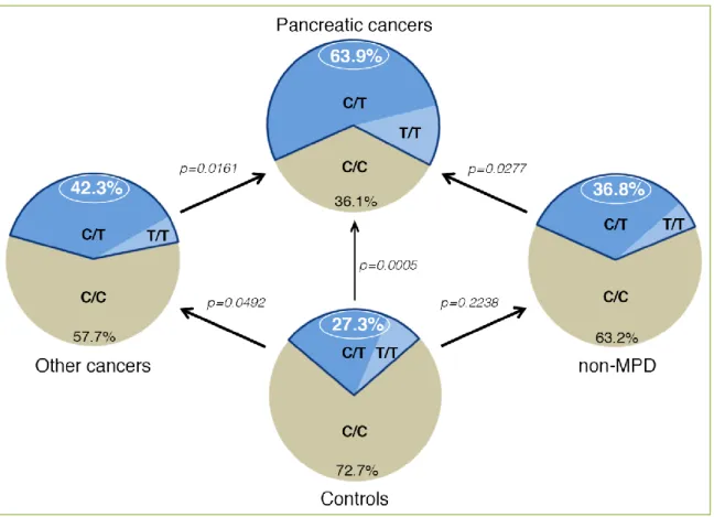
previously determined (Table 1). The SNP occurrence was 63.90% in patients with sporadic PC (n = 36), and decreased to 27.30% in cancer-free control subjects (n = 44) (Figure 1). The frequency of the T allele was 37.50% (n = 72) in the PC cohort versus 17.05% (n = 88) in the control group (P-value = 0.0017). Comparing the T allele frequency in the PC group with the rs488087 SNP data bank (UCSC genome browser, n = 1275) showed that data remained significant (37.50 % versus 23.966%, P-value = 0.0041). In addition, the T allele frequency of the control group showed no significant difference with that of the rs488087 data bank (P-value = 0.0669). Therefore, the limited size of cohorts analyzed by
Martinez et al. [53] did not appear to impair the statistical
significance of their results. Sequence analysis of patients with non-malignant disease of the pancreas (non-MPD) revealed that 36.80 % held the T allele, with no significant difference compared to the control group (P-value = 0.2238). However, occurrence of the polymorphism in the non-MPD group was statistically lower than that in the PC group (36.80% vs 63.90%, P-value = 0.0277). This result based on matched non-MPD and PC populations strongly suggests a link between the c.1719C>T transition and PC. In a cohort of non-pancreatic malignant diseases, i.e. other cancers cohort
(OC) (Figure 1), Martinez et al. [53] found a significant
difference between the occurrence of the c.1719C>T SNP
Figure 1. Schematic representation of SNP rs488087 occurrence in different groups. Non-MPD,
Page 7 of 12 among the OC cohort (42.30%) compared to the PC group
(63.90%, P-value = 0.0161). However, the SNP occurrence among the OC cohort did not differ from that of the control
group. In terms of allelic frequency, Martinez et al. [53] found
no significant difference between the OC group (23.70%) and control group (17.05%) (P-value = 0.1108), or between the OC group and the rs488087 data bank (23.70% versus 23.966%, P-value = 0.4725).
In view of the clinical interest of these results, Martinez et
al. [53] constructed two probes to discriminate between the C
and T SNP alleles of BSDL by droplet digital PCR (ddPCR). Analyses obtained on 143 patients by ddPCR matched 100% of those genotyped by Sanger sequencing. This absolute concordance demonstrates the high specificity of these
probes to rs488087 SNP and their capacity to allow its simple, rapid and specific detection. Martinez et al. also used
specific properties of this ddPCR technique [77] to count the
number of copies of DNA target sequence in each of the DNA samples tested by Sanger sequencing. In the examined heterozygous samples, the fractional abundance of T/T+C was close to 0.5 (i.e. 0.45 +/- 0.07, n = 52) for all cohorts, thus confirming the germline character as the SNP of c.1719C>T transition.
Potential impact of SNP rs488087 on BSDL
function/outcome
The transition c.1719C> T occurs in the third position of a codon which results in no change in terms of encoded amino
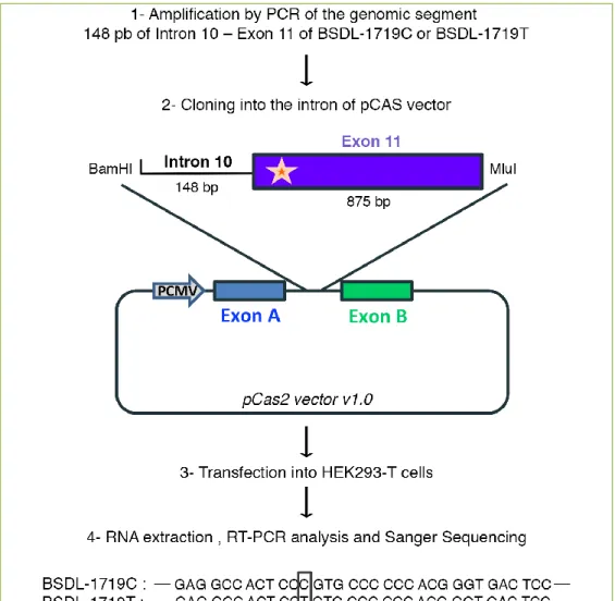
Figure 2. Representation of the functional splicing vector pCAS2. The amplicons (BSDL-1719C
and BSDL-1719T) were cloned into the pCAS reporter vector, based on the pcDNA3.1 plasmid which contained a minigene composed of two exons (named A and B) separated by an intron. The splicing reporter minigene assay pCAS2 was used to evaluate the effect on splicing. After transfection into HEK-293T cells and RNA extraction, analysis of transcripts by Sanger sequencing showed no difference between the BSDL-1719C construction and BSDL-1719T construction by which an ESE sequence could be created. Grey arrows show forward and reverse primers and the star symbolizes the putative ESE sequence and the localization of the SNP rs488087.
Page 8 of 12 acids (synonymous SNP), hence the protein sequence is
unchanged. However, this may not translate to a non-functional effect of this SNP. Indeed, literature based on these particular polymorphisms reveal that synonymous SNPs could have effects on the splicing of pre-messenger RNA, the stability and structure of mRNA, as well as
translation [78]. Pre-messenger RNA may be spliced in
different ways, dependent on whether SNPs occur at donor or acceptor splicing sites, or at the level of regulatory sequences such as exonic splicing enhancer (ESE), exonic splicing silencer (ESS), intronic splicing enhancer (ISE), or intronic splicing silencer (ISS). In the case of cancer, some associations between the presence of a SNP and splicing modification have been identified. This is the case for the gene encoding cyclin D1 (CCND1) which displays an A870G silent polymorphism that alters splicing and therefore induces a susceptibility to the development of lung cancer
[79]. Many SNPs can generate new splicing sites in exons of
the p53 gene, thereby producing a truncated protein [80].
Since the C-terminal domain of BSDL is involved in intracellular processing of BSDL as well as its enzyme
activity, [81] the authors determined whether the SNP
rs488087 could affect BSDL mRNA splicing by an in silico
study (unpublished data). Results obtained using the "Human Splicing Finder” software (http://www.umd.be/HSF/) revealed that the presence of the minor T allele induces the formation of a regulatory sequence of ESE type splicing. The authors therefore examined whether the presence of the SNP rs488087 affects splicing of BSDL mRNA. For this purpose, the cDNA presenting the C or the T polymorphism (150 bp overlapping sequence segment in intron 10 and exon 11 of BSDL-1719C or BSDL-1719T) were cloned into the pCAS2 mini-gene containing 2 artificial exons separated by an intron
[82]. After transfection of plasmid constructs
pCAS2-BSDL-1719C and pCAS2-BSDL-1719T in
HEK293-T cells, RNA was extracted and PCR performed with primers designed to target artificial exons (Figure 2). Results revealed no differences in sequence (and size) between the two transcripts (Figure 2). Therefore, the SNP rs488087 does not appear to induce any alteration of splicing. The second hypothesis that Martinez et al. investigated was whether this SNP affects mRNA stability and structure. To determine this, they conducted an in silico study using the
software "RNA Structure"
(http://rna.urmc.rochester.edu/RNAstructureWeb/). The
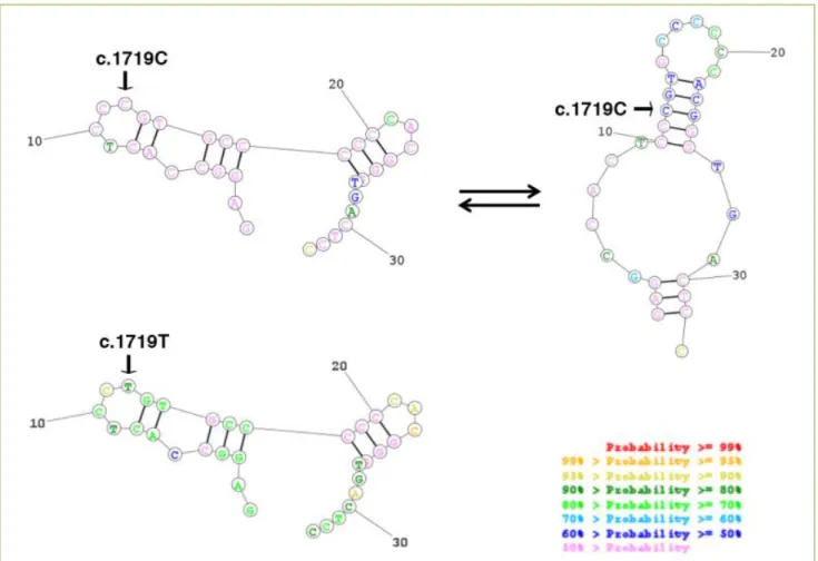
Figure 3. Representation of BSDL VNTR Nb2-mRNA structure and stability. In silico analysis revealed a difference between
Page 9 of 12 mRNA structure at the repeated sequence Nb 2 with C nt
appears to be in equilibrium mainly between two stable structures, whereas the mRNA structure with T nt gains stability with a unique rigid structure (Figure 3). The c.1719C> T transition therefore induces a potential change in the secondary structure of BSDL mRNA and possibly impacts the processing of RNA and translation into protein. This hypothesis agrees with the cellular over-expression of BSDL in various PDAC, partly in the exocrine-PDAC subtype, despite the overexpression of genes encoding lipolytic pancreatic enzymes such as pancreatic lipase (PNLIP) and pancreatic phospholipase A2, group 1B
(PLA2G1B) [83].
A third hypothesis of the potential impact of the c.1719C> T transition was a possible link between this SNP and other gene mutations which could play a role in early tumor
progression in PDAC patients. Martinez et al. [53] examined
Kras point mutations in their cohort of PDAC patients. Among the PDAC patients examined, 66.7 % were T allele holders with Kras mutations. However, the examination of exon 2 Kras mutation subtypes in T allele holders indicated that the T allele in rs488087 SNP was mainly associated (85.7 %) with either the G12D or G12R Kras phenotype. These data clearly suggest that T allele holders of rs488087 SNP favor further somatic mutations in Kras, essentially leading to the G12D or G12R phenotype. Interestingly, these two phenotypes are associated with both the worse
prognostic and lowest patient survival [84, 85].
The final hypothesis is whether the presence of the transition c.1719C> T alters a binding site of a regulating
miRNA. A study by Brest et al. [86] showed in a case of
Crohn's disease that a synonymous SNP (rs10065172; c.313C> T) present within immunity-related GTPase family M protein (IRGM) (also known as interferon-inducible protein 1 (IFI1)) alters the binding of family 196 mi-RNAs
(miR -196A and miR-196B) [86]. Studies are currently in
progress to characterize miRNA binding sites potentially generated by the c.1719C> T transition.
Conclusions
To fully understand the functional impact of the synonymous SNP rs488087, it is essential to thoroughly test each hypothesis to clearly establish any links between the occurrences of the SNP, in part the T allele, and neoplastic diseases of the pancreas. Further investigation is required to confirm the association between the rs488087 SNP and Kras mutations. From a clinical perspective, predictive or diagnostic markers cannot be dualistic (or binary) within the field of biomarkers. More importantly, a single biomarker
alone cannot provide a clinician with definitive black or white answers, therefore SNPs should be associated with other relevant biomarkers, and/or risk factors, to define particular patient populations. This is particularly appropriate for PC patients, as the sr488087 SNP can be associated to other predisposing factors such as hereditary syndromes with BRCA mutations.
Conflicting interests
The authors have declared that no conflict of interests exist.
Acknowledgments
The authors are particularly grateful to Drs. Marc Bartoli (Aix-Marseille Université, INSERM, UMR 910, Marseille, France), Martin Krahn (Assistance Publique Hôpitaux de Marseille, Hôpital de la Timone-Enfants, Département de Génétique Médicale, Marseille, France), Fréderic Fina (LBM, Assistance Publique Hôpitaux de Marseille, Hôpital Nord, Service de transfert d’Oncologie Biologique, Marseille, France), Mehdi Ouaissi (Aix-Marseille Université, CRO2, Centre de Recherche en Oncologie biologique et Oncopharmacologie, INSERM, UMR_S 911, Marseille, France and Assistance Publique Hôpitaux de Marseille, Hôpital de la Timone, Service de Chirurgie Digestive et Viscérale, Marseille, France) and to Prs. Fabrice Barlesi (Assistance Publique Hôpitaux de Marseille, Hôpital Nord, Service d’Oncologie Multidisciplinaire & Innovation
Thérapeutique, Marseille, France), Dominique
Figarella-Branger (Assistance Publique Hôpitaux de
Marseille, Hôpital de la Timone, Service
d’Anatomopathologie, Marseille, France), René Laugier (Assistance Publique Hôpitaux de Marseille, Hôpital de la Timone, Service de Gastroentérologie, Marseille, France) and Juan Iovanna (Aix-Marseille Université, CRCM, Centre de Recherche en Cancérologie de Marseille, Marseille, France) for invaluable discussions. We are deeply indebted to Drs. Marc Bartoli and Martin Krahn for the generous gift of pCAS2 reporter vector.
This work was supported by institutional funding from INSERM (Paris, France), the Aix- Marseille Université (Marseille, France) and a grant INCa-DGSO-INSERM 6038 from Sites de Recherche Intégrée sur le Cancer (SIRIC). Mrs. E. Martinez is a recipient of a fellowship awarded by the French Ministère de la Recherche et de l’Enseignement Supérieur (Paris, France) and the Association pour le développement des recherches biologiques et médicales (La Penne-sur-Huveaune, France).
Page 10 of 12
Abbreviations
BSDL: bile-salt-dependent lipase; PDAC: pancreatic ductal adenocarcinoma; PC: pancreatic cancers.
Author contributions
D.L. and Er.M. write the article. F.S and Em.M conducted the experiment. F.S., Em.M. and Er.M. analysed the results.
References
1. Rahib L, Smith BD, Aizenberg R, Rosenzweig AB, Fleshman JM, Matrisian LM. Projecting cancer incidence and deaths to 2030: the unexpected burden of thyroid, liver, and pancreas cancers in the United States. Cancer Res 2014; 74:2913-2921.
2. Vincent A, Herman J, Schulick R, Hruban RH, Goggins M. Pancreatic cancer. Lancet 2011; 378:607-620.
3. Lockhart AC, Rothenberg ML, Berlin JD. Treatment for pancreatic cancer: current therapy and continued progress. Gastroenterology 2005; 128:1642-1654.
4. Pliarchopoulou K, Pectasides D. Pancreatic cancer: current and future treatment strategies. Cancer Treatment Review 2009; 35:431-436.
5. Jarufe N, McMaster P, Mayer AD, Mirza DF, Buckels JA, Orug T,
et al. Surgical treatment of metastases to the pancreas. Surgeon
2005; 3:79-83.
6. Müller-Nordhorn J, Roll S, Böhmig M, Nocon M, Reich A, Braun C, et al. Health-related quality of life in patients with pancreatic cancer. Digestion 2006; 74:118-125.
7. Amundadottir L, Kraft P, Stolzenberg-Solomon RZ, Fuchs CS, Petersen GM, Arslan AA, et al. Genome-wide association study identifies variants in the ABO locus associated with susceptibility to pancreatic cancer. Nat Genet 2009; 41:986-990.
8. Low SK, Kuchiba A, Zembutsu H, Saito A, Takahashi A, Kubo M, et al. Genome-wide association study of pancreatic cancer in Japanese population. PLOS One 2010; 5:e11824.
9. Aird I, Bentall HH, Roberts JA. A relationship between cancer of stomach and the ABO blood groups. Br Med J 1953; 1:799-801. 10. Marcus DM. The ABO and Lewis blood-group system.
Immunochemistry, genetics and relation to human disease. N Engl J Med 1969; 280:994-1006.
11. Wolpin BM, Kraft P, Gross M, Helzlsouer K, Bueno-de-Mesquita HB, Steplowski E, et al. Pancreatic cancer risk and ABO blood group alleles: results from the pancreatic cancer cohort consortium. Cancer Res 2010; 70:1015-1023.
12. Wolpin BM, Chan AT, Hartge P, Chanock SJ, Kraft P, Hunter DJ,
et al. ABO blood group and the risk of pancreatic cancer. J Natl
Cancer Inst 2009; 10:424-431.
13. Melzer D, Perry JR, Hernandez D, Corsi AM, Stevens K, Rafferty I, et al. A genome-wide association study identifies protein quantitative trait loci (pQTLs). PLoS Genet 2008; 4:e1000072. 14. Paré G, Chasman DI, Kellogg M, Zee RY, Rifai N, Badola S, et
al. Novel association of ABO histo-blood group antigen with
soluble ICAM-1: results of a genome-wide association study of
6,578 women. PLoS Genet 2008; 4:e1000118.
15. Petersen GM, Amundadottir L, Fuchs CS, Kraft P,
Stolzenberg-Solomon RZ, Jacobs KB, et al. A genome-wide association study identifies pancreatic cancer susceptibility loci on chromosomes 13q22.1, 1q32.1 and 5p15.33. Nat Genet 2010; 42:224-228.
16. Wang Y, Broderick P, Webb E, Wu X, Vijayakrishnan J, Matakidou A, et al. Common 5p15.33 and 6p21.33 variants influence lung cancer risk. Nat Genet 2008; 40:1407-1409. 17. Rafnar T, Sulem P, Stacey SN, Geller F, Gudmundsson J,
Sigurdsson A, et al. Sequence variants at the TERT-CLPTM1L locus associate with many cancer types. Nat Genet 2009; 41:221-227.
18. Yamamoto K, Okamoto A, Isonishi S, Ochiai K, Ohtake Y. A novel gene, CRR9, which was up-regulated in CDDP-resistant ovarian tumor cell line, was associated with apoptosis. Biochem Biophys Res Commun 2001; 280:1148-1154.
19. Jia J, Bosley AD, Thompson A, Hoskins JW, Cheuk A, Collins I,
et al. CLPTM1L promotes growth and enhances aneuploidy in
pancreatic cancer cells. Cancer Res 2014; 74:2785-2795.
20. Liu Y, Cao L, Li Z, Zhou D, Liu W, Shen Q, et al. A genome-wide association study identifies a locus on TERT for mean telomere length in Han Chinese. PLoS One 2014; 9:e85043. 21. Wu C, Kraft P, Stolzenberg-Solomon R, Steplowski E, Brotzman
M, Xu M, et al. Genome-wide association study of survival in patients with pancreatic adenocarcinoma. Gut 2014; 63:152-160. 22. He TC, Sparks AB, Rago C, Hermeking H, Zawel L, da Costa LT,
et al. Identification of c-MYC as a target of the APC pathway.
Science 1998; 281:1509-1512.
23. He TC, Chan TA, Vogelstein B, Kinzler KW. PPAR-delta is an APC-regulated target of nonsteroidal anti-inflammatory drugs. Cell 1999; 99:335-345.
24. Lin YM, Ono K, Satoh S, Ishiguro H, Fujita M, Miwa N, et al. Identification of AF17 As a Downstream Gene of the b-catenin/T-Cell Factor Pathway and Its Involvement in Colorectal Carcinogenesis. Cancer Res 2001; 61:6345-6349.
25. Shtutman M, Zhurinsky J, Simcha I, Albanese C, D'Amico M, Pestell R, et al. The cyclin D1 gene is a target of the β-catenin/LEF-1 pathway. Proc Natl Acad Sci U S A 1999; 96:5522-5527.
26. Tetsu O, McCormick F. β-catenin regulates expression of cyclin D1 in colon carcinoma cells. Nature 1999; 398:422-426.
27. Chen JM, Montier T, Férec C. Molecular pathology and
evolutionary and physiological implications of
pancreatitis-associated cationic trypsinogen mutations. Hum Genet 2001; 109:245-252.
28. Kainu T, Juo SH, Desper R, Schaffer AA, Gillanders E, Rozenblum E, et al. Somatic deletions in hereditary breast cancers implicate 13q21 as a putative novel breast cancer susceptibility locus. Proc Natl Acad Sci U S A 2000; 97:9603-9608.
29. Childs EJ, Mocci E, Campa D, Bracci PM, Gallinger S, Goggins M, et al. Common variation at 2p13.3, 3q29, 7p13 and 17q25.1 associated with susceptibility to pancreatic cancer. Nat Genet 2015; 47:911-916.
30. Wu C, Miao X, Huang L, Che X, Jiang G, Yu D, et al. Genome-wide association study identifies five loci associated with
Page 11 of 12
susceptibility to pancreatic cancer in Chinese populations. Nat Genet 2011; 44:62-66.
31. Li D, Duell EJ, Yu K, Risch HA, Olson SH, Kooperberg C, et al. Pathway analysis of genome-wide association study data highlights pancreatic development genes as susceptibility factors for pancreatic cancer. Carcinogenesis 2012; 33:1384-1390. 32. Glucksmann MA, Lehto M, Tayber O, Scotti S, Berkemeier L,
Pulido JC, et al. Novel mutations and a mutational hotspot in the MODY3 gene. Diabetes 1997; 46:1081-1086.
33. Carette C, Vaury C, Barthélémy A, Clauin S, Grünfeld JP, Timsit J, et al. Exonic duplication of the hepatocyte nuclear factor-1beta gene (transcription factor 2, hepatic) as a cause of maturity onset diabetes of the young type 5. J Clin Endocrinol Metab 2007; 92:2844-2847.
34. Holmkvist J, Cervin C, Lyssenko V, Winckler W, Anevski D, Cilio C, et al. Common variants in HNF-1 alpha and risk of type 2 diabetes. Diabetologia 2006; 49:2882-2891.
35. Pierce BL, Ahsan H. Genome-wide "pleiotropy scan" identifies HNF1A region as a novel pancreatic cancer susceptibility locus. Cancer Res 2011; 71:4352-4358.
36. Wolpin BM, Rizzato C, Kraft P, Kooperberg C, Petersen GM, Wang Z, et al. Genome-wide association study identifies multiple susceptibility loci for pancreatic cancer. Nat Genet 2014; 46:994-1000.
37. Giardiello FM, Welsh SB, Hamilton SR, Offerhaus GJ, Gittelsohn AM, Booker SV, et al. Increased risk of cancer in the Peutz-Jeghers syndrome. N Engl J Med 1987; 316:1511-1514. 38. Lee SJ, Kang BW, Chae YS, Kim HJ, Park SY, Park JS, et al.
Genetic variations in STK11, PRKAA1, and TSC1 associated with prognosis for patients with colorectal cancer. Ann Surg Oncol 2014; 21 Suppl 4:S634-9.
39. Bartsch DK, Sina-Frey M, Lang S, Wild A, Gerdes B, Barth P, et
al. CDKN2A germline mutations in familial pancreatic cancer.
Annals Surg 2002; 236:730-737.
40. Di Marco M, Astolfi A, Grassi E, Vecchiarelli S, Macchini M, Indio V, et al. Characterization of pancreatic ductal
adenocarcinoma using whole transcriptome sequencing and copy number analysis by single-nucleotide polymorphism array. Mol Med Rep 2015; 12:7479-7484.
41. Howes N, Lerch MM, Greenhalf W, Stocken DD, Ellis I, Simon P,
et al. Clinical and genetic characteristics of hereditary pancreatitis
in Europe. Clin Gastroenterol Hepatol 2004; 2:252-261.
42. Slater EP, Langer P, Niemczyk E, Strauch K, Butler J, Habbe N,
et al. PALB2 mutations in European familial pancreatic cancer
families. Clin Genet 2010; 78:490-494.
43. Jones S, Hruban RH, Kamiyama M, Borges M, Zhang X, Parsons DW, et al. Exomic sequencing identifies PALB2 as a pancreatic cancer susceptibility gene. Science 2009; 324:217.
44. Huang L, Wu C, Yu D, Wang C, Che X, Miao X, et al. Identification of common variants in BRCA2 and MAP2K4 for susceptibility to sporadic pancreatic cancer. Carcinogenesis 2013; 34:1001-1005.
45. Kastrinos F, Mukherjee B, Tayob N, Wang F, Sparr J, Raymond VM, et al. Risk of pancreatic cancer in families with Lynch syndrome. J Am Med Assoc 2009; 302:1790-1795.
46. Dong X, Li Y, Hess KR, Abbruzzese JL, Li D. DNA mismatch
repair gene polymorphisms affect survival in pancreatic cancer. Oncologist 2011; 16:61-70.
47. Roberts NJ, Jiao Y, Yu J, Kopelovich L, Petersen GM, Bondy ML, et al. ATM mutations in patients with hereditary pancreatic cancer. Cancer Discov 2012; 2:41-46.
48. Thompson D, Easton DF. Breast Cancer Linkage Consortium. Cancer Incidence in BRCA1 mutation carriers. J Natl Cancer Inst 2002; 94:1358-1365.
49. Zhang J, Zhang X, Dhakal IB, Gross MD, Kadlubar FF, Anderson KE. Sequence variants in antioxidant defense and DNA repair genes, dietary antioxidants, and pancreatic cancer risk. Int J Mol Epidemiol Genet 2011; 2:236-244.
50. Nakao M, Hosono S, Ito H, Watanabe M, Mizuno N, Sato S, et al. Selected polymorphisms of base excision repair genes and pancreatic cancer risk in Japanese. J Epidemiol 2012; 22:477-483. 51. Chen H, Zhou B, Lan X, Wei D, Yuan T, Chen P. Association
between single-nucleotide polymorphisms of OGG1 gene and pancreatic cancer risk in Chinese Han population. Tumour Biol 2014; 35:809-813.
52. Hori Y, Miyabe K, Yoshida M, Nakazawa T, Hayashi K, Naitoh I,
et al. Impact of TP53 codon 72 and MDM2 SNP 309
polymorphisms in pancreatic ductal adenocarcinoma. PLos One 2015; 10:e0118829.
53. Martinez E, Silvy F, Fina F, Bartoli M, Krahn M, Barlesi F, et al. Rs488087 single nucleotide polymorphism as predictive risk factor for pancreatic cancers. Oncotarget 2015; 6:39855-39864. 54. Lombardo D. Bile salt-dependent lipase: its pathophysiological
implications. Biochim Biophys Acta 2001; 1533:1-28.
55. Lidberg U, Nilsson J, Strömberg K, Stenman G, Sahlin P, Enerbäck S, et al. Genomic organization, sequence analysis, and chromosomal localization of the human carboxyl ester lipase (CEL) gene and a CEL-like (CELL) gene. Genomics 1992; 13:630-640.
56. Taylor AK, Zambaux JL, Klisak I, Mohandas T, Sparkes RS, Schotz MC, et al. Carboxyl ester lipase: a highly polymorphic locus on human chromosome 9qter. Genomics 1991; 10:425-431. 57. Nilsson J, Bläckberg L, Carlsson P, Enerbäck S, Hernell O, et al.
cDNA cloning of human-milk bile-salt-stimulated lipase and evidence for its identity to pancreatic carboxylic ester hydrolase. Eur J Biochem 1990; 192:543-550.
58. Baba T, Downs D, Jackson KW, Tang J, Wang CS. Structure of human milk bile salt activated lipase. Biochemistry 1991; 30:500-510.
59. Wang CS, Dashti A, Jackson KW, Yeh JC, Cummings RD, Tang J. Isolation and characterization of human milk bile salt-activated lipase C-tail fragment. Biochemistry 1995; 34:10639-10644. 60. Strömqvist M, Hernell O, Hansson L, Lindgren K, Skytt A,
Lundberg L, et al. Naturally occurring variants of human milk bile salt-stimulated lipase. Arch Biochem Biophys 1997; 347:30-36. 61. Swan JS, Hoffman MM, Lord MK, Poechmann JL. Two forms of
human milk bile-salt-stimulated lipase. Biochem J 1992; 283:119-122.
62. Ragvin A, Fjeld K, Weiss FU, Torsvik J, Aghdassi A, Mayerle J,
et al. The number of tandem repeats in the carboxyl-ester lipase
(CEL) gene as a risk factor in alcoholic and idiopathic chronic pancreatitis. Pancreatology 2013; 13:29-32.
Page 12 of 12
63. Caillol N, Pasqualini E, Mas E, Valette A, Verine A, Lombardo D. Pancreatic bile salt-dependent lipase activity in serum of normolipidemic patients. Lipids 1997; 32:1147-1153.
64. Blind PJ, Büchler M, Bläckberg L, Andersson Y, Uhl W, Beger HG, et al. Carboxylic ester hydrolase. A sensitive serum marker and indicator of severity of acute pancreatitis. Int J Pancreatol 1991; 8:65-73.
65. Augé N, Rebaï O, Lepetit-Thévenin J, Bruneau N, Thiers JC, Mas E, et al. Pancreatic bile salt-dependent lipase induces smooth muscle cells proliferation. Circulation 2003; 108:86-91.
66. Rebaï O, Le Petit-Thevenin J, Bruneau N, Lombardo D, Vérine A. In vitro angiogenic effects of pancreatic bile salt-dependent lipase. Arterioscler Thromb Vasc Biol 2005; 25:359-364.
67. Shamir R, Johnson WJ, Morlock-Fitzpatrick K, Zolfaghari R, Li L, Mas E, et al. Pancreatic carboxyl ester lipase: a circulating enzyme that modifies normal and oxidized lipoproteins in vitro. J Clin Invest 1996; 97:1696-1704.
68. Torsvik J, Johansson S, Johansen A, Ek J, Minton J, Raeder H, et
al. Mutations in the VNTR of the carboxyl-ester lipase gene
(CEL) are a rare cause of monogenic diabetes. Hum Genet 2010; 127:55-64.
69. Raeder H, Johansson S, Holm PI, Haldorsen IS, Mas E, Sbarra V,
et al. Mutations in the CEL VNTR cause a syndrome of diabetes
and pancreatic exocrine dysfunction. Nat Genet 2006; 38:54-62. 70. Ræder H, Vesterhus M, El Ouaamari A, Paulo JA, McAllister FE,
Liew CW, et al. Absence of diabetes and pancreatic exocrine dysfunction in a transgenic model of carboxyl-ester lipase-MODY (maturity-onset diabetes of the young). PLoS One 2013; 8:e60229. 71. Torsvik J, Johansson BB, Dalva M, Marie M, Fjeld K, Johansson
S, et al. Endocytosis of secreted carboxyl ester lipase in a syndrome of diabetes and pancreatic exocrine dysfunction. J Biol Chem 2014; 289:29097-29111.
72. Johansson BB, Torsvik J, Bjørkhaug L, Vesterhus M, Ragvin A, Tjora E, et al. Diabetes and pancreatic exocrine dysfunction due to mutations in the carboxyl ester lipase gene-maturity onset diabetes of the young (CEL-MODY): a protein misfolding disease. J Biol Chem 2011; 286:34593-34605.
73. Fjeld K, Weiss FU, Lasher D, Rosendahl J, Chen JM, Johansson BB, et al. A recombined allele of the lipase gene CEL and its pseudogene CELP confers susceptibility to chronic pancreatitis. Nat Genet 2015; 47:518-522.
74. Kamisawa T, Wood LD, Itoi T, Takaori K. Pancreatic cancer. Lancet 2016; pii: S0140-6736 (16) 00141-0.
75. Panicot L, Mas E, Thivolet C, Lombardo D. Circulating antibodies against an exocrine pancreatic enzyme in type 1 diabetes. Diabetes 1999; 48:2316-2323.
76. Kirby A , Gnirke A, Jaffe DB, Barešová V, Pochet N, Blumenstiel B, et al. Mutations causing medullary cystic kidney disease type 1 (MCKD1) lie in a large VNTR in MUC1 missed by massively parallel sequencing. Nat Genet 2013; 45:299-303.
77. Hindson BJ, Ness KD, Masquelier DA, Belgrader P, Heredia NJ, Makarewicz AJ, et al. High-throughput droplet digital PCR system for absolute quantitation of DNA copy number. Anal Chem 2011; 83:8604-8610.
78. Sauna ZE, Kimchi-Sarfaty C, Ambudkar SV, Gottesman MM. Silent polymorphisms speak: how they affect pharmacogenomics and the treatment of cancer. Cancer Res 2007; 67:9609-9612. 79. Gautschi O, Ratschiller D, Gugger M, Betticher DC, Heighway J.
Cyclin D1 in non-small cell lung cancer: a key driver of malignant transformation. Lung Cancer 2007; 55:1-14.
80. Lamolle G, Marin M, Alvarez-Valin F. Silent mutations in the gene encoding the p53 protein are preferentially located in conserved amino acid positions and splicing enhancers. Mut Res 2006; 600:102-112.
81. DiPersio LP, Carter CP, Hui DY. Exon 11 of the rat cholesterol esterase gene encodes domains important for intracellular processing and bile salt-modulated activity of the protein. Biochemistry 1994; 33:3442–3448.
82. Gaildrat P, Killian A, Martins A, Tournier I, Frébourg T, Tosi M. Use of splicing reporter minigene assay to evaluate the effect on splicing of unclassified genetic variants. Methods Mol Biol 2010; 653:249-257.
83. Collisson EA, Sadanandam A, Olson P, Gibb WJ, Truitt M, Gu S,
et al. Subtypes of pancreatic ductal adenocarcinoma and their
differing responses to therapy. Nat Med 2011; 17:500-503. 84. Kawesha A, Ghaneh P, Andrén-Sandberg A, Ograed D, Skar R,
Dawiskiba S, et al. K-ras oncogene subtype mutations are associated with survival but not expression of p53, p16(INK4A), p21(WAF-1), cyclin D1, erbB-2 and erbB-3 in resected pancreatic ductal adenocarcinoma. Int J Cancer 2000; 89:469-474.
85. Rachakonda PS, Bauer AS, Xie H, Campa D, Rizzato C, Canzian F, et al. Somatic mutations in exocrine pancreatic tumors: association with patient survival. PLOS One 2012; 8:e60870. 86. Brest P, Lapaquette P, Mograbi B, Darfeuille-Michaud A, Hofman
P. Risk predisposition for Crohn disease: a "ménage à trois" combining IRGM allele, miRNA and xenophagy. Autophagy 2011; 7:786-787.