HAL Id: hal-00557379
https://hal.archives-ouvertes.fr/hal-00557379
Submitted on 19 Jan 2011HAL is a multi-disciplinary open access archive for the deposit and dissemination of sci-entific research documents, whether they are pub-lished or not. The documents may come from teaching and research institutions in France or abroad, or from public or private research centers.
L’archive ouverte pluridisciplinaire HAL, est destinée au dépôt et à la diffusion de documents scientifiques de niveau recherche, publiés ou non, émanant des établissements d’enseignement et de recherche français ou étrangers, des laboratoires publics ou privés.
INDUCES A DELETERIOUS EXON 23 SKIPPING
BY AFFECTING EXONIC SPLICING REGULATORY
ELEMENTS
Pascaline Gaildrat, Sophie Krieger, Jean-Christophe Thery, Audrey Killian,
Antoine Rousselin, Pascaline Berthet, Thierry Frébourg, Agnès Hardouin,
Alexandra Martins, Mario Tosi
To cite this version:
Pascaline Gaildrat, Sophie Krieger, Jean-Christophe Thery, Audrey Killian, Antoine Rousselin, et al.. THE BRCA1 c.5434C>G (p.PRO1812ALA) VARIANT INDUCES A DELETERIOUS EXON 23 SKIPPING BY AFFECTING EXONIC SPLICING REGULATORY ELEMENTS. Journal of Medical Genetics, BMJ Publishing Group, 2010, 47 (6), pp.398. �10.1136/jmg.2009.074047�. �hal-00557379�
JMG/2009/074047 rev.
The BRCA1 c.5434C>G (p.Pro1812Ala) variant induces a deleterious
exon 23 skipping by affecting exonic splicing regulatory elements
Pascaline GAILDRAT1, Sophie KRIEGER2, Jean-Christophe THERY1, Audrey KILLIAN1, Antoine ROUSSELIN2, Pascaline BERTHET3, Thierry FRÉBOURG1,4, Agnès HARDOUIN2, Alexandra MARTINS1, Mario TOSI1
1
Inserm U614, Faculty of Medicine, University of Rouen, IFRMP, Rouen, Institute for Biomedical Research, Rouen, France
2
Laboratoire de Biologie Clinique et Oncologique, Centre François Baclesse, Caen, France
3
Consultation d’Oncogénétique, Centre François Baclesse, Caen, France
4
Department of Genetics, University Hospital, Rouen, France
Corresponding author:
Mario TOSI, Inserm U614, Faculty of Medicine, 22 Boulevard Gambetta, 76183 Rouen, France, Email: Mario.tosi@univ-rouen.fr, Tel: (33)235-148-311, Fax: (33)235-148-237
ABSTRACT
A large fraction of the sequence variants of unknown significance or unclassified variants (UVs), including exonic variants, could be pathogenic by affecting mRNA splicing. The breast and ovarian cancer susceptibility gene BRCA1 exhibits a large spectrum of sequence variation but only two variants, both located in exon 18, have been shown experimentally to affect splicing regulatory elements. In the present study, we investigate the impact on splicing of the variant BRCA1 c.5434C>G (p.Pro1812Ala), identified in an ovarian cancer patient. This variant has previously been studied at the protein level with inconclusive results concerning its pathogenic role. Here, we show, using patient RNA analyses and hybrid minigene assays, that this variant induces a major splicing defect, with skipping of exon 23, resulting in frameshift and predicted protein termination within the second BRCT domain. This argues for its classification as a pathogenic splicing mutation. Moreover, we demonstrate, using an exonic splicing enhancer-dependent minigene assay, that the segment c.5420-5449 of BRCA1, in the centre of exon 23, exhibits splicing enhancer properties. This enhancement is abolished by the c.5434C>G mutation, indicating that the nucleotide change, in this highly conserved region, affects a splicing regulatory element. Bioinformatics analyses predict that the mutation c.5434C>G creates an hnRNPA1-dependent splicing silencer. These results also suggest that UVs in highly conserved nucleotide sequences of short exons may be good candidates for detecting functionally relevant splicing regulatory elements.
INTRODUCTION
Genetic counseling is often limited by lack of knowledge about the pathogenic role of a large fraction of germline sequence variations. These are usually referred to as variants of unknown significance or unclassified variants (UVs). These types of variants are especially problematic for the breast and ovarian cancer susceptibility gene, BRCA1, which exhibits a large spectrum of sequence variation. Among these variants, most of the missense changes identified in the BRCA1 gene, as well as many intronic sequence changes, are currently listed as UVs in the database of the Breast Cancer Information Core (BIC, http://research.nhgri.nih.gov/projects/bic).
Over the past ten years, it became clear that a large fraction of missense UVs could be deleterious by impacting pre-mRNA splicing.[1,2] Exonic mutations alter either splice site sequences or splicing regulatory elements such as ESE (exonic splicing enhancer) or ESS (exonic splicing silencer). Most ESE elements promote exon inclusion by interacting with activating splicing factors, usually members of the serine/argine-rich protein family. In contrast, ESS elements play a role in splicing repression usually through the binding of proteins, mostly belonging to the heterogenous nuclear ribonucleoprotein (hnRNP) family. Mutations in these cis-acting elements may induce abnormalities in pre-mRNA splicing, that could result either in the degradation of the mature aberrant mRNA by the nonsense mediated decay (NMD) pathway or in the production of an altered protein.[3]
Here, we describe the characterization of the c.5434C>G (p.Pro1812Ala) variant found in the BRCA1 gene in a French ovarian cancer patient. This variant was previously identified in several families with breast and/or ovarian cancers and was reported as an UV but was not listed in the BIC database of BRCA mutations.[4-6] Because it co-segregates with the high risk phenotype at least in one family,[6] this variant was suspected to be deleterious.
It was initially thought that it could interfere with the activity of the BRCA1 protein, considering the high degree of conservation across species of the proline at position 1812 and its location in the second BRCT (BRCA1 carboxyl terminal) domain. However, a previous study showed that the BRCA1 p.Pro1812Ala variant induces a modest decrease in the activity of the BRCA1 protein, based on a transcriptional transactivation assay.[6] More recently, the effects of this variant were also tested on structural integrity, protein stability and interaction with various phosphopeptides but these data were not sufficient to characterize this mutation as pathogenic.[7] Thus, the deleterious nature of this missense mutation was unclear at the protein level and this uncertainty prompted us to investigate its impact on pre-mRNA splicing. Here, we show that the BRCA1 c.5434C>G variant induces a major splicing defect, resulting in skipping of exon 23.
METHODS
Patient
The BRCA1 c.5434C>G variant tested in this study was detected in an index case selected from families undergoing genetic counseling in the Western French breast cancer genetics network. The criteria for diagnostic mutation screening of BRCA1/2 genes in patients were according to the current French recommendations. The patient gave signed informed consent.
Nomenclature
The DNA sequence numbering is based on the cDNA sequence for BRCA1 (NCBI RefSeq NM_007294), following the recommendations of the Human Genome Variation Society (HGVS, first position of the translation initiation codon ATG=1).
Genetic analysis
Patient peripheral blood samples were collected in EDTA tubes. Genomic DNA was isolated using an automated procedure (EZ1 DNA blood kit on biorobot EZ1 workstation, Qiagen, Hilden, Germany). Screening for BRCA1 and BRCA2 mutations was performed using denaturing High Performance Liquid Chromatography (dHPLC) or High Resolution Melting (HRM), followed by direct sequencing. Screening for BRCA1 and BRCA2 large rearrangements was performed using a combination of the multiplex ligation-dependent probe amplification (MLPA) and quantitative multiplex PCR of short fluorescent fragments (QMPSF) methods.
RT-PCR analysis of patient blood RNA
Whole blood samples were collected from patients and voluntary healthy donors in PAXgene Blood RNA tubes (Qiagen). RNA extraction was performed according to the manufacturer’s protocols, including DNase treatment. RT-PCR was performed from 250 ng of total RNA, a forward primer complementary to BRCA1 exon 20
GAAGAAACCACCAAGGTCCAA-3’) and a reverse primer complementary to BRCA1 3’UTR (5’-GAAGAGTGAGAGGAGCTCCCAG-3’), using one-step RT-PCR kit (Qiagen). All amplifications were performed using a touch-down PCR program. The thermocycler parameters for the RT-PCR were 50°C for 30 minutes for the reverse transcription step, 95°C for 15 minutes for the initial DNA polymerase activation, 26 cycles of 94°C for 40 seconds, 63°C for 1 minute with a decrease of 0.5°C/cycle, and 72°C for 45 seconds, followed by 26 cycles of: 94°C for 40 seconds, 50°C for 20 seconds and 72°C for 45 seconds, and a final extension at 72°C for 10 minutes. RT-PCR was also performed using a total of 40 and 46 cycles. RT-PCR products were separated by electrophoresis on a 0.8% agarose gel containing ethidium bromide and visualized by exposure to ultraviolet light. Semi-quantitative analysis
of the RT-PCR products was carried out using a Gel docTM XR 170-8170 instrument (Biorad) and the software Quantity One (Biorad). RT-PCR products were purified and sequenced using a cycle sequencing reaction kit (Big Dye Terminator kit v1.1, Applied Biosystems) on an automated sequencer (ABI 3130, Applied Biosystems).
Bioinformatics predictions of splicing alterations
Exonic variants were analyzed for possible splicing enhancer (ESE) alteration with ESEfinder 3.0 (http://rulai.cshl.edu/cgi-bin/tools/ESE3/esefinder.cgi) [8, 9] and RESCUE-ESE (http://genes.mit.edu/burgelab/rescue-ese) [10] using the integrated software Alamut
(Interactive Biosoftware, http://www.interactive-biosoftware.com) and Human Splicing Finder 2.4 (http://www.umd.be/HSF).[11] Exonic variants were also analyzed for possible splicing silencer (ESS) creation using Human Splicing Finder 2.4.
Splicing minigene reporter assay
Generation of minigene constructs. The splicing minigene assay has been previously
described.[12-14] Wild-type and mutant c.5434C>G BRCA1 exon 23, together with 236 bp of the 5’ flanking intron and 202 bp of the 3’ flanking intron, were amplified by PCR using forward primer (5’-GACCAGATCTATTAGCCAGGGGTGGTGGTA-3’) and reverse primer (5’-GACCACGCGTCACCCCATGGAAACAGTTCA-3’), carrying 5’ tails with BglII and MluI restriction sites (underlined), respectively. After digestion with BglII and MluI, the PCR products were inserted into the intron of the two-exons pCAS1 splicing reporter minigene vector (Fig. 1B).[12-14]
Transfection and RT-PCR analysis. After identification by plasmid sequencing, the wild-type
and mutant minigene constructs were transiently transfected into HeLa cells using FuGENE 6 transfection reagent, according to manufacturer’s instructions (Roche Applied Science). Cells were then collected 24 h post-transfection. Total RNA was extracted using the TriPure Isolation Reagent (Roche Applied Science) and analyzed by RT-PCR. The reverse transcription (RT) step was performed using the Superscript II reverse transcriptase (Invitrogen) primed with oligod(T)18 (New England Biolabs). The resulting cDNA was
amplified by PCR (25, 30, 35 cycles) using the forward primer F CTCCGCAGGTCCGCT-3’) and the reverse primer R (5’-ATTGGTTGTTGAGTTGGTTGTC-3’) (Fig. 1B). RT-PCR products were separated by electrophoresis on 2.5% agarose gel containing ethidium bromide and visualized by exposure
to ultraviolet light. Semi-quantitative analysis of the RT-PCR products was carried out using a Gel docTM XR 170-8170 instrument (Biorad) and the software Quantity One (Biorad). Each DNA band was gel-extracted using Nucleospin Extract II kit (Macherey-Nagel) and sequenced using Big Dye Terminator cycle sequencing kit (Applied Biosystems) on an automated sequencer (ABI 3130, Applied Biosystems).
ESE-dependent splicing assay
Generation of pcDNA-Dup constructs. BRCA1 exon 23 wild-type fragment c.5420-5449 was
tested for the presence of ESE splicing regulatory sequences by using an ESE-dependent splicing assay.[12] Briefly, the exonic fragment was obtained by annealing complementary 5’-phosphorylated oligonucleotides designed to encompass 30 nucleotides around the mutation position and to carry 5’-EcoRI and 3’-BamHI compatibles ends (Fig. 3A). This duplex was inserted into the EcoRI and BamHI sites of the pcDNA-Dup plasmid. This expression vector contains a splicing cassette consisting of a β-globin-derived three-exon minigene (Dup) under the control of the cytomegalovirus (CMV) promoter (Fig. 3A).[12] The effects of the mutations BRCA1 c.5434C>G and c.5434C>T were tested in parallel in the ESE-dependent splicing assay. Control experiments were performed, as previously described,[12] using pcDNA-Dup derived minigenes, including i) the empty pcDNA-Dup plasmid, ii) the pcDNA-Dup-BR2Int11 construct which contains a fragment without known regulatory elements (5’-TGTTGTCCAGGTCACATTCAATAG-3’) derived from BRCA2 intron 11 and iii) the pcDNA-Dup-SF2/ASF-3x construct containing a triplet of binding sites for the SR protein SF2/ASF (5’-AGAAGAACAAGAAGAACAAGAAGAACG-3’).
Transient transfection and RT-PCR analysis. The different pcDNA-Dup constructs have been
post-transfection and total RNA was extracted using the TriPure Isolation Reagent (Roche Applied Science). The RNA preparations were treated by Amplification Grade DNase I (AMP-D1, Sigma-Aldrich), according to the manufacturer’s instructions and subjected to RT-PCR analysis. The one-step RT-PCR reaction was performed from 0.5 µg DNase-treated RNA in a 25 µl reaction volume using the OneStep RT-PCR kit (Qiagen), according to the manufacturer’s instructions. Reverse transcription was done using the reverse primer Dup-2R (5’-GGACTCAAAGAACCTCTGGG-3’) that binds to the last exon of the minigene and PCR amplification was performed with 25 cycles using the forward primer T7-Pro (5’-TAATACGACTCACTATAGGG-3’), which binds immediately upstream of the Dup minigene, and Dup-2R. RT-PCR products were separated by electrophoresis on 2.5% agarose
gel containing ethidium bromide and visualized by exposure to ultraviolet light. Each DNA band was extracted from the gel by using Nucleospin Extract II kit (Macherey-Nagel) and sequenced using Big dye Terminator cycle sequencing kit (Applied Biosystems) on an automated sequencer (ABI 3130, Applied Biosystems).
RESULTS
Identification of the BRCA1 c.5434C>G variant in an ovarian cancer patient
The patient was referred for genetic counseling, based on the diagnosis of ovarian cancer at 53 years and her family history. Her mother and maternal aunt were diagnosed with ovarian cancer at ages 53 and 65 years, respectively. Her paternal aunt was diagnosed with breast cancer at age 65 years. Using the Manchester scoring system,[15] prior probabilities of a pathogenic mutation were 51% in the BRCA1 gene and 32% in the BRCA2 gene. Based on this information, the genomic DNA of this patient was screened for germ-line mutations and large genomic rearrangements in both BRCA1 and BRCA2 genes. No genetic alteration was found in BRCA2. However, a C to G transvertion was identified at position c.5434 of BRCA1. At the time of genetic counseling, this BRCA1 c.5434C>G variant was not listed in the
BRCA1 mutation BIC database nor in the French database of BRCA1 mutation (Dr. Rosette
Lidereau, personal communication). It was therefore considered as an unclassified variant. In order to assess the pathogenicity of this variant, partial segregation analysis was performed. As the mother of the proband was deceased at the time of the analysis, her affected maternal aunt was tested and found to be a carrier of BRCA1 c.5434C>G. This observation suggested a germline transmission of the variant in the maternal branch, indicating that it could represent the disease causing mutation or a genetic marker linked to it.
The BRCA1 c.5434C>G variant is associated with abnormal splicing of BRCA1 mRNA
in patient blood cells
In order to evaluate the consequence of the BRCA1 c.5434C>G sequence variant on BRCA1 pre-mRNA splicing, RT-PCR analysis was performed using total RNA extracted from peripheral lymphocytes of the proband and a control subject (Fig. 1A). A single product with
the expected size (456 bp) was detected in the control subject and sequencing confirmed that it corresponds to the region covering exon 20 to the 3’UTR of the normal BRCA1 transcript, including exon 23 (Fig. 1A). In contrast, the analysis of RNA from the patient carrying the
BRCA1 c.5434C>G variant revealed the presence of an additional RT-PCR product with a
smaller size (395 bp), expressed at a slightly lower level, that was identified by sequencing as an aberrant transcript lacking BRCA1 exon 23 (Fig. 1A). Semi-quantitative analysis indicated that this RT-PCR product accounts for 35% of the total transcripts (data not shown). Therefore, skipping of exon 23 in the transcript corresponding to the mutant allele could be incomplete, or the aberrant mRNA could be degraded in part by the NMD pathway. Sequencing of the top band corresponding to the normal BRCA1 transcript showed a barely
detectable signal from the mutant sequence, strongly suggesting that very low levels of normal mRNA were expressed from the mutant allele. This result demonstrates that the presence of the BRCA1 c.5434C>G variant is associated with a major splicing defect, resulting in the skipping of exon 23.
The BRCA1 c.5434C>G variant induces BRCA1 exon 23 skipping in a minigene splicing
assay
In order to test the hypothesis that this aberrant skipping of BRCA1 exon 23 was directly caused by the substitution C to G at position c.5434, the effect of the variant on splicing was assessed using a minigene splicing assay (Fig. 1B). pCAS minigene constructs carrying either the wild-type or the mutant BRCA1 c.5434C>G exon 23 were transiently expressed in HeLa cells and the minigene transcripts were analyzed by RT-PCR (Fig. 1C). Because the patient is heterozygous for the frequent SNP c.5407-193A>G (rs8176310, minor allele frequency:
0.456) located in intron 22, the wild-type and mutant c.5434C>G minigene constructs carried the G and A polymorphic nucleotides, respectively. All the processed RNA expressed from
the wild-type BRCA1 exon 23 minigene retained BRCA1 exon 23, indicating that the less frequent G nucleotide at the intronic polymorphism rs8176310 has no negative effect on splicing. In contrast, the major spliced transcript expressed from the mutant BRCA1 c.5434C>G exon 23 minigene exhibits skipping of exon 23 (Fig. 1C). Semi-quantitative analysis indicated that, in this monoallelic assay, 75% of the total transcripts have skipped exon 23 (data not shown). These data demonstrate that the variant BRCA1 c.5434C>G is directly responsible for a major splicing defect.
The BRCA1 region c.5420-5449 in the centre of exon 23 contains sequences with global
enhancer properties, which are affected by the c.5434C>G mutation
The c.5434C>G variant is located in the centre (position +28) of the short exon 23 (61 bp) of the BRCA1 gene, in a region highly conserved at nucleotide level across species (Fig. 2). Nucleotide sequence conservation is particularly striking in a stretch of 6 amino acids with highly degenerate codons (IVVVQP). Such an exonic mutation, associated with exon skipping and located at distance from the splice sites, is likely to affect splicing regulatory elements. In order to determine if the BRCA1 exonic region affected by the c.5434C>G mutation contains a splicing enhancer element (ESE), we took advantage of an ESE-dependent splicing assay (Fig. 3A).[12] A ~30 bp fragment was inserted into the middle exon of a three-exon minigene under the control of a CMV promoter. The different minigenes were transiently expressed in HeLa cells and the splicing patterns of the minigene transcripts were analyzed by RT-PCR. Due to a weak 3’ splice site, this middle exon is not recognized by the splicing machinery and therefore is excluded from the processed transcript, unless enhancer elements are inserted within its sequence.[12] As expected, the middle exon was included into the mature transcripts expressed from a positive control minigene (SF2/ASF-3x) carrying triplet of enhancer elements (ESE), known to bind to the splicing regulatory SR protein
SF2/ASF (Fig. 3B), whereas the middle exon without enhancer element was excluded from the mature transcripts expressed from minigenes without an inserted sequence (empty), or containing a randomly chosen intronic sequence derived from the BRCA2 intron 11 (BR2Int11) (Fig. 3B). The wild-type c.5420-5449 segment of BRCA1 exon 23 was able to induce inclusion of the middle exon (Fig. 3B), strongly suggesting that this specific region contains a sequence with enhancer properties. In contrast, the middle exon was excluded from the mature transcripts expressed from the minigene carrying the BRCA1 exon 23 c.5420-5449 region with the c.5434C>G mutation. This result indicates that the c.5434C>G variant induces skipping of BRCA1 exon 23 either by disrupting an enhancer element (ESE) or by creating a silencer element (ESS).
We also analyzed the impact on splicing of a reported SNP at the same position (BRCA1 c.5434C>T; rs1800751) by using the same ESE-dependent assay (Fig. 3B). In contrast to the c.5434C>G mutation, the c.5434C>T variant had modest negative effect on the inclusion of the middle exon. This result suggests that the effect on splicing at position c.5434 is nucleotide-dependent.
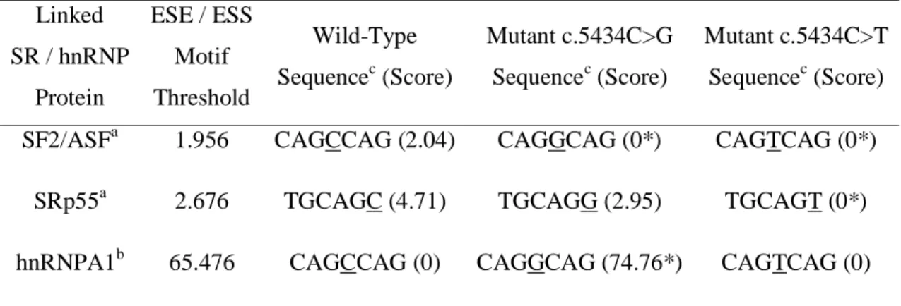
Both exonic variants, BRCA1 c.5434C>G and c.5434C>T, were analyzed by using bioinformatics prediction programs, for possible splicing enhancer (ESE) disruption and silencer (ESS) creation. As shown in Table 1, ESEfinder predicts that both nucleotide changes destroy a SF2/ASF enhancer element and decrease the score or destroy an ESE motif for SRp55. Interestingly, Human Splicing Finder predicts that only the c.5434C>G variant would create a silencer element for hnRNPA1 (Table 1), motif identified using an experimental matrix recently designed for this splicing factor.[11]
Table 1. Bioinformatics predictions of splicing regulatory elements Linked SR / hnRNP Protein ESE / ESS Motif Threshold Wild-Type Sequencec (Score) Mutant c.5434C>G Sequencec (Score) Mutant c.5434C>T Sequencec (Score)
SF2/ASFa 1.956 CAGCCAG (2.04) CAGGCAG (0*) CAGTCAG (0*)
SRp55a 2.676 TGCAGC (4.71) TGCAGG (2.95) TGCAGT (0*)
hnRNPA1b 65.476 CAGCCAG (0) CAGGCAG (74.76*) CAGTCAG (0)
a
, as determined using ESE finder [8, 9]
b
, as determined using hnRNPA1 experimental motif from HSF [11]
c
, the nucleotide underlined indicates the position of the mutation *, scores that differ significantly considering the threshold
DISCUSSION
We have shown, using patient RNA analyses and minigene splicing assays, that the BRCA1 variant c.5434C>G (p.Pro1812Ala) induces a major splicing defect, resulting in the skipping of exon 23. The amount of exon skipping was estimated at 75% in the monoallelic splicing minigene assay, a value that is consistent with the analysis of RT-PCR products obtained from patient blood cell RNA. Translation of the aberrant transcript is predicted to result in a protein with a premature stop codon, p.Gly1803GlnfsX11, which disrupts the second BRCT domain. This functional region is essential for the tumor suppressor activity of the BRCA1 protein, as it is involved in DNA repair and transcriptional control.[16,17] Altogether, these results strongly suggest that the mutation BRCA1 c.5434C>G induces the production of a
non-functional protein, as a consequence of exon 23 skipping, and argue for its classification as a pathogenic mutation. This study adds to the growing list of examples indicating that missense changes, even those affecting extremely conserved amino-acid residues located in functional domains, should not be evaluated exclusively at protein level.[18]
Over the past ten years, exonic mutations identified in genetic screening of several genes have been shown to induce exon skipping by altering splicing regulatory elements.[3] But, so far, in the BRCA1 gene, only two mutations, both located in exon 18, have been shown experimentally to affect splicing regulatory elements: the nonsense variant c.5080G>T (p.Glu1694X) [19-21] and the missense variant c.5123C>T (p.Ala1708Glu).[22] We investigated the mechanism underlying the effect of the mutation BRCA1 c.5434C>G on splicing. Using an ESE-dependent splicing assay, we show that the central 30 nucleotide-long region of exon 23 encloses a sequence with global enhancer properties, which are strongly affected by the c.5434C>G mutation. In contrast, the c.5434C>T variant, which corresponds to a SNP without allele frequencies listed in the dbSNP database of NCBI, has little effect in the ESE-dependent splicing assay and its functional consequences are debated.[23] The
bioinformatics predictions shown in Table I indicate, for both variants, a strong reduction of scores for two ESE elements and predict, only for the c.5434C>G variant, the generation of a new hnRNPA1 splicing silencer motif. Our data and these bioinformatics predictions suggest that the exonic variant BRCA1 c.5434C>G creates a sequence with silencer properties, predicted to bind the splicing repressor hnRNPA1. A similar model has been proposed to explain the impact on splicing of the nonsense BRCA1 c.5080G>T mutation, which causes exon 18 skipping [19] and was initially predicted to disrupt a splicing enhancer element for the SF2/ASF protein.[20] It has recently been shown that this variant creates a silencer element, which interacts with the inhibitory splicing factor hnRNPA1/A2 and DAZAP1.[21] The other variant in exon 18 of BRCA1 reported to induce exon skipping is the missense
variant c.5123C>A, which is thought to increase the RNA binding of the splicing repressors hnRNPA1 and hnRNPH/F.[22] Similar models could explain the effect on exon 23 splicing of the BRCA1 c.5434C>G mutation. However, at this point, we cannot rule out other models, based on the inactivation of an ESE or on a combination of ESE inactivation and generation of an ESS. In the BRCA2 gene, exon skipping has been associated with the missense mutations c.8165C>G (p.Thr2722Arg) in exon 18 [24] and c.6853A>G (p.Ile2285Val) in exon 12 [25], but the molecular bases of these splicing defects have not been described.
Retrospectively, one may ask if bioinformatics predictions could have pointed to the c.5434C>G variant as a splicing mutation. It should be noted that we initially decided to examine the effect of this variant on splicing not because of bioinformatics predictions, but because the available information on the functional consequences of this change at the protein level were inconclusive and family data suggested that it was directly or indirectly associated with the predisposition to breast/ovarian cancer.[6, and the present study] Most published evaluations of bioinformatics predictions of splicing defects deal with UVs that affect splice site changes, either by affecting 5’ or 3’ splice sites, or by creating or activating cryptic splice
sites. These predictions are usually considered sensitive and rather specific.[26] On the other hand, bioinformatics predictions of the effects of UVs on exonic regulatory elements are much less reliable and lack specificity.[12, 27, 28] Therefore, bioinformatics analyses of exonic splicing regulatory elements need the addition of other criteria to improve specificity. Strong sequence conservation at the nucleotide level could be among such additional criteria, as already suggested.[29, 30] Moreover, sequence variations in small size exons could be more prone to affect exonic splicing regulatory elements than those located in large size exons, [31] where compensatory effects may exist.
In summary, our results provide in vivo and ex vivo evidences that the BRCA1 c.5434C>G variant is pathogenic through a major skipping of exon 23. As is often the case
with splicing mutations, the resulting defect is not complete, but was estimated to be present on about 75% of the transcripts from the mutant allele. This finding and the predicted deleterious consequences on the corresponding BRCA1 protein, together with segregation data from this family and from another family [6] places the c.5434C>G variant of BRCA1 into class 4, i.e. “Likely pathogenic” of the classification system that has been recently proposed by Plon et al. [32]
In addition, analyses of the effect of this variant on exonic splicing regulatory elements contribute to a better understanding of the mechanisms underlying splicing regulation.
ACKNOWLEDGEMENTS
We thank the patients and their families who have contributed to this project. This work was supported by the Fondation de France (N°Engt:2004012859) and the French North West Canceropole. P. Gaildrat has received a postdoctoral fellowship from the Fondation de France and is supported by the French North West Canceropole. J.-C. Théry was supported by a fellowship from the Association pour la Recherche sur le Cancer (ARC).
Competing interests:
none declared
The Corresponding Author has the right to grant on behalf of all authors and does grant on behalf of all authors, an exclusive licence (or non-exclusive for government employees) on a worldwide basis to the BMJ Publishing Group Ltd and its Licensees to permit this article (if accepted) to be published in Journal of Medical Genetics and any other BMJPGL products to exploit all subsidiary rights, as set out in our licence
KEYPOINTS:
• The variant BRCA1 c.5434C>G (p.Pro1812Ala) was found to induce a major splicing defect, resulting in exon 23 skipping in hybrid minigene assays and in blood cells from an ovarian cancer patient, arguing for its classification as a likely pathogenic splicing mutation.
• The central segment c.5420-5449 of BRCA1 exon 23 exhibits splicing enhancer properties, which are abolished by the c.5434C>G mutation.
• Bioinformatics analyses predict that the mutation BRCA1 c.5434C>G creates an hnRNPA1-dependent splicing silencer motif.
REFERENCES
1. Cartegni L, Chew SL, Krainer AR. Listening to silence and understanding nonsense:
exonic mutations that affect splicing. Nat Rev Genet 2002;3:285-98.
2. Baralle D, Baralle M. Splicing in action: assessing disease causing sequence changes. J Med Genet 2005;42:737-48.
3. Pagani F, Baralle FE. Genomic variants in exons and introns: identifying the splicing
spoilers. Nat Rev Genet 2004;5:389-96.
4. Martínez-Ferrandis JI, Vega A, Chirivella I, et al. Mutational analysis of BRCA1 and
BRCA2 in Mediterranean Spanish women with early-onset breast cancer: identification of three novel pathogenic mutations. Hum Mutat 2003;22:417-8.
5. Díez O, Osorio A, Durán M, et al. Analysis of BRCA1 and BRCA2 genes in Spanish
breast/ovarian cancer patients: a high proportion of mutations unique to Spain and evidence of founder effects. Hum Mutat 2003;22:301-12.
6. Kaufman B, Laitman Y, Carvalho MA, et al. The P1812A and P25T BRCA1 and the
5164del4 BRCA2 mutations: occurrence in high-risk non-Ashkenazi Jews. Genet Test 2006;10:200-7.
7. Drikos I, Nounesis G, Vorgias CE. Characterization of cancer-linked BRCA1-BRCT
missense variants and their interaction with phosphoprotein targets. Proteins 2009;77:464-76.
8. Cartegni L, Wang J, Zhu Z, et al. ESEfinder: a web resource to identify exonic splicing
9. Smith PJ, Zhang C, Wang J, et al. An increased specificity score matrix for the
prediction of SF2/ASF-specific exonic splicing enhancers. Hum Mol Genet 2006;15:2490-508.
10. Fairbrother WG, Yeh RF, Sharp PA, et al. Predictive identification of exonic splicing
enhancers in human genes. Science 2002;297:1007-13.
11. Desmet FO, Hamroun D, Lalande M, et al. Human Splicing Finder: an online
bioinformatics tool to predict splicing signals. Nucleic Acid Res 2009;37:e67.
12. Tournier I, Vezain M, Martins A, et al. A large fraction of unclassified variants of the
mismatch repair genes MLH1 and MSH2 is associated with splicing defects. Hum Mutat 2008;29:1412-24.
13. Bonnet C, Krieger S, Vezain M, et al. Screening BRCA1 and BRCA2 unclassified
variants for splicing mutations using reverse transcription PCR on patient RNA and an ex vivo assay based on a splicing reporter minigene. J Med Genet 2008;45:438-46.
14. Gaildrat P, Killian A, Martins A, et al. Use of splicing reporter minigene assay to
evaluate the effect on splicing of unclassified genetic variants. Methods Mol Biol 2009; in press.
15. Evans DGR, Eccles DM, Rahman N, et al. A new scoring system for the chances of
identifying a BRCA1/2 mutation, outperforms existing models including BRCAPRO. J
Med Genet 2004;41:474-80.
16. Monteiro AN, August A, Hanafusa H. Evidence for a transcriptional activation function
of BRCA1 C-terminal region. Proc Natl Acad Sci U S A 1996;93:13595-9.
17. Glover JN. Insights into the molecular basis of human hereditary breast cancer from
18. Baralle D, Lucassen A, Buratti E. Missed threads. The impact of pre-mRNA splicing defects on clinical practice. EMBO Rep 2009;10:810-6.
19. Mazoyer S, Puget N, Perrin-Vidoz L, et al. A BRCA1 nonsense mutation causes exon
skipping. Am J Hum Genet 1998;62:713-5.
20. Liu HX, Cartegni L, Zhang MQ, et al. A mechanism for exon skipping caused by
nonsense or missense mutations in BRCA1 and other genes. Nat Genet 2001;27:55-8.
21. Goina E, Skoko N, Pagani F. Binding of DAZAP1 and hnRNPA1/A2 to an exonic
splicing silencer in a natural BRCA1 exon 18 mutant. Mol Cell Biol 2008;28:3850-60.
22. Millevoi S, Bernat S, Telly D, et al. The c.5242C>A BRCA1 missense variant induces
exon skipping by increasing splicing repressors binding. Breast Cancer Res Treat 2009;doi:10.1007/s10549-009-0392-3.
23. Rajasekaran R, Sudandiradoss C, Doss CG, et al. Identification and in silico analysis of
functional SNPs of the BRCA1 gene. Genomics 2007;90:447-52.
24. Fackenthal JD, Cartegni L, Krainer AR, et al. BRCA2 T2722R is a deleterious allele
that causes exon skipping. Am J Hum Genet 2002;71:625-31.
25. Li L, Biswas K, Habib LA, et al. Functional redundancy of exon 12 of BRCA2 revealed
by a comprehensive analysis of the c.6853A>G (p.I2285V) variant. Hum Mutat Published Online First: 4 August 2009. doi:10.1002/humu.21101
26. Vreeswijk MP, Kraan JN, Van der Klift HM, et al. Intronic variants in BRCA1 and
BRCA2 that affect RNA splicing can be reliably selected by splice-site prediction programs. Hum Mutat 2009;30:107-14.
27. Houdayer C, Dehainault C, Mattler C, et al. Evaluation of in silico splice tools for
28. Spurdle AB, Couch FJ, Hogervorst FB, et al. Prediction and assessment of splicing
alterations: implications for clinical testing. Hum Mutat 2008;29:1304-13.
29. Pettigrew C, Wayte N, Lovelock PK, et al. Evolutionary conservation analysis increases
the colocalization of predicted exonic splicing enhancers in the BRCA1 gene with missense sequence changes and in-frame deletions, but not polymorphisms. Breast
Cancer Res 2005;7:R929-39.
30. Pettigrew CA, Wayte N, Wronski A, et al. Colocalisation of predicted exonic splicing
enhancers in BRCA2 with reported sequence variants. Breast Cancer Res Treat 2008;110:227-34.
31. Anczuków O, Buisson M, Salles MJ, et al. Unclassified variants identified in BRCA1
exon 11: Consequences on splicing. Genes Chromosomes Cancer 2008;47:418-26.
32. Plon SE, Eccles DM, Easton D, et al. Sequence variant classification and reporting:
recommendations for improving the interpretation of cancer susceptibility genetic test results. Hum Mutat 2008;29:1282-91.
LEGENDSOFTHEFIGURES
Figure 1. The BRCA1 c.5434C>G variant induces aberrant splicing of exon 23
A. RT-PCR analysis of BRCA1 transcript from patient blood cell RNA. Lane C: a control
subject who does not carry the variant. Lane P: the ovarian cancer patient, carrier of the variant BRCA1 c.5434C>G. Ex20-3’UTR: transcript encompassing BRCA1 exon 20 to the 3’UTR region; ∆Ex23: transcript with deletion of exon 23. Each RT-PCR has been repeated twice, and 2 control subjects have been tested in parallel, with reproducible results.
B. Schematic representation of the pCAS minigene used in the functional splicing assay.
Wild-type and mutant (c.5434C>G) exon 23 of BRCA1 (dark grey box) together with the 5’ and 3’ intronic flanking sequences (thick lines) are cloned into the pCAS minigene, using the BamHI and MluI restriction sites.[12] This plasmid is a pcDNA3.1-based vector that contains 2 exons derived from SERPING1/CINH gene (white boxes), separated by their natural intron (thin line). Transcription is driven by the human cytomegalovirus immediate-early promoter/enhancer (CMV, large black arrow). The location of the primers (F and R) used for the RT-PCR analyses are represented by arrows.
C. RT-PCR analysis of the spliced transcripts expressed from the BRCA1 exon 23 wild-type
and mutant (c.5434C>G) pCAS minigene constructs. HeLa cells were transfected with different pCAS minigene constructs: without insert (pCAS vector), carrying BRCA1 exon 23 wild-type (wt) or mutant (c.5434C>G). Total RNA was collected 24 hr after transfection and subjected to RT-PCR analysis. RT-PCR products were analyzed by ethidium bromide agarose gel separation followed by sequencing of the different bands. The inclusion or the exclusion of BRCA1 exon 23 (dark grey box) in each transcript is schematically indicated on the right.
Figure 2. The central region of BRCA1 exon 23 is highly conserved at the nucleotide
level
The upper panel shows a multiple nucleotide alignment of BRCA1 exon 23 across species. The location of the mutation c.5434C>G is indicated by an arrow, in the boxed codon of Proline 1812. The lower panel shows the corresponding amino acid alignment, with the position of Proline 1812 indicated by an arrow. The nucleotide segment (c.5420-5449) of
BRCA1 exon 23 that has been analyzed in the ESE-dependent splicing assay shown in Figure
3 is indicated by a straight line.
Figure 3. The c.5420-5449 region of BRCA1 exon 23 contains sequences with global splicing enhancer properties, which are affected by the c.5434C>G mutation
A. Schematic representation of the pcDNA-Dup minigene used in the ESE-dependent splicing
assay. The plasmid pcDNA-Dup has a pcDNA3.1(-) backbone and a β-globin-derived three-exon minigene (whites boxes and dark grey box) under the control of the CMV promoter (large black arrow).[12] The middle exon (dark grey box) is flanked upstream and downstream by the 130 nucleotides of human β-globin intron 1 (thin line). The sequences of the intron-exon boundaries of the middle exon are shown, with intronic and exonic sequences indicated in lower and upper case, respectively. The BRCA1 exon 23 (c.5420-5449) wild-type sequence (light grey box) was inserted into the EcoRI and BamHI sites (underlined) of the minigene middle exon, as indicated by the broken lines. The location of the primers (F and R) used for the RT-PCR analyses are represented by arrows.
B. ESE-dependent splicing assay. RT-PCR analysis of the spliced transcripts expressed from
(wt) or mutant (c.5434C>G and c.5434C>T). In parallel, negative controls with the pcDNA-Dup empty vector (empty) or a pcDNA-pcDNA-Dup construct containing a fragment of the BRCA2 intron 11 (BR2Int11), as well as a positive control with a pcDNA-Dup containing triplet of binding sites for the SR protein SF2/ASF (SF2/ASF-3x) were carried out. Spliced transcripts were analyzed on a 2.5% agarose gel and visualized by ethidium-bromide staining. The inclusion or the exclusion of the middle exon (dark grey box) in each transcript is schematically indicated on the right.
Ex20-3’UTR C P Ex20-3’UTR ΔEx23 BamHI BRCA1 Exon 23 F MluI R c.5434C>G B c.5434C >G p C A S vec tor BRCA1 Exon 23 wt
C
Figure 1
C P