CASE REPORT
Spinal epidural abscess from group A Streptococcus
after varicella infection: a case report and review of the literature
G. Cossu&M. A. Farhane&R. T. Daniel&M. Messerer
Received: 16 May 2014 / Accepted: 26 June 2014 / Published online: 8 July 2014 # Springer-Verlag Berlin Heidelberg 2014
Abstract
Introduction Spinal epidural abscess (SEA) is a very rare condition in pediatric patients. Varicella zoster infection could be a predisposing factor, and SEA should be suspected in patients with signs of secondary bacterial infection and even mild neurological signs.
Clinical case We describe here a case of a 30-month-old girl with a history of remitting varicella infection, diagnosed for a lumbar epidural abscess and sacro-ileitis, secondary to group A Streptococcus (GAS).
Discussion This is the third case of SEA from GAS reported in the literature in a pediatric population with varicella infection. We discuss here the clinical presentation and the diagnostic chal-lenges for SEA in childhood through a review of the literature. Keywords Spinal epidural abscess . Varicella infection . Group A Streptococcus . Childhood
Introduction
Spinal epidural abscess (SEA) is a collection of pus or inflam-matory tissue between the dura mater and the vertebral column. It is considered a medical emergency and it is a very rare condition with an incidence of 0.6/10,000 admissions in the
pediatric cohort [2]. SEA is mainly secondary to hematogenous
spread with the primary entry point being skin or soft tissue infections and it is often associated with vertebral osteomyelitis and paraspinal abscess or systemic complications like
endocar-ditis [4]. Predisposing conditions are immunosuppression,
diabetes, spinal surgery, or trauma [2]. The predominant
etio-logical agent is Staphylococcus aureus [2, 11, 21] with an
increasing prevalence of community-acquired methicillin-resistant S. aureus (MRSA) infections in children without risk
factors [9], followed by gram-negative bacteria.
Varicella infection may be a predisposing condition for SEA development in children: the varicella-zoster virus causes generally a self-limiting disease but severe bacterial complications have been reported in previously healthy
chil-dren [6]. S. aureus and group A Streptococcus (GAS) are the
major responsible: hematogenous spreading of GAS most frequently cause musculoskeletal infections, while epidural abscess were described more frequently in an adult population and are generally caused by S. aureus.
In our knowledge, we report here the third case described in the literature of SEA caused by GAS in a pediatric population with a positive history of varicella.
Case report
A previously healthy 30-month-old girl, presented with a two-day history of high-grade fever, right leg pain, and refusal to walk. She had positive history for varicella infection during the last 10 days, followed by abdominal pain and rhinitis. On clinical examination, she was irritable; she showed a normal muscular strength despite her refusal to walk, with normal deep tendon reflexes and no neck stiffness. A throat culture done at admission grew GAS and blood tests showed an inflammatory syndrome and mild coagulopathy (WBC 11 g/l, platelets 458 g/ l, CRP 28 mg/l, TP 65 %, aPTT 74 s, fibrinogen 4.5 g/l). She was hospitalized for further investigations in a peripheral hos-pital with an antibiotic treatment (aqueous penicillin G intrave-nously). Her refusal to walk led to the realization of a lumbar MRI (with no gadolinium administration) showing an anterior epidural collection extending from L3 to S1, interpreted as a G. Cossu (*)
:
M. A. Farhane:
R. T. Daniel:
M. MessererDepartement de Neurosciences Cliniques, Service de Neurochirurgie, Centre Hospitalier Universitaire Vaudois,
Université de Médecine et Biologie de Lausanne, Rue du Bugnon 46, 1011 Lausanne, Switzerland
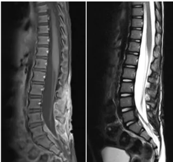
spontaneous epidural hematoma (Fig.1) in the context of her coagulopathy, attributed to vitamin K deficiency.
Standard radiographies and pelvic echocardiography per-formed in the suspicion of osteitis or septic arthritis showed a non-specific peri-articular right sacro-ileal collection.
After her hospital admission of 24 h, she was transferred to our department. A new pelvic and lumbosacral MRI with gadolinium administration revealed a right sacro-iliac osteo-myelitis and a collection extending from the gluteal muscles to the presacral fat and through the right L5 and S1 foramina into the anterior epidural space till the superior plate of L3, with a strong contrast enhancement. The collection showed
progression when compared to the previous MRI and it was
finally interpreted as an epidural abscess (Fig.2).
Because of a rapid neurological deterioration, the patient was taken to the emergency OR, where a bilateral laminoplasty of L3–S1 was performed with abscess drainage. Intraoperative-ly, a purulent material was isolated, surrounded by chronic inflammatory tissue. The initial cultures stayed negative but the gram stain was positive for a coccus and the PCR allowed identifying a GAS, sensitive to the current antibiotic treatment. She was progressively mobilized after the intervention with a rapid recuperation of walking ability and bladder conti-nence. At the end of the intravenous antibiotic course (6 weeks), she had a completely normal clinical status and an oral antibiotic treatment was started for another 6 weeks.
The final lumbar MRI showed complete healing (Fig.3).
Discussion
SEA is more common in the adult population (fifth to seventh decades) and in patients with underlying conditions like
immu-nosuppression, diabetes, spinal surgery, or trauma [2,15]. These
predisposing conditions occur more frequently in adults, and risk factors identified in the literature analysis for pediatric patients are sickle cell disease, Hirschsprung’s disease, or chronic
intes-tinal pseudo-obstruction [2]. Furthermore, varicella infection
may represent an additional risk factor for SEA development. SEA is more likely to develop in a larger epidural space rich in fat: abscesses are more common in posterior areas (above all in children) and in thoracolumbar segment. One
third of pediatric patients had associated osteomyelitis [2]. In
Fig. 1 Sagittal T1-weighted image showing a heterogeneous isointense collection in the anterior epidural space from the sacral column to the superior plate of L3 (left). Sagittal T2-weighted image showing the same isointense collection (right)
Fig. 2 Sagittal (left) and axial (right) T1-weighted images (with gado-linium) showing a collection with strong contrast enhancement, extend-ing into the anterior epidural space till the superior plate of L3, with an important mass effect
Fig. 3 Sagittal T1-weighted (left) and T2-weighted (right) lumbar MRI at 3-month follow-up, after the laminoplasty and the antibiotic therapy, showing a complete radiological healing
50 % of cases, SEA is secondary to hematogenous dissemi-nation from a remote focus (skin or soft tissue, urinary, or
respiratory tract infection), and in 10–30 % of cases, it derives
by contiguous spread (osteomyelitis or muscular abscess). In
another 15 % of cases, invasive procedures are implicated [5].
No source of predisposing illness can be identified in up to 30–40 %, suggesting silent bacteremia seeding the epidural
space [20].
S. aureus is the predominant pathogen in SEA in children
[2] with an increasing prevalence of MRSA during the last
years [9], followed by gram-negative bacteria (especially
Escherichia coli and Pseudomonas), coagulase negative Staphylococci (such as staphylococcus epidermidis), viridans group Streptococci, Enterococci, anaerobes, and
Mycobacte-rium species [4].
Varicella-zoster virus (VZV) may further predispose SEA development. It is a ubiquitous, highly neurotropic,
exclusive-ly humanα-herpesvirus, and varicella infection is generally
considered a mild and self-limiting disease commonly seen in primary care setting. It normally has a benign clinical course, and complications have been reported in 3–5 % of patients,
with an estimated mortality rate at 1.4/100,000 [14]. Bacterial
complications are the most prevalent (45–73 % of reported
complications in varicella-zoster infection) [19] and they
nor-mally consist of skin and soft tissues involvement, with S. aureus and Streptococcus pyogenes being major
responsi-ble [1]. The pathogenesis of bacterial infection is thought to be
due to skin barrier disruption and possibly transient
virus-induced immunosuppression [1].
Our case is the third case described in the literature of pediatric SEA from GAS in the context of varicella infection. In our review of the literature, two cases of SEA from GAS
were reported in two pediatric patients [15] and one case of
SEA caused by S. aureus was reported in a 2-year-old girl
with a positive history of varicella (Table1) [7]. Two other
cases of SEA from GAS were described in two pediatric
patients with no varicella infection [13,16].
Our patient presented with recurrent high fever, she was very irritable, and she refused to walk. The first lumbar MRI without contrast administration showed an anterior epidural collection extending from the sacrum to the superior plate of L3. The signal was isointense in both T1- and T2-weighted images, and the collection was dismissed for a spontaneous
hematoma in the context of her coagulopathy (Fig. 1). A
second pelvic-lumbar MRI with gadolinium administration was performed because of the strong suspicion of an infec-tious focus and it showed a right sacro-iliac osteomyelitis and a lumbar abscess in the anterior epidural space extending by contiguity from the arthritis focus. Urgent surgical decompres-sion was necessary for a rapid neurological deterioration and GAS was isolated from intraoperative material.
The clinical presentation in SEA can be misleading.
Heusner et al. [10] first described the classical symptoms Ta
b le 1 SEA in p edia tr ic pat ient s with posi tive anamnes is for v ari ce lla in fec tion P t Autho rs G ender , age Clinical data V aricella history S EA location an d features P athogen T reat ment Neurologic outcome 1 G re vitt et al. [ 7 ] F , 2 y S tatus epilepticus F ever 38.4 °C No voluntary limb m ovements Absent deep tend on reflexes and posi tive B ab inski A cti ve var ice lla inf ec tion w it h skin ves icles secondarily in fected Pos terior T 5– T7 SE A w ith T6 col la p se S. aur eus -F luc loxa cil lin, F ucidi n , and ge nta m ic in iv and p o (6 w ) -T 6 v er te bre ctomy Residual spasticity in the low er limbs 2 Q ua ch et al . [ 15 ]M , 1 8 m F ev er 4 0 °C Refusal to w al k Sti ff n ec k Brisk reflexes, clonus , and posi tive B abins k i Res o lving v ar ic ell a (d ay 6 o f evolution) Posterior S EA from T3 to T1 1 G AS -Cefotaxime and aqueous pe nic illi n G iv (6 w ) -T 5t oT 1 1 la m in ec to m y No de fic its 3 Q ua ch et al . [ 15 ] M , 5 .5 y F ev er 40 °C Le ft pa ra vert ebr al p ai n Ref u sal to stand, sit, or lie on his b ack Res o lving v ar ic ell a (d ay 7 o f evolution) Cir cum fe re nt ia l S EA T8 –T10 G AS -Ceftriaxone iv (4 w) -T9 to T 1 1 laminotomy No de fic its 4 C os su et al. 2 014 (th is study) F , 30 m F ev er 40 °C Right leg p ain Refusal to w al k Res o lving v ar ic ell a (d ay 10 of evolu tion) An terior S E A from L 3 to S 1 G AS -Aqueous penicillin G iv (6 w ) and po (6 w) -L3 to S 1 laminoplas ty No de fic its F fe ma le , GA S group A S treptoco ccus , iv intravenous, M male, m months, po per o s, S. aur eus S taphylococcus aur eus , w we eks, y year s
associated to epidural abscess: fever and back pain, followed by neurological manifestations. Unfortunately, only a minority of cases presents with the classical triad
[17], and in children, the diagnosis is more challenging.
A high index of suspicion is necessary in pediatric patients with a positive history for varicella infection (either acute or remitting), clinical signs of secondary bacterial infection, high-grade fever, and even mild neu-rological symptoms. With these conditions, it is imper-ative to be attentive to any sign of neurological deteri-oration and it is advisable to investigate the presence of epidural abscess with an adequate imaging.
The differential diagnosis includes other infectious process like myelitis or meningitis and parainfectious
process. Rubin et al. [18] performed lumbar punctions
(LP) in a cohort of patients with SEA and they found clear infectious signs only in 52 % of cases. No specific results were shown in 38 % of cases and CSF was
normal in 10 % of cases. Other authors [3, 15]
per-formed routinely LP for SEA patients. In our opinion, LP is an unnecessary procedure, which stays non infor-mative in half of cases and which may increase the risk of bacterial dissemination in the central nervous system. The therapeutic decisions are not modified by the re-sults of CSF analysis, and according to our point of view, it should not be routinely performed before a correct radiological assessment.
The main determinant of final outcome is the
preop-erative neurologic status [2, 18], and standard approach
lies in surgical decompression and drainage in
combina-tion with several weeks of intravenous (4–6 weeks) [15]
and oral antibiotics (4–6 weeks). Some studies [12, 21]
affirm that a conservative treatment may be the best option for selected candidates with no neurological symptoms or with complete deficits lasting more than 72 h, with extensive abscess or with high surgical risks
[8, 12, 13,17]. However, with conservative treatment, a
risk of deterioration even in patients without neurologic
symp-toms was reported [2,21]. Pediatric patients seem to have a
better prognosis even if they present with a more extended
disease [2].
In our opinion, SEA is a potentially fatal illness necessitat-ing prompt diagnosis, and the best therapeutic option in the pediatric setting is a combination of surgery and long-term antibiotic therapy. In particular, the surgical decompression is advisable even when the neurological deficit is present for more than 72 h.
Conclusion
SEA is a very rare condition in children, and varicella infec-tion may be a predisposing factor. Our report is the third case
described in the literature of pediatric SEA from GAS infec-tion in the context of varicella. Childhood SEA is a challeng-ing diagnosis, and accordchalleng-ing to our findchalleng-ings, spinal MRI should be performed in every patient having varicella infec-tion with bacterial complicainfec-tions or coexisting risk factors and neurological symptoms. The treatment should combine an aggressive surgical approach with a long-course antibiotic therapy.
Conflict of interest The authors declare that they have no conflict of interest.
References
1. Aebi C, Ahmed A, Ramilo O (1996) Bacterial complications of primary varicella in children. Clin Infect Dis 23:698–705
2. Auletta JJ, John CC (2001) Spinal epidural abscesses in children: a 15-year experience and review of the literature. Clin Infect Dis 32:9– 16
3. Danner RL, Hartman BJ (1987) Update on spinal epidural abscess: 35 cases and review of the literature. Rev Infect Dis 9:265–274 4. Darouiche RO (2006) Spinal epidural abscess. N Engl J Med 355:
2012–2020
5. Darouiche RO, Goetz L, Kaldis T, Cerra-Stewart C, AlSharif A, Priebe M (2006) Impact of StatLock securing device on symptomatic catheter-related urinary tract infection: a prospec-tive, randomized, multicenter clinical trial. Am J Infect Control 34: 555–560
6. Fleisher G, Henry W, McSorley M, Arbeter A, Plotkin S (1981) Life-threatening complications of varicella. Am J Dis Child 135:896–899 7. Grevitt MP, Mehdian SH (1998) Epidural abscess in an infant. Eur
Spine J 7:413–415
8. Hawkins M, Bolton M (2013) Pediatric spinal epidural abscess: a 9-year institutional review and review of the literature. Pediatrics 132: e1680–e1685
9. Herold BC, Immergluck LC, Maranan MC, Lauderdale DS, Gaskin RE, Boyle-Vavra S, Leitch CD, Daum RS (1998) Community-acquired methicillin-resistant Staphylococcus aureus in children with no identified predisposing risk. JAMA 279:593–598
10. Heusner AP (1948) Nontuberculous spinal epidural infections. N Engl J Med 239:845–854
11. Karikari IO, Powers CJ, Reynolds RM, Mehta AI, Isaacs RE (2009) Management of a spontaneous spinal epidural abscess: a single-center 10-year experience. Neurosurgery 65:919–923, discussion 923–914
12. Leys D, Lesoin F, Viaud C, Pasquier F, Rousseaux M, Jomin M, Petit H (1985) Decreased morbidity from acute bacterial spinal epidural abscesses using computed tomography and nonsurgical treatment in selected patients. Ann Neurol 17:350–355
13. Nussbaum ES, Rigamonti D, Standiford H, Numaguchi Y, Wolf AL, Robinson WL (1992) Spinal epidural abscess: a report of 40 cases and review. Surg Neurol 38:225–231
14. Preblud SR (1986) Varicella: complications and costs. Pediatrics 78: 728–735
15. Quach C, Tapiero B, Noya F (2002) Group A streptococcus spinal epidural abscess during varicella. Pediatrics 109:E14
16. Renoux MC, Guyon G, Rodiere M (2006) Paravertebral streptococ-cal myositis complicated by an epidural abscess in a 5-year-old girl. Arch Pediatr 13:273–275
17. Rigamonti D, Liem L, Sampath P, Knoller N, Namaguchi Y, Schreibman DL, Sloan MA, Wolf A, Zeidman S (1999) Spinal epidural abscess: contemporary trends in etiology, evaluation, and management. Surg Neurol 52:189–196, discussion 197
18. Rubin G, Michowiz SD, Ashkenasi A, Tadmor R, Rappaport ZH (1993) Spinal epidural abscess in the pediatric age group: case report and review of literature. Pediatr Infect Dis J 12:1007– 1011
19. Saddier P, Floret D, Guess HA, Durr F, Peyrieux JC, Weber DJ, Plotkin SA (1998) Cost of varicella in France: a study in day care centers. J Infect Dis 178(Suppl 1):S58–S63
20. Sendi P, Bregenzer T, Zimmerli W (2008) Spinal epidural abscess in clinical practice. QJM 101:1–12
21. Wheeler D, Keiser P, Rigamonti D, Keay S (1992) Medical manage-ment of spinal epidural abscesses: case report and review. Clin Infect Dis 15:22–27