HAL Id: hal-02512012
https://hal.archives-ouvertes.fr/hal-02512012
Submitted on 19 Mar 2020
HAL is a multi-disciplinary open access
archive for the deposit and dissemination of
sci-entific research documents, whether they are
pub-lished or not. The documents may come from
teaching and research institutions in France or
abroad, or from public or private research centers.
L’archive ouverte pluridisciplinaire HAL, est
destinée au dépôt et à la diffusion de documents
scientifiques de niveau recherche, publiés ou non,
émanant des établissements d’enseignement et de
recherche français ou étrangers, des laboratoires
publics ou privés.
Tetranychidae) from Syria with description of a new
species
Ziad Barbar, Philippe Auger
To cite this version:
Ziad Barbar, Philippe Auger. New records of the genus Bryobia (Acari: Tetranychidae) from Syria
with description of a new species. Acarologia, Acarologia, 2020, 60 (2), pp.268-288.
�10.24349/ac-arologia/20204367�. �hal-02512012�
Acarologia is proudly non-profit,
with no page charges and free open access
Please help us maintain this system by
encouraging your institutes to subscribe to the print version of the journal
and by sending us your high quality research on the Acari
.
Subscriptions: Year 2020 (Volume 60): 450 €
http://www1.montpellier.inra.fr/CBGP/acarologia/subscribe.php
Previous volumes (2010-2018): 250 € / year (4 issues)
Acarologia, CBGP, CS 30016, 34988 MONTFERRIER-sur-LEZ Cedex, France
ISSN 0044-586X (print), ISSN 2107-7207 (electronic)
A quarterly journal of acarology, since 1959
Publishing on all aspects of the Acari
All information:
http://www1.montpellier.inra.fr/CBGP/acarologia/
acarologia-contact@supagro.fr
Acarologia is under free license and distributed under the terms of the
The digitalization of Acarologia papers prior to 2000 was supported by Agropolis Fondation under
the reference ID 1500-024 through the « Investissements d’avenir » programme
Received 18 October 2019 Accepted 17 March 2020 Published 19 March 2020 Corresponding author Ziad Barbar: ziadbarbar88@gmail.com Academic editor Migeon, Alain DOI 10.24349/acarologia/20204367 ISSN 0044-586X (print) ISSN 2107-7207 (electronic) Copyright Barbar Z. and Auger P. Distributed under
Creative Commons CC-BY 4.0
(Acari: Tetranychidae) from Syria with
description of a new species
Ziad Barbar
a, Philippe Auger
baDepartment of Plant Protection, Faculty of Agriculture, Al-Baath University, P.O. Box 77, Al- Sham St.,
Homs, Syria.
bCBGP, INRAE, CIRAD, IRD, Montpellier SupAgro, Univ Montpellier, Montpellier, France.
Original research
ABSTRACT
Three species of Tetranychidae belonging to the genus Bryobia were collected from Latakia governorate, Syria in 2019: Bryobia (Allobia) syriensis n. sp. collected from
Salvia verbenaca L., is described; Bryobia (Allobia) nikitensis and Bryobia (Bryobia) gigas collected from S. verbenaca and from soil litter, respectively, are reported. New
observations of Bryobia specimens previously reported from the same governorate during 2014-2016 revealed that specimens of Bryobia (Bryobia) watersi were misidentified as
Bryobia (Bryobia) graminum and Bryobia (Bryobia) kissophila. Among them, we found
two aberrant females bearing three propodosomal lobes. By analogy, we discussed the cases of the trilobed species, Bryobia bakeri and Bryobia aegyptiacus, and concluded that they could be teratological forms of quadrilobed Bryobia species rather than species with a particular pattern of propodosomal lobes.
Keywords Tetranychid mite; bryobiine mite; new species; new records
Zoobank http://zoobank.org/26A372EC-4BD0-40AB-AA7C-F98BF94299FA
Introduction
Among the spider mite family, the genus Bryobia is the fourth largest genus in terms of species (Migeon and Dorkeld, 2019) and since the revision of the genus Pseudobryobia by Arabuli et
al. (2019), it contains 143 taxa.
Several species belonging to this genus are worldwide distributed, polyphagous, and could be distinctly economically injurious to crops (Vacante, 2010). To date, seven species of the genus Bryobia are known from Syria: Bryobia (Allobia) nikitensis (Livshits and Mitrofanov),
Bryobia (Bryobia) gigas Auger et al., B. (B.) graminum (Schrank), B. (B.) kissophila Eyndhoven, B. (B.) praetiosa Koch, B. (B.) vasiljevi Reck and B. (Lyobia) rubrioculus (Scheuten) (El-Hariri,
1968; Barbar, 2014, 2018; Zeity, 2017; Zeity and Srinivasa, 2019; Zriki et al., 2015). However, some specimens of this genus collected during 2014–2016 still remained unidentified (of which two specimens bearing only three propodosomal lobes each with one seta) and those of B. (B.)
graminum and B. (B.) kissophila seem to be misidentified as the identification was based only
on morphological characteristics of adult females (Barbar, 2014, 2018).
The aims of the current paper are to present the results of some field samplings of Bryobia species undertaken in Latakia governorate in 2019 and to re-examine all specimens of this genus collected in the same governorate over the period 2014–2016 and reported by Barbar (2014, 2018).
The description of a new species of Bryobia is provided. A new record, new host plants and the cases of misidentifications are reported. Finally, the observation of unusual Bryobia specimens’ prodorsal lobes lead us to discuss the identity of Bryobia bakeri (Zaher, Gomaa and El-Enany, 1982) and Bryobia aegyptiacus (Zaher, Gomaa and El-Enany, 1982).
Materials and methods
Field sampling
Samplings of tetranychid fauna were conducted in Latakia governorate in April and June, 2019. Mites were collected from soil litter and from leaves of common weed species within a small forest (about six hectares, 57 m above the sea level) of Pinus halepensis Mill. located at Attabiyyat (35°30’24” N, 35°46’49” E) in the south-western Latakia city, Syria. Mites were removed from leaves using the “dipping-checking-washing-filtering” method (Boller, 1984). For collecting mites from litter, materials were placed on a sieve (25 cm Ø x 10 cm; its screen with 5 mesh/cm) and shaken over a black plastic sheet (1.5 m2). Collected mites were cleared
in lactic acid for 48 hours, mounted on slides in Hoyer’s medium, and then dried in an oven at 40 °C for three days.
Mite observation and description
The specimens were examined using an Olympus® CH20 microscope. Measurements were realized using the scale of a reticle installed on the eyepiece lens. Mite body parts of specimens were pictured using a mobile phone camera (13 megapixels) fixed on the eyepiece lens and images were transferred to the professional quality vector graphics software Inkscape ® 0.92 installed on a computer for drawing with the aid of the computer’s mouse.
All measurements are given in micrometers (µm) and the setal nomenclature used in the de-scription follows Lindquist (1985). The holotype measurements are followed by measurements of the range of paratypes in parentheses. In addition to the key body measurements such as the distance between setae v2and setae h1and members of setae sc2(Saito et al., 1999), body length representing the distance between the tip of palps to the end of idiosoma and body width representing the widest transverse part of the hysterosoma, are also provided. The distance between two setae was measured as the distance from the center of one setal base to the other. Legs were measured from coxae to the distal margin of tarsi (excluding claws and empodia). Leg setal counts are given in the order: coxa-trochanter-femur-genu-tibia-tarsus. Numbers of setae refer to tactile setae, solenidia are given in parentheses and alternative counts are given in brackets. When asymmetry in number of setae on a leg segment was present, only the maximal number was considered.
Systematics
Family Tetranychidae Donnadieu, 1875
Subfamily Bryobiinae Berlese, 1913
Tribe Bryobiini Reck, 1952
Genus Bryobia Koch, 1836
Bryobia (Allobia) nikitensis Livshits and Mitrofanov, 1969
Two female specimens of this species were recorded on a new host plant: it was found on
Salvia verbenaca L., Attabiyyat, south-west of Latakia city, Syria (35°30’24” N, 35°46’49” E,
23 April 2019). This species has been previously collected from Sarcopoterium spinosum (L.) in Syria (Barbar, 2018; Zeity and Srinivasa, 2019).
Bryobia (Allobia) syriensis n. sp.
Zoobank: A423D4AA-D815-4BEF-94B8-47FD082F3ED3
Figures 1–10
Type material — Holotype (female), 23 female and 2 male paratypes on 10 microscopic
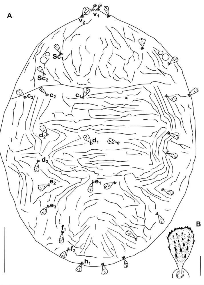
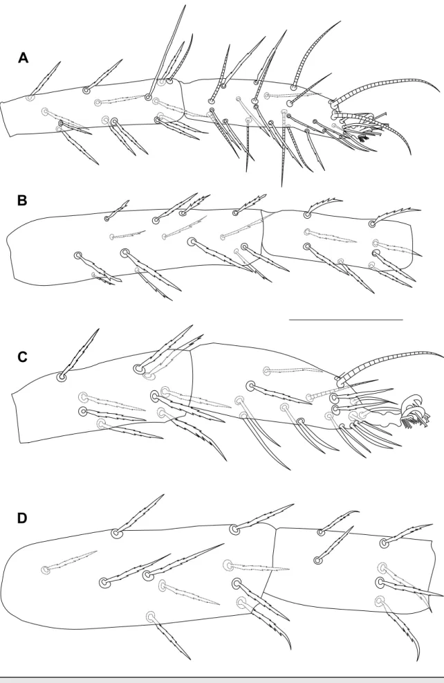
v
1v
2Sc
1Sc
2c
1c
2c
3d
1d
2d
3e
1e
2e
3f
1f
2h
1A
B
A
D
B
C
Figure 2 Bryobia syriensis n. sp., female: A, B, C – variation in propodosomal lobes; D – detail of the prodorsal striation of the area
N, 35°46’49” E), 23 April and 2 June, 2019, coll. Z. Barbar. The holotype and paratypes are deposited in the Arthropod Collection of the Department of Plant Protection, Faculty of Agriculture, Albaath University, Homs, Syria.
Diagnosis — This species belongs to Bryobia (Allobia) Livshitz and Mitrofanov (1971)
which is characterized by the following: propodosomal lobes absent or poorly developed, if present, outer and inner lobes not separated by deep incision; distance between f1members longer than distance between f2 members; f1 and f2 well separated laterally. It can be
distinguished from others Byrobia species by a combination of the following characteristics: females with weakly developed median propodosomal lobes with small and shallow incision, outer lobes reduced to very small tubercles; setae v2 about twice as long as setae v1; dorsal body setae short, spatulate, serrate and inserted in small tubercles; peritreme ends in a tiny anastomosis consisting of 3–4 loges; tarsus III with tactile seta and solenidion subequal in length forming duplex; tarsus IV with solenidion well-separated from tactile, proximal, about 3/4 the length of tactile; leg I shorter than body length, empodial claws I-IV uncinate, each with one pair of tenent hairs; empodium I with one pair of tenent hairs and empodia II-IV with two rows of tenent hairs.
Female description — (n = 24). Relatively small, 548 (488–548) long excluding
gnatho-soma, 663 (575–663) including gnathognatho-soma, and 384 (327–384) wide.
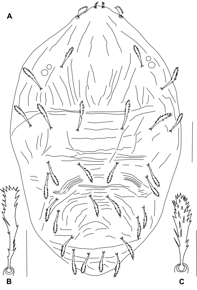
Dorsum – Prodorsum with four pairs of setae (Figure 1A); median propodosomal lobes weakly developed with fused base (variations in propodosomal lobes due to mounting are presented in Figure 2A-C); outer propodosomal lobes reduced to small tubercles; setae v2about
twice as long as setae v1(Figure 2). Distance between members of first (v1) and second (v2) pair of propodosomal setae insertions 10 (8–12) and 47 (45–50), respectively. Dorsal body setae short, spatulate and serrate, 12–17 width, subequal in length except for v1far smaller, inserted on small tubercles (Figure 1B). Measurements of dorsal setae: v111 (10–11); v223 (22–26);
sc121 (21–25); sc223 (21–25); c1 25 (23–29); c223 (21–26); c323 (21–25); d123 (21–25);
d223 (21–25); d323 (21–23); e123 (21–23); e223 (21–23); e323 (21–23); f123 (21–25); f2 21 (21–23); h118 (18–23). Distances between setae: sc2–sc2237 (202–237), c1–c183 (64–83),
d1–d1 60 (55–64), e1–e151 (41–51), f1–f1 148 (126–148), f2–f2 115 (110–115), h1–h1 46 (40–50), c1–d1101 (81–101), d1–e181 (69–81), v2–h1535 (470–535). Setae f1and f2lateral, distance between f1members larger than between f2members. Dorsal body surface granulated with longitudinal and irregular folds on propodosoma (Figure 2D) and with transverse spaced granulate striae on medial hysterosoma irregularly arched posteriorly (Figure 1).
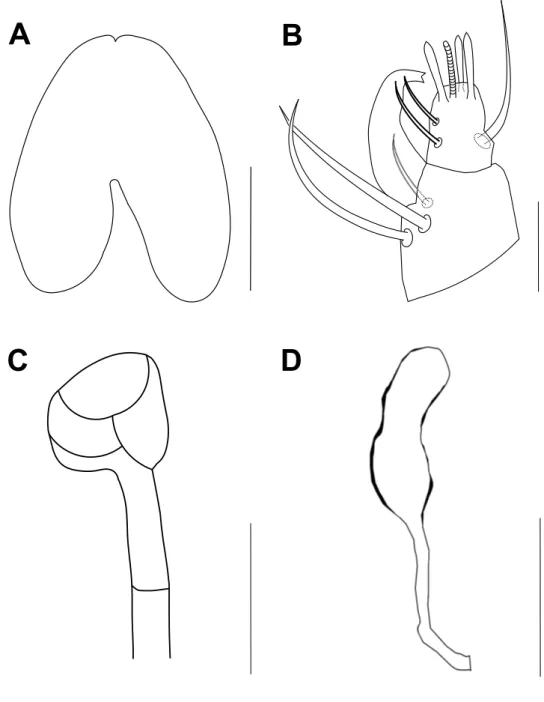
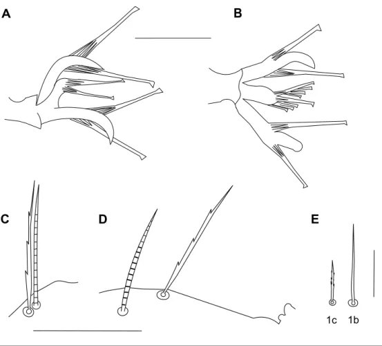
Gnathosoma – Stylophore slightly notched anteriorly, longer than wide (Figure 3A). Tibial claw of palpus bidentate. Palptarsus smaller than tibial claw, with three tactile setae, three eupathidia and one solenidion; eupathidia and solenidion subequal in length (Figure 3B). Peritreme ends in a tiny bulbous anastomosis (about 7 in diameter) consisting of 3–4 loges (Figure 3C).
Venter – Striation transverse, sparse and broken between 1a and 3a pairs of setae, irregularly longitudinal, sparse and broken between 3a and 4a pairs of setae, transverse and broken between
4a and aggenital (ag) pairs of setae, irregular in the area anterior to genital flap and longitudinal
between (ag) setae. Genitoanal region with two pairs of genitals (g1-2), three pairs of pseudanal
(ps1-3) and two pairs of ventrocaudal (h2-3) setae. Sacculus of spermatheca elongate, with smooth surface, seems constricted in its distal third, 15 (15–16) long, 7 (6–7) wide (Figure 3D).
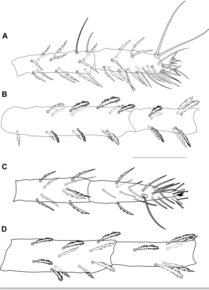
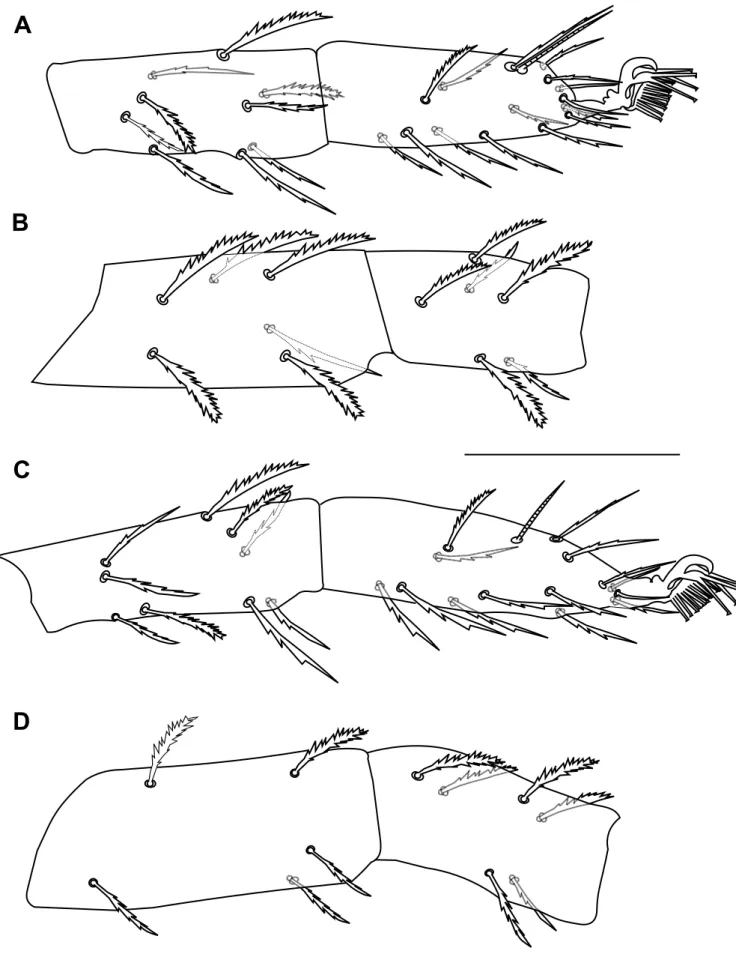
Legs – (Figures 4, 5, 6). Leg I shorter than body length, 389 (381–391) long, leg II 290 (253–290), leg III 290 (255–290), leg IV 306 (299–322). Length of segments of leg I as follows: trochanter 18 (18–24), femur 115 (108–115), genu 64 (57–69), tibia 74 (64–78), tarsus 69 (60–69). Leg setal counts as follows:
I: 2 – 1 – 14[15] – 8 – 13 + (1) – 18 + (3) + 2 duplexes II: 1 – 1 – 10 – 5 – 9 – 15 + (1) + 1 duplex
III: 1 − 1 − 6 – 6 – 9 –14 + 1 duplex IV: 1 – 1 – 5 – 6 – 9 − 14 + (1)
A
B
C
D
Figure 3 Bryobia syriensis n. sp., female: A – stylophore; B – palpal tibia and tarsus; C – peritreme;
D – spermatheca. Scale bars = 50 µm (A), 10 µm (B, C), 15 µm (D).
True claws uncinate, each with one pair of tenent hairs. Empodium I with one pair of tenent hairs, empodia II-IV each with two rows of tenent hairs (Figure 6A, B). Tarsus III associated tactile seta and solenidion forming duplex sub-equal in length (Figure 6C), on tarsus IV solenidion well-separated from tactile, proximal, about 3/4 the length of tactile (Figure 6D). Coxisternal setae 1b, 44, smooth about twice as long as setae 1c, 24, serrate (Figure 6E).
Male description — (n = 2). Small, 311–317 long excluding gnathosoma and 366–377
including gnathosoma, 207–210 wide.
Dorsum – Without prodorsal projections, setae v1and v2located on tiny tubercles (Figure 7A). Setae v1and v2short, subspatulate, serrate, v2twice as long as v1(Figure 8A). Distance between first (v1) and second (v2) pair of propodosomal setae insertions 14–18 and 54–56,
A
B
C
D
Figure 4 Bryobia syriensis n. sp., female: A – tibia and tarsus I; B – femur and genu I; C – tibia and tarsus II; D – femur and genu II. Scale
A
B
C
D
Figure 5 Bryobia syriensis n. sp., female: A – tibia and tarsus III; B – femur and genu III; C – tibia and tarsus IV; D – femur and genu IV.
A
B
C
D
E
1b 1c
Figure 6 Bryobia syriensis n. sp., female: A – claw and empodium I; B – claw and empodium II;
C – duplex setae on tarsus III; D – solenidion and associated tactile seta on tarsus IV; E – coxisternal setae 1b and 1c. Scale bars = 10 µm (A, B), 20 µm (C, D), 25 µm (E).
respectively. Other dorsal body setae subequal in length, elongate subspatulate, enlarged distally (setae c1less enlarged), serrate, all inserted on tiny tubercles (Figure 7B, C). Dorsocentral setae (c1, d1and e1) shorter than distances between consecutive setae. Measurements of dorsal setae:
v17; v214–15; sc128; sc228–32; c130–35; c230–35; c3 28; d128–30; d2 28–30; d328; e1 30–32; e230–32; e328–30; f125–30; f230; h123. Distances between setae: sc2–sc2150–170,
c1–c155–60, d1–d1 41–51, e1–e123–25, f1–f164–68, f2–f2 48–53, 64–68, h1–h120, c1–d1 44–48, d1–e139–46, v2–h1274–304. Dorsal body surface granulate with few longitudinal and
irregular folds on propodosoma, with transverse spaced striae on medial hysterosoma, and with arched striae on the surface between d1–d3and e1–e3(Figures 7A).
Gnathosoma – Stylophore slightly notched anteriorly, longer than wide, as in female. Tibial claw of palpus bidentate. Palptarsus as in female with solenidion shorter than eupathidia (Figure 8B). Peritreme ends in small bulb with four pointed denticles at its distal margin (Figure 8C).
Aeadeagus – Without knob, bent dorsad near at right angle, weakly sigmoid, with tapering distal part and acute tip pointing caudad, shaft dorsal margin curved (Figure 8D).
Legs – (Figures 9, 10). Leg I longer than body length 451 long, leg II 290–304, leg III 260–281, leg IV 288–304. Length of segments of leg I as follows: trochanter 18–23, femur 133–150, genu 69–76, tibia 81, tarsus 81. Leg setal counts as follows:
I: 2 – 1 – 14[15] – 8 – 13 + (1) – 18 + (8) + 2 duplexes II: 1 – 1 – 10[11] – 6[5] – 9 – 15 + (1) + 1 duplex III: 1 – 1 – 7 – 6 – 9 – 14 + 1 duplex
A
B
C
A
D
C
B
Figure 8 Bryobia syriensis n. sp., male: A – propodosomal lobes; B – palpal tibia and tarsus; C –
peritreme; D – aeadeagus. Scale bars = 20 µm (A), 10 µm (B, C, D).
IV: 1 − 1 − 5 – 6 – 9 – 14 + (1)
True claws uncinate. Claws I-IV each with one pair of tenent hairs. Empodia I-IV each with two rows of tenent hairs. Tarsus III associated tactile seta and solenidion forming duplex sub-equal in length; tarsus IV with solenidion well-separated from tactile, proximal, about 2/3 the length of tactile.
Etymology — This species was named after the country, Syria, where it was collected. Remarks — Among the species that belong to the subgenus Bryobia (Allobia) (as defined
by Livshitz and Mitrofanov, 1971) to which B. (A.) syriensis n. sp. also belongs, it is very close to B. (A.) livschitzi Mitrofanov and Strunkova, 1968 (Livshitz and Mitrofanov, 1971), B. (A.) ziziphorae Strunkova and Mitrofanov, 1983, B. (A.) tuttlei Smiley and Baker, 1995, B. (A.)
reckiana Mitrofanov and Strunkova, 1968, B. (A.) strunkovae Mitrofanov, 1968 (Livshitz and
Mitrofanov, 1971), B. (A.) montana Mitrofanov, 1973 and B. (A.) giannitensis Hatzinikolis and Panou, 1996. However B. (A.) syriensis differs from B. (A.) livschitzi by having dorsal body
A
B
C
D
Figure 9 Bryobia syriensis n. sp., male: A – tibia and tarsus I; B – femur and genu I; C – tibia and tarsus II; D – femur and genu II. Scale bar
A
B
C
D
Figure 10 Bryobia syriensis n. sp., male: A – tibia and tarsus III; B – femur and genu III; C – tibia and tarsus IV; D – femur and genu IV. Scale
setae spatulate (vs. subsaptulate with pointed or rounded tip in B. (A.) livschitzi); by differences in femoral setal count 14[15] – 10 – 6 – 5 and 12 – (7-8) – 4 – 4 in B. (A.) syriensis and B. (A.)
livschitzi, respectively; and by differences in tibial setal count on legs II-IV, 9 – 9 – 9 and 7 – 7
– 7 in B. (A.) syriensis and B. (A.) livschitzi, respectively.
Bryobia (A.) syriensis can be separated from B. (A.) ziziphorae by the following characters:
the outer prodorsal lobes are more developed in B. (A.) ziziphorae and the shape of setae v2is also different; the distal end of the peritreme consists of a tiny anastomosis of 3–4 loges in B. (A.) syriensis whereas it is simple in B. (A.) ziziphorae; solenidion and tactile seta on tarsus IV are well–separated in B. (A.) syriensis vs. a duplex setae is present in B. (A.) ziziphorae; and the femoral, tibial and tarsal setal counts obviously differ between the two species.
The main differences between females of B. (A.) syriensis and B. (A.) tuttlei are the following: the outer propodosomal lobes are very reduced in B. (A.) syriensis but well developed in B. (A.) tuttlei; setae v1are about half the length of v2in B. (A.) syriensis whereas they are quite similar in size in B. (A.) tuttlei; the spermatheca has a smooth surface in B. (A.)
syriensis vs. reticulate in B. (A.) tuttlei; obvious differences in leg setal counts are observed
between the two species (with the exception of genu II and femur IV). In the male of B. (A.)
syriensis dorsal body setae are elongated, subspatulate but in males of B. (A.) tuttlei they are
spatulate (similar to those of female).
Bryobia (A.) syriensis clearly differs from B. (A.) reckiana by having spatulate, serrate and
distally rounded dorsal body setae vs. lanceolate in B. (A.) reckiana; solenidion and tactile seta on tarsus IV are well-separated in B. (A.) syriensis vs. a duplex seta is present in B. (A.)
reckiana; the empodium I of B. (A.) syriensis bears only one pair of tenent hairs whereas two
rows of tenent hairs are present in B. (A.) reckiana; leg setal counts are very different between the two species.
Bryobia (A.) syriensis and B. (A.) strunkovae can be separated by: a smaller body width of B. (A.) syriensis, 327–384 vs. 572 in B. strunkovae; the leg I length that is far shorter (381–391)
than the body length in B. (A.) syriensis whereas it is about as long as the body length (640) in
B. (A.) strunkovae; the distal end of the peritreme is less developed in B. (A.) syriensis than in B.
(A.) strunkovae; solenidion and tactile seta on tarsus IV are well-separated in B. (A.) syriensis but a duplex seta is present in B. (A.) strunkovae; finally, obvious differences between the setal counts of the two species are present.
Bryobia (A.) syriensis differs from B. (A.) montana by having one pair of tenent hairs on
empodium I vs. two rows in B. (A.) montana; solenidion and tactile seta on tarsus IV are well–separated in B. (A.) syriensis vs. a duplex seta is present in B. (A.) montana; and by very different leg setal counts.
The main differences between B. (A.) syriensis and B. (A.) giannitensis are: a smaller body size of B. (A.) syriensis, 575–663 long and 327–384 wide vs. 789–809 and 650–559; a far shorter leg I in B. (A.) syriensis (381–391) compared to B. (A.) giannitensis (603–609); setae v1 and v2are spatulate in B. (A.) syriensis but lanceolate in B. (A.) giannitensis; in B. (A.) syriensis one pair of tenent hairs is present on claws II-IV but each of them bears two pairs of tenent hairs in B. (A.) giannitensis; obvious differences in leg setal counts between the two species are also observed.
Bryobia (Bryobia) gigas Auger, Arabuli and Migeon, 2015
Four females of this species were collected from soil litter, Attabiyyat, south-west of Latakia city, Syria (35°30’24” N, 35°46’49” E), 23 April 2019. This species has been already collected from Bituminaria bituminosa (L.) in Syria (Zeity and Srinivasa, 2019) but this host plant was no present in the place where we collected these specimens.
Bryobia (Bryobia) watersi Manson, 1967
The specimens of this species were identified as Bryobia sp. and misidentified as B. (B.)
Peritreme
A
B
C
D
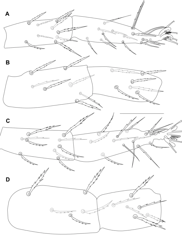
Figure 11 Bryobia watersi, female: A-D – variations in propodosomal lobes. Scale bars = 25 µm (A, B, C, D).
due to species identification only based on the morphological characteristics of females and particularly on variations in the shape of propodosomal lobes. In the present study, comparisons of all the specimens collected (including females, a male and juveniles, Figures 11-15) with the detailed redescriptions of B. (B.) graminum, B. (B.) kissophila and B. (B.) praetiosa by Mathys (1957) and the original description of B. (B.) watersi by Manson (1967) were undertaken. This led us to the conclusion that the morphological characteristics of the Syrian specimens fit well with those provided in the original description of B. (B.) watersi by Manson (1967) even if few morphological small differences were found: the dorsal body setae are slightly longer in Syrian females (there are no obvious differences in juveniles); genu II and tarsus IV of Syrian females with 6 and 14 setae respectively vs. 5 and 15 setae; genu II-III of Syrian male with 6 – 5 setae
vs. 4[5] – 6; and tarsus III of Syrian deutonymphs with 10 setae vs. 11[12].
Remarks — This species was collected from several host plants surrounding a citrus
orchard located at Al-ya’robiyah, Latakia governorate, Syria (35°30’24” N, 35°48’33” E) and was found on: Amaranthus retroflexus L., a new host plant (five females, 15 April 2014, previously identified as Bryobia sp.), Malva sylvestris L. (five females, two deutonymphs, three protonymphs and two larvae, 1 January 2014; four females, 15 February 2014; three females, 7 April 2015; two females, two protonymphs and one larva, 18 February 2016, previously misidentified as B. (B.) graminum; six females and one larva, 16 April 2016, previously misidentified as B. (B.) kissophila), Urtica urens L., a new host plant (three females and one protonymph, 15 April, 2014, previously identified as Bryobia sp.) and Trifolium sp., a new host plant (four females, three deutonymphs and one male, 14 April, 2015, previously identified as
Bryobia sp.).
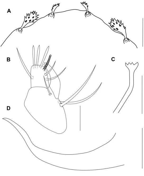
Among the specimens of B. (B.) watersi mentioned above, two females collected from
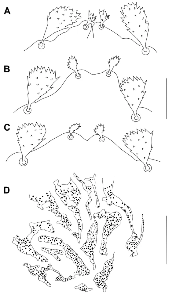
A. retroflexus have only three propodosomal lobes each with a single seta (Figures 15 A-B).
Initially, the specific identification of these specimens led to a result that they are either new specimens (or closely related species) of the two “trilobed” species B. bakeri Zaher et al. (1982)
A
B
C
D
Figure 12 Bryobia watersi, female: A-D – variations in spermatheca. Scale bars = 20 μm (A, B, C, D).
and B. aegyptiacus Zaher et al. (1982) or they are aberrant Bryobia specimens. Actually, the overall propodosomal lobe shapes of the trilobed Syrian specimens are closer to those of B.
bakeri (Figure 15A) than those of B. aegyptiacus. Nevertheless, the Syrian specimens differ
from those two species by the palptarsus setal count [unusual counts are found in the two species described by Zaher et al. (1982)] and by small differences in the global leg setal counts. These results led to conclude that the Syrian trilobed specimens did not belong to B. bakeri nor to B. aegyptiacus.
Figure 13 Bryobia watersi, male: propodosomal lobes. Scale bar = 25 µm.
in Figure 15B (typical of abnormal ”asymmetrical” lobes; the outer lobe of the left side is obviously missing) and those of the specimen presented in Figure 15A (the axis of symmetry passes through the middle of the single inner lobe), we concluded that these females are abnormal individuals of B. (B.) watersi for several reasons:
(1) Both specimens are morphologically identical to B. (B.) watersi [i.e. having similar leg ambulacra, leg chaetotaxy and articles dimensions, same shape and length of dorsal body setae and peritreme etc.].
(2) Both specimens were collected together with specimens of B. (B.) watersi (same host plant, same date and place of collection).
(3) Several attempts carried out in 2015 and 2016 to re-collect additional trilobed individu-als (in the same location where they were collected first) were unsuccessful and all re-collected individuals were B. (B.) watersi.
(4) Propodosomal lobe aberrations have already been reported in several Bryobia species (Arabuli and Auger, 2016; Fashing et al. 2016; Smiley and Baker, 1995).
This variability in the propodosomal lobe shape found in the two Syrian trilobed individuals of B. (B.) watersi guided us to question about the taxonomical value of the number of propodosomal lobes used to separate B. bakeri and B. aegyptiacus from other Bryobia species. Several arguments tend to show that these specimens could be teratological forms rather than species with a particular propodosomal lobes pattern:
(1) Specimens are rare: like the Syrian trilobed specimen of B. (B.) watersi of the Figure 15A, only one specimen of B. bakeri and one of B. aegyptiacus are known. Although Smiley and Baker (1995) reported a possible additional female of B. bakeri, it could belong to another species because its leg setal count is far different from that of the type specimen of B. bakeri (it shares the same setal count only on five leg articles; as a comparison, the Syrian trilobed B. (B.) watersi are closer to B. bakeri for the reason that they share the same setal count on 12 leg articles).
(2) Specimens with three propodosomal lobes (each bearing one seta) are known to occur in several species of Bryobia: Smiley and Baker (1995) mentioned that in a few species of
Figure 14 Bryobia watersi, immature stages: A – dorsal seta e1of larva; B-C – propodosomal lobes of protonymph; D – propodosomal lobes of deutonymph. Scale bars = 20 µm (A), 50 µm (B, C, D).
Bryobia some aberrant females (with two or three propodosomal lobes) appear sometimes.
Since that work, several cases of Bryobia species with three propodosomal lobes have been reported (Arabuli and Auger, 2016; Fashing et al. 2016). In the detailed study by Fashing et al. 2016, it was demonstrated that both morphotypes (with three or four propodosomal lobes) of Bryobia abyssiniae Fashing and Ueckermann, 2016 belong to the same species, and about 9.5% of observed specimens had a single propodosomal inner lobe (with a single seta v1).
(3) The two Syrians trilobed specimens are conspecific despite the fact that one of them is obviously an aberrant form (asymmetry) and the other has a symmetrical propodosomal lobe pattern similar to that found in B. bakeri.
This tends to show that a bryobiine mite with an unpaired inner propodosomal lobe bearing a unique seta v1, can be an aberrant specimen despite a symmetrical propodosomal trilobed lobe pattern.
In our opinion, all these elements together strongly suggest that B. bakeri and B. aegyptiacus would be more aberrant individuals of two species of Bryobia (four-lobed) than species characterized by unpaired inner propodosomal lobe. Even if the data are insufficient to assign these “trilobed” species to an existing four-lobed Bryobia species, the demonstration presented here is consistent with the synonymy of the genus Septobia with the genus Bryobia by Bolland
et al. (1998).
Acknowledgements
The first author would like to thank Professor Hassan Khalil for his valuable help in the identification of host plants. Thanks are due to Dr. Mahran Zeity and Dr. Mohamed W. Negm for supplying scientific papers to the first author.
References
Arabuli T., Auger P. 2016. Intraspecific morphological variability in Bryobia rubrioculus (Scheuten, 1857) (Acari: Tetranychidae) from Georgia (Caucasus). 8th Symposium of the European Association of Acarologists. Valencia, Spain, 11th -15th July 2016. Oral presentation.
Arabuli T., Maric I., Auger P. 2019. Revision of the genus Pseudobryobia McGregor, 1950 (Acari, Tetranychidae), Acarologia, 59(3): 291-300.doi:10.24349/acarologia/20194331
Barbar Z. 2014. Occurrence, population dynamics and winter phenology of spider mites and their phytoseiid predators in a citrus orchard in Syria. Acarologia, 54: 409-423. doi:10.1051/acarologia/ 20142143
Barbar Z. 2018. New mite records (Acari: Mesostigmata, Trombidiformes) from soil and vegetation of some Syrian citrus agrosystems, Acarologia, 58(4): 919-927.doi:10.24349/acarologia/20184298
Bolland H.R., Gutierrez J., Flechtmann C.H.W. 1998. World catalogue of the spider mite family (Acari: Tetranychidae). Leiden: Brill Academic Publishers. pp. 392.
Boller E.F. 1984. Eine anfache Ausschwemm-Methode zur schellen Erfassung von Raumilben, Trips und anderen Kleinathropoden im Weinbau, Schweiz Zeitschrift für Obst-und Weinbau, 120: 249-255. El-Hariri G. 1968. A list of recorded Syrian insect and Acari. Faculty of Agriculture, University of
Aleppo. pp.160.
Fashing, N.J., Ueckermann, E.A., Fashing, P.J., Nguyen, N., Back, A.M. Allison L.A. 2016. Bryobia
abyssiniae (Prostigmata: Tetranychidae), a new species from the highlands of Ethiopia. Int. J. Acarol.,
42: 366-376.doi:10.1080/01647954.2016.1194891
Hatzinikolis E.N., Panou H.N. 1996. Three new species of Bryobia (Acari, Tetranychidae) from fruit trees in Greece. Acarologia, 37: 107-113.
Lindquist, E.E. 1985. External anatomy. In: Helle, W. & Sabelis, M.W. (Eds.), Spider mites. Their Biology, natural enemies and control. Amsterdam: Elsevier Science Publishing. p. 3-28.
Livshits I.Z., Mitrofanov V.I. 1971. The mites of the genus Bryobia C.L. Koch, 1836 (Acari-formes,Bryobiidae). Trudy Gosudarstvennogo Nikitskogo Botanicheskogo Sada, 51: 1-112. Manson D.C.M. 1967. The spider mite family Tetranychidae in New Zealand. I. The genus Bryobia.
Acarologia, 9: 76-123.
Mathys G. 1957. Contribution à la connaissance de la systématique et de la biologie du genre Bryobia en Suisse romande. Mitteilungen der Schweizerischen Entomologischen Gesellschaft, 30: 189-284. Migeon A., Dorkeld F. 2006-2019. Spider Mites Web: a comprehensive database for the Tetranychidae.
Mitrofanov V.I. 1973. Three new species of mites of the genus Bryobia C.L. Koch, 1836 (Acariformes, Tetranychoidea) from the Pamir. Trudy Gosudarstvennogo Nikitskogo Botanicheskogo Sada, 51: 12-14.
Saito Y., Mori K., Chittenden A.R. 1999. Body characters reflecting the body size of spider mites in flattened specimens (Acari, Tetranychidae). Applied Entomology and Zoology, 34: 383-386.
doi:10.1303/aez.34.383
Smiley R.L., Baker E.W. 1995. A report on some tetranychid mites (Acari: Prostigmata) from Yemen. Int. J. Acarol., 21: 135-164.doi:10.1080/01647959508684055
Strunkova Z.I., Mitrofanov V.I. 1983. New species of the family Bryobiidae (Acariformes) from Middle Asia. Zool. Zh., 62: 464-468.
Vacante V. 2010. Citrus mites, Identification, bionomy and control. CABI Head Office, Oxfordshire, UK. pp. 378.doi:10.1079/9781845934989.0000
Zaher M.A., Gomaa E.A., and El-Enany M.A. 1982. Spider mites of Egypt (Acari: Tetranychidae). Int. J. Acarol., 8: 91-114.doi:10.1080/01647958208683284
Zeity M. 2017. Some new records of spider mites (Acari, Tetranychidae) from Syria. Acarologia, 57(3): 651-654.doi:10.24349/acarologia/20174184
Zeity M., Srinivasa N. 2019. Updated contribution to the knowledge of Tetranychoidea (Acari: Tetrany-chidae, Tenuipalpidae) from Syria with reinstatement of genus Nuciforaella Vacante. Syst. Appl. Acarol., 24(4): 529-543.doi:10.11158/saa.24.4.1
Zriki G., Shaabo A., Boubou A. 2015. A preliminary survey of the spider mites (Acari: Tetranychidae) in Latakia Governorate of Syria. Acarologia, 55: 303-309.doi:10.1051/acarologia/20142173