3D matrix#embedding inhibits cycloheximide#
mediated sensitization to TNF#alpha#
induced apoptosis of human endothelial cells
The MIT Faculty has made this article openly available.
Please share
how this access benefits you. Your story matters.
Citation
Saemisch, Michael, et al., "3D matrix#embedding inhibits
cycloheximide#mediated sensitization to TNF#alpha#induced
apoptosis of human endothelial cells." Journal of tissue engineering
and regenerative medicine 12, 4 (November 2017) doi 10.1002/
TERM.2609 ©2017 Author(s)
As Published
10.1002/TERM.2609
Publisher
Wiley
Version
Author's final manuscript
Citable link
https://hdl.handle.net/1721.1/124512
Terms of Use
Creative Commons Attribution-Noncommercial-Share Alike
3D matrix embedding inhibits cycloheximide-mediated
sensitization to TNF-alpha-induced apoptosis of human
endothelial cells
Michael Saemisch1,2, Markus Nickmann3, Lisa Riesinger2, Elazer R. Edelman4,5, and Heiko
Methe2,3,5
1Department of Internal Medicine II, University Hospital Regensburg, Germany 2Department of Cardiology, Ludwig-Maximilians-University Munich, Germany 3Kliniken an der Paar, Aichach, Germany
4Division of Cardiovascular Medicine, Department of Medicine, Brigham and Women’s Hospital,
Harvard Medical School, Boston, MA, USA
5Institute for Medical Engineering and Science, Massachusetts Institute of Technology,
Cambridge, MA, USA
Abstract
The programmed form of cell death (apoptosis) is essential for normal development of
multicellular organisms. Dysregulation of apoptosis has been linked with embryonal death and is involved in the pathophysiology of various diseases. Others and we previously demonstrated endothelial biology being intertwined with biochemical and structural composition of the subendothelial basement membrane.
We now demonstrate that a three-dimensional growing environment significantly shields endothelial cells from cytokine-induced apoptosis. Detailed analysis reveals differences in
intracellular signaling pathways in naive endothelial cells and cytokine-stimulated endothelial cells when cells are grown within a three-dimensional collagen-based matrix compared to cells grown on two-dimensional tissue culture plates. Main findings are significantly reduced p53 expression and level of p38-phosphorylation in three-dimensional grown endothelial cells. Despite similar concentrations of focal adhesion kinase three-dimensional matrix embedded endothelial cells express significantly less tyrosine-phosphorylated focal adhesion kinase. Pretreatment with antibodies against integrin αvβ3 partially reversed the protective effect of three-dimensional
matrix-embedding on endothelial apoptosis.
Our findings provide detailed insights into the mechanisms of endothelial apoptosis with respect to the spatial matrix environment. These results enhance our understanding of endothelial biology and may otherwise help in the design of tissue-engineered materials. Furthermore, findings on focal adhesion kinase phosphorylation might enhance our understanding of clinical studies with tyrosine kinase inhibitors.
HHS Public Access
Author manuscript
J Tissue Eng Regen Med
. Author manuscript; available in PMC 2018 May 24.Published in final edited form as:
J Tissue Eng Regen Med. 2018 April ; 12(4): 1085–1096. doi:10.1002/term.2609.
A
uthor Man
uscr
ipt
A
uthor Man
uscr
ipt
A
uthor Man
uscr
ipt
A
uthor Man
uscr
ipt
1. Introduction
Apoptosis is the programmed suicide of a cell. It serves as an important tool during
development and in maintaining homeostasis in adult tissue remodeling (Raff, 1992; Weil et al., 1996; E. White, 1996). Furthermore, diseases like cancer and foregoing metastasis would not be possible without apoptotic processes of otherwise healthy tissue. In addition,
apoptosis plays a significant role in atherosclerotic disease progression. Within this process vascular endothelial cells (ECs) are important regulators within overall vessel structure. Most ECs in adult blood vessels are relatively quiescent and resistant to apoptosis. However, they are thought to retain the latent capacity for proliferation and apoptosis to mediate angiogenesis and regression, respectively. EC apoptosis can be detected in atherosclerotic plaques and may provide an important step in transition from stableMehta, Kang, Bansal, & Bansal, 2002; Norata, Tonti, Roma, & Catapano, 2002). The tissue environment of an atherosclerotic plaque may promote EC apoptosis. Several factors promoting apoptosis of ECs in the context of atherosclerosis have been identified over the last couple of years including biochemical (e.g., oxidized low density lipoprotein) as well as biomechanical factors (e.g., turbulent flow) (Dimmeler, Hermann, & Zeiher, 1998; Sata & Walsh, 1998). Endothelial biology is to a great extent regulated by the subendothelial basement membrane. This is mediated by integrins via outside-in and inside-out signaling in form of a crosstalk between components of the subendothelial basement membrane, attaching cells, as well as direct EC-EC contact (Niland & Eble, 2012). ECs are linked to the underlying basement membrane by focal adhesion complexes. Formation of focal adhesion complexes is initiated by linking extracellular matrix protein ligands in the basement membrane with specific integrin receptors on ECs (Hynes, 1992). Growing evidence indicates that within this adhesion complex the focal adhesion kinase (FAK) is important in maintenance of normal cell survival. Disruption of FAK signaling results in loss of substrate adhesion and apoptosis of anchorage-dependent cells (Lu & Rounds, 2012). In two-dimensional (2D) growing conditions FAK autophosphorylates at tyrosine in ECs, thereby exerting anti-apoptotic influences (Bellas et al., 2002; Ilic et al., 1998; Renshaw, Price, & Schwartz, 1999). Others and we demonstrated that three-dimensional (3D) matrix-embedding influences the pheno- and genotype of a variety of human cells mimicking the in vivo cell type to a greater extent than simply 2D tissue culture plating (Baharvand, Hashemi, Kazemi Ashtiani, & Farrokhi, 2006; Benya & Shaffer, 1982; Luca et al., 2013; Mabry, Payne, & Anseth, 2016; Methe et al., 2005; Nelson & Bissell, 2005). 3D cell culture systems have gained increasing interest in drug discovery and tissue engineering due to their evident advantages in providing more physiologically relevant information and more predictive data for in vivo tests
(Edmondson, Broglie, Adcock, & Yang, 2014). Kim et al. demonstrated that in 3D collagen matrices human platelet lysate promotes cell survival and enhance vasculogenesis of endothelial colony forming cells via upregulation of pro-survival molecules (Kim et al., 2015). Others demonstrated that integration of ECs in 3D spheroids prevented apoptosis (Korff & Augustin, 1998). Other authors also demonstrated the importance of the spatial environment on regulation of the apoptotic signaling pathway (Dangles et al., 1997; Du et al., 2016; Gilmore, 2005). Interestingly, Baldo et al. demonstrated that induction of EC
A
uthor Man
uscr
ipt
A
uthor Man
uscr
ipt
A
uthor Man
uscr
ipt
A
uthor Man
uscr
ipt
apoptosis by the snake venom jararhagin is higher in ECs cultured in 3D collagen enriched matrices as compared to ECs cultured on gelatin-coated plastic dishes (Baldo et al., 2015). Previous own studies have shown that xenogenic and allogenic matrix-embedded ECs can be perivascularly delivered to tailor the response to injury and the remodeling of arteriovenous anastomoses. The matrix used was a 3D matrix of denaturated collagen (Gelfoam; Pfizer, New York, USA) (Conte et al., 2009; Nugent & Edelman, 2001; Nugent et al., 2009; Nugent, Rogers, & Edelman, 1999; Nugent et al., 2007; Zani, Kojima, Vacanti, & Edelman, 2008). A fascinating aspect of these Gelfoam embedded ECs is that these cells produce enhanced levels of soluble factors that regulate both the local arterial homeostasis and immunobiology (Methe, Hess, & Edelman, 2007) and even xenogeneic ECs do not induce a significant host immune response (Methe et al., 2005). Interestingly, the effect of these non-vascularized matrix-embedded EC implants lasted for several months (Nugent & Edelman, 2001) which let us to speculate that ECs within this matrix must be protected from apoptosis to a certain degree.
As we have previously shown that the Gelfoam scaffolds used in our study have topological features on the order of the EC size and consequently facilitate a 3D cellular morphology unseen before that includes bending and deformation during attachment, thus eliciting strong alterations in cytoskeletal organization (Indolfi, Baker, & Edelman, 2012) we now aimed to explore if 3D matrix-embedding within this Gelfoam scaffold influences human aortic EC apoptosis in vitro when compared to ECs plated on conventional 2D polystyrene culture plates. We correlated these results with pivotal components of the intracellular apoptotic signaling pathways.
2. Material and Methods
2.1 Cell culture on 3D-matrices and on 2D-tissue culture plates
Human aortic ECs were obtained from Promocell (Heidelberg, Germany) and grown in optimized endothelial growth medium-2 (Promocell) supplemented with 5% fetal bovine serum either on polystyrene-coated tissue culture plates or embedded within 3D matrices of denaturated collagen (Gelfoam; Pfizer, New York, USA) as previously published (Methe et al., 2005; Nugent & Edelman, 2001): compressed sponges were cut into 1 × 1 × 0.3 cm blocks and hydrated in culture medium at 37 °C for ≥4 h. Then 4.5 × 10⁴ ECs (suspended in ~50 μL media) were seeded onto one surface of the hydrated matrix, allowed 1.5 h to attach before turning the matrix over and seeding an additional 4.5 × 10⁴ in growth media. After a further 1.5 h of incubation each piece was added to a separate 30 mL polypropylene tube containing 10 mL of culture medium. Matrices were cultured for up to 3 weeks, with media changed every 48–72 h, under standard culture conditions (37 °C humidified environment with 5% CO2). Homogeneous distribution of ECs within the Gelfoam was demonstrated by
scanning electron microscopy (data not shown) and biosecretory function through assays for production of a standard panel of factors. Cell count was determined using trypan blue and a Neubauer counting chamber after dissolution of Gelfoam blocks through addition of collagenase type I as previously described (Methe et al., 2005). All of the assays were conducted on confluent ECs.
A
uthor Man
uscr
ipt
A
uthor Man
uscr
ipt
A
uthor Man
uscr
ipt
A
uthor Man
uscr
ipt
2.2 Induction of endothelial apoptosis
Apoptosis of confluent ECs was induced by adding a combination of 10 ng/ml tumor necrosis factor(TNF)-α (R&D Systems, Wiesbaden, Germany) and 1 μg/ml cycloheximide (CHX; Sigma-Aldrich, Steinheim, Germany) to the cell culture medium.
2.3 Quantification of enzymatic activity via ELISA (for caspase 3 and lactate dehydrogenase)
The commercial ELISA-Kit ApoTarget™ (Invitrogen, Carlsbad, US) was used to determine the proteolytic activity of caspase 3 according to the manufacturer’s instructions. Each sample was analyzed in comparison to an unstimulated control. Before analysis all samples were normalized to equal protein concentrations ranging from 50 - 200 μg per 50 ml of reaction solution. For analysis of caspase activity 5 μl of DEVD-pNA were added to each sample as substrate. After 2 hours of incubation in the dark at 37°C enzymatic activity was determined at a wavelength of 405 nm in a microplate reader.
The enzymatic activity of lactate dehydrogenase (LDH) was also determined by enzyme-linked immunosorbent assay (Invitrogen, Carlsbad, US) in a similar fashion.
2.4 Annexin V staining and fluorescence activated cell sorting (FACS)
The apoptotic fraction of ECs [%] was determined by Annexin V staining with the Vybrant® Apoptosis Assay Kit #2 (Molecular Probes, Carlsbad, US). During apoptosis phosphatidylserine is translocated to the outer cell membrane. Annexin V is a calcium dependent phospholipid binding protein with affinity for phosphatidylserine. Therefore, Annexin V, which had previously been marked with Alexa Fluor 488 is used to identify apoptotic ECs by binding the translocated phosphatidylserine. Before analysis by FACS trypsin was added to detach the cells from the TCPS. Afterwards ECs were washed in MACS buffer, centrifuged and resuspended in Annexin-binding buffer. Cell count was normalized to 106 ECs/ml Annexin-binding buffer. For each assay 100 μl of sample solution were used after 5 μl of Fluor 488 Annexin V and 1 μl of PI working solution had been added. All samples were then incubated for 15 minutes at room temperature. After
incubation 400 μl of Annexin-Binding buffer were added and the samples were analyzed by FACS analysis with CellQuest software (Becton Dickinson, New Jersey, US).
2.5 Immunoprecipitation
For isolation of particular proteins from a cell lysate immunoprecipitation using magnetic Dynabeads was performed. The immunoprecipitation kit DYNAL (Invitrogen, Carlsbad, US) was used to quantify the association of receptor-interacting protein (RIP) and focal adhesion kinase (FAK). Prior to immunoprecipitation 50 μl of resuspended dynabeads were exposed to a separation magnet. The G-protein-coupled Dynabeads were then incubated slowly rotating at room temperature for 10 minutes with the first antibody against FAK (Santa Cruz, Dallas, US), which had been dissolved in antibody binding and washing buffer. Afterwards the solution was twice exposed to the separation magnet for 1 minute, the supernatant was discarded and the Dynabeads-antigen-antibody-complex was resuspended in 200 μl antibody binding and washing buffer. In the next step the sample in form of a cell lysate was given to the purified antigen-antibody-complexes and incubated under slow rotation for 10 minutes at
A
uthor Man
uscr
ipt
A
uthor Man
uscr
ipt
A
uthor Man
uscr
ipt
A
uthor Man
uscr
ipt
room temperature. After four further steps of purification using the separation magnet and resuspension in washing buffer 20 μl of elution buffer were added to each sample. Then the samples were heated to 70°C for 10 minutes and exposed to the separation magnet to remove the dynabeads. Afterwards the samples were used for western blot using an anti-RIP IgG antibody (GeneTex, Irvine, US).
2.6 Determination of protein concentration using a bradford protein assay
To determine the protein concentration of the samples an assay based on the Bradford method was performed for each sample (Biorad, Hercules, US). A dilution series of a protein aliquot of bovine serum albumin (BSA) was prepared as reference standard. After addition of Coomassie brilliant blue every sample of the dilution series was measured via photometry at a wavelength of 595 nm to create a BSA standard curve. Afterwards at minimum three different dilutions of the cell sample were made (regularly 1:1000 and 1:2000 and depending on the expected protein concentration 1:500 or 1:4000) since the protein concentration of the sample to be analyzed had to lie in the range of the BSA standard curve. Before the diluted samples were analyzed via photometry the same amount of dye (200 μl) was added. After its determination the protein concentration was equilibrated for all samples by addition of aqua bidest.
2.7 Western blot
Cell monolayers or cells digested from Gelfoam matrixes by collagenase treatment were washed in PBS buffer. Dissolved cells were washed with PBS and centrifuged multiple times to create cell pellets. By adding RIPA buffer (Pierce, Waltham, US) and a protease- and a phosphatase-inhibitor (Halt Protease and Phosphatase Inhibitor Cocktail, Thermo Scientific, Waltham, US) cell pellets were lysed and the phosphorylation status preserved. Cell pellets were incubated with RIPA buffer for 45 minutes on ice and afterwards centrifuged at 15.000 rpm to free the sample solution from cell debris. The protein concentration of the western blot samples was equilibrated by adding aqua bidest as described above. In preparation for analysis to each sample 4x NuPage sample buffer, NuPage antioxidant and NuPage reducing agent (Invitrogen, Carlsbad, US) were added and then incubated at 70° C for 10 minutes in a dry bath heating block. For final purification, the samples were centrifuged at 1000 rpm for 3 minutes before applying them into the gel lanes. Through gel electrophoresis using gradient gels the samples were analyzed (running at 200 V and 2 A for 1 hour). The samples from the different compared cell culture conditions (i. e. different tissue culture plate coatings) were always given into the same gradient gel to allow comparison and analysis under the exact same conditions. Afterwards proteins were transferred onto a carrier membrane via electrophoretic transfer and then incubated with a specific antibody. Previous to incubation with the antibody the membrane was blocked with blocking buffer (Invitrogen, Carlsbad, US) at room temperature for 1 hour to prevent unspecific binding. Incubation time of the first antibody (rabbit anti-Bax and rabbit anti-Bcl2 BioLegend, San Diego, USA; rabbit anti-FAK, rabbit anti-FAK (Tyr-397), and rabbit anti-FAK (Tyr-925) Santa Cruz Biotechnology, Heidelberg, Germany; rabbit anti-Akt and rabbit anti-RIP Biozol, Munich, Germany; rabbit AKT (Ser-473), rabbit ERK 1/2 (pan), and rabbit anti-ERK 1/2 (phospho) MBL Intl., Woburn, MA, USA; rabbit anti-p38 and rabbit anti-p38
A
uthor Man
uscr
ipt
A
uthor Man
uscr
ipt
A
uthor Man
uscr
ipt
A
uthor Man
uscr
ipt
(T180, Y182) Abcam, Cambridge, UK; mouse anti-p53 Merck, Darmstadt, Germany) varied between 1 and 24 hours depending on our experience from previous studies and the
manufacturers’ instructions. For this incubation the carrier membrane was kept freely swimming in a sufficient amount of washing buffer (Invitrogen, Carlsbad, US) with a distinct concentration of the chosen antibody. Upon incubation the carrier membrane was washed three times with 15 ml of washing buffer for 5 minutes in a shaking water bath. In the next step the membrane was incubated with the secondary antibody at room temperature for 1 hour. The secondary antibody binds the first antibody and offers a G-protein-coupled binding site for Q-dots (Qdot® 625 Streptavidin Conjugate, Invitrogen, Carlsbad, US). After washing the membrane again for 3 times with washing buffer it was incubated in a solution of Qdot-625-streptavidin-conjugate and 8 ml of blocking buffer for 1 hour. After a final washing step in aqua bidest the protein bands were imaged in a darkroom using ultraviolet light and then photographed in high resolution with exposure times ranging from 0.0125 to 8.0 seconds to optimize imaging of the antigen of interest. The western blot images were then analyzed using the open source program ImageJ (NIH).
2.8 Microarray Analysis and RT-PCR
Integrin expression was compared between human aortic ECs grown to confluence on polystyrene tissue culture plates and EC embedded within Gelfoam matrices using the extracellular matrix and adhesion molecule microarray assay (SuperArray Biosciences, Frederick, USA). Final hybridization was performed with CDP-Star chemiluminescent substrate followed by exposure and analysis on a FluorChem SP (Alpha Innotech, San Leandro, Calif, USA).
Total RNA was extracted from ECs grown on TCPS or matrix-embedded using the RNeasy Mini Kit (Qiagen, Valencia, CA) according to the manufacturer’s instructions.
Complementary DNA was synthesized using the TaqMan reverse transcription reagents from Applied Biosystems (Foster City, CA). Real-time PCR analysis was performed with an Opticon Real Time PCR Machine (MJ Research) using SYBR Green PCR Master Mix Reagent Kit (Applied Biosystems). Cycling conditions were as follows: step 1, 10 min at 95°C; step 2, 15 s at 94°C; step 3, 1 min at 60°C; and step 4, 2 min at 72°C, with repeat from step 2 to step 4 for 45 cycles. Data from the reaction were collected and analyzed by the complementary Opticon computer software (MJ Research). Relative quantitations of gene expression were calculated with standard curves and normalized to GAPDH.
In some experiments, matrix-embedded ECs (n=4) were pretreated with antibodies against integrin αvβ3 (Abcam, Cambridge, UK) before stimulation with TNF-α and CHX.
2.9 Statistical analysis
Results are given as arithmetic mean with standard deviation (SD). The program Prism 7® (Graphpad, La Jolla, US) was used for analysis and all calculations. For analyzing
differences for more than 2 groups ANOVA was used. P-values < 0.05 were considered statistically significant and marked with * for p < 0.05 and ** for p < 0.01. Using Bonferroni correction post hoc testing was performed to determine between which conditions (i. e.
A
uthor Man
uscr
ipt
A
uthor Man
uscr
ipt
A
uthor Man
uscr
ipt
A
uthor Man
uscr
ipt
samples) the relevant differences were detected. For analysis of correlation Spearman’s rank correlation coefficient was used.
3. Results
3.1 Culturing human aortic endothelial cells in a 3-dimensional matrix reduces cytokine-induced apoptosis
Based on Annexin V staining, human aortic ECs cultured in 3D Gelfoam matrices display a significant reduced level of apoptosis when compared to EC plated on 2D polystyrene tissue culture plates (p<0.01, Figure 1A). This associated with a strong decrease in enzymatic activity of lactate dehydrogenase (LDH) in cell culture supernatants of EC plated on 3D matrices when compared to ECs on 2D culture plates upon cytokine stimulation (Figure 1B). Using spearman’s correlation coefficient a significant correlation between the apoptotic EC and LDH-activity in the cell culture supernatant was observed after CHX-mediated sensitization of ECs to TNF-α (rs = 0,81; p < 0.05).
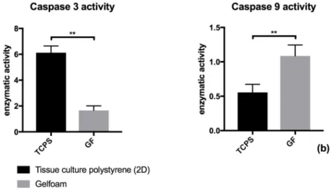
3.2 Activity of caspase 3 is altered by cell culture of human aortic endothelial cells in a 3-dimensional Gelfoam matrix
When cultured in a 3D Gelfoam matrix ECs showed a significant decreased caspase 3 activity compared to ECs cultured on 2D plates (p<0.01, Figure 2). Using spearman’s correlation coefficient a significant correlation (rs = 0,73; p<0.05) between caspase 3 activity and apoptotic EC fraction after Annexin V staining was found in both growing conditions.
3.3 Impact of 3-dimensional cell culture of human aortic ECs on basal protein expression and phosphorylation of anti-apoptotic signaling molecules
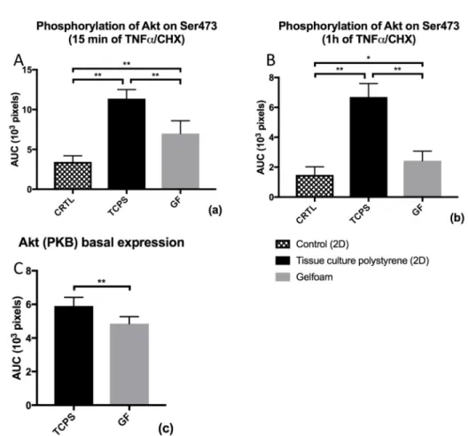
To investigate if the anti-apoptotic effect of a 3D culture of ECs in a Gelfoam matrix is mediated via differences in intracellular signaling basal expression of AKT, ERK, and Bcl-2 and respective degree of activation (level of AKT- and ERK- phosphorylation) were
analyzed.
Expression of Akt in naive ECs was significantly lower in ECs grown in a Gelfoam matrix when compared to ECs grown on polystyrene culture plates (p < 0.01; Figure 3C). Furthermore, cytokine stimulation for 15 minutes (Figure 3A) and 1 hour (Figure 3B) resulted in reduced Akt phosphorylation at Ser473 when EC were grown in a 3D compared to a 2D environment (p < 0.01).
While basal protein expression (without prior induction of apoptosis) of ERK-1 showed a significant increase (p < 0.01; Figure 4A) in human aortic ECs when cultured in a 3D Gelfoam matrix, expression of ERK-2 in non-stimulated ECs grown in Gelfoam was markedly reduced in comparison to cell culture on conventional tissue culture plates (p < 0.01; Figure 4A). Compared to 2D growing conditions, phosphorylation of both ERK isoforms was significantly reduced when human aortic ECs were cultured in 3D Gelfoam matrices (p < 0.01; Figure 4B).
A
uthor Man
uscr
ipt
A
uthor Man
uscr
ipt
A
uthor Man
uscr
ipt
A
uthor Man
uscr
ipt
Compared to human aortic ECs grown on tissue culture plates expression of Bcl-2 isoform 1 was significantly lower in ECs grown in a Gelfoam matrix, whereas expression of isoform 2 in matrix-embedded ECs exceeded expression in 2D cultured ECs (p<0.01; Figure 5).
3.4 Impact of 3-dimensional cell culture of human aortic ECs on basal protein expression and phosphorylation of pro-apoptotic signaling molecules
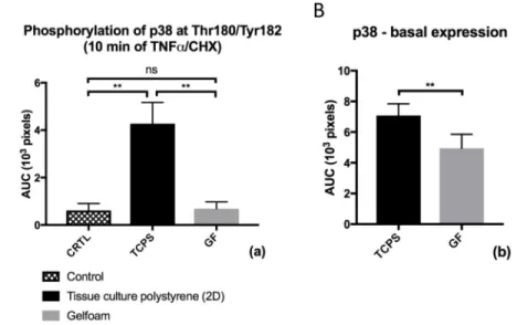
To investigate if the anti-apoptotic effect of a 3D culture of ECs in a Gelfoam matrix is mediated via differences in intracellular signaling basal expression of p38-MAPK, Bax, and p53 and respective degree of activation via phosphorylation of p38 were analyzed.
Expression of p38-MAPK in non-stimulated ECs was significantly lower in ECs grown in a Gelfoam matrix when compared to ECs grown on polystyrene culture plates (p < 0.01; Figure 6B). Upon CHX-mediated sensitization to TNF-α phosphorylation of p38-MAPK at Thr180/Tyr182 was significantly reduced (p < 0.01) in ECs in a 3D environment when compared to human aortic ECs grown on conventional 2D culture plates (Figure 6A). To determine whether 3D culture of human aortic ECs in a Gelfoam matrix had an influence on protein expression of p53 and Bax Western blots were performed. Expression of p53 was significantly lower in ECs grown in a 3D Gelfoam matrix compared to ECs grown on 2D culture plates (p<0.01; Figure 7). Interestingly, cytokine treatment resulted in a significant increased p53 expression in 2D-EC (p<0.01) whereas no significant change could be observed in cytokine-treated 3D-EC. On the contrary, we could not observe significant differences in expression of Bax in unstimulated ECs between the two culture conditions (data not shown).
3.5 Influence of 3D matrix embedding on endothelial FAK expression and cytokine-induced phosphorylation
Basal expression of FAK did not differ in human aortic ECs when compared between ECs grown on polystyrene coated tissue culture plates or within 3D Gelfoam matrices (Figure 8B). Compared to culture of ECs on 2D culture plates phosphorylation of FAK at Tyr397 was significantly reduced when human aortic ECs were cultured in a 3D Gelfoam matrix (p<0.01; Figure 8A). Interestingly the level of tyrosine phosphorylation in matrix embedded EC was even significantly lower than in naive EC grown on 2D culture plates (p<0.01; Figure 8A).
3.6 Association of RIP and FAK in human aortic endothelial cells is reduced through addition of TNF-α and CHX
Considering the important role of the extracellular matrix as key regulator of EC fate, we next analyzed if culturing human aortic ECs in a 3D matrix would influence association of FAK with receptor-interacting protein (RIP). Using immunoprecipitation and Western blot we found that treatment of ECs with a combination of TNF-α and CHX resulted in similar rates of FAK-RIP association in EC grown on 2D-TCPS and within 3D-Gelfoam matrices (Figure 9). Interestingly, FAK-RIP association in both growing conditions was significantly lower when compared to naive EC grown on 2D-TCSP (p < 0.01; Figure 9).
A
uthor Man
uscr
ipt
A
uthor Man
uscr
ipt
A
uthor Man
uscr
ipt
A
uthor Man
uscr
ipt
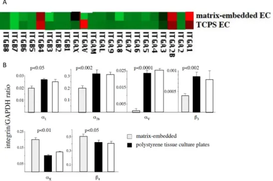
3.7 Matrix-Embedded Endothelial Cells Exhibit an Altered Integrin Expression
Microarray analysis revealed significant differences in integrin expression between 3D matrix-embedded ECs and ECs grown to confluence on 2D tissue culture plates (Figure 10A). RT-PCR confirmed higher expression of integrins α1 (p<0.05), α2b (p<0.002), αv, (p<0.0001) and β3 (p<0.002) on ECs grown on tissue culture plates, whereas matrix-embedded ECs displayed higher expression of αx (p<0.01) and β4 (p<0.05; Figure 10B). Interestingly, TNF-α treatment enhanced expression of integrin αvβ3 in matrix-embedded EC to a greater extent than in 2D-EC (data not shown). Pretreatment of matrix-embedded EC with antibodies against integrin αvβ3 resulted in significantly higher rate of apoptosis and increased p53 expression upon cytokine stimulation compared to cytokine-stimulated matrix-embedded EC without antibody-pretreatment. Yet, level of apoptosis (Annexin V staining: anti-integrin αvβ3 pretreated cytokine–stimulated 3D-EC 12±0.9% vs. 3.2±1.9% cytokine–stimulated 3D-EC; p<0.05; data not shown) and of p53 expression still remained lower than in cytokine-treated EC cultured in 2D (p<0.05; Figure 7). Pretreatment with antibodies against integrin αvβ3 did not affect basal FAK expression or level of tyrosine phosphorylation in 3D matrix-embedded ECs (data not shown).
4. Discussion
In the last few years, it has become increasingly apparent that cell survival and death, especially apoptosis, strongly depend on cell adhesion and the extracellular matrix. Recently, a growing body of evidence has suggested that 3D cell culture systems, in contrast to 2D culture system, represent more accurately the actual microenvironment where cells reside in tissues. Thus, the behavior of 3D cultured cells is more reflective of in vivo cellular
responses. In fact, research has found that cells in a 3D culture environment differ morphologically and physiologically from the same cells in a 2D culture environment (Baharvand et al., 2006; Benya & Shaffer, 1982; Methe et al., 2005; Nelson & Bissell, 2005). Already in 1996, Boudreau et al. demonstrated that suppression of apoptosis by basement membrane requires 3D tissue organization (Boudreau, Werb, & Bissell, 1996). Adding to the existing literature on EC apoptosis we now demonstrate that culturing human aortic ECs in a 3D denatured collagen matrix significantly attenuates cytokine-induced apoptosis in vitro. In line with our result, Chitalia et al. demonstrated that uremic serum reduced viability and number of live thereby increasing dead ECs when grown on 2D tissue culture plates, but not in 3D matrix-embedded ECs (Chitalia et al., 2011).
To the best of our knowledge, influence of spatial environment on endothelial apoptosis has not been studied in extent. It is the additional dimensionality of 3D cultures that is the crucial feature leading to the differences in cellular responses because not only does it influence the spatial organization of the cell surface receptors engaged in interactions with surrounding cells, but it also induces physical constraints to cells (Santini, Rainaldi, & Indovina, 2000). These spatial and physical aspects in 3D cultures affect the signal
transduction from the outside to the inside of cells, and ultimately influence gene expression and cellular behavior (Lee, Cuddihy, & Kotov, 2008; Shield, Ackland, Ahmed, & Rice, 2009; Zietarska et al., 2007). Our findings are of special interest as we not only demonstrated long-lasting therapeutic efficacy of Gelfoam embedded ECs but also that
A
uthor Man
uscr
ipt
A
uthor Man
uscr
ipt
A
uthor Man
uscr
ipt
A
uthor Man
uscr
ipt
seeding ECs on this specific matrix resembles configuration of the actin cytoskeleton, interaction with integrins and focal adhesion complexes more similar to that induced in healthy endothelial monolayer in in-vivo settings (Indolfi et al., 2012; G. E. White, Gimbrone, & Fujiwara, 1983). Matrix embedding ECs in Gelfoam therefore seems to resemble the in vivo phenotype of ECs to a much greater extent than comparable 3D matrices so far used.
As analyzed by Annexin V staining ECs grown within the 3D Gelfoam matrix display a significant lower level of apoptosis upon cytokine treatment than ECs grown to confluence on 2D culture plates. This tracked with LDH release by TNF-α- and CHX-treated ECs. We next analyzed differences in caspase 3, one of three effector caspases (caspase-3, 6, 7) (Deveraux et al., 1998; Earnshaw, Martins, & Kaufmann, 1999). Caspase 3 activity matched our results with significantly reduced activity in ECs grown within a 3D environment compared to ECs cultured in a 2D environment.
In a next step we analyzed activity of established anti- (AKT, ERK, Bcl-2) and pro-apoptotic (p38-MAPK, Bax; p53) effector molecules (el-Deiry, 1998; Reed, 1998; Reed, Zha, Aime-Sempe, Takayama, & Wang, 1996) in EC plated in 2D and 3D growing conditions. The influence on cytokine-mediated EC apoptosis of these molecules remains a matter of debate. Interestingly, 3D embedding of human aortic ECs influences basal expression of most of the molecules studied, e.g. decreased expression of Akt, ERK, p53, and p38-MAPK. Overall, we found that differences in pro-apoptotic p53 (factor 3.4) and p38-MAPK (factor 8.2) between EC grown within a 3D matrix environment and on 2D-TCPS exceeded differences of anti-apoptotic molecules between the two growing conditions. p38-MAPK and p53 have been demonstrated to be key pro-apoptotic molecules in EC apoptosis. As such Grethe et al. demonstrated that the p38 MAPK inhibitor SB203580 significantly attenuated TNF-α induced apoptosis in ECs (Grethe, Ares, Andersson, & Porn-Ares, 2004). p53 stimulates the transcription of a set of genes thereby promoting cell death and growth arrest (Golubovskaya & Cance, 2007). Sequestering p53 by tyrosine phosphorylated FAK has been shown to mediate cell survival (Cance & Golubovskaya, 2008).
The role of Akt and Bcl-2 in cytokine-induced endothelial apoptosis remains a matter of uncertainty; whereas Madge et al. demonstrated it to be unlikely that Akt is involved in this pathway as an anti-apoptotic player because EC could not be sensitized to cytokine-mediated cell death following inhibition of Akt with K179M (Madge, Li, Choi, & Pober, 2003). On the other hand, it has been demonstrated that transduction of ECs with an adenovirus expressing dominant-negative Akt enhanced TNF-α induced endothelial apoptosis (Shen et al., 2010).
In addition overexpression of anti-apoptotic Bcl-2 or Bcl-xL proteins has been demonstrated to inhibit EC apoptosis (Schechner et al., 2000; Zheng et al., 2000). On the other hand it has been demonstrated that in EC the activation of the mitochondrial pathway was not essential for the progression of apoptosis in response to cytokine plus CHX as shown by the fact that overexpression of Bcl-2 had no effect on cell survival (Madge et al., 2003). Therefore, our results of significantly reduced expression of anti-apoptotic Bcl-2 isoform 1 in Gelfoam
A
uthor Man
uscr
ipt
A
uthor Man
uscr
ipt
A
uthor Man
uscr
ipt
A
uthor Man
uscr
ipt
embedded EC compared to EC grown on 2D culture plates with higher expression of isoform 2 expression in the former may be without relevant information for the observed reduced apoptosis in EC when embedded within a 3D environment.
ERK-1 phosphorylation in EC cultured on 2D tissue culture plates significantly exceeded level of phosphorylation in TNF-α treated and CHX-sensitized 3D matrix-embedded EC; indeed, compared to differences in p53 and p38-MAPK expression, ERK-1 phosphorylation in 2D cultured EC exceeded phosphorylation by only a factor of 2.4.
Our findings indicate that phosphorylation of ERK in 3D embedded EC is without direct anti-apoptotic effect on cytokine-induced apoptosis in ECs in this environmental setting. These results are in line with results by Yang et al. demonstrating a major functional switch of the ERK pathway in regulation of EC apoptosis cultured in 2D and 3D culture
environments (Yang, Cao, Sainz, Colman, & Guo, 2004).
Based on our results, the anti-apoptotic effect of 3D matrix-embedding seems to be independent of FAK since we were not able to detect differences in basal expression levels and FAK-RIP association between ECs grown on 2D-TCPS and within a 3D Gelfoam matrix. Moreover, level of FAK tyrosine phosphorylation was significantly lower in TNF-α treated and CHX-sensitized ECs in a 3D environment when compared to cytokine-induced 2D-EC and even significantly lower when compared to non-treated human aortic ECs in a 2D environment. In a 2D environment, tyrosine-phosphorylated FAK acts anti-apoptotic via different pathways, e.g. inhibition of p53 (Ilic et al., 1998), activation of PI3/Akt pathway (Bellas et al., 2002), or RAS GTPase signaling (Renshaw et al., 1999). Kabir et al. demonstrated that staurosporine induces EC apoptosis via focal adhesion kinase
dephosphorylation in a 2D environment (Kabir, Lobo, & Zachary, 2002). Tyrosine kinase inhibitors have found its way into clinical applications as cancer drugs (Sulzmaier, Jean, & Schlaepfer, 2014). Our findings of reduced FAK tyrosine phosphorylation in cytokine-treated EC in a 3D environment are in line with previous results by Cukierman
demonstrating that adhesion of cells within a 3D matrix differ significantly from adhesions characterized on 2D substrates. This is mainly mirrored by an absence of
tyrosine-phosphorylated FAK from most of the length of 3D-matrix adhesions delineating a non-significant role of tyrosine-phosphorylation of FAK within an healthy 3D matrix
environment (Cukierman, Pankov, Stevens, & Yamada, 2001) – and thus in marked contrast to the established role of FAK tyrosine phosphorylation in apoptosis regulation in 2D environments.
In addition, we analyzed differences in integrin expression in ECs between the two growing conditions. Whereas we observed reduced expression of integrin αvβ3 in naive EC when embedded within a 3D matrix, TNF-α treatment resulted in increased expression of integrin
αvβ3 when compared to cytokine-treated EC grown to confluence on 2D tissue culture plates. Our results of increased p53 expression upon pretreatment with an integrin αvβ3 -antibody are in line with findings by Strömland et al. demonstrating that a blocking -antibody to integrin αvβ3 induce EC apoptosis, increase the activity of the tumour suppressor p53, increase levels of the cell cycle inhibitor p21WAF1/CIP1 and decrease levels of the anti-apoptotic protein bax (Stromblad, Becker, Yebra, Brooks, & Cheresh, 1996).
A
uthor Man
uscr
ipt
A
uthor Man
uscr
ipt
A
uthor Man
uscr
ipt
A
uthor Man
uscr
ipt
Ligation of endothelial integrin αvβ3 in 2D has also been shown to activate MAP kinase, focal adhesion kinase (FAK) and Src, among other kinases, resulting in cell proliferation, differentiation and migration (Eliceiri, Klemke, Stromblad, & Cheresh, 1998). In marked contrast, we saw no difference in FAK expression in 3D-EC when pretreated with antibodies against integrin αvβ3.
In conclusion, we are able to demonstrate that 3D Gelfoam embedding ECs reduces CHX-mediated sensitization to TNF-α-induced apoptosis when compared to level of apoptosis in 2D cultured ECs. This is a result of higher activity of pro-apoptotic molecules in ECs cultured on 2D tissue culture plates. In contrast to findings in 2D cultured ECs regulation of apoptosis by FAK and tyrosine-phosphorylated FAK seems to have no anti-apoptotic effect on ECs grown in a 3D matrix in vitro. However, we found an influence of integrin αvβ3– mediated signaling on CHX-mediated sensitization to TNF-α-induced apoptosis in 3D matrix embedded ECs indicating a role of cell-matrix interaction in regulation of endothelial apoptosis.
Our findings are in line with our in vivo data demonstrating long-term clinical effectiveness of perivascular implanted 3D Gelfoam embedded EC to prevent hemodialysis vascular access failure in humans (Conte et al., 2009), to limit neointima formation upon stent placement in peripheral artery disease (Nugent et al., 2009), and to decrease tumor growth and invasiveness in mice (Franses, Baker, Chitalia, & Edelman, 2011).
Our findings provide detailed insights into the mechanisms of endothelial apoptosis with respect to the spatial matrix environment. These results enhance our understanding of endothelial biology on the one side and may otherwise help in the design of tissue
engineered materials. Furthermore, findings on focal adhesion kinase phosphorylation might enhance our understanding of clinical studies with tyrosine kinase inhibitors.
References
Baharvand H, Hashemi SM, Kazemi Ashtiani S, Farrokhi A. Differentiation of human embryonic stem cells into hepatocytes in 2D and 3D culture systems in vitro. Int J Dev Biol. 2006; 50(7):645–652. DOI: 10.1387/ijdb.052072hb [PubMed: 16892178]
Baldo C, Lopes DS, Faquim-Mauro EL, Jacysyn JF, Niland S, Eble JA, … Moura-da-Silva AM. Jararhagin disruption of endothelial cell anchorage is enhanced in collagen enriched matrices. Toxicon. 2015; 108:240–248. DOI: 10.1016/j.toxicon.2015.10.016 [PubMed: 26528579] Bellas RE, Harrington EO, Sheahan KL, Newton J, Marcus C, Rounds S. FAK blunts
adenosine-homocysteine-induced endothelial cell apoptosis: requirement for PI 3-kinase. Am J Physiol Lung Cell Mol Physiol. 2002; 282(5):L1135–1142. DOI: 10.1152/ajplung.00174.2001 [PubMed: 11943680]
Benya PD, Shaffer JD. Dedifferentiated chondrocytes reexpress the differentiated collagen phenotype when cultured in agarose gels. Cell. 1982; 30(1):215–224. [PubMed: 7127471]
Boudreau N, Werb Z, Bissell MJ. Suppression of apoptosis by basement membrane requires three-dimensional tissue organization and withdrawal from the cell cycle. Proc Natl Acad Sci U S A. 1996; 93(8):3509–3513. [PubMed: 8622967]
Cance WG, Golubovskaya VM. Focal adhesion kinase versus p53: apoptosis or survival? Sci Signal. 2008; 1(20):pe22.doi: 10.1126/stke.120pe22 [PubMed: 18493017]
Chitalia VC, Murikipudi S, Indolfi L, Rabadi L, Valdez R, Franses JW, Edelman ER. Matrix-embedded endothelial cells are protected from the uremic milieu. Nephrol Dial Transplant. 2011; 26(12):3858–3865. DOI: 10.1093/ndt/gfr337 [PubMed: 21795755]
A
uthor Man
uscr
ipt
A
uthor Man
uscr
ipt
A
uthor Man
uscr
ipt
A
uthor Man
uscr
ipt
Conte MS, Nugent HM, Gaccione P, Guleria I, Roy-Chaudhury P, Lawson JH. Multicenter phase I/II trial of the safety of allogeneic endothelial cell implants after the creation of arteriovenous access for hemodialysis use: the V-HEALTH study. J Vasc Surg. 2009; 50(6):1359–1368. e1351. DOI: 10.1016/j.jvs.2009.07.108 [PubMed: 19958986]
Cukierman E, Pankov R, Stevens DR, Yamada KM. Taking cell-matrix adhesions to the third dimension. Science. 2001; 294(5547):1708–1712. DOI: 10.1126/science.1064829 [PubMed: 11721053]
Dangles V, Femenia F, Laine V, Berthelemy M, Le Rhun D, Poupon MF, … Schwartz-Cornil I. Two- and three-dimensional cell structures govern epidermal growth factor survival function in human bladder carcinoma cell lines. Cancer Res. 1997; 57(16):3360–3364. [PubMed: 9269996]
Deveraux QL, Roy N, Stennicke HR, Van Arsdale T, Zhou Q, Srinivasula SM, … Reed JC. IAPs block apoptotic events induced by caspase-8 and cytochrome c by direct inhibition of distinct caspases. EMBO J. 1998; 17(8):2215–2223. DOI: 10.1093/emboj/17.8.2215 [PubMed: 9545235]
Dimmeler S, Hermann C, Zeiher AM. Apoptosis of endothelial cells. Contribution to the
pathophysiology of atherosclerosis? Eur Cytokine Netw. 1998; 9(4):697–698. [PubMed: 9889419] Du Y, Herath SC, Wang QG, Wang DA, Asada HH, Chen PC. Three-Dimensional Characterization of
Mechanical Interactions between Endothelial Cells and Extracellular Matrix during Angiogenic Sprouting. Sci Rep. 2016; 6:21362.doi: 10.1038/srep21362 [PubMed: 26903154]
Earnshaw WC, Martins LM, Kaufmann SH. Mammalian caspases: structure, activation, substrates, and functions during apoptosis. Annu Rev Biochem. 1999; 68:383–424. DOI: 10.1146/
annurev.biochem.68.1.383 [PubMed: 10872455]
Edmondson R, Broglie JJ, Adcock AF, Yang L. Three-dimensional cell culture systems and their applications in drug discovery and cell-based biosensors. Assay Drug Dev Technol. 2014; 12(4): 207–218. DOI: 10.1089/adt.2014.573 [PubMed: 24831787]
el-Deiry WS. Regulation of p53 downstream genes. Semin Cancer Biol. 1998; 8(5):345–357. [PubMed: 10101800]
Eliceiri BP, Klemke R, Stromblad S, Cheresh DA. Integrin alphavbeta3 requirement for sustained mitogen-activated protein kinase activity during angiogenesis. J Cell Biol. 1998; 140(5):1255– 1263. [PubMed: 9490736]
Franses JW, Baker AB, Chitalia VC, Edelman ER. Stromal endothelial cells directly influence cancer progression. Sci Transl Med. 2011; 3(66):66ra65.doi: 10.1126/scitranslmed.3001542
Gilmore AP. Anoikis. Cell Death Differ. 2005; 12(Suppl 2):1473–1477. DOI: 10.1038/sj.cdd.4401723 [PubMed: 16247493]
Golubovskaya VM, Cance WG. Focal adhesion kinase and p53 signaling in cancer cells. Int Rev Cytol. 2007; 263:103–153. DOI: 10.1016/S0074-7696(07)63003-4 [PubMed: 17725966] Grethe S, Ares MP, Andersson T, Porn-Ares MI. p38 MAPK mediates TNF-induced apoptosis in
endothelial cells via phosphorylation and downregulation of Bcl-x(L). Exp Cell Res. 2004; 298(2): 632–642. DOI: 10.1016/j.yexcr.2004.05.007 [PubMed: 15265709]
Hynes RO. Integrins: versatility, modulation, and signaling in cell adhesion. Cell. 1992; 69(1):11–25. [PubMed: 1555235]
Ilic D, Almeida EA, Schlaepfer DD, Dazin P, Aizawa S, Damsky CH. Extracellular matrix survival signals transduced by focal adhesion kinase suppress p53-mediated apoptosis. J Cell Biol. 1998; 143(2):547–560. [PubMed: 9786962]
Indolfi L, Baker AB, Edelman ER. The role of scaffold microarchitecture in engineering endothelial cell immunomodulation. Biomaterials. 2012; 33(29):7019–7027. DOI: 10.1016/j.biomaterials. 2012.06.052 [PubMed: 22796162]
Kabir J, Lobo M, Zachary I. Staurosporine induces endothelial cell apoptosis via focal adhesion kinase dephosphorylation and focal adhesion disassembly independent of focal adhesion kinase
proteolysis. Biochem J. 2002; 367(Pt 1):145–155. DOI: 10.1042/BJ20020665 [PubMed: 12084011]
Kim H, Prasain N, Vemula S, Ferkowicz MJ, Yoshimoto M, Voytik-Harbin SL, Yoder MC. Human platelet lysate improves human cord blood derived ECFC survival and vasculogenesis in three dimensional (3D) collagen matrices. Microvasc Res. 2015; 101:72–81. DOI: 10.1016/j.mvr. 2015.06.006 [PubMed: 26122935]
A
uthor Man
uscr
ipt
A
uthor Man
uscr
ipt
A
uthor Man
uscr
ipt
A
uthor Man
uscr
ipt
Korff T, Augustin HG. Integration of endothelial cells in multicellular spheroids prevents apoptosis and induces differentiation. J Cell Biol. 1998; 143(5):1341–1352. [PubMed: 9832561] Lee J, Cuddihy MJ, Kotov NA. Three-dimensional cell culture matrices: state of the art. Tissue Eng
Part B Rev. 2008; 14(1):61–86. DOI: 10.1089/teb.2007.0150 [PubMed: 18454635]
Lu Q, Rounds S. Focal adhesion kinase and endothelial cell apoptosis. Microvasc Res. 2012; 83(1):56– 63. DOI: 10.1016/j.mvr.2011.05.003 [PubMed: 21624380]
Luca AC, Mersch S, Deenen R, Schmidt S, Messner I, Schafer KL, … Stoecklein NH. Impact of the 3D microenvironment on phenotype, gene expression, and EGFR inhibition of colorectal cancer cell lines. PLoS One. 2013; 8(3):e59689.doi: 10.1371/journal.pone.0059689 [PubMed: 23555746] Mabry KM, Payne SZ, Anseth KS. Microarray analyses to quantify advantages of 2D and 3D hydrogel
culture systems in maintaining the native valvular interstitial cell phenotype. Biomaterials. 2016; 74:31–41. DOI: 10.1016/j.biomaterials.2015.09.035 [PubMed: 26433490]
Madge LA, Li JH, Choi J, Pober JS. Inhibition of phosphatidylinositol 3-kinase sensitizes vascular endothelial cells to cytokine-initiated cathepsin-dependent apoptosis. J Biol Chem. 2003; 278(23): 21295–21306. DOI: 10.1074/jbc.M212837200 [PubMed: 12663669]
Mehta U, Kang BP, Bansal G, Bansal MP. Studies of apoptosis and bcl-2 in experimental
atherosclerosis in rabbit and influence of selenium supplementation. Gen Physiol Biophys. 2002; 21(1):15–29. [PubMed: 12168721]
Methe H, Hess S, Edelman ER. Endothelial immunogenicity--a matter of matrix microarchitecture. Thromb Haemost. 2007; 98(2):278–282. [PubMed: 17721607]
Methe H, Nugent HM, Groothuis A, Seifert P, Sayegh MH, Edelman ER. Matrix embedding alters the immune response against endothelial cells in vitro and in vivo. Circulation. 2005; 112(9
Suppl):I89–95. DOI: 10.1161/01.CIRCULATIONAHA.105.524991 [PubMed: 16159871] Nelson CM, Bissell MJ. Modeling dynamic reciprocity: engineering three-dimensional culture models
of breast architecture, function, and neoplastic transformation. Semin Cancer Biol. 2005; 15(5): 342–352. DOI: 10.1016/j.semcancer.2005.05.001 [PubMed: 15963732]
Niland S, Eble JA. Integrin-mediated cell-matrix interaction in physiological and pathological blood vessel formation. J Oncol. 2012; 2012:125278.doi: 10.1155/2012/125278 [PubMed: 21941547] Norata GD, Tonti L, Roma P, Catapano AL. Apoptosis and proliferation of endothelial cells in early atherosclerotic lesions: possible role of oxidised LDL. Nutr Metab Cardiovasc Dis. 2002; 12(5): 297–305. [PubMed: 12616810]
Nugent HM, Edelman ER. Endothelial implants provide long-term control of vascular repair in a porcine model of arterial injury. J Surg Res. 2001; 99(2):228–234. DOI: 10.1006/jsre.2001.6198 [PubMed: 11469891]
Nugent HM, Ng YS, White D, Groothius A, Kanner G, Edelman ER. Delivery site of perivascular endothelial cell matrices determines control of stenosis in a porcine femoral stent model. J Vasc Interv Radiol. 2009; 20(12):1617–1624. DOI: 10.1016/j.jvir.2009.08.020 [PubMed: 19854069] Nugent HM, Rogers C, Edelman ER. Endothelial implants inhibit intimal hyperplasia after porcine
angioplasty. Circ Res. 1999; 84(4):384–391. [PubMed: 10066672]
Nugent HM, Sjin RT, White D, Milton LG, Manson RJ, Lawson JH, Edelman ER. Adventitial endothelial implants reduce matrix metalloproteinase-2 expression and increase luminal diameter in porcine arteriovenous grafts. J Vasc Surg. 2007; 46(3):548–556. DOI: 10.1016/j.jvs.2007.04.074 [PubMed: 17826244]
Raff MC. Social controls on cell survival and cell death. Nature. 1992; 356(6368):397–400. DOI: 10.1038/356397a0 [PubMed: 1557121]
Reed JC. Bcl-2 family proteins. Oncogene. 1998; 17(25):3225–3236. DOI: 10.1038/sj.onc.1202591 [PubMed: 9916985]
Reed JC, Zha H, Aime-Sempe C, Takayama S, Wang HG. Structure-function analysis of Bcl-2 family proteins. Regulators of programmed cell death. Adv Exp Med Biol. 1996; 406:99–112. [PubMed: 8910675]
Renshaw MW, Price LS, Schwartz MA. Focal adhesion kinase mediates the integrin signaling requirement for growth factor activation of MAP kinase. J Cell Biol. 1999; 147(3):611–618. [PubMed: 10545504]
A
uthor Man
uscr
ipt
A
uthor Man
uscr
ipt
A
uthor Man
uscr
ipt
A
uthor Man
uscr
ipt
Santini MT, Rainaldi G, Indovina PL. Apoptosis, cell adhesion and the extracellular matrix in the three-dimensional growth of multicellular tumor spheroids. Crit Rev Oncol Hematol. 2000; 36(2– 3):75–87. [PubMed: 11033298]
Sata M, Walsh K. Oxidized LDL activates fas-mediated endothelial cell apoptosis. J Clin Invest. 1998; 102(9):1682–1689. DOI: 10.1172/JCI3531 [PubMed: 9802882]
Schechner JS, Nath AK, Zheng L, Kluger MS, Hughes CC, Sierra-Honigmann MR, … Pober JS. In vivo formation of complex microvessels lined by human endothelial cells in an immunodeficient mouse. Proc Natl Acad Sci U S A. 2000; 97(16):9191–9196. DOI: 10.1073/pnas.150242297 [PubMed: 10890921]
Shen B, Gao L, Hsu YT, Bledsoe G, Hagiwara M, Chao L, Chao J. Kallistatin attenuates endothelial apoptosis through inhibition of oxidative stress and activation of Akt-eNOS signaling. Am J Physiol Heart Circ Physiol. 2010; 299(5):H1419–1427. DOI: 10.1152/ajpheart.00591.2010 [PubMed: 20729399]
Shield K, Ackland ML, Ahmed N, Rice GE. Multicellular spheroids in ovarian cancer metastases: Biology and pathology. Gynecol Oncol. 2009; 113(1):143–148. DOI: 10.1016/j.ygyno. 2008.11.032 [PubMed: 19135710]
Stromblad S, Becker JC, Yebra M, Brooks PC, Cheresh DA. Suppression of p53 activity and p21WAF1/CIP1 expression by vascular cell integrin alphaVbeta3 during angiogenesis. J Clin Invest. 1996; 98(2):426–433. DOI: 10.1172/JCI118808 [PubMed: 8755653]
Sulzmaier FJ, Jean C, Schlaepfer DD. FAK in cancer: mechanistic findings and clinical applications. Nat Rev Cancer. 2014; 14(9):598–610. DOI: 10.1038/nrc3792 [PubMed: 25098269]
Weil M, Jacobson MD, Coles HS, Davies TJ, Gardner RL, Raff KD, Raff MC. Constitutive expression of the machinery for programmed cell death. J Cell Biol. 1996; 133(5):1053–1059. [PubMed: 8655578]
White E. Life, death, and the pursuit of apoptosis. Genes Dev. 1996; 10(1):1–15. [PubMed: 8557188] White GE, Gimbrone MA Jr, Fujiwara K. Factors influencing the expression of stress fibers in vascular
endothelial cells in situ. J Cell Biol. 1983; 97(2):416–424. [PubMed: 6684121]
Yang B, Cao DJ, Sainz I, Colman RW, Guo YL. Different roles of ERK and p38 MAP kinases during tube formation from endothelial cells cultured in 3-dimensional collagen matrices. J Cell Physiol. 2004; 200(3):360–369. DOI: 10.1002/jcp.20025 [PubMed: 15254963]
Zani BG, Kojima K, Vacanti CA, Edelman ER. Tissue-engineered endothelial and epithelial implants differentially and synergistically regulate airway repair. Proc Natl Acad Sci U S A. 2008; 105(19): 7046–7051. DOI: 10.1073/pnas.0802463105 [PubMed: 18458330]
Zheng L, Dengler TJ, Kluger MS, Madge LA, Schechner JS, Maher SE, … Bothwell AL. Cytoprotection of human umbilical vein endothelial cells against apoptosis and CTL-mediated lysis provided by caspase-resistant Bcl-2 without alterations in growth or activation responses. J Immunol. 2000; 164(9):4665–4671. [PubMed: 10779771]
Zietarska M, Maugard CM, Filali-Mouhim A, Alam-Fahmy M, Tonin PN, Provencher DM, Mes-Masson AM. Molecular description of a 3D in vitro model for the study of epithelial ovarian cancer (EOC). Mol Carcinog. 2007; 46(10):872–885. DOI: 10.1002/mc.20315 [PubMed: 17455221]
A
uthor Man
uscr
ipt
A
uthor Man
uscr
ipt
A
uthor Man
uscr
ipt
A
uthor Man
uscr
ipt
Figure 1. Apoptotic fraction [%] of ECs determined by FACS after Annexin V staining (Figure 1A) and LDH-activity [U/l] in the cell culture supernatant determined by ELISA (Figure 1B)
Human aortic ECs were cultured either on conventional 2D-polystyrene tissue culture plates (TCPS) or within 3D Gelfoam matrices (GF). A CHX-mediated sensitization to TNF-α was used to induce EC apoptosis. Results are given as the mean SD of n = 8 independent experiments. **p < 0.01.
A
uthor Man
uscr
ipt
A
uthor Man
uscr
ipt
A
uthor Man
uscr
ipt
A
uthor Man
uscr
ipt
Figure 2. Alteration of enzymatic activity of caspase 3 through culture of ECs in a 3-dimensional gelfoam matrix (GF) compared to culture of ECs on conventional uncoated 2D-tissue culture plates (TCPS)
Results are given as fold times of enzymatic activity of an unstimulated control. Results show the mean ± SD of n = 8 independent experiments. EC-apoptosis was induced by a combination of TNF-α/CHX for a period of 8 hours. **p < 0.01.
A
uthor Man
uscr
ipt
A
uthor Man
uscr
ipt
A
uthor Man
uscr
ipt
A
uthor Man
uscr
ipt
Figure 3. Basal protein expression and phosphorylation of Akt at Ser473
ECs were either cultured on conventional 2D-polystyrene tissue culture plates with (TCPS) or without (CRTL) addition of TNF-α and CHX or within a Gelfoam matrix with addition of TNF-α and CHX. Results show the mean ± SD of n = 8 independent Western blots without induction of EC-apoptosis (Figure 3C), after 15 minutes (Figure 3A) and 1 hour (Figure 3B) of stimulation TNF-α and CHX. *p < 0.05; **p < 0.01.
A
uthor Man
uscr
ipt
A
uthor Man
uscr
ipt
A
uthor Man
uscr
ipt
A
uthor Man
uscr
ipt
Figure 4. Basal protein expression and phosphorylation of ERK-1/2 at Thr202/Tyr204 and Thr185/Tyr187
Results show the mean ± SD of n = 8 independent Western blots without induction of EC-apoptosis (Figure 4A) and after stimulation with TNF-α and CHX for 60 minutes (Figure 4B). **p < 0.01
A
uthor Man
uscr
ipt
A
uthor Man
uscr
ipt
A
uthor Man
uscr
ipt
A
uthor Man
uscr
ipt
Figure 5. Basal protein expression of Bcl-2
Results show the mean ± SD of n = 8 independent Western blots after 48 hours of cultivation without CHX-mediated sensitization to TNF-α induced EC apoptosis. **p < 0.01; ns = not significant.
A
uthor Man
uscr
ipt
A
uthor Man
uscr
ipt
A
uthor Man
uscr
ipt
A
uthor Man
uscr
ipt
Figure 6. Basal protein expression and phosphorylation of p38-MAPK at Thr180/Tyr182
Figure 6A shows phosphorylation of p38-MAPK at Thr180/Tyr182 after treatment with a combination of TNF-α and CHX for 10 minutes for ECs on 2D-TCPS and in a Gelfoam matrix (GF). An unstimulated 2D-control culture (CRTL) without addition of cytokines was evaluated. Figure 6B shows basal protein expression of p38-MAPK as determined by western blot for culture of ECs on conventional 2D-polystyrene tissue culture plates (TCPS) and in a 3D Gelfoam matrix (GF). For evaluation of basal expression no cytokines were added. Results show the mean ± SD of n = 8 independent western blots. *p < 0.05; **p < 0.01; ns = not significant.
A
uthor Man
uscr
ipt
A
uthor Man
uscr
ipt
A
uthor Man
uscr
ipt
A
uthor Man
uscr
ipt
Figure 7. Expression of p53 in naïve and cytokine treated EC
grown to confluence on conventional 2D-polystyrene tissue culture plates (TCPS) or in a 3D Gelfoam matrix (GF). In some experiments EC were pretreated with antibodies against integrin αvβ3 before cytokine-treatment (n=4). Results show the mean ± SD of n = 8 independent Western blots. *p < 0.05; **p < 0.01
A
uthor Man
uscr
ipt
A
uthor Man
uscr
ipt
A
uthor Man
uscr
ipt
A
uthor Man
uscr
ipt
Figure 8. Basal protein expression and phosphorylation of FAK at Tyr397
Figure 8A shows phosphorylation of FAK at Tyr397 as determined by Western Blot (n=8). ECs cultured on conventional 2D-tissue culture plates (TCPS) or in a 3D Gelfoam matrix (GF) were stimulated with a combination of TNF-α and CHX for 10 minutes. An
unstimulated culture of ECs on conventional 2D-polystyrene culture plates served as control (CRTL). Figure 8B shows basal protein expression of FAK as determined by Western blot, comparing culture of ECs on 2D-TCPS and in a Gelfoam matrix (GF). When comparing basal protein expression no cytokines were added. Results show the mean ± SD of n = 8 independent western blots. *p < 0.05; **p < 0.01; ns = not significant.
A
uthor Man
uscr
ipt
A
uthor Man
uscr
ipt
A
uthor Man
uscr
ipt
A
uthor Man
uscr
ipt
Figure 9. RIP-Western blot after immunoprecipitation with anti-FAK-antibodies
Association of RIP with FAK were analyzed by Western Blot using anti-RIP-IgG-antibodies was compared in ECs cultured on TCPS with ECs cultured within a 3D Gelfoam matrix (GF) after addition of TNF-α and CHX. Additionally, both conditions were compared to an unstimulated control 2D-culture on TCPS (CRTL) without addition of cytokines. Results show the mean ± SD of n = 8 independent western blots. **p < 0.01; ns = not significant.
A
uthor Man
uscr
ipt
A
uthor Man
uscr
ipt
A
uthor Man
uscr
ipt
A
uthor Man
uscr
ipt
Figure 10. Matrix-embedded ECs display an altered integrin expression
Microarray analysis (Figure 10A) and RT-PCR (Figure 10B) revealed significant differences in integrin expression between matrix-embedded ECs and ECs grown to confluence on tissue culture polystyrene plates.
![Figure 1. Apoptotic fraction [%] of ECs determined by FACS after Annexin V staining (Figure 1A) and LDH-activity [U/l] in the cell culture supernatant determined by ELISA (Figure 1B) Human aortic ECs were cultured either on conventional 2D-polystyrene tis](https://thumb-eu.123doks.com/thumbv2/123doknet/13801889.441162/17.918.266.759.94.414/apoptotic-fraction-determined-annexin-supernatant-determined-conventional-polystyrene.webp)