HAL Id: hal-02107292
https://hal.archives-ouvertes.fr/hal-02107292
Submitted on 23 Apr 2019HAL is a multi-disciplinary open access archive for the deposit and dissemination of sci-entific research documents, whether they are pub-lished or not. The documents may come from teaching and research institutions in France or abroad, or from public or private research centers.
L’archive ouverte pluridisciplinaire HAL, est destinée au dépôt et à la diffusion de documents scientifiques de niveau recherche, publiés ou non, émanant des établissements d’enseignement et de recherche français ou étrangers, des laboratoires publics ou privés.
Fear extinction learning improvement in PTSD after
EMDR therapy: an fMRI study
Pierre-François Rousseau, Myriam El Khoury-Malhame, Emmanuelle
Reynaud, Sarah Boukezzi, Aida Cancel, Xavier Zendjidjian, Valérie Guyon,
Jean-Claude Samuelian, Eric Guedj, Thierry Chaminade, et al.
To cite this version:
Pierre-François Rousseau, Myriam El Khoury-Malhame, Emmanuelle Reynaud, Sarah Boukezzi, Aida Cancel, et al.. Fear extinction learning improvement in PTSD after EMDR therapy: an fMRI study. European Journal of Psychotraumatology , Taylor & Francis, 2019, 10 (1), pp.1568132. �10.1080/20008198.2019.1568132�. �hal-02107292�
Full Terms & Conditions of access and use can be found at
https://www.tandfonline.com/action/journalInformation?journalCode=zept20
European Journal of Psychotraumatology
ISSN: 2000-8198 (Print) 2000-8066 (Online) Journal homepage: https://www.tandfonline.com/loi/zept20
Fear extinction learning improvement in PTSD
after EMDR therapy: an fMRI study
Pierre-François Rousseau, Myriam El Khoury-Malhame, Emmanuelle
Reynaud, Sarah Boukezzi, Aïda Cancel, Xavier Zendjidjian, Valérie Guyon,
Jean-Claude Samuelian, Eric Guedj, Thierry Chaminade & Stephanie Khalfa
To cite this article: Pierre-François Rousseau, Myriam El Khoury-Malhame, Emmanuelle Reynaud, Sarah Boukezzi, Aïda Cancel, Xavier Zendjidjian, Valérie Guyon, Jean-Claude Samuelian, Eric Guedj, Thierry Chaminade & Stephanie Khalfa (2019) Fear extinction learning improvement in PTSD after EMDR therapy: an fMRI study, European Journal of Psychotraumatology, 10:1, 1568132, DOI: 10.1080/20008198.2019.1568132To link to this article: https://doi.org/10.1080/20008198.2019.1568132
© 2019 The Author(s). Published by Informa UK Limited, trading as Taylor & Francis Group.
Published online: 29 Jan 2019.
Submit your article to this journal
Article views: 198
BASIC RESEARCH ARTICLE
Fear extinction learning improvement in PTSD after EMDR therapy: an fMRI
study
Pierre-François Rousseaua, Myriam El Khoury-Malhameb, Emmanuelle Reynaudc, Sarah Boukezzic,
Aïda Cancel c, Xavier Zendjidjiand, Valérie Guyond, Jean-Claude Samueliand, Eric Guedje,
Thierry Chaminadecand Stephanie Khalfaa
aLaboratoire de Neurosciences Sensorielles et Cognitives, UMR 7260 CNRS, Marseille, France;bSchool of Arts and Sciences,
Neurosciences, Neuropsychology, Lebanese American University, Beirut, Lebanon;cTimone Institute of Neuroscience, UMR 7289 CNRS, Marseille, France;dDepartment of Psychiatry, La Conception University Hospital, Marseille, France;eBiophysics and Nuclear Medicine Department, Timone Hospital, Marseille, France
ABSTRACT
Objective: Neurobiological models of Posttraumatic Stress Disorder (PTSD) implicate fear processing impairments in the maintenance of the disorder. Eye Movement Desensitization and Reprocessing (EMDR) is one of the most efficient psychotherapies to treat PTSD. We aimed at exploring the brain mechanisms of the fear circuitry involved in PTSD patients’ symptom remission after EMDR therapy.
Method: Thirty-six PTSD participants were randomly assigned to either EMDR group receiv-ing EMDR therapy or Wait-List (WL) group receivreceiv-ing supportive therapy. Participants under-went a behavioural fear conditioning and extinction paradigm during functional magnetic resonance (fMRI). In the EMDR group, patients were scanned at baseline, before EMDR and one week after remission. In the WL group, patients were scanned at baseline and within the same time interval as the EMDR group.
Results: In the EMDR group after treatment, fear responses in the late extinction were significantly lower than before therapy. In parallel, significant functional activity and con-nectivity changes were found in the EMDR group versus the WL during the late extinction. These changes involve the fear circuit (amygdalae, left hippocampus), the right inferior frontal gyrus, the right frontal eye field and insula (pFWE < .05).
Conclusion: These functional modifications underlie a significant improvement of fear extinction learning in PTSD patients after EMDR therapy.
El aprendizaje de extinción del miedo mejora en el TEPT después de la terapia EMDR: un estudio con imágenes por resonancia magnética funcional (fMRI en sus siglas en inglés)
Objetivo: Los modelos neurobiológicos del TEPT implican deficiencias en el procesamiento del miedo en el mantenimiento del trastorno. EMDR es una de las psicoterapias más eficaces para tratar el TEPT. Nuestro objetivo fue explorar los mecanismos cerebrales de los circuitos de miedo implicados en la remisión de los síntomas de los pacientes con el TEPT después de la terapia EMDR.
Método: Treinta y seis participantes con el TEPT fueron asignados aleatoriamente a un grupo EMDR que recibió terapia EMDR o un grupo de Lista de Espera (LE) que recibió terapia de apoyo. Los participantes se sometieron a un paradigma de condicionamiento y extinción del miedo conductual durante la resonancia magnética funcional (fMRI). En el grupo EMDR, los pacientes fueron escaneados al inicio del estudio, antes de EMDR y una semana después de la remisión. En el grupo LE, los pacientes fueron escaneados al inicio y en el mismo intervalo de tiempo que el grupo EMDR.
Resultados: En el grupo EMDR después del tratamiento, las respuestas de miedo en la extinción tardía fueron significativamente más bajas que antes de la terapia. En paralelo, se encontraron cambios significativos en la actividad funcional y en la conectividad en el grupo EMDR v/s el grupo LE durante la extinción tardía. Estos cambios involucran el circuito de miedo (amígdala, hipocampo izquierdo), el giro frontal inferior derecho, los campos del ojo frontal derecho y la ínsula (pFWE < .05).
Conclusión: Estas modificaciones funcionales subyacen a una mejora significativa del aprendizaje de extinción del miedo en pacientes con el TEPT después de la terapia EMDR.
在EMDR治疗PTSD后恐惧消退学习的改善:一项fMRI研究 目的:创伤后应激障碍的神经生物学模型暗示该疾病维持涉及恐惧加工障碍。 EMDR是治 疗创伤后应激障碍的最有效的心理疗法之一。我们的目的是探索EMDR治疗后PTSD患者症 状缓解所涉及的恐惧回路的脑机制。 ARTICLE HISTORY Received 3 April 2018 Revised 25 October 2018 Accepted 29 December 2018 KEYWORDS EMDR; PTSD; fear conditioning PALABRAS CLAVES EMDR; TEPT;
condicionamiento del miedo
关键词 EMDR; PTSD;恐惧条件反应 HIGHLIGHTS • PTSD patients have a deficit to extinguish a conditioned fear • This deficit is reversible after treatment
• EMDR therapy restoration of the fear conditioning ability in PTSD relies upon fear circuitry (amygdala, hippocampus) and other cortical brain structures (insula, posterior cingulate cortex, right frontal eye field, right inferior frontal gyrus and left Heschl gyrus).
CONTACT Pierre-François Rousseau [email protected] Laboratoire de Neurosciences Sensorielles et Cognitives, UMR 7260 CNRS, Fédération 3C, 3 place Victor Hugo, Marseille 13331, France
EUROPEAN JOURNAL OF PSYCHOTRAUMATOLOGY 2019, VOL. 10, 1568132
https://doi.org/10.1080/20008198.2019.1568132
© 2019 The Author(s). Published by Informa UK Limited, trading as Taylor & Francis Group.
This is an Open Access article distributed under the terms of the Creative Commons Attribution-NonCommercial License (http://creativecommons.org/licenses/by-nc/4.0/), which permits unrestricted non-commercial use, distribution, and reproduction in any medium, provided the original work is properly cited.
方法:将36名PTSD被试随机分配到EMDR组(接受EMDR治疗的)或接受支持治疗的等候 名单(WL)组。被试在fMRI期间接受了行为恐惧条件反射和消退范式。在EMDR组中,患 者在基线、在EMDR之前和一周后进行扫描。在WL组中,患者在基线和与EMDR组相同的 时间间隔内进行扫描。 结果:在治疗后的EMDR组中,消退后期的恐惧反应显著低于治疗前。同时在消退后期, 相比WL组,EMDR组中出现了显著的功能活动和连接性变化。这些变化涉及恐惧回路(杏 仁核,左侧海马)、右侧额下回、右侧额叶和岛叶(pFWE < .05)。 结论:这些功能改变是EMDR治疗后PTSD患者恐惧消退学习显著改善的基础。 1. Introduction
Posttraumatic Stress Disorder (PTSD) occurs in the aftermath of a traumatic event (American Psychiatric Association, 2013). The fear conditioning paradigm is used to mimic PTSD acquisition (Hamner, Lorberbaum, & George,1999). Alterations in fear conditioning, extinc-tion learning and extincextinc-tion retenextinc-tion are likely to be involved in the development and maintenance of PTSD (Peri, Ben-Shakhar, Orr, & Shalev,2000). Studies have found modified fear conditioning, extinction and/or extinction recall in PTSD in comparison to trauma exposed individuals or healthy controls (Blechert, Michael, Vriends, Margraf, & Wilhelm, 2007; Milad et al., 2007). Results remain discrepant as to which of the three aforementioned stages of the fear conditioning is altered: the conditioning, extinction or recall. Discrepancies are due to variation in the protocols used. When reproducing the Blechert et al. protocol (2007), we confirmed a deficit in fear extinction in PTSD patients (Wurtz et al.,2016). Using a contextual fear conditioning protocol, Milad et al. (2009) found a deficit in the extinction recall in PTSD. Contextual fear conditioning involves taking a subject and placing this subject into a novel environment while providing an aversive stimulus. When the subject is again put into the same environment, a fear response occurs. Cued fear conditioning is similar to contextual conditioning with one notable exception: the conditioned stimulus (CS) is added to the context but is not the context (Curzon, Rustay, & Browman,2009; Sehlmeyer et al.,2009).
Imaging studies have started investigating fear conditioning, extinction and recall in PTSD patients. The most robust results report an increased amygdala activity during fear conditioning and decreased Anterior Cingulate Cortex (ACC) activity during extinction (Bremner et al., 2005), suggesting insuffi-cient inhibitory inputs from the medial Prefrontal Cortex (PFC) to amygdala. In comparison to trauma-exposed controls, PTSD patients showed a failure to consolidate extinction learning, mediated by hypoac-tivity of ventromedial Prefrontal Cortex (vmPFC) and hippocampus, and also by hyperactivity in the dorsal ACC at recall of extinction (Milad et al.,2009). These studies suggest that dysfunctional amygdala– vmPFC interactions could be at the core of PTSD disorders (Parsons & Ressler, 2013). In such
a model, the persistent conditioned fear in PTSD patients would be related to a decreased activation of hippocampus and vmPFC in addition to an increased activation of dorsal ACC and amygdala (Dejean et al.,2015).
Eye Movement Desensitization and Reprocessing (EMDR) is among the recommended first line psy-chotherapies for PTSD (WHO,2013). EMDR consists of accessing cognitive, emotional and physical aspects of actual distress to traumatic scenes. Imaginal expo-sure to the traumatic event is then after proposed in association with bilateral alternating stimulations (BAS) (auditory, visual or somatosensory stimuli; Servan-Schreiber, Schooler, Dew, Carter, & Bartone,
2006). This results in a change of cognitive processing of memory and cessation of trauma-related distress, while eliminating physical discomfort associated with the initial memory and establishing a positive cogni-tion about the self (Shapiro, 1989). In one study (Wurtz et al., 2016), EMDR treatment for PTSD achieved symptom remission and restored normal fear conditioning and extinction learning, as assessed by objective (physiological) and subjective (verbal) measures. However, this result has never been repli-cated and the neural underpinnings of EMDR-driven remission remain unknown.
To explore the mechanisms involved in fear pro-cessing that might underlie symptom remission in PTSD, patients performed a classical fear condition-ing and extinction protocol. They were scanned in an fMRI before (T0) and after (T1) EMDR therapy (EMDR group) and their results were compared to patients who were included in a wait-list group and were only offered supportive psychotherapy for the duration of the study (WL group).
Our first hypothesis is that, after treatment, the EMDR therapy group would show decreased PTSD symptoms, relative to the WL group. Our second hypothesis is that EMDR therapy would restore nor-mal behavioural fear conditioning and extinction learning in PTSD patients only in the EMDR group as compared to the WL group. Our third hypothesis is that major brain structures known to regulate the fear conditioning and/or extinction learning would be modified post treatment in the EMDR group as com-pared to the WL group.
2. Materials and method
2.1. Procedure
Patients were randomly attributed to one of the two groups. The EMDR group was given EMDR therapy until remission whereas the other group only received supportive therapy (WL group). The EMDR therapy was done according to the standard protocol (Shapiro, 1989) by two psychologists trained and accredited by EMDR Europe. Therapists used hori-zontal hand movements to be visually followed by the patients. All traumatic targets related to the traumatic event at the origin of PTSD were treated until reach-ing a subjective unit of discomfort (SUD) of zero, and having completely true positive cognition about the trauma event and no body discomfort while mentally scanning it. EMDR therapy was stopped when all traumatic targets were treated and the subsequent PCLS scores no longer meet PTSD criteria. The sup-portive therapy was ensured by two other (non-EMDR) psychologists and two psychiatrists from the two recruiting centres. For both therapies, one hour sessions were planned every 7–15 days according to the availabilities of the patients and the therapists. At the end of the protocol, patients of the WL group were offered EMDR therapy.
2.2. Participants
The study was reviewed and approved by the local ethics committee (CPP South Mediterranean 2), and all participants provided written informed consent. Participants were recruited by psychiatrists in univer-sity hospitals in Marseille, France. Diagnosis of PTSD was established according to the DSM-IV TR (American Psychiatric Association, 2000). We excluded patients with present and past neurological or psychiatric conditions, with the exception of anxi-ety and depressive disorders, if their occurrence was related to PTSD. Patients with an addictive disorder, even if related to PTSD, were excluded. Patients could keep their psychotropic medication as long as it did not change during the trial. Therefore, the population included in this study is fairly representa-tive of that found in the medical practice. Diagnoses and clinical interviews were carried out by psychia-trists not otherwise engaged in the study. All partici-pants were assessed by a psychiatrist for PTSD and other mental health disorders using the structured Mini-International Neuropsychiatric Interview (MINI; Lecrubier, Weiller, Hergueta, Bonora, & Lepine,n.d.). This allowed us to diagnose PTSD and screen for potential premorbid or comorbid psychia-tric disorders. Participants at T0 completed the Beck Depression Inventory (Collet & Cottraux, 1986), PTSD Check List Scale (Ventureyra, Yao, Cottraux, Note, & De Mey-Guillard, 2002) and the Impact of
Event Scale Revised (IES-R) (Weiss & Marmar,1997). For the EMDR group a total of 18 adult patients were originally included. Three patients were later excluded because they did not succeed in properly conditioning within experimental design and three others had their data removed due to excessive head motion in the fMRI scanner. Hence the final EMDR group included 12 patients (six men and six women) who were in remission and no longer diagnosed with PTSD after the EMDR therapy, as assessed by psy-chiatrist diagnosis with DSM-IV criteria. For the WL group a total of 18 adult patients were originally included. Six patients were later excluded because they did not succeed in properly conditioning within experimental design. Hence the final WL group included 12 patients (five men and seven women) who were still symptomatic and diagnosed with PTSD at the end of the study. At T1, after therapy (EMDR and supportive), participants were assessed again by a psychiatrist for PTSD symptoms with the MINI. Patients filled the same clinical scales than at T0. The groups did not differ on demographics or severity of symptoms (seeTable 1).
2.3. fMRI procedures
All participants were scanned twice, at T0 (prior to treatment) and T1. In the EMDR group, the T1 scan was conducted one week after remission, which was, on average, three months after the first scan (96.75 ± 95.23 days). In the WL group, the T1 scan was conducted within a week when a EMDR patient was in remission. There was no difference between the two groups for the duration between T0 and T1 (seeTable 1).
2.4. Image acquisition
Data were acquired on a 3-Tesla MEDSPEC 30/80 AVANCE imager (Bruker) at the fMRI centre of Marseille, France. Head movements were restricted with foam cushions. After an initial localizing scan, functional data were acquired using a T2*-weighted gradient-echoplanar imaging (EPI) sequence (TR = 2530 ms, TE = 30 ms; FOV = 19.2 × 19.2; 64 × 64 matrix; flip angle 82.4; voxel size 3 × 3 × 3 mm3). Volumes comprised 38 interleaved axial slices were acquired along anterior-posterior com-missure plane with a continuous slice thickness of 3 mm to cover all the brain. One functional run consisted of 205 volumes. After the fMRI scans, high-resolution images were acquired for the pur-pose of anatomical identification with a sagittal T1-weighted MP-RAGE sequence (TR = 9.4 ms; TE = 4.42 ms; TI = 800 s; 256 × 256 × 180 Matrix; Flip angle 30; voxel size 1 × 1 × 1 mm3).
2.5. Fear conditioning and extinction procedure Fear conditioning and extinction were conducted as part of the fMRI scanning protocol, using electric shocks as the unconditioned stimulus (US) paired with neutral visual stimuli to be the conditioned stimu-lus (CS). All subjects pre-selected the shock level they perceived as highly annoying but not painful (up-down staircase method). Once determined the shock intensity was kept constant for the rest of the conditioning/ extinction task. There were two types of trials consisting of an image of a house in its original version (CS+) and its negative version (CS-), used in a counter-balanced order. The habituation phase started with written instructions telling participants that two pictures would be shown on the screen and that there will be no shock delivery. It consisted of six trials of each to be CS+ and to be CS-. Images were presented for 4 s. At the conditioning phase, instruction informed participants that two pictures will be shown on the screen and that images could be occasionally followed by the electric shock. It consisted of 24 CS+ and 24 CS-. The CS+ were paired with the US at a partial reinforcement rate of 60%. As soon as they saw a CS, subjects had to answer as fast as possible to the question‘ Do you think that you will receive an electric shock after this picture?’ by ‘yes’ or ‘no’ using a two-button key-pad. The subject’s responses were recorded for each trial. No instructions were shown when the extinction phase started. In the extinction phase, the same stimuli for conditioning were presented to patients. It consisted of 24 CS+ and 24 CS-. The only difference between the conditioning and extinction phases was that the CS+ during
extinction was no longer followed by electrical stimula-tion. The US shock occurred for 500 ms immediately at CS+ offset with an electric stimulator. Setting a cyclic ratio of a pulse train allows controlling the frequency and the intensity of the 500 msec transcutaneous elec-trical stimulation. The electrodes delivering the electric stimulation remained attached to the subject’s left ankle throughout the experiment.
3. fMRI data analysis
3.1. Preprocessing
We used the SPM8 software (http://www.fil.ion.ucl.ac. uk/spm/software/spm8). The first four functional volumes were discarded, corresponding to signal stabili-zation. For the functional images, slice timing was used to correct slice acquisition order, realigned was used to control motion effects and to estimate the six head motion parameters. For normalization, the T1-wheighted structural images were co-registered to the EPI mean images and segmented into white matter, grey matter and cerebrospinal fluid. The functional images were next normalized to MNI space using a 3 × 3 × 3 mm3voxel resolution. The normalized data were spacially smoothed using an 8 mm Gaussian kernel.
3.2. First-level analysis
CS+ and CS- trials were separately modelled and convolved with a canonical hemodynamic response function to form regressors. The six movement para-meters were included in the analysis as regressors of
Table 1.Socio-demographic characteristic for the sample.
EMDR Group Wait-list Group Significativity Socio-demographic characteristic
Sex ratio (m/w) 6/6 5/7 NS
Age, years 45.25 (7.83) 40.08 (10.54) NS Education level, years 8.08 (2.71) 8.08 (2.9) NS Duration of illness, days 365 426 NS Duration of therapy, hours 2.83 (0.38) 2.75 (0.45) NS Delay between T0 and T1 96.75 (95.23) 149.25 (122.42) NS Psychometric scales PCLS at T0 59.5 (12.93) 60.75 (13.05) NS IES at T0 48.25 (17.31) 58.41 (11.18) NS BECK at T0 13.5 (5.02) 12.17 (7.33) NS PCLS at T1 28.41 (7.73) 53.58 (18.77) p < .001 IES at T1 9 (7.9) 43.5 (24.39) p < .001 BECK at T1 5.75 (4.49) 11.58 (8.37) p < .05 Type of trauma Accident 4 3 NS Holdup 1 2 NS Physical assault 7 7 NS
Use of psychiatric medications 6/12 9/12 NS Selective serotonin reuptake inhibitors 4 6
Hypnotic 2 3
Psychiatric comorbidities in the MINI at T0
Major depressive episode 8/12 8/12 NS
Suicidality 5/12 2/12 NS
Manic or hypomanic episode 0/12 0/12 NS
Anxiety disorder 10/12 9/12 NS
Substance abuse or dependence 2/12 0/12 NS Values in bold represent significant results.
no interest to model residual effects due to head motion. A 128 s high-pass filter was applied to the data to remove low-frequency noise. For each parti-cipant at the first level, contrast images were calcu-lated to estimate BOLD signal changes due to variation in each phase of the run (conditioning and extinction) for the contrast CS+ vs CS- (dCS) for the two times (T0 and T1). Behavioural responses to each CS-type (CS+, CS-) were averaged on six consecutive presentations for the habituation and four consecu-tive presentations for the conditioning and the extinction, resulting in one value per habituation phase and six values per each of the conditioning (C1 to C6) and extinction phases (E1 to E6). Then, we created the contrast early C1 (four first CS during conditioning) minus late C6 (four last CS during conditioning) conditioning for CS+ vs CS- and the contrast early E1 (four first CS during extinction) minus late E6 (four last CS during extinction) extinc-tion for CS+ vs CS- (dCS).
3.3. Second-level analysis
The individual contrast images were then entered into a second-level model to compare between the two groups (EMDR and WL) the evolution at T1 minus T0 for the early minus late conditioning C1 dCS– C6 dCS and for the early minus late extinction E1 dCS– E6 dCS. This would be best illustrated by the following formula: (C1 dCS– C6 dCS) T1 – (C1 dCS – C6 dCS) T0 for the conditioning part, and (E1 dCS– E6 dCS) T1 – (E1 dCS – E6 dCS) T0 for the extinction part. fMRI brain activity data were analysed by a flexible factorial design which used three factors: Subjects, Group (EMDR or WL) and Time (T0 or T1). We tested the Group × Time interaction to analyse the results. We created one flexible factorial design for the conditioning phase and another one for the extinction phase. We performed whole brain analysis for each contrast. Statistical maps of interest were created using a threshold of uncorrected p < .001. A significant cluster-level defined as cluster p-values < .05 after correction for family-wise error (FWE).
3.4. Connectivity analysis
Following the preprocessing in SPM, connectivity analysis was performed using the functional Connectivity Toolbox (Conn) for MATLAB. Functional volumes were band pass filtered at 0.008–0.09 Hz (default values). Subjects specific nui-sance regressors included six movements and their derivatives and five regressors pertaining to white matter and CSF signals, respectively. The seeds and Regions of Interest (ROI) used for this analysis are those from Conn’s cerebral parcelization. This parce-lization includes an atlas of cortical and subcortical
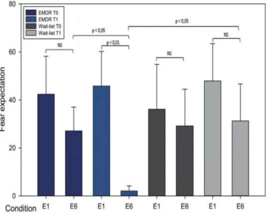
areas from the FSL Harvard-Oxford Atlas, as well as cerebellar areas from the Anatomical Automatic Labelling (AAL) atlas. First-level analysis was done correlating time course from the seeds to whole brain voxels creating connectivity maps for each seed region, using bivariate correlations. These connectiv-ity maps were then passed up to group-level analyses (ROI to ROI module) comparing differences in con-nectivity among EMDR in T1 versus WL in T1 group for the late extinction E6 (last four CS in the extinc-tion). We choose as significant level of connectivity for a p corrected < .05 for the False Discovery Rate (FDR). We have chosen the late extinction E6 in line with the behavioural results, since at that stage the most significant difference is observed in fear expec-tation for the EMDR group at T0 than at T1 and as compared to the WL group (seeFigure 1).
3.5. Statistical analysis
To quantitatively analyse the behavioural results we attributed numerical values to the answers given in the scanner by patients to the question‘do you think you will receive an electric shock’ for each CS. The ‘yes’ was equivalent to ‘1’ and the ‘no’ to ‘0’. For each pair of stimuli (CS+ and CS-) we subtracted the responses for CS+ minus CS-. We multiply this result by 100 to obtain an expected percentage of fear per stimuli. Results clo-ser to 100 indicated learning that shock would follow the image and so indicated and acquisition of the con-ditioned fear whereas results closer to 0 indicated that no electric stimulation was expected. Behavioural results for responses for the fear conditioning phase and the fear extinction phase were separately analysed by two-way repeated measures ANOVA with Group (EMDR or WL) as a between factor and Time (T0 and T1) as a within factor. When significant effects were obtained, t-tests or paired t-test with Bonferroni correc-tions were used as post-hoc comparisons.
4. Results
4.1. Clinical scores
Table 1displays the types of trauma in each group, as well as group mean age, education, duration of ill-ness, duration of therapy, PCLS, IES-R and BECK scores before (T0) and after therapy (T1). There was a significant group × time interaction for the PCLS scale scores (F = 17.09 and p < .001), the IES-R scale scores (F = 8.98 and p < .007) and the BECK scale scores (F = 13.74 and p < .001). PCLS, IES-R and BECK scores were significantly lower in the EMDR than in the WL group at T1 (p < .001, .001 and .05, respectively). PCLS, IES-R and BECK scores in the EMDR group significantly decreased between T0 and T1 (p < .001 for the three scales). There was not any
significant change in clinical scores (PCLS, IES-R and BECK) in the WL group from T0 to T1.
4.2. Fear expectation results
During fear conditioning, there was no significant group × time interaction for the behavioural responses (see Figure 1). During fear extinction, there was a significant group × time interaction for the beha-vioural responses (F = 5.27 and p < .05). In the EMDR group at T1 fear responses in the late extinction (E6) were significantly lower than in the early extinction (p < .01) as displayed inFigure 1. Fear responses in E6 at T1 were significantly lower than at T0 (p < .05). Fear expectation in E6 at T1 was lower in the EMDR than in the WL group (p < .05). Fear expectations for each condition are displayed inTable 2.
4.3. fMRI data
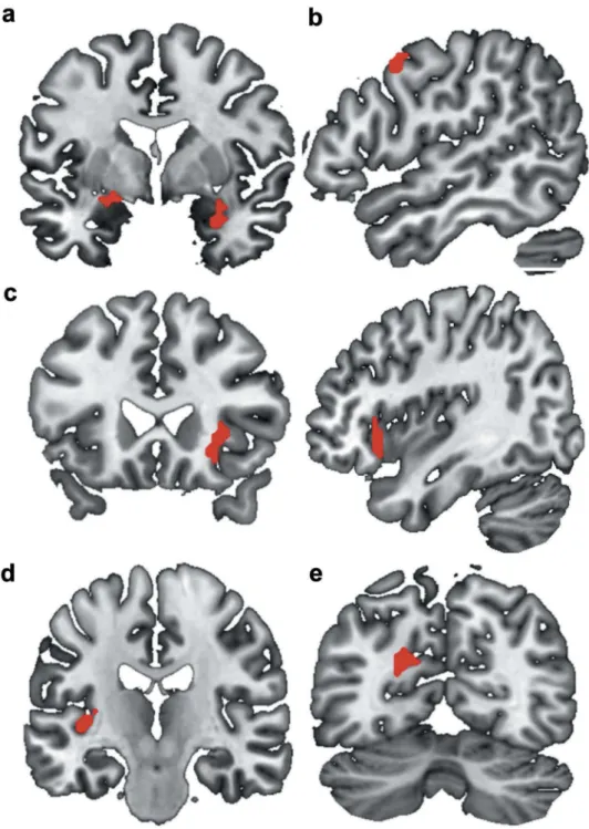
The factorial design analysis has evidenced six significant clusters when considering the EMDR vs the WL group for the contrast T1 minus T0 for E1 minus E6 (CS+ minus CS-). These clusters correspond to the right amyg-dala, the left amygdala and hippocampus, the right frontal eye fields (BA 8), the right inferior frontal gyrus (BA 47) and insula, the left Heschl gyrus and the left dorsal poster-ior cingulate cortex (BA 31). Characteristics of the six
significant clusters are represented inFigure 2. We did not observe any correlation between the evolution of clinical scores (PCLS, IES-R and BECK) and the
Figure 1.Changes in behavioural responses during extinction before and after recovery.
There was a significant group × time interaction for the behavioural responses during fear extinction (F = 5.27 and p < .05). In the EMDR group after treatment, fear responses in the late extinction (E6) were significantly lower than the early extinction (p < .01). Fear responses in E6 after treatment were significantly lower than before treatment (p < .05). Fear expectation in E6 at T1 was lower in the EMDR than in the WL group (p < .05).
Table 2.Fear expectations during the fear condition and extinction protocol for the two groups before (T0) and after (T1) EMDR therapy.
EMDR group Wait-list group C1 T0 0.125 (0.22) 0.35 (0.18) T1 0.5 (0.12) 0.46 (0.14) C2 T0 0.55 (0.15) 0.42 (0.14) T1 0.65 (0.11) 0.57 (0.22) C3 T0 0.475 (0.15) 0.42 (0.23) T1 0.62 (0.1) 0.6 (0.13) C4 T0 0.45 (0.11) 0.6 (0.17) T1 0.67 (0.09) 0.57 (0.16) C5 T0 0.4 (0.15) 0.53 (0.14) T1 0.62 (0.13) 0.25 (0.21) C6 T0 0.42 (0.14) 0.35 (0.22) T1 0.75 (0.09) 0.39 (0.21) E1 T0 0.55 (0.16) 0.64 (0.13) T1 0.45 (0.15) 0.42 (0.2) E2 T0 0.6 (0.13) 0.71 (0.15) T1 0.25 (0.14) 0.26 (0.19) E3 T0 0.47 (0.14) 0.67 (0.10) T1 0.15 (0.13) 0.17 (0.16) E4 T0 0.2 (0.11) 0.42 (0.13) T1 0.02 (0.12) 0.15 (0.18) E5 T0 0.32 (0.12) 0.25 (0.17) T1 0.08 (0.09) 0.21 (0.19) E6 T0 0.25 (0.11) 0.28 (0.16) T1 0.07 (0.05) 0.53 (0.16) Behavioural responses for the difference between CS+ minus CS- were
averaged on four consecutive presentations for the conditioning and the extinction, resulting in six values per conditioning and extinction phases. Values represent average and standard error on fear expecta-tion per group.
evolution of the BOLD signal in the significant clusters during extinction.
4.4. Functional connectivity
Significant differences for the Group × Time interac-tion was only observed during the end of the extinc-tion (E6) for the CS+ minus CS-.
4.5. Positive connectivity
At T1, at the late extinction E6, the left amygdala in the EMDR group showed an increased connec-tivity with the left posterior division of the inferior temporal gyrus, a part of the temporal pole (F = 0.87; intensity = 4.41; p FDR < .022) as compared to the WL group, as displayed in
Figure 3.
Figure 2.Brain representation of the significant clusters for the EMDR group (T1 (E1(CS+ vs CS-)-E6(CS+ vs CS-)))-T0(E1(CS+ vs CS-)-E6) minus wait-list group (T1 (E1(CS+ vs CS-)-E6(CS+ vs CS-)))-T0(E1(CS+ vs CS-)-E6) contrast. (a): right amygdala; left amygdala and hippocampus. (b): right BA8. (c): right BA47 and insula. (d): left Heschl gyrus. (e): left BA31. The factorial design analysis has evidenced six significant clusters when considering the EMDR group vs the Wait-List group for the contrast after minus before therapy for early E1 minus late E6 (CS+ minus CS-). Significance level was defined as clusterp-values < .05 after correction for family-wise error (FWE).
4.6. Negative connectivity
At T1, the EMDR group showed a connectivity decrease as compared to the WL group between the left hippocampus and the left superior parietal lobule (F = 1.13; intensity = 4.73; p FDR < .01) and between the right insula and the right ventral entorhinal cor-tex (BA 28) (F = 1.95; intensity = 4.8; p FDR < .008).
5. Discussion
Patients who received EMDR improved their fear extinction learning as compared to the WL group. This improvement was underlined by functional modifications in the right and left amygdala, hip-pocampus, the right frontal eye fields (BA 8), the right inferior frontal gyrus (BA 47) and insula, left Heschl gyrus and the left dorsal posterior cingulate cortex (BA 31). These functional adaptations were coupled with increased connectivity between left amygdala and the left posterior division of the inferior temporal gyrus and with decreased connec-tivity between the left hippocampus and the left superior parietal lobule and between the right insula and the right ventral entorhinal cortex (BA 28). At T0, the two PTSD populations were com-parable. Thus, the modifications in clinical, beha-vioural and neural results seem to be driven by the therapy rather than by intergroup differences.
5.1. Behaviour results
Our results support the Wurtz et al. (2016) and Blechert et al. (2007) findings, as fear extinction learn-ing was impaired in PTSD patients and was restored after EMDR therapy (at the end of the extinction).
5.2. Functional brain modifications
Our second hypothesis was confirmed since the fear extinction learning improvements in the EMDR group after therapy were indeed paralleled by mod-ifications of brain structures known to be involved in the fear circuitry and in the fear extinction mechan-isms. Other cerebral structures were also highlighted. First, changes observed in the structures convention-ally involved in the extinction of fear will discussed, and then we will focus on the other structures mod-ified by the PTSD remission.
5.3. Structures related to fear extinction
Our results are in line with previous studies. After EMDR therapy, PTSD patients demonstrated a deactivation in the right frontal lobe during an attentional task (Lansing, Amen, Hanks, & Rudy,
2005). A SPECT study has evidenced a deactivation in the temporal pole, medial temporal cortex and orbitofrontal cortex while PTSD patients listened to a script portraying the traumatic event in comparison to control. These differences were restored after symptom remission (Pagani et al., 2007). To the best of our knowledge this is the first-time BOLD activity in limbic and frontal regions change along-side symptoms improvement in fear network at the end of extinction in PTSD. Decreasing symptomatic reaction after individual EMDR therapy seems to enhance the fear extinction ability of PTSD patients. Such enhanced performances of fear processing most likely recruit modified functional involvement of the amygdalae, prefrontal cortex and left hippocampus, all of which regulate the neural fear network (Quirk, Garcia, & González-Lima,2006) and all of which are disrupted in fear extinction learning in PTSD patients (Lonsdorf, Haaker, & Kalisch, 2014). These same
Figure 3.Functional connectivity.
Positive connectivity After treatment, at the end of the extinction the left amygdala in the EMDR group shows an increase of its connectivity with the left posterior division of the inferior temporal gyrus, a part of the temporal pole (F = 0.87; intensity = 4.41; p FDR < .022) compared to the wait-list group. Lateral and anterior view. Negative connectivity After treatment, at the end of the extinction, the left hippocampus in the EMDR group shows a decrease of its connectivity with the left superior parietal lobule (F = 1.13; intensity = 4.73; p FDR < .01) compared to the wait-list group. Lateral and anterior view. After treatment, at the end of the extinction, the right insula in the EMDR group shows a decrease of its connectivity with the right ventral entorhinal cortex (BA 28) (F = 1.95; intensity = 4.8; p FDR < .008) compared to the wait-list group. Anterior and lateral view. Fear extinction learning improvement in PTSD after EMDR therapy: an fMRI study.
structures were also found to be dysfunctional in PTSD in other paradigms such as in script driven imagery (Dahlgren et al., 2017) or in negative emo-tional tasks (Bisby, Horner, Hørlyck, & Burgess,
2016). The decreased activity of the insular cortex activity observed along the fear extinction in the EMDR compared to the WL group could be related to the improvement in patients’ ability to manage negative pictures and their association to inner nega-tive feeling. The insular cortex is indeed involved in monitoring internal bodily states (Pitman et al.,
2012). Individuals with PTSD generally exhibit greater insular cortex activation during the anticipa-tion of aversive images and in response to fearful facial expression, memories and painful stimuli as compared to controls (Aupperle et al., 2012). We found no changes in the medial PFC after EMDR therapy.
5.4. Structures not classically involved in fear extinction learning
Our results suggest that the cerebral modification of activity after symptom remission correspond to func-tional modifications of neural networks involved not only in fear processing but also in processing of negative emotions. Our results evidenced the involve-ment of brain structures neither classically described to intervene in PTSD nor in fear extinction learning such as the right frontal eye field (BA 8), the dorsal posterior cingulate cortex (BA 31), the left Heschl gyrus and the right inferior frontal gyrus (BA 47). The right frontal eye field is implicated in oculomotor control and also in the horizontal saccadic eye move-ment (Miki, Nakajima, Miyauchi, Takagi, & Abe,
1996). Such a visual neuronal plasticity (Vernet et al., 2013) seems to be mostly modulated at the extinction phases. The BA 31 is a part of the posterior cingulate cortex (Leech & Sharp, 2014). This struc-ture is associated with learning complex motor tasks (Tracy et al., 2003) and is involved in control-ling self-determined finger movements (Schubert, von Cramon, Niendorf, Pollmann, & Bublak, 1998). These movements could be correlated with the extinction learning, since they could be faster to per-form the fear evaluation task and perhaps more auto-mated when extinction is better learnt. The left Heschl gyrus has not previously been described as being part of the fear extinction learning. However, Quirk et al.’s model (2006) seems to suggest that fear extinction learning involves not only the vmPFC but also its interactions with other neocortical structures, such as the ones we listed. PTSD patients often pre-sent a decrease of safety cue processing frequently associated with impaired fear inhibition. This deficit to distinguish safe from threatening cues in their environment was modelled in a stop signal task by
van Rooij et al. in 2015 (van Rooij, Geuze, Kennis, Rademaker, & Vink,2015), and involves a reduction of the right inferior frontal gyrus activity in a PTSD group as compared to a control group. These results could explain the post-EMDR functional modifica-tion of the right BA 47 which is a part of the inferior frontal gyrus and as such could allow gaining safety during extinction when viewing the CS+ (that is no longer coupled with the shock at that stage).
We have demonstrated significant changes in con-nectivity patterns after EMDR. After EMDR, at the end of the extinction phase, the left amygdala shows an increase of its connectivity with the left temporal pole in the EMDR group. Given the anatomical and functional relationships between the amygdalae and the temporal pole (Hortensius et al.,2017), and their common involvement in emotional processes as part of the extended limbic system (Olson, Plotzker, & Ezzyat,2007), this increased connectivity may reflect the enhancement of fear conditioning. The EMDR therapy may have restored the amygdalae-temporal network ability to accurately participate in the fear extinction processing by fine-tuning its processing of emotional stimuli.
The left hippocampus and the right ventral entorhinal cortex (BA28) in the EMDR group both show a decreased connectivity with the left superior parietal lobule and the right insula, respectively. Connectivity decreases between the insula, the left superior parietal lobe and structures involved in memory processes in particular in memory for unpleasant or fearful emotional stimuli (Albouy et al., 2008) have to be further replicated and explained. These connectivity modifications could be related to the role of the insula in emotion proces-sing (Pitman et al.,2012) and the role of the superior parietal lobe in saccadic eye movement (Heide et al.,
2001).
5.5. Limitations
This study has some limitations. That EMDR was conducted by only two therapists is a limitation to the generalizability of the results even if the same EMDR protocol was used. Although 18 subjects were initially recruited for each group, a large num-ber was dropped out for various reasons including head movement in the scanner due to the electric stimulation or inability to respond properly to the guidelines. Our final sample is small, which cannot rule out the possibility that the activations found are due to chance. Another limitation is the use of psychiatric medications, the type of trauma included and the presence of comorbidities that may influ-ence the results. Yet, our groups had no statistical differences when tested for use of psychiatric med-ication, presence of psychiatric comorbidities
according to the MINI and type of trauma. Patients were aware of the existence of two treatment groups before starting the study. The decrease in symptoms in the EMDR group and their maintenance in the supportive psychotherapy group may be due to the effect expected by the patients of the treatment received.
6. Conclusions
Our experiment has replicated fear extinction learning improvement in PTSD patients after EMDR therapy and has shown that this improvement seems to be underlined by functional modification of the main brain structures known to be involved in fear extinc-tion learning and neocortical interconnected struc-tures. Modification of connectivity between structures involved in emotion and memory processing further contributes to the improved behavioural performance of participants after EMDR therapy. These results sug-gest that symptoms amelioration in PTSD patients and enhanced fear extinction learning rely upon complex modifications of brain structures of the fear circuitry and their connectivity with networks involved in emo-tion and memory. The study design barely addresses the question whether these modifications are correlated with mere symptoms decrease or whether these are a trademark of the mechanism of action EMDR ther-apy as it could have direct specific effects such as those observed on the frontal eye field.
Disclosure statement
No potential conflict of interest was reported by the authors.
ORCID
Aïda Cancel http://orcid.org/0000-0002-6236-4999
References
Albouy, G., Sterpenich, V., Balteau, E., Vandewalle, G., Desseilles, M., Dang-Vu, T., … Maquet, P. (2008). Both the hippocampus and striatum are involved in consolidation of motor sequence memory. Neuron, 58 (2), 261–272.
American Psychiatric Association. (2000). Diagnostic and statistical manual of mental disorders. Text revision (4th ed.). Washington, DC: American Psychiatric Association.
American Psychiatric Association. (2013). Diagnostic and statistical manual of mental disorders (5th ed.). Washington, DC: American Psychiatric Association. Aupperle, R. L., Allard, C. B., Grimes, E. M.,
Simmons, A. N., Flagan, T., Behrooznia, M., … Stein, M. B. (2012). Dorsolateral prefrontal cortex acti-vation during emotional anticipation and neuropsycho-logical performance in posttraumatic stress disorder. Archives of General Psychiatry, 69(4), 360–371.
Bisby, J. A., Horner, A. J., Hørlyck, L. D., & Burgess, N. (2016). Opposing effects of negative emotion on amyg-dalar and hippocampal memory for items and associations. Social Cognitive and Affective Neuroscience, 11(6), 981–990.
Blechert, J., Michael, T., Vriends, N., Margraf, J., & Wilhelm, F. H. (2007). Fear conditioning in posttrau-matic stress disorder: Evidence for delayed extinction of autonomic, experiential, and behavioural responses. Behaviour Research and Therapy, 45(9), 2019–2033. Bremner, J. D., Vermetten, E., Schmahl, C., Vaccarino, V.,
Vythilingam, M., Afzal, N., … Charney, D. S. (2005). Positron emission tomographic imaging of neural corre-lates of a fear acquisition and extinction paradigm in women with childhood sexual-abuse-related post-traumatic stress disorder. Psychological Medicine, 35(6), 791–806.
Collet, L., & Cottraux, J. (1986). The shortened Beck depression inventory (13 items). Study of the concurrent validity with the Hamilton scale and Widlöcher’s retar-dation scale. L’Encephale, 12(2), 77–79.
Curzon, P., Rustay, N. R., & Browman, K. E. (2009). Cued and contextual fear conditioning for rodents. In J. J. Buccafusco (Ed.), Methods of behavior analysis in neuroscience (2nd ed., pp. 27–73). Boca Raton, FL: CRC Press/Taylor & Francis. Consulté à l’adresse.http://www. ncbi.nlm.nih.gov/books/NBK5223/.
Dahlgren, M. K., Laifer, L. M., VanElzakker, M. B., Offringa, R., Hughes, K. C., Staples-Bradley, L. K., … Shin, L. M. (2017). Diminished medial prefrontal cortex activation during the recollection of stressful events is an acquired characteristic of PTSD. Psychological Medicine, 1–13. doi:10.1017/S003329171700263X
Dejean, C., Courtin, J., Rozeske, R. R., Bonnet, M. C., Dousset, V., Michelet, T., & Herry, C. (2015). Neuronal circuits for fear expression and recovery: Recent advances and potential therapeutic strategies. Biological Psychiatry, 78(5), 298–306.
Hamner, M. B., Lorberbaum, J. P., & George, M. S. (1999). Potential role of the anterior cingulate cortex in PTSD: Review and hypothesis. Depression and Anxiety, 9(1), 1–14.
Heide, W., Binkofski, F., Seitz, R. J., Posse, S., Nitschke, M. F., Freund, H. J., & Kömpf, D. (2001). Activation of frontoparietal cortices during memorized triple-step sequences of saccadic eye movements: An fMRI study. The European Journal of Neuroscience, 13 (6), 1177–1189.
Hortensius, R., Terburg, D., Morgan, B., Stein, D. J., van Honk, J., & de Gelder, B. (2017). The basolateral amyg-dalae and frontotemporal network Functions for threat perception. ENeuro, 4(1). doi: 10.1523/ENEURO.0314-16.2016
Lansing, K., Amen, D. G., Hanks, C., & Rudy, L. (2005). High-resolution brain SPECT imaging and eye move-ment desensitization and reprocessing in police officers with PTSD. The Journal of Neuropsychiatry and Clinical Neurosciences, 17(4), 526–532.
Lecrubier, Y., Weiller, E., Hergueta, P., Bonora, L., & Lepine, J. (n. d.). M.I.N.I 5.0.0/DSM-IV. French version. Paris, France: INSERM.
Leech, R., & Sharp, D. J. (2014). The role of the posterior cingulate cortex in cognition and disease. Brain: A Journal of Neurology, 137(Pt 1), 12–32.
Lonsdorf, T. B., Haaker, J., & Kalisch, R. (2014). Long-term expression of human contextual fear and extinction memories involves amygdala, hippocampus and
ventromedial prefrontal cortex: A reinstatement study in two independent samples. Social Cognitive and Affective Neuroscience, 9(12), 1973–1983.
Miki, A., Nakajima, T., Miyauchi, S., Takagi, M., & Abe, H. (1996). [Functional magnetic resonance imaging of the frontal eye fields during saccadic eye movements]. Nippon Ganka Gakkai Zasshi, 100(7), 541–545.
Milad, M. R., Pitman, R. K., Ellis, C. B., Gold, A. L., Shin, L. M., Lasko, N. B., … Rauch, S. L. (2009). Neurobiological basis of failure to recall extinction memory in posttraumatic stress disorder. Biological Psychiatry, 66(12), 1075–1082.
Milad, M. R., Wright, C. I., Orr, S. P., Pitman, R. K., Quirk, G. J., & Rauch, S. L. (2007). Recall of fear extinc-tion in humans activates the ventromedial prefrontal cortex and hippocampus in concert. Biological Psychiatry, 62(5), 446–454.
Olson, I. R., Plotzker, A., & Ezzyat, Y. (2007). The Enigmatic temporal pole: A review of findings on social and emotional processing. Brain: A Journal of Neurology, 130(Pt 7), 1718–1731.
Pagani, M., Högberg, G., Salmaso, D., Nardo, D., Sundin, O., Jonsson, C., … Hällström, T. (2007). Effects of EMDR psychotherapy on 99mTc-HMPAO distribution in occupation-related post-traumatic stress disorder. Nuclear Medicine Communications, 28(10), 757–765.
Parsons, R. G., & Ressler, K. J. (2013). Implications of memory modulation for post-traumatic stress and fear disorders. Nature Neuroscience, 16(2), 146–153.
Peri, T., Ben-Shakhar, G., Orr, S. P., & Shalev, A. Y. (2000). Psychophysiologic assessment of aversive conditioning in posttraumatic stress disorder. Biological Psychiatry, 47(6), 512–519.
Pitman, R. K., Rasmusson, A. M., Koenen, K. C., Shin, L. M., Orr, S. P., Gilbertson, M. W., … Liberzon, I. (2012). Biological studies of post-traumatic stress disorder. Nature Reviews. Neuroscience, 13(11), 769–787.
Quirk, G. J., Garcia, R., & González-Lima, F. (2006). Prefrontal mechanisms in extinction of conditioned fear. Biological Psychiatry, 60(4), 337–343.
Schubert, T., von Cramon, D. Y., Niendorf, T., Pollmann, S., & Bublak, P. (1998). Cortical areas and the control of self-determined finger movements: An fMRI study. Neuroreport, 9(14), 3171–3176.
Sehlmeyer, C., Schöning, S., Zwitserlood, P., Pfleiderer, B., Kircher, T., Arolt, V., … Gendelman, H. E. (2009). Human fear conditioning and extinction in neuroima-ging: A systematic review. PloS One, 4(6), e5865. Servan-Schreiber, D., Schooler, J., Dew, M. A., Carter, C., &
Bartone, P. (2006). Eye movement desensitization and reprocessing for posttraumatic stress disorder: A pilot blinded, randomized study of stimulation type. Psychotherapy and Psychosomatics, 75(5), 290–297. Shapiro, F. (1989). Eye movement desensitization: A new
treat-ment for post-traumatic stress disorder. Journal of Behavior Therapy and Experimental Psychiatry, 20(3), 211–217. Tracy, J., Flanders, A., Madi, S., Laskas, J., Stoddard, E.,
Pyrros, A., … DelVecchio, N. (2003). Regional brain activation associated with different performance patterns during learning of a complex motor skill. Cerebral Cortex (New York, N.Y.: 1991), 13(9), 904–910.
van Rooij, S. J. H., Geuze, E., Kennis, M., Rademaker, A. R., & Vink, M. (2015). Neural correlates of inhibition and contextual cue processing related to treatment response in PTSD. Neuropsychopharmacology: Official Publication of the American College of Neuropsychopharmacology, 40 (3), 667–675.
Ventureyra, V. A. G., Yao, S.-N., Cottraux, J., Note, I., & De Mey-Guillard, C. (2002). The validation of the post-traumatic stress disorder checklist scale in postpost-traumatic stress disorder and nonclinical subjects. Psychotherapy and Psychosomatics, 71(1), 47–53.
Vernet, M., Bashir, S., Yoo, W.-K., Perez, J. M., Najib, U., & Pascual-Leone, A. (2013). Insights on the neural basis of motor plasticity induced by theta burst stimulation from TMS-EEG. The European Journal of Neuroscience, 37(4), 598–606.
Weiss, D., & Marmar, C. (1997). The impact of event scale – revised. In J. P. Wilson & T. M. Keane (Eds.), Assessing psychological trauma and PTSD (pp. 399–411). New York: Guilford Press.
WHO. (2013). Guidelines for the management of conditions specifically related to stress. Geneva, Switzerland: World Health Organisation.
Wurtz, H., El-Khoury-Malhame, M., Wilhelm, F. H., Michael, T., Beetz, E. M., Roques, J., … Herry, C. (2016). Preventing long-lasting fear recovery using bilat-eral alternating sensory stimulation: A translational study. Neuroscience, 321, 222–235.