HAL Id: hal-02492417
https://hal.archives-ouvertes.fr/hal-02492417
Submitted on 26 Feb 2020
HAL is a multi-disciplinary open access
archive for the deposit and dissemination of
sci-entific research documents, whether they are
pub-lished or not. The documents may come from
teaching and research institutions in France or
abroad, or from public or private research centers.
L’archive ouverte pluridisciplinaire HAL, est
destinée au dépôt et à la diffusion de documents
scientifiques de niveau recherche, publiés ou non,
émanant des établissements d’enseignement et de
recherche français ou étrangers, des laboratoires
publics ou privés.
biliary tree communication
F Panaro, H Habibeh, Patrick Pessaux, F Navarro
To cite this version:
F Panaro, H Habibeh, Patrick Pessaux, F Navarro. Navigation liver surgery for complex hydatid cyst
with biliary tree communication. International Journal of Surgery Case Reports, Elsevier, 2015, 12,
pp.112-116. �10.1016/j.ijscr.2015.05.030�. �hal-02492417�
CASE REPORT – OPEN ACCESS
International Journal of Surgery Case Reports 12 (2015) 112–116
Contents lists available atScienceDirect
International Journal of Surgery Case Reports
j o u r n a l h o m e p a g e :w w w . c a s e r e p o r t s . c o mNavigation liver surgery for complex hydatid cyst with biliary tree
communication
Fabrizio Panaro
∗, Hussein Habibeh, Patrick Pessaux
1, Francis Navarro
Department of General Liver Transplant Surgery, University of Montpellier, Hôpital Saint Eloi, 80 avenue Augustin Fliche 34295, Montpellier-Cedex 5, France
a r t i c l e i n f o
Article history: Received 24 March 2015
Received in revised form 21 May 2015 Accepted 25 May 2015
Available online 29 May 2015 Keywords: Hydatid disease Cystectomy Bile ducts Image-guided surgery Navigated ultrasound
a b s t r a c t
INTRODUCTION: Today, liver surgery navigation is utilized only in high-specialized centers for patients affected by malignant diseases. However, navigated surgery may also be of great interest for benign diseases such as hydatidosis in particular if the hydatid cyst is communicating with the biliary tree. With navigation we know exactly in each moment during the surgery the relationship of the cyst with the vascular/biliary structures around it.
PRESENTATION OF CASE: Herein, we report a case of a 20-year-old W/M affected by hepatic hydatid cyst communicating with the right bile duct, causing recurrent cholangitis. The diagnosis was confirmed by endoscopic retrograde cholangiography and magnetic resonance imaging. The liver cystectomy was easily performed using a navigation system incorporating instrument tracking and three-dimensional CT-reconstruction, thus permitting a selective suture of the bile duct communicating with the cyst. CONCLUSIONS: The navigated system may guide the surgeon in patients with severe and complicated hydatid cysts.
© 2015 The Authors. Published by Elsevier Ltd. on behalf of Surgical Associates Ltd. This is an open access article under the CC BY-NC-ND license (http://creativecommons.org/licenses/by-nc-nd/4.0/).
1. Introduction
In general navigation systems could be of great importance in guiding in real time the surgeon during various procedures for different diseases. These navigations systems represent a major innovation in the field of liver surgery during the last five years. Their application is reserved quite exclusively to malignant dis-eases due to the high costs[1]. However, some severe benign disease could be more easily treated using these innovations. An example of a possible application is the guidance of procedures in case of complicated hydatic cyst.
Hydatid disease is a parasitic disease caused by Echinococcus granulosus and is endemic in Eastern Europe, the Mediterranean coast, and South Africa. The liver is the organ most frequently affected (50–70%)[2,3].
The complications of hydatid disease include rupture, infection, or anaphylaxis. Rupture of hydatid cyst of the liver is the most com-mon complication[4,5]. Furthermore, the liver hydatid cyst could communicate with any part of biliary tree, causing potentially acute
∗ Corresponding author at: Department of Surgery, Liver/Pancreas Transplant Unit Montpellier University Hospital, College of Medicine 80, avenue Augustin Fliche 34295, Montpellier Cedex 5, France. Tel.: +33 4 67 33 67 33; fax: +33 4 67 33 76 23.
E-mail address:[email protected](F. Panaro).
1 Hepato-Biliary and Pancreatic Surgical Unit, General, Digestive and Endocrine
Surgery, IRCAD, IHU MixSurg, Institute for Minimally Invasive Image-Guided Surgery, University of Strasbourg, 1 place de l’Hôpital, 67091, Strasbourg, France.
cholangitis or jaundice, this could occurs in only 5–15% of cases
[6,7].
Despite many advances in medical treatment and radiological intervention methods, the main therapy is still surgery[5]. The surgical treatment depends on the patient’s general condition, the location and number of cysts, and the surgeon’s experience. The main complications of the surgery are cyst rupture with anaphylac-tic shock or dissemination, uncontrolled bleeding and bile leakage
[5]. Image-guided surgery (IGS) provides guidance information via the display of tracking surgical devices overlaid on preoperative tomograms, such as those provided by computed tomography (CT) or magnetic resonance imaging (MRI), which are 3-D in nature and of high resolution. Herein, we present a case of hydatid cyst of the liver with communication with the biliary tree causing recurrent cholangitis successfully operated with IGS devices.
2. Presentation of case
A 20-year-old W/M patient (of Armenian origin) was admit-ted to the hospital complaining of abdominal pain and jaundice since 8 days with no fever. Physical examination revealed yellow-ish discoloration of the sclera and abdominal tenderness in the right subcostal area.
On day 9 he had fever with chills and highly colored urine. Liver function test showed a total bilirubin of 12 mg/dl, ALT 82 U/L, and ALP 125 U/L. Abdominal ultrasonography showed a 110× 98 mm cystic mass in the right lobe.
http://dx.doi.org/10.1016/j.ijscr.2015.05.030
2210-2612/© 2015 The Authors. Published by Elsevier Ltd. on behalf of Surgical Associates Ltd. This is an open access article under the CC BY-NC-ND license (http://creativecommons.org/licenses/by-nc-nd/4.0/).
Fig. 1. (a–b) CT-scan before surgery, (c–d) MRI confirms the vascular rapport of the cyst and its communication between with the biliary tree (d). CYST: hydatid cyst, IVC:
inferior vena cava; RHV: right hepatic vein.
Fig. 2. Pre-operative imaging planning. CYST: hydatid cyst, IVC: inferior vena cava; RHV: right hepatic vein; PV: portal vein. Volumetry of the liver (right below).
CT-scan showed a uniloculated cyst of the right liver, contigu-ous with the retro-hepatic vena cava and the right hepatic vein (Fig. 1a). Abdominal magnetic resonance imaging (MRI) conducted the same day showed a unioculated mass that was hypointense on T1-weighted images, with no contrast uptake in T2 hyperin-tense secants with a suspected biliary fistula (Fig. 1b). The ERCP cholangiopancreatography showed communication of the hidatic cyst with the right biliary tree and the absence of parasites in the common bile duct.
The serological investigation was positive for hydatid disease and surgical intervention was planned for peri-cystectomy to reduce the risks of rupture and parasitic dissemination. The surgery was performed with the aid of the liver navigation system during the open surgery. The follow up was uneventful, and the radiologic post-operative study (CT-scan) was good (Fig. 5). The patient was discharged on postoperative day 15 and albendazole wasadminis-tered for 3 months. Histological investigation confirmed a hydatid cyst.
CASE REPORT – OPEN ACCESS
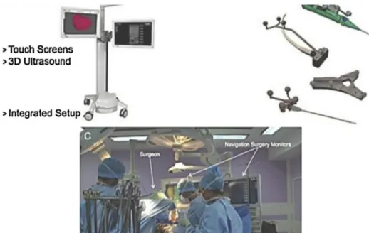
114 F. Panaro et al. / International Journal of Surgery Case Reports 12 (2015) 112–116Fig. 3. (a) Navigation platform, (b) navigated instruments and (c) operating room setup.
3. Surgical innovation 3.1. Preoperative planning
Prior to surgery, a commercially available software package (MeVis Medical Solutions AG, Caroline-Herschel-Str. 1, Bremen, Deutschland) was used to generate 3-D surfaces of anatomic structures from preoperative images producing semi-automatic segmentation of the liver, portal, hepatic veins, biliary tree and liver volumetry (Fig. 2).
To aid in intraoperative navigation, identification of salient anatomic features including the falciform ligament and inferior ridges (segments III, IVb, V and VI), is performed by the experienced surgeon. Finally, the re-constructed images were transferred to the navigation system via USB drive.
3.2. Intraoperative set-up
The navigation system set-up requires the presence of one addi-tional person in the operating room (OR) to control the operating interface. Initial setup takes a few minutes and comprises place-ment of the system, camera carts and cable connections.
A passive optical tracking system is utilized in conjunction with the IGS platform (CAScination AG, Stauffacherstrasse 78, Bern) and a set of disposable surgical tools (Fig. 3).
3.3. Intraoperative ultrasound
An optically tracked adapter is attached to the instrument via a universal clamp and a tracking calibration is performed via a refer-ence device (Fig. 4). When the surgeon is ready to utilize navigation, the liver is prepared so that movement is minimized. Tracking adapters are placed on the relevant surgical instruments.
In the OR, Navigated US projected both the standard B-mode US view and the analogous CT scan slice. The cyst was visualized on the CT plane projected by the navigation system onto the 3-D model, thus confirming that the US transducer was in the correct position to assist with visualization of the cyst once detected.
Fig. 4. (a) Intraoperative ultrasound (right upper: vascular rapport of the cyst), (b) surgical device (dissectron) guided by imaging navigation. CYST: hydatid cyst, IVC: inferior vena cava; RHV: right hepatic vein.
3.4. Surgical procedure
(1) Mobilization of the right liver utilizing a monopolar scalpel, (2) vascular control of the hepatic pedicle and of the right hep-atic vein, (3) parenchymal dissection of the liver utilizing a guided-Cavitron®CUSA (Dissectron®, Integra NeuroSciences Lim-ited Newbury Road Andover – Hampshire SP10 DR, UK) device connected to the IGS system, (4) liver cut surface hemostasis by bipolar grasper, (5) intra-hepatic ligature of the vascular elements (artery, portal and hepatic veins), (6) selective ligature of the intra-hepatic bile ducts communicating with the hydatid cyst.
3.5. Postoperative complications and their management
The postoperative period was uneventful. A right subcostal abdominal pain and a pleural effusion were reported and medically treated.
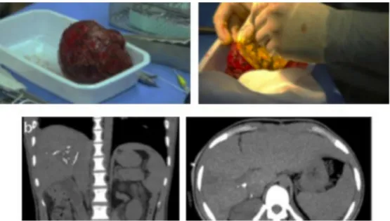
Fig. 5. (a) Specimen (entire hydatid cyst), (b) CT-scan after surgery.
4. Discussion
This navigation device can be safely utilized to guide the sur-geon to remove complicated liver hydatic cyst minimizing the risks of this procedure. Furthermore, in case of communication between the cyst and the biliary tree the IGS may reduce the risks of bile leak-age. Today, we routinely perform hepatectomies without a hepatic pedicle control, therefore the utilization of a IGS device may con-tribute to decrease the risks of vascular and biliary injury. The “static” images (CT-scan or MRI) traditionally used to guide the surgeon during the hepatectomy are not in real-time and so easy to manipulate by the surgeon during the surgery. During a surgical procedure guided by an IGS device we know in advance real time the vascular/biliary structures that we are approaching.
Despite advances in medical treatment and radiological inter-ventions, the main treatment remains surgery [5,8,9]. Surgical procedures range from simple drainage to complex resections and transplantation[8–17]. In all procedures, the objective is to remove the entire hydatid mass without causing any spillage[12,13].
Although drainage procedures are widely used, the results are controversial [8,12,15]. Some authors recommend more radical interventions, such as cystectomy or hepatectomy, especially for smaller, peripherally located cysts[12]. With these procedures, the morbidity and mortality are expected to be more favorable[12,17]. Recent progress in computer science enables the use of instru-ment guidance systems for open and endoscopic liver surgery
[18–20]. These real-time guided surgery systems combine 3 dimen-sional preoperative reconstruction images (CT-scan, MRI) with an intraoperative echography. Currently, the utilization of the IGS devices is limited to the surgery of the solid organs (kidney, liver) due to their limits. In fact, IGS system reflects the relative positions of anatomic structures acquired in preoperative scan-ning and thus does not take into account deformations caused by posture or surgical traction or changes in anatomy since tomo-graphic scan acquisition[19,20]. However, the challenge remains when precise alignment between the pre-operative image data and the intra-operative situation is required, since the liver is subject to deformation and movements during the surgical treatment. A fusion of intra-operative navigated US with the pre-operative data is one solution to improve this situation.
Our first experience is that navigation technology for hydatid liver surgery appears to be a promising approach. In fact, the IGS enabled real-time control of dissection planes will lead to a greater
precision in resection margins and ensure total resection of the cyst sparing the vascular and biliary structures, thus limiting the complications.
Conflicting interests
The authors declared no potential conflicts of interest with respect to the research, authorship, and/or publication of this arti-cle.
Funding
The authors received no financial support for the research, authorship, and/or publication of this article.
References
[1] S. Marubashi, K. Gotoh, H. Akita, H. Takahashi, K. Sugimura, N. Miyoshi, M. Motoori, K. Kishi, S. Noura, Y. Fujiwara, M. Ohue, M. Yano, O. Ishikawa, M. Sakon, Navigation guidance using polyglycolic acid felt in pure laparoscopic partial hepatectomy, Surg. Innov. (August (29)) (2014), pii:
1553350614547772.
[2] G. Yagci, B. Ustunsoz, N. Kaymakcioglu, U. Bozlar, S. Gorgulu, A. Simsek, A. Akdeniz, S. Cetiner, T. Tufan, Results of surgical, laparoscopic, and
percutaneous treatment for hydatid disease of the liver: 10 years experience with 355 patients, World J. Surg. 29 (December) (2005) 1670–1679. [3] I. Pedrosa, A. Saiz, J. Arrazola, J. Ferreiros, C.S. Pedrosa, Hydatid disease:
radiologic and pathologic features and complications, Radiographics 20 (3) (2000) 795–817.
[4] O. Yüksel, N. Akyürek, T. Sahin, B. Salman, C. Azili, H. Bostanci, Efficacy of radical surgery in preventing early local recurrence and cavity-related complications in hydatic liver disease, J. Gastrointest. Surg. 12 (March) (2008) 483–489.
[5] I. Sayek, M.B. Tirnaksiz, R. Dogan, Cystic hydatid disease: current trends in diagnosis and management, Surg. Today 34 (2004) 987–996.
[6] R. Kumar, S.N. Reddy, S. Thulkar, Intrabiliary rupture of hydatid cyst: diagnosis with MRI and hepatobiliary isotope study, Br. J. Radiol. 75 (891) (2002) 271–274.
[7] A. Kumar, D.N. Upadhyaya, S. Singh, M. Kumar, M.A. Ansari,
Cholecysto-hydatid cyst fistula, Indian J. Gastroenterol. 23 (2) (2004) 76–77. [8] J. Richter, E. Profis, M.C. Holtfreter, A. Orhun, I. Müller-Stöver, H. Dedelen, R. Kubitz, Anaphylactic shock ensuing therapeutic puncture of an echinococcal cyst, Parasitol Res. 2014 (December (31)) (2014).
[9] A.N. Yücesoy, S. Poc¸an, Secondary gallbladder hydatidosis and nonfragmanted germinative membrane sourced obstructive jaundice caused by intrabiliary ruptured hepatic hydatid cyst (a case report): two rare complication of the intrabiliary ruptured hepatic hydatid cyst, Hepatobiliary Surg. Nutr. 3 (August (4)) (2014) 209–211.
[10] W.H. Kitchens, C. Liu, E.T. Ryan, C. Fernandez-del Castillo, Hepatic hydatid cyst: a rare cause of recurrent pancreatitis, J. Gastrointest. Surg. 18 (November (11)) (2014) 2057–2059.
CASE REPORT – OPEN ACCESS
116 F. Panaro et al. / International Journal of Surgery Case Reports 12 (2015) 112–116[11] P. Shalayiadang, I. Muzaffar, Yusp-Yimit, A. Turxun, C. Nannan, H. Wen, Comparison of post-operative short-term and long-term outcomes between occult and frank biliary rupture of hydatid disease, Hepatogastroenterology 61 (March–April (130)) (2014) 431–435.
[12] H.O. El Malki, A. Souadka, A. Benkabbou, R. Mohsine, L. Ifrine, R. Abouqal, A. Belkouchi, Radical versus conservative surgical treatment of liver hydatid cysts, Br. J. Surg. 101 (May (6)) (2014) 669–675.
[13] W. Liu, É. Delabrousse, O. Blagosklonov, J. Wang, H. Zeng, Y. Jiang, J. Wang, Y. Qin, D.A. Vuitton, H. Wen, Innovation in hepatic alveolar echinococcosis imaging: best use of old tools, and necessary evaluation of new ones, Parasite 21 (2014) 74.
[14] F. Acar, M. Sahin, H. Alptekin, H. Yılmaz, M.E. Kafalı, Surgical treatment of giant liver hydatid cysts: comparison of cystojejunostomy and partial cystectomy, Surg. Today 44 (November (11)) (2014)
2065–2071.
[15] E. Sozuer, M. Akyuz, S. Akbulut, Open surgery for hepatic hydatid disease, Int. Surg. 99 (November–December (6)) (2014) 764–769.
[16] W. Liu, Delabrousse É, O. Blagosklonov, J. Wang, H. Zeng, Y. Jiang, J. Wang, Y. Qin, D.A. Vuitton, H. Wen, Laparoscopic versus open hepatic resections for benign and malignant neoplasms-a metaanalysis, Surgery 2007 (February (141)) (2007) 203–211.
[17] H. Li, Y. Shao, T. Aji, J. Zhang, K. Kashif, Q. Ma, B. Ran, H. Wen, Laparoscopic approach for total cystectomy in treating hepatic cystic echinococcosis, Parasite 21 (2014) 65.
[18] P. Pessaux, M. Diana, L. Soler, T. Piardi, D. Mutter, J. Marescaux, Towards cybernetic surgery: robotic and augmented reality-assisted liver segmentectomy, Langenbecks Arch. Surg. 400 (April (3)) (2015) 381–385. [19] Y. Morita, K. Takanishi, J. Matsumoto, A new simple navigation for anatomic
liver resection under intraoperative real-time ultrasound guidance, Hepatogastroenterology 61 (September (34)) (2014) 1734–1738.
[20] S. Satou, T. Aoki, J. Kaneko, Y. Sakamoto, K. Hasegawa, Y. Sugawara, O. Arai, T. Mitake, K. Miura, N. Kokudo, Initial experience of intraoperative
three-dimensional navigation for liver resection using real-time virtual sonography, Surgery 155 (February (2)) (2014) 255–262.
Open Access
This article is published Open Access atsciencedirect.com. It is distributed under theIJSCR Supplemental terms and conditions, which permits unrestricted non commercial use, distribution, and reproduction in any medium, provided the original authors and source are credited.