Publisher’s version / Version de l'éditeur:
PLoS ONE, 10, 9, 2015-09-17
READ THESE TERMS AND CONDITIONS CAREFULLY BEFORE USING THIS WEBSITE. https://nrc-publications.canada.ca/eng/copyright
Vous avez des questions? Nous pouvons vous aider. Pour communiquer directement avec un auteur, consultez la
première page de la revue dans laquelle son article a été publié afin de trouver ses coordonnées. Si vous n’arrivez pas à les repérer, communiquez avec nous à PublicationsArchive-ArchivesPublications@nrc-cnrc.gc.ca.
Questions? Contact the NRC Publications Archive team at
PublicationsArchive-ArchivesPublications@nrc-cnrc.gc.ca. If you wish to email the authors directly, please see the first page of the publication for their contact information.
Archives des publications du CNRC
This publication could be one of several versions: author’s original, accepted manuscript or the publisher’s version. / La version de cette publication peut être l’une des suivantes : la version prépublication de l’auteur, la version acceptée du manuscrit ou la version de l’éditeur.
For the publisher’s version, please access the DOI link below./ Pour consulter la version de l’éditeur, utilisez le lien DOI ci-dessous.
https://doi.org/10.1371/journal.pone.0138565
Access and use of this website and the material on it are subject to the Terms and Conditions set forth at
Control of Francisella tularensis intracellular growth by pulmonary
epithelial cells
Maggio, Savannah; Takeda, Kazuyo; Stark, Felicity; Meierovics, Anda I.;
Yabe, Idalia; Cowley, Siobhan C.
https://publications-cnrc.canada.ca/fra/droits
L’accès à ce site Web et l’utilisation de son contenu sont assujettis aux conditions présentées dans le site LISEZ CES CONDITIONS ATTENTIVEMENT AVANT D’UTILISER CE SITE WEB.
NRC Publications Record / Notice d'Archives des publications de CNRC:
https://nrc-publications.canada.ca/eng/view/object/?id=05d42fa9-085b-4694-9427-b06be84c7cb6 https://publications-cnrc.canada.ca/fra/voir/objet/?id=05d42fa9-085b-4694-9427-b06be84c7cb6Control of Francisella tularensis Intracellular
Growth by Pulmonary Epithelial Cells
Savannah Maggio1, Kazuyo Takeda2, Felicity Stark3, Anda I. Meierovics1, Idalia Yabe4, Siobhan C. Cowley1*
1Laboratory of Mucosal Pathogens and Cellular Immunology, Division of Bacterial, Parasitic and Allergenic Products, Center for Biologics Evaluation and Research, U.S. Food and Drug Administration, Silver Spring, Maryland, United States of America, 2 Microscopy and Imaging Core Facility, Division of Bacterial, Parasitic and Allergenic Products, Center for Biologics Evaluation and Research, U.S. Food and Drug Administration, Silver Spring, Maryland, United States of America, 3 National Research Council Canada, Human Health Therapeutics Portfolio, Ontario, Canada, 4 National Heart, Lung, and Blood Institute, National Institutes of Health, Bethesda, Maryland, United States of America
*siobhan.cowley@fda.hhs.gov
Abstract
The virulence of F. tularensis is often associated with its ability to grow in macrophages, although recent studies show that Francisella proliferates in multiple host cell types, includ-ing pulmonary epithelial cells. Thus far little is known about the requirements for killinclud-ing of F. tularensisin the non-macrophage host cell types that support replication of this organism. Here we sought to address this question through the use of a murine lung epithelial cell line (TC-1 cells). Our data show that combinations of the cytokines IFN-γ, TNF, and IL-17A acti-vated murine pulmonary epithelial cells to inhibit the intracellular growth of the F. tularensis Live Vaccine Strain (LVS) and the highly virulent F. tularensis Schu S4 strain. Although paired combinations of IFN-γ, TNF, and IL-17A all significantly controlled LVS growth, simultaneous treatment with all three cytokines had the greatest effect on LVS growth inhibi-tion. In contrast, Schu S4 was more resistant to cytokine-induced growth effects, exhibiting significant growth inhibition only in response to all three cytokines. Since one of the main antimicrobial mechanisms of activated macrophages is the release of reactive nitrogen intermediates (RNI) via the activity of iNOS, we investigated the role of RNI and iNOS in Francisellagrowth control by pulmonary epithelial cells. NOS2 gene expression was signifi-cantly up-regulated in infected, cytokine-treated pulmonary epithelial cells in a manner that correlated with LVS and Schu S4 growth control. Treatment of LVS-infected cells with an iNOS inhibitor significantly reversed LVS killing in cytokine-treated cultures. Further, we found that mouse pulmonary epithelial cells produced iNOS during in vivo respiratory LVS infection. Overall, these data demonstrate that lung epithelial cells produce iNOS both in vitroand in vivo, and can inhibit Francisella intracellular growth via reactive nitrogen intermediates.
OPEN ACCESS
Citation: Maggio S, Takeda K, Stark F, Meierovics AI, Yabe I, Cowley SC (2015) Control of Francisella tularensisIntracellular Growth by Pulmonary Epithelial Cells. PLoS ONE 10(9): e0138565. doi:10.1371/journal.pone.0138565
Editor: Dennis W Metzger, Albany Medical College, UNITED STATES
Received: June 5, 2015 Accepted: August 31, 2015 Published: September 17, 2015
Copyright: This is an open access article, free of all copyright, and may be freely reproduced, distributed, transmitted, modified, built upon, or otherwise used by anyone for any lawful purpose. The work is made available under theCreative Commons CC0public domain dedication.
Data Availability Statement: All relevant data are within the paper and its Supporting Information files. Funding: This work was supported by the FDA Medical Countermeasures Initiative. The funder had no role in study design, data collection and analysis, decision to publish, or preparation of the manuscript. Competing Interests: The authors have declared that no competing interests exist.
Introduction
Francisella tularensis is a zoonotic facultative intracellular bacterium that causes a lethal febrile illness in humans known as tularemia. F. tularensis can infect the host via multiple routes, including the respiratory and gastrointestinal tracts, as well as through broken skin. Respira-tory tularemia is the most lethal form of the disease; inhalation of as few as 10 CFU of the highly virulent F. tularensis subspecies tularensis (F.t. tularensis) is sufficient to establish infec-tion, and the mortality rate of untreated pneumonic tularemia is estimated to be as high as 60% [1]. For these reasons, virulent F.t. tularensis was developed as a biological weapon in the mid-20thcentury, and remains a high priority agent identified as a risk to national security by the United States Centers for Disease Control.
The attenuated live vaccine strain (LVS) of F. tularensis was derived by repeated passage of a virulent F. tularensis subspecies holarctica strain on agar; LVS has been studied as an investi-gational product but is not currently licensed for use in humans in the United States [2]. Thus far the protective efficacy of LVS and the key mechanisms of immunity to tularemia remain only partially characterized. In order to better understand the LVS vaccine, and to facilitate the development of new vaccines and therapies against highly lethal pneumonic tularemia, it is important to identify the immune mechanisms that limit respiratory F. tularensis infection. As an additional benefit, discoveries defining immunity to pulmonary F. tularensis infection may be applied to other respiratory intracellular pathogens, such as Mycobacterium tuberculosis.
F. tularensis has been detected within alveolar macrophages and airway dendritic cells within one hour after murine pulmonary infection, although the bacteria quickly invade a myriad of other cell types, including lung monocytes, neutrophils, and alveolar type II epithe-lial (ATII) cells [3]. The majority of these cell types are professional phagocytes that produce multiple anti-microbial factors, such as degradative enzymes, reactive oxygen and nitrogen intermediates, and cationic peptides to inhibit pathogen growth. In particular, macrophages are well known to become activated by interferon-gamma (IFN-γ) and tumor necrosis factor alpha (TNF) to produce reactive nitrogen intermediates (RNI) through induction of the enzyme inducible nitric oxide synthase (iNOS) [4–7]. iNOS produces nitric oxide (NO), which together with other oxidative products such as peroxynitrite and S-nitrosothiols, exert microbiocidal activities [8]. The importance of iNOS to immune defense is reflected by the fact that iNOS-deficient mice are susceptible to sublethal LVS infections [9], and chemical inhibition of iNOS activity significantly inhibits IFN-γ-induced killing of LVS and virulent F. tularensis in peritoneal exudate macrophages in vitro [10,11]. Overall, macrophage-derived nitric oxide production is considered an important mechanism by which macrophages kill intracellular pathogens, including Mycobacterium tuberculosis, Salmonella typhimurium, and Leishmania donovani.
In contrast to macrophages, ATII cells are not professional phagocytes, although they read-ily support Francisella growth both in vitro and in vivo [3,12]. Since ATII cells comprise 15% of all lung cells [13], they have the potential to provide a significant cellular niche for Franci-sella replication during pulmonary infection. Importantly, a ΔpyrF FranciFranci-sella mutant that grew poorly in macrophages but vigorously in other cell types retained full virulence in the murine pulmonary infection model, demonstrating that growth in non-macrophage cell types significantly contributes to Francisella virulence [14]. Despite the fact that pulmonary epithelial cells are a potentially unique replication site for Francisella in the lungs, little is known about their capacity to inhibit Francisella intracellular growth.
Since the immune mechanisms involved in control of F. tularensis growth in pulmonary epithelial cells will likely provide insights into defense against respiratory Francisella infec-tion, here we sought to investigate the possibility that cytokines can activate these cells to
produce anti-microbial factors that inhibit Francisella growth. Indeed, we show that combi-nations of the cytokines IFN-γ, TNF, and IL-17A activate murine pulmonary epithelial cells to inhibit the intracellular growth of LVS and the highly virulent F. tularensis Schu S4 strain. Gene expression analyses revealed up-regulation of NOS2 in F. tularensis-infected, cytokine-treated cells in a manner that correlated with Francisella growth control. Treatment of LVS-infected pulmonary epithelial cells with an iNOS inhibitor significantly reversed LVS killing in cytokine-treated cultures, establishing iNOS as a major antimicrobial mechanism used by these cells to inhibit bacterial intracellular growth. Further, mouse lung ATII cells produced iNOS following in vivo sublethal LVS intranasal infection. Collectively, the data presented here demonstrate that lung epithelial cells actively produce iNOS both in vitro and in vivo, and that these cells possess the capacity to restrict Francisella intracellular growth via reactive nitrogen intermediates.
Materials and Methods
Bacteria
F. tularensis LVS (ATCC 29684) was obtained from the American Type Culture Collection. F. tularensis Schu S4 was obtained from the Francisella Strain Collection, Swedish Defense Research Agency, Sweden. Viable bacteria were quantified by plating serial dilutions on supple-mented Mueller-Hinton agar (MHA) plates.
TC-1 cells
The TC-1 cell line was purchased from ATCC, and can now be obtained from Johns Hopkins Technology Ventures. TC-1 cells are pulmonary epithelial cells derived from primary lung cells of C57BL/6 mice cotransformed with HPV-16 E6 and E7 and c-Ha-ras oncogenes [15].
In vitro
assessment of control of intracellular bacterial growth in TC-1
cells
TC-1 cells were cultured in Dulbecco minimal essential medium (DMEM; Life Technologies) supplemented with 10% heat-inactivated fetal calf serum (FCS; HyClone), 1 mM HEPES buffer (Life Technologies), and 0.1 mM nonessential amino acids (Life Technologies) (com-plete DMEM; cDMEM), and plated at 2 x 10⁵viable cells per well in 24-well plates. After overnight culture, TC-1 cells were infected with F. tularensis LVS or SchuS4 at a multiplicity of infection (MOI) of 1:1 (bacterium-to-TC-1 cell ratio) for 2 hours, washed with PBS, incu-bated with 50 μg/ml gentamicin for 45 minutes, washed twice with warm PBS, and cDMEM with or without recombinant cytokines was added to the wells, as indicated. All cytokines (IFN-γ, TNF, IL-17A, and IL-22) were obtained from BioLegend, and used in experiments at a concentration of 100 ng/ml, with the exception of TNF, which was used at a concentration of 50 ng/ml. Cultures were incubated at 37°C in 5% CO2for the remainder of the experiment. Bacterial uptake or recovery was determined after initial infection, and after 72 hours of cul-ture, by washing the monolayers once with warm PBS, followed by lysis of infected cells with sterile distilled water, plating on agar, and counting. Supernatants were obtained from the cultures after 72 hours for nitrite analyses.
RNA extraction and quantitative real time RT-PCR
Total RNA was prepared using RNeasy Mini Kit (Qiagen), and first-strand cDNA was synthe-sized using the Retroscript Kit (Ambion) according to the manufacturer's instructions. Real time PCR was performed using the ABI Prism ViiA7 instrument (Applied Biosystems). NOS2
transcripts were quantified using TaqMan1Pre-developed Assay Kits (Applied Biosystems) according to the manufacturer's instructions, and the results normalized to expression of GAPDH. The relative standard curve method was used to derive normalized expression for all samples, and “NOS2 fold induction” was calculated as the ratio of cytokine-treated samples to “LVS alone” or “Schu S4 alone” control samples.
Quantitation of RNI in alveolar epithelial cell culture supernatants
Culture supernatants were assayed for nitrite by the Griess reaction using a commercial Griess reagent according to the manufacturer's instructions (Sigma).
In vivo
mouse infections
Male specific pathogen free C57BL/6J mice were purchased from Jackson Laboratory. Animals were housed in a barrier environment at CBER/FDA, and the FDA Institutional Animal Care and Use Committee approved this study. Intranasal infections were performed by delivering 2 x 10²LVS CFUs in a volume of 25 μl per nare to anesthetized mice. Bacteria for infection were diluted in PBS (Cambrex) containing <0.01 ng/ml endotoxin.
Immunohistochemistry
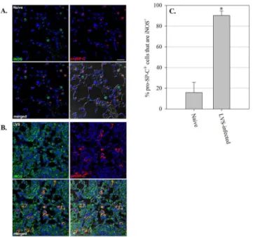
Six C57BL/6 strain mice were intranasally infected with 2 x 10²CFU of LVS and six uninfected mice were used as controls (naïve). Seven days after infection, lungs were harvested from the mice. Mouse lungs were inflated with 4% paraformaldehyde installed via the trachea, and fixed for 24 hours at room temperature. The paraffin sections were made by standard procedure for immunofluorescence staining. A dual immunofluorescence staining method was performed to demonstrate immunoreactivity for a combination of mouse monoclonal and rabbit polyclonal antibodies against iNOS and pro-Surfactant protein C (ab129372, Abcam, and Ab3786, Milli-pore, respectively). Briefly, the sections were deparaffinized, rehydrated and then incubated with 5% normal donkey serum for 30 min at room temperature. Sections were subsequently incubated with primary antibodies against iNOS and pro-SP-C overnight at 4°C. The sections were then incubated with secondary antibodies conjugated with Alexa fluor488 and Alexa fluor594 (Jackson Immunoresearch Laboratories). Hoechst 33258 (Life Technologies) was used for counterstaining at 0.5ug/ml concentration. Fluorescence microscopic images were obtained using a Leica TCS_SP8 DMI6000 confocal microscope system (Leica Microsystems). Images were acquired at 63 x Objective lens (N.A. 1.4) for Alexa 488 and 594 emission wave-lengths and stored as lif format for further analyses.
Statistical Analyses
All experiments were repeated at least two to three times to assess reproducibility. Statisical analyses were performed on each experiment, and one representative experiment is shown for publication. All data were tested for equal variance and normal distribution (Shapiro-Wilk) prior to statistical testing. Following confirmation of equal variance and normal distribution, data were analyzed via one-way ANOVA followed by the Student-Newman-Keuls multiple stepwise comparison (for experiments with >2 experimental groups; Figs1and2). For experi-ments with only two experimental groups (Fig 3), data were analyzed using a two-tailed Stu-dent’s t-test; P-values of 0.05 or less were considered significant.
Results
Cytokine-induced control of F. tularensis LVS growth in pulmonary
epithelial cells is partially dependent upon RNI
Previous studies have shown that F. tularensis LVS invades and replicates in several lung epi-thelial cell lines, including the murine lung epiepi-thelial cell line TC-1, the human lung ATII cell line A459, and the mouse ATII-derived cell line MLE-12 [12,16]. Of the three aforementioned cell lines, TC-1 cells exhibited the highest LVS invasion frequency [12], and have been used to define the intracellular trafficking of LVS during infection of pulmonary epithelial cells [16]. Since TC-1 cells are the best characterized pulmonary epithelial cell line with respect to Franci-sella infection, we used TC-1 cells to examine the impact of cytokines on control of LVS intra-cellular growth in pulmonary epithelial cells.
Fig 1. Cytokine-induced control of F. tularensis LVS growth in pulmonary epithelial cells is partially dependent upon RNI.(A) TC-1 lung epithelial cells were infected with LVS, and intracellular bacteria were enumerated after 72 h. Cultures were treated with recombinant cytokines immediately after infection as indicated, or untreated (‘LVS alone’). * = significantly different from ‘LVS alone’ cultures (P<0.05). (B) Changes in expression of NOS2 in TC-1 cell cultures infected with LVS and treated with recombinant cytokines. Fold induction values are expressed relative to ‘LVS alone’ cultures. * = significantly different from cultures treated only with single cytokines (P<0.05). (C) Nitrite production in TC-1 cell cultures infected with LVS and treated with recombinant cytokines after 72 h. * = significantly different from ‘LVS alone’ cultures (P<0.05). (D) 72 h growth of LVS in cytokine-treated TC-1 cultures in the presence of NMMA. * = significantly different from LVS growth in identically treated cultures in the absence of NMMA (P<0.05). Data are
expressed as the mean ± SD of triplicate cultures, and plots are representative of three experiments of similar design.
Fig 2. F. tularensis subspecies tularensis Schu S4-infected pulmonary epithelial cells are relatively resistant to the antimicrobial effects of cytokine treatment.(A) TC-1 lung epithelial cells were infected with Schu S4, and intracellular bacteria were enumerated after 72 h. Cultures were treated with recombinant cytokines immediately after infection as indicated, or untreated (‘Schu S4 alone’). (B) Changes in NOS2 gene expression in TC-1 cell cultures infected with Schu S4 and treated with recombinant cytokines. Fold induction values are expressed relative to ‘Schu S4 alone’ cultures. * = significantly different from cultures treated only with individual cytokines (P<0.05). Data are expressed as mean ± SD of triplicate cultures, and plots are representative of two experiments of similar design.
doi:10.1371/journal.pone.0138565.g002
Fig 3. ATII cells in the lungs of mice produce iNOS during in vivo pulmonary F. tularensis LVS infection.(A) Naïve control mice, and (B) mice intranasally inoculated with 2x10²CFU of F. tularensis LVS
(lungs collected on day 7 after infection). Lungs were harvested and prepared for immunofluorescence analysis. (C) Percentage of pro-SP-C+cells that stained positively for iNOS in naïve and LVS-infected mouse lungs. Five separate images were quantitated per mouse. Data are expressed as mean ± SD of individual mice. Nuclei were stained with Hoechst 33258 (blue) to visualize lung cells. Sections were probed with fluorescently labeled antibody to pro-SP-C (red) and iNOS (green) to identify lung epithelial cells expressing iNOS. The lower right corner of each panel is a DIC/merged image. Scale bar, 250 μm. Representative images are shown. * = P<0.001 as compared to naïve mice.
We first chose to focus our efforts on cytokines known to be essential for survival of primary murine LVS respiratory infection: IL-17A, IFN-γ, and TNF. Since all three of these cytokines are produced by both innate and adaptive immune cells in the lungs, they have the potential to influence pulmonary epithelial cell activities throughout LVS pulmonary infection. Recombi-nant cytokines were added to LVS-infected TC-1 cells immediately after infection, and growth of LVS in cytokine-treated and control cultures was evaluated after 72 hours. As seen inFig 1A, LVS growth in TC-1 cells was only marginally affected by addition of either IL-17A or TNF individually (not significant; P > 0.05). In contrast, cultures containing IFN-γ demon-strated significant LVS growth inhibition (0.6 log10CFU; P = 0.02), while cultures containing a combination of IL-17A and IFN-γ exhibited an enhanced effect on LVS growth control that was significantly greater than cultures treated with either IFN-γ or IL-17A alone (2.4 log10 CFU as compared to untreated cultures; P = 0.003 as compared to IFN-γ alone). Similarly, the combination of IFN-γ and TNF significantly reduced LVS growth (2.1 log10CFU as compared to untreated cultures; P = 0.003 as compared to IFN-γ alone). Further, the combination of IL17A and TNF reduced LVS growth to a significantly greater extent than cultures treated with either TNF or IL-17A alone (1.5 log10CFU as compared to untreated cultures; P = 0.02 as com-pared to IL-17A alone). Finally, the triple combination of IFN-γ, TNF, and IL-17A yielded maximal LVS growth inhibition (3.5 log10CFU as compared to untreated cultures; P = 0.02 as compared to the IFN-γ and IL-17A combination). Of note, addition of recombinant IL-22 to the LVS-infected TC-1 cultures, either alone or in combination with the other cytokines, had no significant effect on LVS growth in TC-1 cells (data not shown).
Macrophages are well known to up-regulate expression of the inducible nitric oxide synthase gene (NOS2) in response to IFN-γ, but the ability of pulmonary epithelial cells to express NOS2 and associated RNI has not been extensively investigated. Therefore we next considered the possibility that cytokine treatment may induce expression of NOS2 transcripts in LVS-infected pulmonary epithelial cells. To this end, TC-1 cells were infected with LVS and treated with recombinant cytokines as described above; RNA was harvested on day 2 after infection and NOS2 transcripts were evaluated by Taqman qRT-PCR. Although IFN-γ is well known to induce NOS2 gene expression in macrophages, IFN-γ treatment of LVS-infected TC-1 cells had no substantial effect on NOS2 gene expression (Fig 1B); similarly, no nitrite was detected in the supernatant of these cultures (Fig 1C). However, NOS2 expression was signifi-cantly up-regulated in LVS-infected TC-1 cells treated with the double cytokine combinations (IFN-γ + TNF, IFN-γ + IL-17A, and IL-17A + TNF) as compared to cultures containing the single cytokine treatments; correspondingly, low but significant levels of nitrite were detected in the supernatants of these cultures (Fig 1B and 1C). Consistent with the high level of LVS growth inhibition observed in the triple cytokine combination cultures (containing IFN-γ, IL-17A, and TNF;Fig 1A), these cultures also exhibited the highest level of NOS2 up-regulation and nitrite production as compared to the other cytokine-treated cultures (Fig 1B and 1C). Of note, NOS2 expression was undetectable in uninfected TC-1 cultures treated with the afore-mentioned cytokine combinations (data not shown).
We next sought to determine whether the observed up-regulation of NOS2 gene expression and RNI production was sufficient to control LVS growth in cytokine-treated pulmonary epi-thelial cells. To this end, the iNOS inhibitor NG-monomethyl L-arginine (NMMA) was added to LVS-infected TC-1 cultures treated with recombinant cytokines, and LVS growth was deter-mined after 72 hours. As seen inFig 1D, the presence of NMMA resulted in a small but signifi-cant reversal of LVS growth control in the IFN-γ + IL-17A cultures (approximately 0.7 log10, P<0.05) and IL-17A + TNF cultures (approximately 0.7 log10, P<0.05), but had no detectable effect on the IFN-γ + TNF combination; in contrast, addition of NMMA to TC-1 cells contain-ing the triple cytokine combination resulted in a much larger 2.1 log10reversal of LVS killing
(P = 0.006). Importantly, although NMMA had a significant effect on LVS killing by three of the cytokine combinations, substantial and significant LVS growth control remained in some of the cytokine combinations despite the presence of NMMA. Indeed, control of LVS growth in the presence of IFN-γ + TNF appeared to be completely independent of iNOS and RNI. Overall, these results demonstrate that, similar to macrophages, pulmonary epithelial cells pro-duced RNI in response to cytokines; however, a significant amount of cytokine-inpro-duced LVS growth inhibition in these cells was RNI-independent.
Highly virulent F. tularensis Schu S4 is relatively resistant to the
antimicrobial activities of cytokine treated pulmonary epithelial cells
The virulent F. tularensis subspecies tularensis (F.t. tularensis) has been classified as a Tier 1 select agent due to its low infectious dose and ability to cause a lethal infection in humans when acquired via aerosol [1]. F.t. tularensis possesses several mechanisms by which its high virulence may be attributed, including the capacity to quickly suppress host inflammatory responses. Since F.t. tularensis Schu S4 has been shown to infect ATII cells in mouse lungs fol-lowing intranasal infection [3], we were interested in determining whether virulent F.t. tularen-sis Schu S4 growth in pulmonary epithelial cells could, similar to LVS, be controlled by
cytokine treatment. TC-1 cells were infected with Schu S4 at an MOI of 1:1 and treated with recombinant cytokines as described above; bacterial growth in the TC-1 monolayers was deter-mined after 72 hours. Similar to LVS, Schu S4 grew unrestricted in TC-1 cells over the 72 hour time period (Fig 2A). However, unlike LVS, the double cytokine combinations did not signifi-cantly limit Schu S4 growth; only the triple cytokine combination (IFN-γ, TNF, and IL-17A) significantly reduced Schu S4 growth in TC-1 cells (approximately 2 log10CFU for Schu S4, as compared to 3.5 log10CFU for LVS). Thus, F.t. tularensis Schu S4 is relatively resistant to the growth inhibitory effects of cytokine-treated pulmonary epithelial cells.
We were next interested in determining whether the observed resistance of Schu S4 to cyto-kine-induced growth inhibition by TC-1 cells occurred despite NOS2 gene expression. As seen inFig 2B, NOS2 expression was significantly up-regulated in Schu S4-infected cells treated with all of the double cytokine combinations, and appeared to be synergistically up-regulated in the triple cytokine combination. The magnitude of NOS2 gene up-regulation was compara-ble between similarly treated LVS-infected and Schu S4-infected TC-1 cells, suggesting that the lack of Schu S4 growth inhibition in these cells is not due to impaired NOS2 gene expression. Overall, these data demonstrate that the resistance of Schu S4 to the growth inhibitory effects of cytokine-activated ATII cells occurs despite robust expression of NOS2.
Pulmonary epithelial cells produce iNOS during F. tularensis LVS
intranasal infection in vivo
To determine whether lung epithelial cells produce iNOS during pulmonary F. tularensis LVS infection in vivo, we harvested lungs from mice 7 days after sublethal LVS intranasal infection, and examined co-localization of iNOS with pro-Surfactant Protein-C (pro-SP-C), a protein shown to be specifically produced by ATII cells [17]. The lungs of naïve uninfected mice exhib-ited marginal iNOS staining, and little co-localization of iNOS with pro-SP-C-positive cells (15.95 ± 9.74%;Fig 3A and 3C). However, as seen inFig 3B and 3C, numerous ATII cells posi-tive for pro-SP-C in the lungs of mice 7 days after LVS intranasal infection were also strongly positive for expression of iNOS (90.25 ± 4.31%). Not surprisingly, we further observed numer-ous cells in the lungs of infected mice that were positive for iNOS, but negative for pro-SP-C. Collectively, these results show that ATII cells, in addition to other lung cell types that likely
include macrophages, produce iNOS during in vivo LVS pulmonary infection, suggesting that pulmonary epithelial cells use RNI to control LVS growth in vivo as well as in vitro.
Discussion
The virulence of F. tularensis has long been associated with its ability to grow in macrophages, although it recently has become evident that Francisella proliferates in a wide variety of different host cell types both in vitro and in vivo. Although it is well known that IFN-γ activation severely limits F. tularensis replication in macrophages, little is known about the requirements for killing of F. tularensis in the non-macrophage host cell types that support replication of this organism. Here we sought to address this question through the use of a murine pulmonary epithelial cell line that has been well characterized with respect to LVS intracellular localization and growth. Using this cell line, we found that murine pulmonary epithelial cells possess the ability to inhibit intracellular growth of F. tularensis following activation with combinations of the cytokines IFN-γ, TNF, and IL-17A. Cytokine treatment induced RNI production by murine pulmonary epithelial cells in sufficient quantities to limit LVS growth in vitro. We further found that pul-monary epithelial cells produced iNOS protein during LVS respiratory infection in vivo.
Although macrophages have frequently been shown to produce RNI, reports of pulmonary epithelial cell RNI production are less frequent. A number of human and rodent pulmonary epithelial cell types have been shown to express NOS2 and RNI under various conditions such as lung injury [18], RSV infection [19], or cytokine and LPS treatment [20], but relatively few studies have examined pulmonary epithelial cell expression of NOS2 in the context of bacterial infection and cytokine activation. Roy et al. demonstrated that the A549 human alveolar epi-thelial cell line produced RNI following M. tuberculosis infection and treatment with IFN-γ and TNF [21]. Although the observed reduction in mycobacterial CFUs correlated with cyto-kine treatment, the role of RNI in mycobacterial growth inhibition was not assessed. However, consistent with our own findings, IL-17A and IFN-γ synergistically induced NOS2 expression and mediated RNI-dependent control of Chlamydia muridarum growth in a murine pulmo-nary epithelial cell line in vitro [22]. Here we show for the first time that the cytokine-induced production of RNI by murine pulmonary epithelial cells is an important mechanism by which these cells inhibit F. tularensis LVS growth. The double cytokine combinations of IFN-γ + TNF, IFN-γ + IL-17A and IL-17A + TNF, as well as the triple cytokine combination of IFN-γ + TNF + IL-17A, resulted in significant up-regulation of NOS2 transcripts in LVS-infected pul-monary epithelial cells. We further found that the triple cytokine combination had a synergistic effect on pulmonary epithelial cell RNI production that correlated with increased control of LVS growth. Importantly, addition of the iNOS inhibitor NMMA significantly reversed control of LVS growth in most of these cultures, confirming the role of RNI in LVS killing in cytokine-activated pulmonary epithelial cells.
In contrast to LVS, Schu S4 was much less susceptible to the antimicrobial activities of cyto-kine-treated pulmonary epithelial cells, despite robust levels of NOS2 gene expression by these cells. Correspondingly, Schu S4 has previously been shown to be more resistant to RNI than LVS [11]. Several genes have been identified in Francisella that can mediate resistance to RNI and reactive oxygen intermediates (ROI) in vitro, including catalase (KatG), alkyl-hydroperox-ide reductase (AhpC), glutathione reductase (GpX), and a DyP-type peroxidase (FTT0086) [11,23]. Since ROI can combine with nitric oxide to form peroxynitrite, a key mediator of IFN-γ-induced killing of LVS in macrophages in vitro [10], enzymes that neutralize ROI likely also contribute to Francisella resistance to RNI. Interestingly, a Schu S4 AhpC mutant was highly susceptible to in vitro killing by SIN-1, a chemical compound that spontaneously gener-ates peroxynitrite [23]. This mutant also exhibited impaired growth in organs during in vivo
intradermal infection of mice, suggesting that AhpC contributes to Schu S4 virulence [23]. However, possible differences in AhpC expression or activity between Schu S4 and LVS have yet to be investigated, so it remains unknown whether this gene contributes to the heightened resistance of Schu S4 to RNI. Regardless of the mechanism, it appears that the quantities of RNI generated by the cytokine-treated pulmonary epithelial cells were, in contrast to LVS, largely insufficient to limit Schu S4 intracellular growth.
In many extracellular bacterial infections, IL-17A modulates neutrophil activity by inducing production of cytokines that promote neutrophil expansion and survival (G-CSF and
GM-CSF) in addition to chemokines that induce neutrophil recruitment (CXC chemokines) [24–26]. Moreover, IL-17A promotes production of antimicrobial peptides that directly con-tribute to pathogen destruction [27]. In contrast, IL-17A appears to have a more complex role in defense against Francisella infection. The primary function for IL-17A in F. tularensis LVS infection is proposed to be induction of IL-12 and IFN-γ by dendritic cells and macrophages, thus driving the development of critical Th1 responses required for clearance of the pathogen [28]. The resulting IFN-γ production subsequently acts on macrophages to mediate LVS killing [28]. Interestingly, the data presented here showed that IL-17A worked synergistically with IFN-γ and TNF to induce control of F. tularensis growth in pulmonary epithelial cells. Indeed, IFN-γ and TNF, either alone or in combination, were not sufficient to induce maximal killing and RNI production by pulmonary epithelial cells, but instead these cells also required the presence of IL-17A. Thus IL-17A may have an additional role in defense against F. tularensis infection that involves direct activation of pulmonary epithelial cells to produce optimal levels of bactericidal RNI that aid in bacterial clearance.
Although our results demonstrated that pulmonary epithelial cells utilized RNI to control LVS intracellular growth, we further observed that a significant proportion of cytokine-induced LVS growth inhibition was RNI-independent. In particular, pulmonary epithelial cells treated with IFN-γ + TNF produced minor amounts of RNI, but exhibited potent control of LVS intra-cellular growth that was unaffected by treatment with NMMA. This suggests that other unidentified antimicrobial mechanisms contribute to control of F. tularensis LVS intracellular growth in pulmonary epithelial cells. Interestingly, previous studies have reported RNI-inde-pendent control of F. tularensis growth in alveolar macrophages and bone-marrow-derived macrophages [29,30], although the alternative mechanisms responsible for this activity have yet to be identified. Potential RNI-independent mechanisms that may limit F. tularensis intra-cellular growth include anti-microbial peptides [31], depletion of tryptophan via the activity of indolamine-2,3-dioxygenase [32], and iron sequestration [33]. Future studies will investigate the contribution of these and other mechanisms to control of F. tularensis intracellular growth in pulmonary epithelial cells.
Overall, our data demonstrate that pulmonary epithelial cells require a combination of IFN-γ, TNF and IL-17A in order to exert maximal control of F. tularensis intracellular growth. In response to these cytokines, pulmonary epithelial cells up-regulate NOS2 and produce RNI that inhibit Francisella growth. The ability of these cells to express iNOS both in vitro and during in vivo respiratory LVS infection revealed that pulmonary epithelial cells actively contribute to the control of Francisella infection through the production of antimicrobial products. Further, the observation that IL-17A was necessary to elicit maximum iNOS activity and Francisella killing by these cells underscores the importance of Th17 responses in defense against Francisella infection.
Acknowledgments
We gratefully acknowledge Dr. J. Wayne Conlan for his generosity and thoughtful review of the manuscript.
Author Contributions
Conceived and designed the experiments: SCC KT FS SM. Performed the experiments: SCC KT FS SM AM IY. Analyzed the data: SCC KT FS SM AM IY. Wrote the paper: SCC.
References
1. Dennis DT, Inglesby TV, Henderson DA, Bartlett JG, Ascher MS, Eitzen E, et al. Tularemia as a biologi-cal weapon: medibiologi-cal and public health management. JAMA. 2001; 285(21):2763–73. PMID:
11386933.
2. Conlan JW. Vaccines against Francisella tularensis—past, present and future. Expert review of vac-cines. 2004; 3(3):307–14. Epub 2004/06/05. doi:10.1586/14760584.3.3.307PMID:15176947. 3. Hall JD, Woolard MD, Gunn BM, Craven RR, Taft-Benz S, Frelinger JA, et al. Infected-host-cell
reper-toire and cellular response in the lung following inhalation of Francisella tularensis Schu S4, LVS, or U112. Infect Immun. 76. United States2008. p. 5843–52. doi:10.1128/IAI.01176-08PMID:18852251 4. Anthony LS, Morrissey PJ, Nano FE. Growth inhibition of Francisella tularensis live vaccine strain by
IFN-gamma-activated macrophages is mediated by reactive nitrogen intermediates derived from L-argi-nine metabolism. J Immunol. 1992; 148(6):1829–34. Epub 1992/03/15. PMID:1541823.
5. Fortier AH, Polsinelli T, Green SJ, Nacy CA. Activation of macrophages for destruction of Francisella tularensis: identification of cytokines, effector cells, and effector molecules. Infect Immun. 1992; 60 (3):817–25. Epub 1992/03/01. PMID:1541555; PubMed Central PMCID: PMCPmc257560.
6. Liew FY, Li Y, Millott S. Tumor necrosis factor-alpha synergizes with IFN-gamma in mediating killing of Leishmania major through the induction of nitric oxide. J Immunol. 1990; 145(12):4306–10. Epub 1990/ 12/15. PMID:2175327.
7. Denis M. Interferon-gamma-treated murine macrophages inhibit growth of tubercle bacilli via the gener-ation of reactive nitrogen intermediates. Cellular immunology. 1991; 132(1):150–7. Epub 1991/01/01. PMID:1905984.
8. Prolo C, Alvarez MN, Radi R. Peroxynitrite, a potent macrophage-derived oxidizing cytotoxin to combat invading pathogens. BioFactors (Oxford, England). 2014; 40(2):215–25. Epub 2013/11/28. doi:10. 1002/biof.1150PMID:24281946; PubMed Central PMCID: PMCPmc3997626.
9. Lindgren H, Stenmark S, Chen W, Tarnvik A, Sjostedt A. Distinct roles of reactive nitrogen and oxygen species to control infection with the facultative intracellular bacterium Francisella tularensis. Infect Immun. 2004; 72(12):7172–82. PMID:15557642.
10. Lindgren H, Stenman L, Tarnvik A, Sjostedt A. The contribution of reactive nitrogen and oxygen species to the killing of Francisella tularensis LVS by murine macrophages. Microbes Infect. 2005; 7(3):467–75. PMID:15788155.
11. Lindgren H, Shen H, Zingmark C, Golovliov I, Conlan W, Sjostedt A. Resistance of Francisella tularensis strains against reactive nitrogen and oxygen species with special reference to the role of KatG. Infect Immun. 2007; 75(3):1303–9. Epub 2007/01/11. doi:10.1128/iai.01717-06PMID:17210667; PubMed Central PMCID: PMCPmc1828546.
12. Hall JD, Craven RR, Fuller JR, Pickles RJ, Kawula TH. Francisella tularensis replicates within alveolar type II epithelial cells in vitro and in vivo following inhalation. Infect Immun. 2007; 75(2):1034–9. PMID: 17088343.
13. Mason RJ. Biology of alveolar type II cells. Respirology (Carlton, Vic). 2006; 11 Suppl:S12–5. PMID: 16423262.
14. Horzempa J, O'Dee DM, Shanks RM, Nau GJ. Francisella tularensis DeltapyrF mutants show that repli-cation in nonmacrophages is sufficient for pathogenesis in vivo. Infect Immun. 2010; 78(6):2607–19. PMID:20385757. doi:10.1128/IAI.00134-10
15. Lin KY, Guarnieri FG, Staveley-O'Carroll KF, Levitsky HI, August JT, Pardoll DM, et al. Treatment of established tumors with a novel vaccine that enhances major histocompatibility class II presentation of tumor antigen. Cancer Res. 1996; 56(1):21–6. Epub 1996/01/01. PMID:8548765.
16. Craven RR, Hall JD, Fuller JR, Taft-Benz S, Kawula TH. Francisella tularensis invasion of lung epithelial cells. Infect Immun. 76. United States2008. p. 2833–42. doi:10.1128/IAI.00043-08PMID:18426871 17. Kalina M, Mason RJ, Shannon JM. Surfactant protein C is expressed in alveolar type II cells but not in Clara cells of rat lung. Am J Respir Cell Mol Biol. 1992; 6(6):594–600. Epub 1992/06/01. doi:10.1165/ ajrcmb/6.6.594PMID:1591008.
18. Toga H, Tobe T, Ueda Y, Yang GH, Osanai K, Ishigaki M, et al. Inducible nitric oxide synthase expres-sion and nuclear factor-kappaB activation in alveolar type II cells in lung injury. Experimental lung research. 2001; 27(6):485–504. Epub 2001/09/18. PMID:11558966.
19. Tsutsumi H, Takeuchi R, Ohsaki M, Seki K, Chiba S. Respiratory syncytial virus infection of human respiratory epithelial cells enhances inducible nitric oxide synthase gene expression. J Leukoc Biol. 1999; 66(1):99–104. Epub 1999/07/20. PMID:10410996.
20. Asano K, Chee CB, Gaston B, Lilly CM, Gerard C, Drazen JM, et al. Constitutive and inducible nitric oxide synthase gene expression, regulation, and activity in human lung epithelial cells. Proc Natl Acad Sci U S A. 1994; 91(21):10089–93. Epub 1994/10/11. PMID:7524082; PubMed Central PMCID: PMCPmc44963.
21. Roy S, Sharma S, Sharma M, Aggarwal R, Bose M. Induction of nitric oxide release from the human alveolar epithelial cell line A549: an in vitro correlate of innate immune response to Mycobacterium tuberculosis. Immunology. 2004; 112(3):471–80. Epub 2004/06/16. doi:10.1046/j.1365-2567.2004. 01905.xPMID:15196216; PubMed Central PMCID: PMCPmc1782514.
22. Zhang Y, Wang H, Ren J, Tang X, Jing Y, Xing D, et al. IL-17A synergizes with IFN-gamma to upregu-late iNOS and NO production and inhibit chlamydial growth. PLoS One. 2012; 7(6):e39214. Epub 2012/ 06/30. doi:10.1371/journal.pone.0039214PMID:22745717; PubMed Central PMCID:
PMCPmc3379979.
23. Binesse J, Lindgren H, Lindgren L, Conlan W, Sjostedt A. Roles of Reactive Oxygen Species-Degrad-ing Enzymes of Francisella tularensis SCHU S4. Infect Immun. 2015; 83(6):2255–63. Epub 2015/03/25. doi:10.1128/iai.02488-14PMID:25802058; PubMed Central PMCID: PMCPmc4432764.
24. Ye P, Rodriguez FH, Kanaly S, Stocking KL, Schurr J, Schwarzenberger P, et al. Requirement of inter-leukin 17 receptor signaling for lung CXC chemokine and granulocyte colony-stimulating factor expres-sion, neutrophil recruitment, and host defense. J Exp Med. 2001; 194(4):519–27. Epub 2001/08/22. PMID:11514607; PubMed Central PMCID: PMCPmc2193502.
25. Yu JJ, Ruddy MJ, Conti HR, Boonanantanasarn K, Gaffen SL. The interleukin-17 receptor plays a gen-der-dependent role in host protection against Porphyromonas gingivalis-induced periodontal bone loss. Infect Immun. 2008; 76(9):4206–13. Epub 2008/07/02. doi:10.1128/iai.01209-07PMID:18591228; PubMed Central PMCID: PMCPmc2519446.
26. Yu JJ, Ruddy MJ, Wong GC, Sfintescu C, Baker PJ, Smith JB, et al. An essential role for IL-17 in pre-venting pathogen-initiated bone destruction: recruitment of neutrophils to inflamed bone requires IL-17 receptor-dependent signals. Blood. 2007; 109(9):3794–802. Epub 2007/01/05. doi: 10.1182/blood-2005-09-010116PMID:17202320; PubMed Central PMCID: PMCPmc1874584.
27. Kolls JK, McCray PB Jr, Chan YR. Cytokine-mediated regulation of antimicrobial proteins. Nature reviews Immunology. 2008; 8(11):829–35. Epub 2008/10/25. doi:10.1038/nri2433PMID:18949018; PubMed Central PMCID: PMCPmc2901862.
28. Lin Y, Ritchea S, Logar A, Slight S, Messmer M, Rangel-Moreno J, et al. Interleukin-17 is required for T helper 1 cell immunity and host resistance to the intracellular pathogen Francisella tularensis. Immunity. 2009; 31(5):799–810. PMID:19853481. doi:10.1016/j.immuni.2009.08.025
29. Edwards JA, Rockx-Brouwer D, Nair V, Celli J. Restricted cytosolic growth of Francisella tularensis subsp. tularensis by IFN-gamma activation of macrophages. Microbiology. 156. England2010. p. 327– 39. doi:10.1099/mic.0.031716-0PMID:19926654
30. Polsinelli T, Meltzer MS, Fortier AH. Nitric oxide-independent killing of Francisella tularensis by IFN-gamma-stimulated murine alveolar macrophages. J Immunol. 1994; 153(3):1238–45. Epub 1994/08/ 01. PMID:8027551.
31. Amer LS, Bishop BM, van Hoek ML. Antimicrobial and antibiofilm activity of cathelicidins and short, syn-thetic peptides against Francisella. Biochemical and biophysical research communications. 2010; 396 (2):246–51. Epub 2010/04/20. doi:10.1016/j.bbrc.2010.04.073PMID:20399752.
32. Beatty WL, Belanger TA, Desai AA, Morrison RP, Byrne GI. Tryptophan depletion as a mechanism of gamma interferon-mediated chlamydial persistence. Infect Immun. 1994; 62(9):3705–11. Epub 1994/ 09/01. PMID:8063385; PubMed Central PMCID: PMCPmc303021.
33. Paradkar PN, De Domenico I, Durchfort N, Zohn I, Kaplan J, Ward DM. Iron depletion limits intracellular bacterial growth in macrophages. Blood. 2008; 112(3):866–74. Epub 2008/03/29. doi: 10.1182/blood-2007-12-126854PMID:18369153; PubMed Central PMCID: PMCPmc2481528.