HAL Id: tel-02476456
https://tel.archives-ouvertes.fr/tel-02476456
Submitted on 12 Feb 2020HAL is a multi-disciplinary open access
archive for the deposit and dissemination of sci-entific research documents, whether they are pub-lished or not. The documents may come from teaching and research institutions in France or abroad, or from public or private research centers.
L’archive ouverte pluridisciplinaire HAL, est destinée au dépôt et à la diffusion de documents scientifiques de niveau recherche, publiés ou non, émanant des établissements d’enseignement et de recherche français ou étrangers, des laboratoires publics ou privés.
Exploring new paradigms of translational control in
eukaryotes
Manuel Bulfoni
To cite this version:
Manuel Bulfoni. Exploring new paradigms of translational control in eukaryotes. Human health and pathology. Université Sorbonne Paris Cité, 2018. English. �NNT : 2018USPCC230�. �tel-02476456�
1
Thèse de doctorat
de l’Université Sorbonne Paris Cité
Préparée à l’Université Paris Diderot
HOB (n°561)
Laboratoire : UMR7216, Epigenetique et Destin Cellulaire Equipe: Post-translational and epigenetic regulations
Exploring new paradigms of
translational control in eukaryotes
Manuel Bulfoni
Thèse de doctorat de biologie
Dirigée par Bertrand Cosson
Présentée et soutenue publiquement à Paris le 29/11/2018
Président du jury : Janel, Nathalie, PR. HDR, UMR825, Paris Diderot, CNRS Rapporteurs : Morales, Julia, CR HDR, UMR8227, Sorbonne Université, CNRS Rapporteurs : Audic, Yann, CR HDR, UMR6290, Université Rennes 1, CNRS Directeur de thèse : Cosson, Bertrand, PR HDR, UMR7216, Paris Diderot, CNRS Examinateurs : Lelandais, Gaëlle, PR HDR, UMR9198, Université Paris Saclay, CNRS Examinateurs : Palancade, Benoit, DR2 HDR, UMR7592, Paris Diderot, CNRS
Acknowledgements
I would like to start by thanking Julia Morales and Yann Audic to have accepted to judge my PhD work. I would also like to thank Nathalie Janel, Gaëlle Lelandais and Benoît Palancade to have to have accepted to be part of my jury.
I also would like to thank the members of my committee Vincent Anquetil, David Garrick and Slimane AIT-SI-ALI that have helped me with useful advices during these year
I would like to thank my team: Bertrand Cosson for the guidance and presence during these three years, Costas for the time spent trying to teach me a bit of bioinformatic and Fabienne for all the big laughs in the lab.
I would like to thank all the people of the unit for the help and the useful advices during these three years. Special thanks to the lunch group Baptiste, Miguel Luis Nikhil and the others. The friendly discussions and the questionable topics have always made my day
Thank you, Martina and Bojana for sharing this adventure in Paris since the beginning, to be there for me always, to support and push me during these years.
Thanks to Giorgio and Claudio for these three years in Paris. I am glad to have met you (you too, Chiaretta and Isabella).
Thanks to all the friends from the Chevaleret residence: Umberto, Isabel, Nikola, Swati and Michi. Thanks for the brunches at the barge and all the nice moments spent together. Thank you, Anna, Federico and Nico, for the funny holidays around Europe! Thank you, Alberto, Lorenzo, Fabio and Elisa, we don’t see each other much but is always a pleasure to see you when I come back to Italy. Thank you, Elena, Fabrizio and Annnaclaudia for have always believed in me.
Abstract
EXPLORATION DE NOUVEAUX PARADIGMES DE RÉGULATION DE
LA TRADUCTION CHEZ LES EUCARYOTES
La régulation de la synthèse des protéines est une étape clé de la régulation de l'expression des gènes dans de nombreux processus cellulaires, permettant à la cellule de s'adapter rapidement au changement d’environnement en particulier en réponse à des stimuli externes et au stress. La majeure partie de la régulation de la traduction se produit à l'étape d'initiation, lorsque les ribosomes sont recrutés sur les ARNm, en perturbant eIF4F, le complexe fixé à l’extrémité 5' de l’ARNm via eIF4E, ou en réduisant la disponibilité du complexe ternaire lié au facteur d’initiation eIF2 (eIF2-GTP-Met-tARNMet).
Au cours de ma thèse, j’ai montré comment ces étapes universelles sont régulées pour moduler spécifiquement les taux de traduction de différents ARNm.
Nous avons montré qu'Angel1, une protéine interagissant avec eIF4E, est spécifiquement localisée dans le compartiment périnucléaire où elle régule la traduction d’ARNm spécifiques.
Nous avons aussi décrit un nouvel opéron ARN caractérisé par la liaison spécifique de Hek2, une protéine de type hnRNP K de levure, à un sous-ensemble d'ARNm codant pour les pores nucléaires et régulant leur traduction. De plus, nous avons montré que la liaison de Hek2 à l'ARNm est empêchée par SUMOylation, une modification post-traductionnelle qui est contrecarrée par Ulp1, une SUMO protéase. Enfin, nous avons observé que la perturbation de l'intégrité des pores nucléaires suite à des mutations ou à un stress induisait l'accumulation de la forme SUMOylée de Hek2. Hek2-SUMO est incapable de se lier aux ARNm, dont la traduction se trouve ainsi augmentée dans un processus de rétroaction.
Dans la dernière partie de ma thèse, nous avons réalisé la toute première étude de traductome d'une lignée de cellules β pancréatiques humaines en réponse à une stimulation par le glucose. Nous avons observé que le glucose stimule la traduction d'un ensemble défini d'ARNm pour lesquels nous avons identifié des caractéristiques spécifiques. Ces avancées sont importantes pour mieux comprendre la régulation de l’expression des gènes par le glucose.
L’ensemble de nos résultats nous ont permis d’établir de nouveaux paradigmes de régulation traductionnelle.
Mots clefs : Régulation traductionnelle, 4E-IPs, Interaction protéine-ARN, Traductome, cellules bêta pancréatiques, Opéron ARN, Pore nucléaire.
EXPLORING NEW PARADIGMS OF TRANSLATIONAL REGULATION
IN EUKARYOTES
Regulation of protein synthesis is a key regulatory step of gene expression of many cellular processes allowing the cell to quickly adapt to the changing environment including external stimuli and stresses. Most of the translational regulation occurs at the initiation step when ribosomes are recruited to the mRNAs, by disrupting eIF4F, the complex bound to the 5’ mRNA extremity through eIF4E, or by reducing the availability of the eIF2 ternary complex (eIF2-GTP-Met-tRNAMet). During my thesis I showed
how these universal steps are regulated to specifically modulate the translation rates of different mRNAs.
We showed that Angel1, an eIF4E interacting protein, is specifically localized to the perinuclear compartment where it regulates mRNA translation of specific mRNAs.
We also described a novel RNA operon characterized by the specific binding of Hek2, a yeast hnRNP K-like protein, to a subset of nuclear-pore-encoding mRNAs regulating their translation. Moreover, we showed that Hek2 binding to the mRNA is impeded by the SUMOylation, a post-translational modification which is counteracted by Ulp1, a SUMO-protease. Finally, we reported that perturbation of the nuclear pore integrity by either mutations or stress, induced the accumulation of the SUMOylated form of Hek2. Hek2-SUMO is unable to bind to the mRNAs whose translation is thereby enhanced in a feedback process.
In the last part of my thesis, we performed the first ever translatome study of a human pancreatic β cell line in response to glucose stimulation. We report that glucose stimulates translation of a defined set of mRNAs and identified some specific features providing important advances to better understand regulation of gene expression by glucose. Taken together our results allowed us to establish new paradigms of translational regulation.
Key words: Translation regulation, 4E-IPs, RNA-protein interactions, Translatome, pancreatic β cells, RNA operon, Nuclear pore.
Table of contents
Acknowledgements ... 5
Abstract ... 7
Table of contents ... 9
List of figures and tables ... 11
I.
Introduction ... 13
A. Gene expression regulation ... 13
Epigenetic regulation and transcription ... 13
Co-transcriptional pre-mRNA processing... 13
The Nuclear Pore Complex ... 16
Nuclear export of mRNAs ... 18
Pioneer round of Translation ... 19
mRNP granules biogenesis and function ... 19
mRNP transport granules and mRNA localization ... 20
Interplay between translation and decay ... 21
Post-translational regulation of proteins ... 21
Importance of post-transcriptional regulations ... 21
B. Players and mechanism of translation initiation ... 23
1. The players of translation process ... 23
mRNA ... 23
Ribosomes ... 24
Eukaryotic Factors for translation ... 24
2. The translation initiation process ... 25
Translation initiation requires primed ribosomal subunits ... 25
Formation of the pre-initiation complex ... 25
Formation of the eIF4F complex ... 25
Formation of the 48S initiation complex ... 27
60S joining and eIFs displacement ... 27
3. A new complex for cap-dependent translation ... 27
C. Regulation of eIFs ... 29
1. eIFs phosphorylation and dephosphorylation ... 29
a) Regulation of eIF2 and eIF2B ... 32
2. eIFs regulation by protein partners: the case of eIF4E ... 34
The eIF4E interacting partners ... 36
3. Specialized family members: eIF4E paralogs ... 38
a) The eIF4E2 ... 39
b) The eIF4E3 ... 40
D. The mRNA features drive translation initiation ... 42
1. Cis-driven regulation ... 42
a) Linear elements: The upstream Open Reading Frame ... 43
b) Structured elements ... 44
G-quadruplex and pseudoknots ... 44
IRESs ... 45
CITE (Cap-Independent Translation Enhancers) ... 47
2. Trans-driven regulation ... 48
a) RNA Binding Proteins ... 48
TOP mRNAs ... 48
PolyA binding proteins couple translation with RNA stability ... 50
The CCR4-NOT complex ... 51
The CCR4 family... 53
RBPs decipher a new layer of code to regulate translation: the epitranscriptome ... 55
a) Non-coding RNA ... 55
Long non-coding RNAs ... 55
tRNA fragments ... 56
E. Glucose: a paradigm of translation regulation ... 57
1. The integrated stress response ... 57
2. The mTOR signaling pathway ... 57
3. MAPK pathways and interplay with mTOR... 61
F. Thesis objectives ... 62
II.
Results ... 64
A. Functional characterization of an eIF4E interacting protein: Angel1 ... 65
B. Translatome analysis of glucose stimulated human pancreatic β-cells ... 102
C. A novel RNA regulon: Hek2 regulates translation of Nuclear Pore Proteins ... 126
III.
Conclusion and perspectives... 141
Bibliography ... 145
List of figures and tables
FIGURE 1.GENE EXPRESSION REGULATION ... 15
FIGURE 2.THE ARCHITECTURE OF THE YEAST NUCLEAR PORE COMPLEX ... 17
FIGURE 3.SCHEMATIC REPRESENTATION OF STRUCTURAL ORGANIZATION OF A MATURE MRNA ... 24
FIGURE 4.CAP-DEPENDENT EUKARYOTIC TRANSLATION INITIATION ... 26
FIGURE 5.POSSIBLE INITIATION COMPLEX FORMED BY EIF3D-DAP5 ON 5' CAP OF THE MRNAS ... 28
FIGURE 6.SCHEMATIC VIEW OF THE REGULATION OF TERNARY COMPLEX FORMATION ... 33
FIGURE 7.4E-BP IS PHOSPHORYLATED BY MTORC1 TO BLOCK CAP-DEPENDENT TRANSLATION ... 35
FIGURE 8.REGULATION OF EIF4E1 AND EIF4E3 DRIVEN TRANSLATION ... 41
FIGURE 9.POSSIBLE MECHANISMS OF REGULATION BY UORFS ... 44
FIGURE 10.CLASSIFICATION OF THE IRESS BASED ON THE RECRUITED FACTORS ... 46
FIGURE 11.LARP1 REGULATES TOP MRNAS IN A MTORC1 DEPENDENT MANNER ... 50
FIGURE 12.CNOT1 SERVES AS SCAFFOLD PROTEIN TO ALLOW THE ASSEMBLY OF THE CCR4-NOT COMPLEX ... 52
FIGURE 13.SCHEMATIC REPRESENTATION OF CCR4-NOT COMPLEX DEADENYLATION ACTIVITY ... 53
FIGURE 14.ARCHITECTURE OF THE DOMAINS OF THE CCR4 DEADENYLASE FAMILY ... 54
FIGURE 15.SCHEMA OF THE MAIN MTORC1 TARGETS IN TRANSLATION REGULATION ... 60
TABLE 1.KNOWN PHOSPHORYLATION SITE FOR THE EIFS ... 31
TABLE 2. EIF4ES INTERACTING PARTNERS ... 37
13
I. Introduction
A.Gene expression regulation
A single cell is capable of generating a multicellular organism in which genetically homogeneous cells differ dramatically in both structure and function. To achieve this, cells have developed a complex system of regulation able to modify their patterns of gene expression in response to internal or external cues with a precise spatial and temporal coordination (Fig. 1).
Epigenetic regulation and transcription
The activity of genes and their DNA is tightly linked to a myriad of epigenetic modifications. These reversible and inheritable modifications alter gene expression without modifying the genetic code and are, in great part, responsible for the complexity of cellular identities found in adult organisms. Epigenetic changes can affect the DNA (e.g. DNA-methylation) or DNA associated proteins, known as histones, by decorating them with different post-translational modifications (e.g. acetylation, methylation, phosphorylation, ubiquitination, biotinylation, etc.). These modulate the accessibility of the transcription machinery to genes, allowing for a RNA molecule to be synthetized from its template DNA sequence. Transcription is orchestrated by a cohort of factors (e.g. transcription factors) which regulate the recruitment and activity of the transcriptional machinery, known as RNA polymerases. Eukaryotes possess three different RNA polymerases: Pol I, Pol II, Pol III. Each polymerase has specific targets, activities and regulation; Pol I and III are mainly responsible for the synthesis of non-coding RNAs (e.g. ribosomal RNA, t-RNA). Pol II is responsible for the transcription of some non-coding RNAs (e.g. siRNAs, snRNAs, lncRNAs) but also of protein coding RNAs, called messenger RNAs (mRNAs). As a nascent RNA molecule is being transcribed, it readily associates with proteins to form Ribonucleoprotein (RNP) complexes. When these include a messenger RNA they are named mRNPs. The generation of mRNPs from transcribed genes is essential for coupling transcription with diverse mechanisms of pre-mRNA maturation, essential to produce mature, functional RNAs. The RNA Pol II plays a pivotal role during the maturation of the RNAs that it transcribes (Bentley, 2014).
Co-transcriptional pre-mRNA processing
Soon after the Pol II has begun to transcribe, the 5’ end of the nascent mRNA is readily capped by addition of a methylated guanosine (m7GTP). This uniquely modified nucleotide protects
14
the transcript from 5’-3’ exonucleases but also participates in the recruitment of different factors required for splicing, export and translation (Ramanathan et al., 2016). For example, the recruitment of the cap-binding complex (CBC, composed of CBP80 and CBP20) promotes the assembly of the spliceosome, a rather large and dynamic complex responsible for the splicing of precursor RNAs. Splicing removes parts of the transcribed RNA, defined as introns, while the remaining segments, called exons, are joined together to form the processed mRNA. Interestingly, exons can either be included or excluded from the final mRNA, a process known as alternative splicing. Thus, through alternative splicing each gene generates different mRNA isoforms that increase the complexity of gene expression patterns in a cell. Indeed, the different mRNA products can code for different protein isoforms, differing in their amino acid composition and thus having different cellular functions. In other cases, due to a variation in the untranslated regions of the mRNA (UTRs), the mRNA isoforms can form different mRNPs that will have different fates. Another process, that participates in increasing the variation of gene expression during splicing, is the retention of introns on the matured mRNA. Intron retention was considered as an erroneous splicing event, but it has recently been described as a widespread mechanism involved in the regulation of gene expression (Middleton et al., 2017). Splicing also favors the 3’ end cleavage of the nascent transcript and its polyadenylation. Polyadenylation consists in the synthesis of an adenosine rich nucleotide sequence (poly-A) at the 3’ end of the transcript. The poly-A is found in most mRNAs apart from histone transcripts (Gilmartin, 2005). The poly-A protects the mRNA from 3’-5’ nucleolytic degradation and its length is an important determinant of the fate of the mRNA, influencing its export, stability and translation (Eckmann et al., 2011). Finally, splicing also favors the deposition of a protein complex, called the exon junction complex (EJC), at the splice sites. The EJC will remain associated with the mature mRNA until the first round of translation, known as the pioneer round of translation. Taken together, these examples help to understand how the mRNP components and their continuous remodelling interconnect the various steps of maturation of an RNA molecule.
15
Figure 1. Gene expression regulation
A network of transcription factors and epigenetic regulators, determine the transcriptional program of a cell. The nascent transcripts readily associate with different RNA biding proteins forming the messenger ribonucleoproteins (mRNPs). The generated mRNPs allow coupling of transcription with the pre-mRNA maturation steps such as 5’ capping, splicing, 3’ cleavage and polyadenylation. The mRNPs containing fully matured mRNAs are then transported to the cytosol where the mRNPs are remodeled in order to enter active translation or to form various type of granules regulating storage, transport and degradation of the mRNAs. Newly synthetized proteins can be regulated by post-translational modifications (PTMs) that alter the protein function and/or stability.
16
The Nuclear Pore Complex
The mechanisms of gene expression control described above, all take place in the nuclear compartment in a co-transcriptional manner. However, the regulation of gene expression extends way beyond the place where RNAs are produced, occurring during the export of RNA into the cytosol compartment, in which, different regulatory steps will determine the localization, the stability and the translation efficiency of the mRNAs. Nucleus and cytosol are physically separated by the nuclear envelope, a membrane composed of a double lipid bilayer. One of the biggest macromolecular protein complexes present in the cell, called the Nuclear Pore Complex (NPC), connects the two compartments. The NPC is composed of more than 30 different proteins known as nucleoporins (Nups), each of which present in multiple copies but respecting a very precise stoichiometry (Beck and Hurt, 2017). Nups have been shown to form stable sub-complexes, essential for the assembly of the full NPC: the inner pore ring, the nuclear and cytoplasmic rings and the nuclear basket and the cytoplasmic filaments. The full NPCs are assembled mainly via two different pathways: the mitotic and the interphase pathways. The mitotic pathway takes place during active mitosis: at the end of the mitotic stage, both the nuclear envelope and the NPCs are quickly reassembled (Dultz et al., 2008). Post-translational modifications, such as phosphorylation of Nups, are of critical importance in the regulation of the disassembly and reassembly of the NPCs during the mitotic assembly pathway (Weberruss and Antonin, 2016). The interphase pathway is responsible for the increase in the number of NPCs from the G1 to the G2 phase when the nuclear envelope is intact. This pathway has much slower kinetics compared to the mitotic pathway (Dultz and Ellenberg, 2010).
The number of NPCs per cell is highly variable and it is regulated in a cell type specific manner, spanning from a few hundred to more than tens of thousands (Maul and Deaven, 1977). To date, the exact mechanisms by which cells regulate the number of NPCs remain poorly understood. A recent report has shed some light in this matter, by showing that the number of NPCs is under a negative control by the ERK pathway (McCloskey et al., 2018). Thus, while we have a clear picture of the structure and the processes required for the assembly of NPCs, very little is known about the mechanisms sensing and adjusting the biogenesis of the NPCs in response to changes in the physiological context and how Nups are produced in precise stochiometric amounts.
The NPCs form a selective barrier between the nucleus and cytosolic compartments allowing for the passive diffusion of small molecules across the nuclear membrane, while achieving selective transport for larger molecules such as proteins or mRNPs. This is achieved by the concerted action of a subset of Nups containing an intrinsically disordered domain, rich in
17
phenylalanine-glycine (FG) repeats (Fig. 2, FG-Nups are underlined in bold). These FG domains form a disordered web that fills the internal channel creating a barrier in the NPC. To be transported through the nuclear pore, proteins must contain either the nuclear localization or the export signals (NLS/NES). Specialized proteins, known as importins and exportins, are able to recognize these signal sequences and dynamically interact with the FG repeats of the Nups allowing the transport of their cargo through the NPCs. Importantly, the formation and dissociation of these complexes is tightly regulated on both side of the nuclear pore to achieve directionality and irreversibility of the transport (Wente and Rout, 2010). Despite being tightly regulated, the process of nucleo-cytoplasmic transport remains an incredibly fast process. In which manner cells achieve such efficiency, known as the “transport paradox”, is still largely ununderstood (reviewed in (Beck and Hurt, 2017)).
Figure 2. The architecture of the yeast nuclear pore complex
Schematic representation of the yeast nuclear pore complex and its constituent nucleoproteins (Nups). FG-Nups are highlighted in bold and underlined. Taken from Rouvière et al., 2018. The Nups circled in green belong to the outer ring subcomplex, while those circled in pink compose the inner ring. The Nups circled with light green form the nuclear basket and the external FG-Nups, while those circled in purple form the cytoplasmic filaments. Moreover, circle in red are the transmembrane Nups, while in dark yellow the central FG-Nups.
18
Besides regulating nucleo-cytoplasmic transport, the NPC participates in the regulation of other cellular processes such as cell cycle progression (Rodriguez-Bravo et al., 2014), genome stability (Bukata et al., 2013) and gene expression. For all these processes, Nups play an essential role in bridging transcription, nuclear export and cellular function (Raices and D’Angelo, 2017).
A particularly family of post-translational modifications, has been shown to be important for the regulation of many of the NPC functions mentioned above: SUMOylation. SUMO (Small Ubiquitin-like Modifier) proteins are a family of small peptides (conserved among eukaryotes) that are covalently bound to other proteins, affecting their function. SUMOylation of target proteins has been shown to affect protein activity, stability, or interactions with other proteins and to be essential for various cellular processes, like nuclear-cytosolic transport, transcriptional regulation, apoptosis, response to stress, cell cycle progression, heterochromatin formation, etc. (Flotho and Melchior, 2013). Notably, some of the enzymes implicated in the SUMO pathway are found in association with nuclear pores. For example, the SUMO proteases Ulp1 (Ubl-specific protease Ulp1) in yeast (Fig. 2) and the human ortholog SENP2 (sentrin-specific proteases), are anchored to the inner side of the nuclear envelope which appears to be a conserved eukaryotic feature (Palancade and Doye, 2008). Therefore, the localization of the SUMO proteases at the NPC seem important in the modulation of the function and activity of NPCs.
Nuclear export of mRNAs
A fully matured mRNA, as described in the sections above, is an essential pre-requisite for allowing its export from the nucleus to the cytosol. In turn, the transport of mRNAs to the cytoplasm is essential for the mRNA to continue its journey until it is translated into a protein. To begin their export, mRNPs must recruit specialized mRNA export receptors. In metazoans, two different pathways have been described; the first and most studied mRNA export pathway involves the receptor heterodimer NXF1–NXT1 (Nuclear RNA export factor 1 and NTF2-related export protein 1 (reviewed in Carmody and Wente, 2009)). This heterodimer is recruited onto mRNAs via interactions with different trans-acting factors located along the mRNA sequence. For example, the NXF1–NXT1 complex is recruited by a yet unknown trans-acting element, onto the mRNA region that codes for the signal peptide of ER and Mitochondria-resident proteins (Cenik et al., 2011); the second export pathway depends on the exportin CRM1 (chromosomal maintenance 1, also known as Exportin 1 (XPO1)). CRM1 is recruited onto mRNPs by different RBPs recognizing specific cis-elements (reviewed in (Natalizio and Wente, 2013)).
19
Once the mRNPs reach the cytosolic side of the NPC, they undergo a general remodelling where the export receptors and other RBPs quickly dissociate in order to prevent their re-entry into the nucleus. Importantly, not all the trans-acting factors that associate with mRNPs in the nucleus, are removed when the mRNPs first reach the cytosol (Carmody and Wente, 2009) due to their important function in regulating mRNA localization and translation. For example, mRNPs containing mRNAs that require transport to specific sites of translation are kept in a translationally inactive state by the concerted action of different RBPs. A well-studied example found in yeast is the ASH1 mRNP. The ASH1 mRNA associates with different RBPs in the nucleus, which are essential for the transport of the mRNP to its correct cytosolic localization and for maintaining its translational repression during transport (Niedner et al., 2014).
Pioneer round of Translation
Once in the cytoplasm, the mRNAs are subjected to a quality control step defined as the pioneer round of translation (Isken and Maquat, 2008). The nuclear cap-binding complex (CBC) helps loading of one or more ribosome onto the mRNA. The ribosome in association with SURF complex, which is composed by SMG1 (Nonsense Mediated mRNA Decay Associated PI3K Related Kinase), UPF1 (up-frameshift 1), and the two translation termination factors eRF1 and eRF3 (eukaryotic release factor 1 and 3), read the mRNA and actively remove the protein complexes deposited at the exon junction sites (EJCs). If the translating ribosomes recognize a premature stop codon located more than 50 nucleotides upstream of an EJC, the UPF1 and SMG1 proteins are transferred to the downstream EJC and trigger the nonsense mediated decay pathway (NDM), inducing the degradation of the mRNA. The CBC complex, by interacting with UPF1 strongly enhances the activation of the NMD pathway (Hwang et al., 2010). Given the need to ensure the fidelity and quality of the mRNAs that will be translated into proteins, several other EJC-independent mRNA surveillance mechanisms are found in cells:
- No-go decay detects stalled ribosomes (Harigaya and Parker, 2010);
- Non-stop decay detects mRNAs missing a stop codon (Klauer and van Hoof, 2012); - Non-EJC-dependent NMD mechanism is triggered by premature translation
termination codons (PTCs) (Wen and Brogna, 2010);
If none of these surveillance pathways are engaged, the nuclear CBC is replaced by the cytosolic cap-binding protein eIF4E, a step that will allow the mRNA to finally enter active translation.
mRNP granules biogenesis and function
The mRNPs that are not actively translated often assemble together into non-membranous cytoplasmic structures termed mRNPs granules. Despite different roles and compositions, the
20
cytoplasmic mRNPs granules share some common features: they can re-enter active translation (Brengues, 2005) and they can share the same mRNAs (which can dynamically change from one type of granule to another) (Buchan and Parker, 2009). Depending on the composition of the mRNPs, these granules have been described to modulate transport (e.g. neuronal transport granules (Kiebler and Bassell, 2006)), storage and degradation. Two types of granules have been identified in the cells: Stress granules (SGs) and P-bodies (PBs). While PBs are constitutively found in the cells, SGs form under stress situations which provoke inhibition of translation (e.g. arsenate, viral infection, heat-shock). In these condition SGs form next to pre-existing PBs by an unknown mechanism. Unlike PBs, SGs contain mRNA blocked at the initiation stage of translation. Indeed, despite containing a shared core of elements PBs and SGs are distinguished by a set of proteins which are specific or preferentially localized to one of the two types of granules (Youn et al., 2018) (Hubstenberger et al., 2017).
mRNP transport granules and mRNA localization
An increasing amount of evidence shows that accurate localization of specific mRNAs is achieved in different organisms: for example, ASH1 in S. Cerevisiae (Niedner et al., 2014), oskar mRNA in embryos of D. Melanogaster (Kim-Ha et al., 1995), cytoskeletal mRNAs in mammalian fibroblast cells (Willett et al., 2011) and specific mRNAs in neurons (Jung et al., 2006). It is now clear that cis-acting elements, mostly found in the 3’ UTR of these mRNA, recruit trans-acting factors that will drive the formation of transport granules. These granules will transport mRNAs to a specific localization by either passive diffusion or active transport along cytoskeleton filaments. A striking feature of these granules is that they are always in a translational inert state, albeit containing both ribosomes and decay factors (e.g. neural granules). Usually, functionally related mRNAs are co-transported by the same particles. The mRNAs coding for membrane embedded proteins, organelle resident proteins (Endoplasmic reticulum, Golgi apparatus, lysosomes…) usually use a well describe pathway, known as the co-translational translocation, which allows transfer of the nascent polypeptide chain into the ER lumen or membrane while they are being synthetized. The first bases of the 5’ end of these mRNAs code for a “signal peptide” that is recognized by the signal recognition particle (SRP), a specialized RNP complex composed of different proteins associated to the signal recognition particle RNA, (also known as 7SL). Next, the SRP interact with the membrane-bound SRP Receptor (SR) that will facilitate the delivery of the nascent polypeptide to the Sec61 translocation complex which forms a channel on the ER membrane (reviewed in (Luirink, 2004). A specific pathway is adopted by the nuclear transcribe mitochondrial mRNAs (Eliyahu et al., 2010)
Thus, the purpose of the specialized complexes is to finely regulate the localization where the protein will be synthetized.
21
Interplay between translation and decay
After its long journey from its inception in the nucleus, until reaching the cytoplasm or its final destination, the mRNAs can finally associate with the ribosomes, which are able to read the genetic information contained in the mRNA translating it in an amino acid chain. Translation is divided in three steps: initiation: recruitment and full assembly of the translational machinery at the initiation codon of the mature mRNA, elongation: the ribosomes and aminoacyl tRNAs read the genetic code to synthetize a polypeptide and termination: the ribosome encounters a stop-codon, leading to its disassembly and dissociation from the mRNA.
Here, further mechanisms come into play, to fine-tune the expression of mRNAs. Namely, a fine interplay between translation and decay, will dictate the fate of the mRNA and further contribute to the regulation of gene expression. In the past, over-simplistic views about this question, suggested that translation and decay were directly linked and mutually exclusive: “translation protects from degradation”. We now know that these two processes are closely interconnected. All cellular mRNAs have a limited half-life spanning from a few minutes to more than 24 hours and their half-life is not directly proportional to the rate of translation (Schwanhäusser et al., 2011). Moreover, the pool of mRNAs coding for the same protein can exist in different types of mRNPs: actively translating, transiently silenced or targeted for decay. The interchange between these different states is possible because two features of the mRNAs, the 5’cap and the poly A tail, are targeted by machinery regulating both translation and decay. Indeed, translation is highly dependent on the formation of a closed loop conformation, bringing together the two extremities of the mRNA. On the other hand, the rate of mRNA decay is limited (and dependent) by the shortening of the poly A tail and the decapping of the 5’ end.
Regulation of mRNA stability by mRNA decay, and regulation of translation, which define the rate of newly synthetized proteins, are two key events in gene expression.
Post-translational regulation of proteins
Once proteins are synthesized, they are continuously modified by diverse post-translational modifications (PTMs), significantly increasing the diversity and complexity of the proteome. These PTMs regulate proteins at diverse levels, influencing its localization, structure, function and half-life. A staggering amount of different modifications has been described and new modification keep being reported and documented in protein data repositories like UniProt (Khoury et al., 2011).
Importance of post-transcriptional regulations
Transcription is a significant contributor of gene expression and has been the main focus of study for many years. Recent technological advancements, highlighted the complexity and
22
importance of the post-transcriptional events, in which RNA binding proteins and processing factors coordinate regulation of functionally related transcripts, defining the RNA operons (Keene, 2007).
To test the contribution of these post-transcriptional regulations recent studies tried to quantify the contribution of the four major events regulating gene expression: transcription, mRNA half-life, translation and protein degradation. Translation regulation emerged to account for protein levels to a similar extent as transcription (Schwanhäusser et al., 2011) (Kristensen et al., 2014).
In energetics terms, translation is one of most expensive process in the cell, accounting to almost 90% of energy consumption in mammalian cells (Buttgereit and Brand, 1995). As consequence, cells, are able to quickly downregulate their rates of translation in stress condition to avoid energy-consumption and increasing their survival rates by enhancing translation of specific stress responsive genes.
The need of post-transcriptional regulation is also apparent in cellular types transcriptional control is not possible such as in reticulocytes, which are enucleated cells. Post-transcriptional regulation account also for the correct spatial and temporal expression of the coded proteins. Finally, all the gene expression levels described before ranging from regulation of transcriptional activity until protein degradation all function in a unique system that finely regulates gene expression.
During my PhD I focused on the study of post-transcriptional regulations with particular attention towards regulations at the translation level. Although the three steps characterizing translation (initiation, elongation and termination) are all open to regulation, most of it takes place at the initiation phase which will be the topic of the next chapter.
23
B.Players and mechanism of translation
initiation
1. The players of translation process
Translation process is carried out by a specialized mRNP that requires the participation of three fundamental elements: the mRNA, the ribosomes and accessory proteins, known as the eukaryotic initiation factors (eIFs).
mRNA
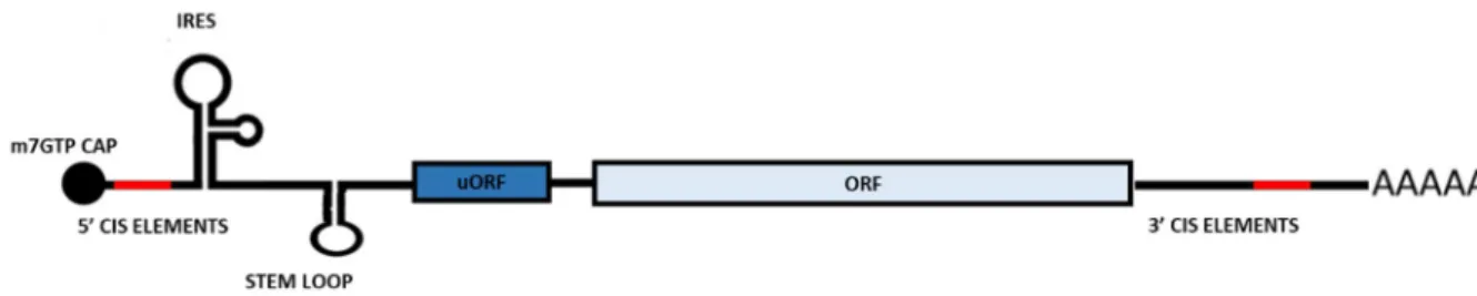
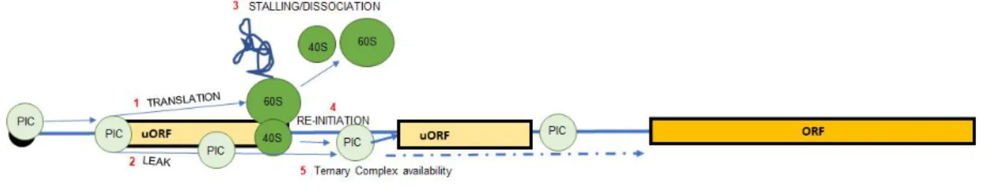
The fully mature mRNA possesses two characteristics shared by almost all the mRNAs: the 5’ cap and the poly-A tail. Each mRNA can be further divided in three components: a 5’ untranslated sequence (5’ UTR), a central coding sequence and 3’ untranslated sequence (3’ UTR). During evolution, the UTRs of the mRNA, have acquired a series of distinctive features that influence the stability and functionality of mRNA (Fig. 3). These features are referred as cis-elements and can be either recognized by regulatory factors such as RNA-binding proteins and miRNA. Further, merely their presence can influence the stability and/or translation of the mRNA. The 5’ UTR features mainly regulate translation: for example, ribosome internal entry site (IRES) allows a cap-independent translation initiation and, in some cases, generation of two completely different proteins from the same mRNA defined as polycistronic mRNA (Karginov et al., 2017). Another example is the upstream open reading frame (uORFs) which are important regulators of translation efficiency (McGillivray et al., 2018). The 3’ UTR contains cis-element that recruit trans-active factors involved in mRNPs transport and regulate stability (e.g. miRNA binding site and AU-rich elements).
The focus of recent research is to characterize the chemical modifications of the mRNAs (e.g. methylation), driven by technological advancement. This has allowed the establishment of a new layer of post-transcriptional regulation called the “epitranscriptome”. As for epigenetic regulation, the epitranscriptome, has its own readers, writers and erasers which are able to depose remove and read the modifications on the RNAs. The epitranscriptome has been implicated in regulating many post-transcriptional processes including translation (Lewis et al., 2017).
24
Figure 3. Schematic representation of structural organization of a mature mRNA
Ribosomes
The Ribosome is a macromolecular complex composed by the ribosomal proteins (RPs) and non-coding ribosomal RNA (rRNA). In eukaryotic cells, ribosomes are composed by two subunits: the small subunit (40S) and the large subunit (60S). Together, they constitute the complete ribosome (80S). The large subunit consists of three rRNA: 5S, 5.8S and 28S and 50 proteins. The small subunit is composed of one rRNA (18S) and 33 proteins (Garrett and Grisham, 2010). Importantly, single-copy mutations in specific ribosomal proteins or defects in ribosome biogenesis factors are known to cause a heterogeneous group of human disorders, called ribosomopathies (Mills and Green, 2017). Recent studies also claim the existence of heterogeneous ribosomes at the level of core ribosomal proteins which can affect translation of specific sub pools of mRNAs (Shi et al., 2017). For example, ribosomal protein L38 selectively facilitates the translation of subsets of Hox mRNAs (Kondrashov et al., 2011).
Eukaryotic Factors for translation
The eukaryotic translation factors are proteins which help structurally and enzymatically during several phases of translation, therefore they have been named accordingly to the translation phase they participate: eukaryotic initiation factors (eIFs), eukaryotic elongation factors (eEFs), eukaryotic release factors (eRFs) and ribosome recycling factors (RRF) which are needed to release the ribosome after protein synthesis. These factors are also the main targets that regulate translation as discussed in subsequent sections.
25
2. The translation initiation process
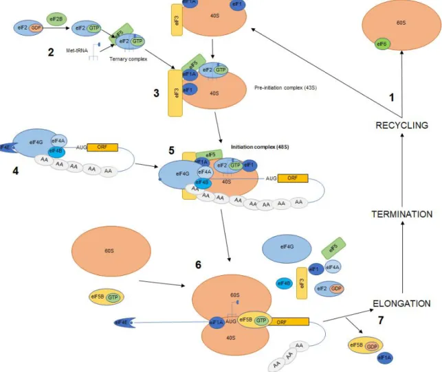
Generally, translation is a sequence of of three steps: initiation, elongation and termination. Although they are all susceptible to regulation, most of which takes place at the initiation step which is rate limiting step. In eukaryotes, cells usually initiate translation via a cap-dependent mechanism named Cap dependent translation (von der Haar et al., 2004). The cap is a guanosine methylated in position N7 linked to the first 5’ nucleotide by an unusual 5’ to 5’ triphosphate bond. The interaction of eIFs factor with the cap allow the recruitment of the ribosomal subunits onto the mRNA. Translation initiation is a multi-step process (overview in Fig. 4) that involves twenty-five eIFs factor and the auxiliary factor PABP (poly-A binding protein). The process can be divided in different steps as described in subsequent sections.
Translation initiation requires primed ribosomal subunits
Translation initiation requires a pool of non-associated 60S and 40S ribosomal subunits. To this end, the large 60S ribosomal subunit is bound by eIF6 to block its interaction with 40S subunit. The small ribosomal subunit is bound by eIF1A, eIF1, eIF3 and eIF5 that induce a conformational change that opens the 40S mRNA binding channel. Moreover, their association is required for 40S priming, which is necessary for the upcoming translation initiation steps (Hinnebusch, 2014).
Formation of the pre-initiation complex
A key step in translation initiation is the recycling of eIF2-GDP by eIF2B to the eIF2-GTP activated form. Once eIF2 is bound to GTP, it can recruit a Met-tRNAiMet to form the ternary complex (eIF2-GTP-Met-tRNAiMet). The ternary complex associates with a primed 40S subunit forming the 43S preinitiation complex (PIC). Importantly, eIF1 and eIF1A enhance the recruitment and stability of the ternary complex on the mRNA (Passmore et al., 2007). The eIF3 complex, by interacting with eIF1, eIF1A, eIF2, eIF5 and the rRNA of the 40S, stabilizes the newly formed 43S complex (Querol-Audi et al., 2013).
Formation of the eIF4F complex
The 5’ cap of the mRNA is bound by the eukaryotic Initiation Factor 4E (eIF4E) (Sonenberg et al., 1978), which replaces the nuclear CBC during the pioneer round of translation. eIF4E recruits eIF4G, a scaffold protein that on one hand enhances eIF4E affinity for the cap structure (Haghighat and Sonenberg, 1997), and on the other hand recruits eIF4A, an RNA helicase that unwinds the 5’ UTR secondary structures. The activity of eIF4A is modulated by two other factors: eIF4B and eIF4H (Rogers et al., 2001). Together, these factors form a complex known as eIF4F. Moreover, eIF4G binds to the PABPs (PolyA binding proteins) family members, that cover the poly-A tail at the 3’ end of the mRNA. As a consequence, the two
26
mRNA extremities are brought together forming the closed loop conformation which enhances translation efficiency and recycling of the components involved in the initiation step (Kahvejian et al., 2001).
Figure 4. Cap-dependent eukaryotic translation initiation
1. During the recycling step, the 60S interacts with eIF6 which impedes its association with the 40S while the latter is bound by eIF1A, eIF3 and eIF5. 2. eIF2B enhances the recycling of eIF2-GDP to eIF2-GTP which forms the ternary complex with the Met-tRNAiMet. 3. Ternary
complex associates with the recycled 40S forming the preinitiation complex 43S. 4. Meanwhile, the eIF4F complex (eIF4E, eIF4G, eIF4A) binds to the 5’ cap of the mRNA through bound PABPs hence forming the close loop conformation. 5. The preinitiation complex is recruited onto the mRNA by the eIF4F complex forming the 48S complex which scans the mRNA for the start codon, which when recognized induces eIF5-mediated hydrolysis of eIF2-bound GTP and inorganic phosphate (Pi) release. 6. 60S is recruited causing displacement of the eIFs factors. 7. Hydrolysis of eIF5B-bound GTP results in its release of eIF1A from the assembled 80S ribosomes, which will commence the Elongation stage. After termination ribosomes subunits re-enter the cycle (modified/adapted from Nature Reviews Molecular Cell Biology 11, 113–127 (2010)).
27
Formation of the 48S initiation complex
Both eIF4G and the eIF3 are required for the following step where the 43S is recruited onto the mRNA to form the 48S complex, also known as the pre-initiation complex (PIC). The 48S complex will then start scanning the 5’UTR for the proper AUG triplet in an optimum context, defined Kozak sequence (Kozak, 1987). Importantly, to allow the proper reading from the first nucleotide, the eIF4E is likely to be displaced from the cap structure (Kumar et al., 2016). While the PIC complex scans the 5’UTR of the mRNA, eIF1 and eIF1A are responsible for the fidelity of the recognition of the start codon. Their concomitant absence induces recognition of erroneous starting codons (Pestova et al., 1998). Once the proper starting codon is recognized, the PIC complex undergoes a structural change to switch to closed conformation of the mRNA binding channel. This conformational change allows eIF5B-GTP to interact with eIF2-GTP causing its hydrolysis to eIF2-GDP and Pi (Algire et al., 2005).
60S joining and eIFs displacement
The eIF5B-GTP through its interaction with eIF1A, allows the joining of the large subunit of the ribosome with the PIC causing dissociation of the eIFs from the complex (Acker et al., 2006). Before its dissociation eIF5B-GTP gets hydrolyzed to eIF5B-GDP thus causing release of eIF1A from the assembled ribosome (Allen and Frank, 2007). Furthermore, eIF6 interacts with RACK1 (receptor for activated protein kinase C) that, when activated, causes eIF6 release from the 60S thus allowing formation of the 80S (Ceci et al., 2003). Ribosome can now enter the elongation stage.
3.
A new complex for cap-dependent
translation
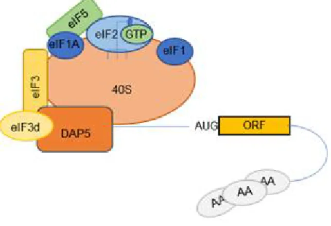
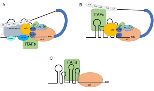
It has been recently demonstrated that eIF3d, a subunit of the eIF3 complex, can recognize the 5’ cap of the mRNAs (Fig. 5). Importantly, the region which binds to the cap is conserved in three different kingdoms: plants, fungi and animals suggesting an evolutionary conservation (Lee et al., 2016). A follow up study published recently identified DAP5 as protein partner of eIF3d when bound to the cap (ref). DAP5 is a member of the eIF4G family which has the ability to interact with the eIF3 complex and eIF4A, but it has lost the N-terminal domain, through which the other two eIF4Gs (4G1 and 4G2) interact with eIF4E and the PABPs (Imataka, 1997). DAP5 was previously known to participate in a cap-independent translation which require particular RNA structures known as IRESs (Internal Ribosome entry sites) (Lewis et al., 2007). However, silencing of DAP5 induces a 20% loss of protein synthesis rates which
28
does not reflect the number of mRNAs translated via IRES elements. Moreover, genome wide studies demonstrated that DAP5 affect translation of large number of genes that does not possess a known IRES element (de la Parra et al., 2018). It is thus likely that some mRNAs could use both eIF4E or DAP5 cap-complexes to initiate translation. This could be a major breakthrough in understanding why and how many mRNAs are translated under condition in which mTOR signalling inhibits eIF4E-4G interaction (See Chapter B, “4E-BPs”).
Figure 5. Possible initiation complex formed by eIF3d-DAP5 on 5' cap of the mRNAs The eIF3d subunit of the eIF3 complex is able to interact with the cap at the 5’UTR of the mRNAs and together with DAP5, homolog of eIF4G possibly allows recruitment of the pre-initiation complex.
The mechanisms regulating translation initiation can be divided in two categories: the first where the activity of the initiation factors is modulated by post-translational modification or by binding partners, while the second depends on the mRNA characteristic, named cis-elements, which modulate the composition of the mRNP and therefore its own fate. This classification is arbitrary, since the two groups are intertwined, and its only scope is to aid describing the various actors of translation initiation. In the following chapters, I will discuss the regulation of the eIFs and the pathways involved. In the second chapter I will discuss the different ways by which mRNA cis elements affect translation initiation.
29
C. Regulation of eIFs
As described in the previous chapter, eIFs are the main actors in the initiation of translation and thus, unsurprisingly, most of the regulation of translation initiation focuses on the modulation of their activities. The regulation of eIFs is known to occur by two major mechanisms: post-translational modifications, among which phosphorylation is by far the most important, and intervention of protein partners. The following section focuses on these important modes of regulation and brings to light some key examples.
1. eIFs phosphorylation and dephosphorylation
Phosphorylation consists of the addition of a phosphoryl group (PO3-) by enzymes called kinases to the side chains of specific amino acids which, in eukaryotes, are usually serine, threonine, tyrosine and, to a lesser extent, histidine. Phosphorylation is the most common post-translational modification found in the cells and plays a key role in many cellular processes including translation, where if a key modulator of the activity of the eIFs. A good effort has been made to identify the phosphorylated eIFs, the sites or residues phosphorylated and the responsible kinases. The functional roles of some of these eIF phosphorylations have been investigated thoroughly, especially those regulated by the mammalian target of rapamycin (mTOR) pathway (Masvidal et al., 2017). However, the mechanisms and roles of newly discovered phosphorylation sites identified by genome wide methods, are yet to be uncovered. Particularly, the importance of these phosphorylation sites in regulating the translatome (the ensemble of mRNA being translated in the cells) is still largely unknown (table 1, summarize all the known phosphorylation sites of the different eIFs). Importantly, the phosphorylation of the eIFs is counterbalanced by enzymes known as phosphatases that remove the phosphoryl group from the modified amino acids. Phosphatases behave like buffering systems that avoid over-signalling via feedback loops or by directly counteracting phosphorylation.The phosphorylation dynamics of eIF2 are major modulators of translation initiation due to the central role of eIF2 in enhancing the rate limiting step of ternary complex assembly. Importantly, both kinases and phosphatases have been shown to regulate eIF2 phosphorylation status. In the following paragraph I will enter in the details of eIF2 regulation.
30
Protein Phosphorylation site(s), [kinase(s)]
Biological function(s)
eIF4E Ser209 [MNK1/2] increases oncogenic activity and promotes
translation of a subset of mRNAs (e.g., Mcl-1, MMPs, CCLs)
eIF4GI Ser1185 [PKCα, TBK1 ?]
Ser1106, Ser1147, Ser1194
[mTORC1]
Ser896 [Pak2] Ser1231 [CDK1?]
Modulates MNK binding
Stimulation of translation of mRNAs containing uORFs
Inhibition of cap-dependent translation Inhibition of eIF4A/mRNA binding?
eIF2α Ser52 [HRI, PKR, GCN2, PERK] Stabilizes the eIF2/GDP/eIF2B complex, thus preventing recycling of eIF2
eIF2β S2, S67 [CK2, mTORC1?] Stimulates translation and proliferation; stimulates eIF2α dephosphorylation
eIF4B Ser406, Ser422 [S6K1/2, AKT, RSK], Ser422 [MELK?]
Increases binding to eIF3, affects translation and proliferation
eIF4H Tyr12, Tyr45, Tyr101, Ser193 Unknown eIF2Bε Ser540 [GSK3]
Ser544 [DYRK] Ser717/718 [CK2]
Inhibits recycling of eIF2 Priming site for GSK3 Facilitates eIF2 binding eIF3 Subunit? [S6K1/2]
eIF3b: Ser83, Ser85, Ser125
eIF3c: Ser39, Ser166, Thr524, Ser909 eIF3f: Ser46, Thr119 [CDK11] eIF3g: Thr41, Ser42 eIF3h: Ser 183 eIF3i: Tyr 445 Paip1-eIF3 interaction Unknown Unknown
Regulation of protein synthesis and apoptosis Unknown
Increased oncogenic activity Unknown
eIF1 Tyr 30 Tyr 72
Unknown
Stimulation of mRNA translation
31
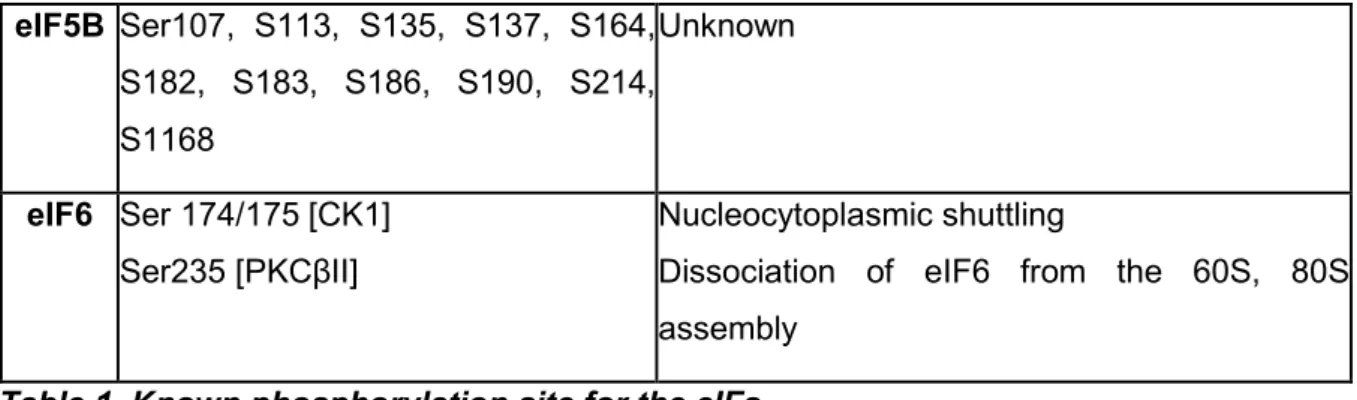
eIF5B Ser107, S113, S135, S137, S164, S182, S183, S186, S190, S214, S1168 Unknown eIF6 Ser 174/175 [CK1] Ser235 [PKCβII] Nucleocytoplasmic shuttlingDissociation of eIF6 from the 60S, 80S assembly
Table 1. Known phosphorylation site for the eIFs
List of all known phosphorylation sites on each of the eIFs factors and possible function. Between square brackets the kinase responsible (if known). Adapted from (Roux and Topisirovic, 2018).
32
a) Regulation of eIF2 and eIF2B
eIF2 is a highly conserved protein composed of three subunits (α, β and γ). The α and β subunits have regulatory function while the γ subunit interacts with both the Met-tRNAiMet and the GTP to form a ternary complex (TC). During translation initiation eIF2-GTP is hydrolyzed to eIF2-GDP to allow 60S joining. To re-form the TC, eIF2 is recycled to eIF2-bound GTP by the decameric complex eIF2B which possess guanine nucleotide exchange (GEF) function (Bogorad et al., 2018). eIF2α phosphorylated at Ser 51 sequesters eIF2B, inhibiting TC recycling and thus shutting down global translation (Sonenberg and Hinnebusch, 2009). eIF2α could be phosphorylated by either of the four kinases mentioned below, that respond to a wide array of cellular stresses, in a pathway known as the integrated stress response (ISR):
i. Protein kinase RNA-like endoplasmic reticulum kinase (PERK) is activated by hypoxia and endoplasmic reticulum stress
ii. Protein kinase RNA-activated (PKR) is activated by viral infections
iii. General control non-de-repressible 2 (GCN2) senses nutrients and UV irradiation iv. Heme-regulated inhibitor kinase (HRI) is regulated by heme deficiency and heat and
osmotic shocks.
As depicted in figure 6, there are many other regulations affecting the activity of these two factors. For example, eIF2β can also be phosphorylated at Ser2/67 by CK2 and by the mTOR complex 1 (mTORC1) (Llorens et al., 2006, Gandin et al., 2016). Phosphorylated eIF2β favors the interaction of the tyrosine kinase adaptor protein 1 (NCK1). NCK1 then recruits protein phosphatase 1 (PP1) complex which results in the dephosphorylation of eIF2α.
The activity of PP1 is regulated by different regulatory subunit: GADD34 (growth arrest and DNA damage-inducible protein 34), and the constitutive repressor of eIF2 phosphorylation (CreP) (Rojas et al., 2015).
Phosphorylation sites have also been identified on the ε subunit of eIF2B. While most of these phosphorylation sites are required for GEF (Guanine nucleotide exchange factors) activity of eIF2B and its interaction with eIF2 (Wang, 2001), phosphorylations at Ser544 or at Ser 540 by the GSK3β (Glycogen synthase kinase 3 beta) inhibit the activity of eIF2Bε (Welsh et al., 1998).
33
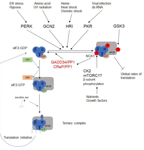
Figure 6. Schematic view of the regulation of Ternary Complex formation
Upon different stresses four different kinases (PERK, GCN2, HRI, PKR) can be activated and phosphorylate eIF2α subunit at Ser51 which increase its affinity for eIF2B. This conformation inhibits eIF2B activity, preventing the recycling of eIF2-GDP to eIF2-GTP. The phosphorylation status of eIF2α is counterbalanced by phosphatases recruited by NCK1 along with the phosphorylation of eIF2β by CK2 and possibly mTORC1.
34
2. eIFs regulation by protein partners: the case
of eIF4E
The activity and the recruitment of the eIFs during translation initiation is also modulated by some of their protein partners.
Most eukaryotic mRNAs are translated by the cap-dependent mechanism, that requires eIF4E to be bound to the 5’ cap structure of the mRNA. In this conformation, eIF4E recruits eIF4G onto the mRNA paving the way for ribosome recruitment. Therefore, the bond between eIF4E and eIF4G has become a core regulatory mechanism of translation initiation. Indeed, various proteins, termed the eIF4E-interacting proteins (4E-IPs), are able to compete with eIF4G for binding to eIF4E. We have suggested that the 4E-IPs evolved independently in the different taxonomic groups through a process of molecular tinkering (Hernández et al., 2016).
Most of the 4E-IPs interact with eIF4E via the consensus motif YXXXXLϕ (where ϕ is an aliphatic amino acid (L, F or M)), while few of them have opted for different strategies: the human promyelocytic leukemia (PML) and the arenavirus Z protein interact through a RING motif (Kentsis et al., 2001). Instead, the Cytoplasmic FMR1-interacting protein 1 (CYFIP1), interacts via a peptide which tertiary structure mimics the tertiary structure of the YXXXXLϕ motif (Napoli et al., 2008). The following section addresses the role and regulation of the most studied family of 4E-IPs: the 4E-BP family. This is continued by a further discussion of the roles of other 4E-IPs.
a) 4E-BPs
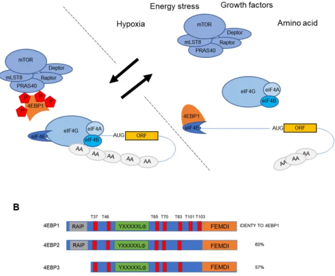
Three members of the 4E-BP family have been identified in vertebrates: 4E-BP1, 4E-BP2 and 4E-BP3 (Table 2). They share approximately 60% protein identity (Fig. 6B) and they are expressed in all tissues in varying amounts, depending largely on tissue-specific contexts. Mechanistically, hyperphosphorylated 4E-BPs (phosphorylation on multiples sites) cannot interact with eIF4E, while hypophosphorylated they sequester eIF4E from eIF4G (Gingras et al., 1998). Under favorable physiological context the two isoforms 4E-BP1 and 4E-BP2 are recruited to the mTORC1 complex (mammalian target of rapamycin complex 1) via the interaction between the Raptor, subunit of mTORC1, and their C-terminal TOS (TOR signaling) motifs (also known as FEMDI) (Fig 6A). In this conformation, the kinase subunit mTOR phosphorylates 4E-BP1 and 4E-BP2 in a hierarchical way: phosphorylation of Thr37 and Thr46 precedes phosphorylation of Thr70 and Ser65 (Gingras, 2001).
Upon a wide range of stimuli including growth factors, hormones, cellular energy status, and oxygen availability, mTORC1 is inhibited resulting in a quick dephosphorylation of the 4E-BPs. Importantly, the phosphatases belonging to two different families PP1/PP2 and PPM, have
35
been shown to be important players in this process (Gardner et al., 2015). Once, hypophosphorylated, the 4E-BPs are free to sequester eIF4E shutting down translation initiation.
More importantly, other kinases have been shown to be capable of 4E-BPs phosphorylation on identical or dissimilar residues. However, their physiological relevance is not yet well understood (reviewed in (Batool et al., 2017)).
Strikingly 4E-BP3 is likely regulated in a unique manner since rapamycin, a specific inhibitor of mTORC1 (blocks mTOR-Raptor interaction) does not affect 4E-BP3 binding to eIF4E. Indeed, in a recent work, 4E-BP3 was shown to play an important role as translation repressor when mTORC1 remained inhibited for a prolonged period of time (Tsukumo et al., 2016).
Figure 7. 4E-BP is phosphorylated by mTORC1 to block cap-dependent translation A. mTORC1 in normal conditions binds and phosphorylates the 4E-BPs impeding their binding to eIF4E. Upon cellular stress such as hypoxia, energetic stress (e.g. glucose deprivation), amino acid starvation and growth factors mTORC1 is sequestered or inactivated. 4E-BPs are then free to sequester eIF4E thus inhibiting cap-dependent translation. B. Overview of the protein structure of the 4E-BP family with representation of the various domains and phosphorylation sites.
36
The eIF4E interacting partners
4E-IPs have the most disparate roles in different cellular processes including functions outside translation regulation. Notably,
They act as transacting factors by either directly binding RNA elements (e.g. LRPPRC and DDX3) or by contacting others RNA binding proteins (Neuroguidin and CYFIP1) They also regulate eIF4E localization: PML sequesters eIF4E in the nuclear bodies. In
fact, in many cells, a prominent part of eIF4E is in the nuclear compartment (Dostie et al., 2000). DDX3 interacts with eIF4E in the P-bodies. 4E-T is responsible for the shuttling of eIF4E between nucleus and cytosol, but it also causes eIF4E re-localization to P-bodies. They regulate nuclear export: LRPPRC regulates nuclear export by binding mRNAs bearing on their 3’ UTR a motif of 50 to 100 nucleotides with a highly conserved secondary structure, called 4E-SE (4E-sensitivity element) (Topisirovic et al., 2009).
A complete overview of the known mammalian 4E-IPs is listed in following Table 2.
Protein Binding
partner
Biological process Reference
4E-BP1; 4E-BP2; 4E-BP3;
eIF4E1 Cell cycle progression, cell growth and proliferation; Synaptic plasticity and memory formation.
(Haghighat et al., 1995); (Pause et al., 1994); (Lin et al., 1994); (Poulin et al., 1998) Neuroguidin (Ngd)
eIF4E1 Neurogenesis (Jung et al., 2006)
Angel1 eIF4E1 Endo/exo-nuclease-phosphatase
domain-containing protein; No known biological role.
(Gosselin et al., 2013)
Promyelocytic leukemia protein (PML)
eIF4E1 Nuclear mRNA export and DNA repair; Cell growth and apoptosis.
(Kentsis et al., 2001)
GYGYF2b eIF4E2 GRB10-interacting GYF protein 2. (Morita et al., 2012) Cytoplasmic
FMR1-interacting
protein 1
(CYFIP1)
eIF4E1 FMRP-interaction factor during
neuronal activity; Actin
polymerization.
(Napoli et al., 2008)
Leucine-rich pentatricopeptide
eIF4E1 Mitochondrial RNA transport and expression; Nuclear mRNA
metabolism; Neurogenesis;
37
repeat containing protein (LRPPRC)
Mitochondrial unfolded protein response.
PRH eIF4E1 Homeobox transcription factor;
Haematopoiesis.
(Topisirovic, 2003)
HOXA9 eIF4E1 Homeobox transcription factor;
Haematopoiesis.
(Topisirovic et al., 2005)
DEAD-box
helicase 3 (DDX3)
eIF4E1 DEAD box RNA helicase; (Shih et al., 2008)
EMX2 eIF4E1 Homeobox transcription factor;
Neurogenesis.
(Nedelec et al., 2004)
PREP1 eIF4E2 Homeobox transcription factor;
Embryo development;
Hematopoietic stem cell biology.
(Villaescusa et al., 2009) eIF4E transport (4E-T) eIF4E1 eIF4E2 Nucleo-cytoplasmic transport protein; P-body formation in human
(Kamenska et al., 2014)
(Kubacka et al., 2013)
Z protein eIF4E1 Viral life cycle. (Kentsis et al., 2001)
TRIM22 eIF4E1 mRNA translation in response to
p53 and/or IFN signalling
(Petersson et al., 2012)
HIF-2α/RBM4 eIF4E2 mRNA translation in response to hypoxia
(Uniacke et al., 2012)
ARIH1 eIF4E2 IGS15 modification, modulation of
cap affinity?
(Tan et al., 2003) (Okumura et al., 2007)
Table 2. eIF4Es interacting partners
List of known proteins to interact with one or more eIF4E family members with known role. Adapted from (Hernández, 2008)
38
3. Specialized family members: eIF4E paralogs
Gene duplication events are a major driving force of evolution in eukaryotes. Gene descending from a unique common ancestor are defined as paralogs. The most important characteristic of paralogs genes is to code for proteins during evolution have acquired new independent functions. This is also the case also for many of the eIF factors (Hernández et al., 2005). For example, the family of eIF4E is composed of three paralogs genes (eIF4E1, eIF4E2 and eIF4E3) which originated from one common ancestor and they share 30% sequence similarity (Joshi et al., 2005). The three classes are defined based on presence of tryptophan at position 43 et 56 (human sequence) and the latter reside in the interaction domain with the cap structure. The three classes of eIF4E have substantially different characteristics (summarized in table 3): they differ for their ability to interact with the cap structure,
not all the three classes interact with the two major protein interactors (eIF4G and 4E-BPs),
they also differ for their expression levels and distribution among the different tissues which strongly indicates specific and independent roles between the three classes. In tetrapoda eIF4E1 is present in two copies eIF4E1a and eIF4E1b that arose from gene duplication. In mammals, the eIF4E1b expression is confined to oocytes (Evsikov et al., 2006) and has 3-fold reduced affinity for the cap compared to eIF4E1a (Kubacka et al., 2015). To date, eIF4E1b functional role remains a mystery.
Instead, eIF4E1a is ubiquitously expressed and since it has always been the center of researcher’s attention is historically referred as eIF4E.
Despite being the general regulator of cap-dependent translation, eIF4E has been shown to exert a stronger translational control on those mRNA containing a long and highly structured 5’ UTR. These features suggest that translation initiation for these mRNA is strongly influenced by the helicase activity of eIF4A, which is strongly enhanced when part of eIF4F complex (formed by eIF4E, eIF4G and eIF4A).
Importantly, many of the genes affected by eIF4E are known proto-oncogenes involved in important hallmarks of cancer such as cell cycle regulation, survival and angiogenesis (De Benedetti and Harris, 1999) (Mamane et al., 2007). Accordingly, over-expression of eIF4E by 2-3 folds is sufficient to induce tumorigenesis (Ruggero et al., 2004) and indeed is a common feature of many types of cancers (reviewed in (Hsieh and Ruggero, 2010)). Surprisingly eIF4E down-regulation in cellular models did not affect global proteins synthesis rates (Yanagiya et al., 2012) and in mouse models its downregulation to 50% of initial levels did not affect normal development (Truitt et al., 2015) but affected stress responses and increased cancer rates. In
39
the next paragraph I will focus on the recent advances in understanding the role of the two less studied classes eIF4E2 and 4E3.
Interactions Function
Cap eIF4G
4E-BPs
Role Expression
eIF4E1a strong strong strong General regulator of cap dependent translation
Ubiquitously expressed
eIF4E1b weak ? ? ? Oocytes
eIF4E2 weak weak No? Regulation of subset of mRNA
under particular stress conditions
Ubiquitously expressed
eIF4E3 weak yes No Selective translation Tissue specific
Table 3. Main properties of the eIF4E family members
a) The eIF4E2
The paralog eIF4E2 is ubiquitously expressed albeit at 10-fold lower levels of expression than eIF4E1. Its affinity for the cap structure is one hundred times less strong than eIF4E1 (Zuberek et al., 2007). The role of eIF4E2 has remained elusive until recent years when it has been reported to regulate translation of a subset of genes during hypoxic stress, where eIF4E2 is brought to the 5’ cap structure by RBM4 (RNA-Binding Motif protein 4) and HIF-2α (Hypoxia Inducible Factor 2α) that bind to the RNA hypoxia response element (rHRE) present on the 3’UTRs of specific mRNAs. Then, eIF4E2 forms an alternative eIF4F complex by recruiting eIF4G3 and eIF4A. Thus, eIF4E2 induces an adaptive, cap-dependent hypoxic translatome (Uniacke et al., 2012). The mRNAs translationally regulated by eIF4E2 have been shown to be involved in cancer progression. Interestingly, eIF4E2 has been found to be mutated in many cancers (Melanson et al., 2017) where hypoxia is a driving force that favors angiogenesis, metastasis, and resistance to therapy. Moreover, another report has shown that during embryonic development, a process driven by hypoxic condition, eIF4E2 forms a complex with GYGYF2 (GRB10-interacting GYF protein 2) and the zinc finger protein 598 (ZNF598). Knock out of either of these proteins lead to perinatal mortality (Morita et al., 2012). Taken together, these results highlighted that eIF4E2 is able to form different cap binding complexes that shape the translatome depending on the proteins partners and the cellular context.
Beside eIF4E2 role under hypoxic condition, it has been described to participate also under other cellular stresses: