Surprising lack of liposome-induced complement activation by artificial
1,3-diamidophospholipids in vitro
Simon Bugna, MSc
a, Marzia Buscema, MSc
a, Sofiya Matviykiv, MSc
a,
Rudolf Urbanics, MD, PhD
b, Andreas Weinberger, PhD
c, Tamas Meszaros, MSc
b,
Janos Szebeni, MD, PhD, DSc
b, Andreas Zumbuehl, PhD
c,⁎
, Till Saxer, MD
d,⁎
,
Bert Müller, PhD
a,⁎
a
Biomaterials Science Center, University of Basel, Allschwil, Switzerland
bNanomedicine Research and Education Center, Semmelweis University, Budapest, Hungary cDepartment of Chemistry, University of Fribourg, Fribourg, Switzerland dCardiology Division, University Hospitals of Geneva, Geneva, Switzerland
Abstract
Cardio-vascular diseases are the main cause of death, emphasizing the need to improve patient treatment and survival. One therapeutic approach is a liposome-based drug carrier system specifically targeting constricted arteries. The recently discovered mechano-sensitive liposomes use hemodynamic shear-stress differences between healthy and constricted blood vessels as trigger for drug release. Liposomes are promising delivery containers but are being recognized as foreign by the immune system. Complement activation as essential factor of the recognition leads to adverse effects. Here, we tested complement activation by liposomes formulated from the artificial phospholipid Pad-PC-Pad in vitro. Surprisingly no complement activation was detected in human sera and porcine plasma. In in vivo experiments with three pigs, neither anaphylactic reactions nor other significant hemodynamic changes were observed even at comparably high liposome doses. The pilot study holds promise for an absence of complement-mediated adverse effects of Pad-PC-Pad liposomes in human.
From the Clinical Editor: A lot of research has been done on new treatment for cardiovascular diseases. Liposome-based carrier systems have also shown promises. In this article, the authors studied the potential risks of complement activation by liposomes in in-vivo experiments. The absence of complement activation by Pad-PC-Pad liposomes may indicate its use in humans.
Key words: Pad-PC-Pad; CARPA; Pig study; Shear sensitive nano-container; Physical trigger
According to the World Health Organization, cardiovascular diseases are responsible for 30% of worldwide deaths.1Many of them arise during transfer to hospital, motivating the increasing interest in pre-hospital drug delivery to critically constricted vessels.2,3 One strategy involves targeting the constricted arteries by capitalizing on the endogenous, elevated shear stresses present in diseased artery segments and using the changes as a purely physical trigger for drug release.4
Liposomes composed of the artificial phospholipid Pad-PC-Pad can release their payload in areas of increased shear stress.5In order to push this development towards clinical application, the liposomes need to be further characterized to exclude non-desired systemic effects. Here, predicting the risk of complement-activation-related-pseudo-allergy (CARPA) is fundamental, as it characterizes the hypersensitivity towards liposomal formulations.6,7 This phenomenon also occurs with clinically approved lipid-based drugs and creates adverse effects in 2% to 30% of people, occasionally leading to anaphylactic reaction or even death.7,8
Previous studies have shown that liposomal formulations including Doxil9,10and AmBisome11lead to lethal effects, if 0.5 to 5.0 mg/kg phospholipids are applied. First cell experiments with Pad-PC-Pad, however, have indicated a reasonable tolerance.5 Therefore, we propose in vitro tests with human sera and porcine plasma to characterize the CARPA reaction and animal studies to search for liposome-induced blood-pressure Disclosure: The study was supported by the Swiss National Science
Foundation within the project NO stress of the National Research Programme 62 Smart Materials. The authors report no conflicts of interest.
Acknowledgments: We acknowledge the experimental support of Dennis Müller as well as the help of Hans Deyhle and Georg Schulz in preparing Figure 3. ⁎Corresponding authors at: Biomaterials Science Center, University of Basel, Gewerbestrasse 14, Allschwil, Switzerland.
E-mail address:[email protected](B. Müller).
http://doc.rero.ch
Published in "Nanomedicine: Nanotechnology, Biology and Medicine 12(3): 845–849, 201
6"
changes; both are fundamental to bridge the gap between bench and bedside. We hypothesize that contrary to Doxil and AmBisome the liposomes from the artificial phospholipid Pad-PC-Pad without an encapsulated active drug are reaction-free at comparable dose. Revealing such lack of complement activation, the native drug carrier could be assumed as safe from CARPA.
Methods
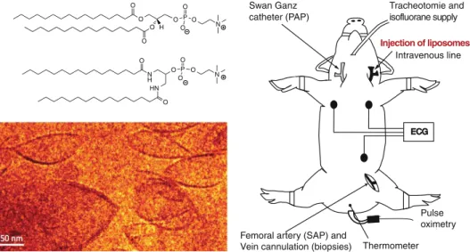
Pad-PC-Pad (1,3-palmitoylamido-1,3-deoxy-sn-glycero-2-phosphatidyl-choline) has been prepared according to the established protocol.5 Figure 1 displays its structure in comparison to the natural 1,2-diesterphospholipid DPPC.
The liposomes were formulated using the thin-film method.12 The phospholipids hydrated with PBS were extruded at a temperature between 65 and 70 °C through 100 nm track-edged filters at a concentration of 20 mg/mL. The liposome stock suspension was stored without light at a temperature of 4 °C. The experiments were conducted within 48 h after formulation. DLS measurements have shown no liposomes aggregation for in vitro testing, while their aggregation and sedimentation were observed after 12 h of storage in case of in vivo experiment (see Figure S1). Therefore, before bolus injection, the storage vial was repeatedly inverted to re-suspend the sediments, and, subsequently, the suspension was passed through a 0.22-μm syringe filter.
Five human sera samples, labeled H1 to H5, were tested with the Quidel MicroVue SC5b-9 ELISA kit. A volume of 20μL Pad-PC-Pad suspension was added to each serum at a temperature of 37.1 °C. Additionally, five pig hirudinized plasma samples, labeled P1 through P5 were tested with the Quidel MicroVue Pan-specific C3 reagent kit. A volume of 25μL of Pad-PC-Pad suspension was added to each plasma. After 0, 20, 40, and 60 min, EDTA was added to stop the reaction. Each series contained controls of PBS only and 20 or 25μL zymosan (1.2 mg/mL), purchased from Sigma Chemical Co. (St Louis, MO). The optical density of the
samples was measured with a plate reader at a wavelength of 450 nm. The concentration of the SC5b-9 complex was calculated from a linear calibration curve.
Three female 12- to 14-week-old Yorkshire pigs with a weight of 18.4, 22.8 and 20.2 kg were injected with various bolus doses of Pad-PC-Pad vesicle suspensions and with zymosan, while monitor-ing the hemodynamic parameters. The protocol was approved by the local Animal Subject Review Committees; for details see Appendix C of supplementary information.
Results and discussion
Figure 2summarizes the results of the Pad-PC-Pad liposome tests using human sera in the ELISA assay for SC5b-9. Within P O O O N O H O O O O N H O HN P O O O N O O ECG ECG
Femoral artery (SAP) and Vein cannulation (biopsies)
Pulse oximetry Intravenous line Tracheotomie and Swan Ganz catheter (PAP) Thermometer isofluorane supply Injection of liposomes
Figure 1. Structures from the natural 1,2-diesterphospholipid DPPC, the major component of FDA-approved liposomal formulations and below of 1,3-diamidophospholipid Pad-PC-Pad that can form the lentil-shaped liposomes used in this study. The scheme explains the surgery procedure. The details are described in the supplementary material, Appendix C.
Figure 2. The complement activation of Pad-PC-Pad liposomes with five human sera labeled H1 to H5 is comparable to the negative control PBS for the incubation times indicated. The bars are the standard deviations between the donors. Significance of differences among the groups was determined by Two-Way analysis of Variance (ANOVA), followed by the Newmann–Keuls test (Pb 0.05).
the error bars, the liposomes do not induce any complement activation, whereas the complement activator zymosan triggered an immediate complement reaction more than 20 times higher than the negative control. The variability between the five donors is given in Figure S2. The results are in line with placebo Doxil (Doxebo).13The soluble terminal complement complex SC5b-9 is formed from C5 to C9 as the end result of complement activation.14An elevation of SC5b-9 against artificial phospholipid liposomes is therefore indicative for the in vivo reaction. Conse-quently, one does not expect in vivo reactions in pig experiments.
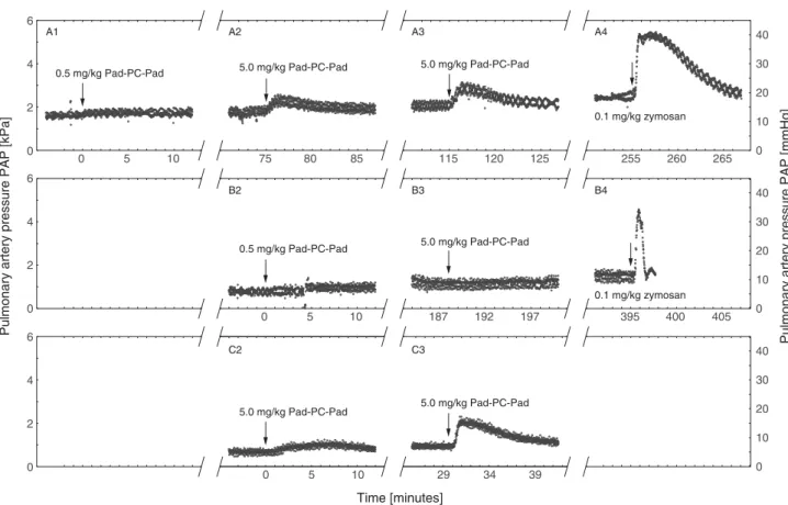
Indeed,Figure 3demonstrates that the in vivo reactions have been consistent with the ELISA tests. Globally, the three pigs showed a stable hemodynamic behavior during anesthesia and after injection of Pad-PC-Pad. Since SAP, heart rate, and ECG were constant within the error bars,Figure 3only displays PAP. Two pigs, Figure 3, A and C, were injected with unfiltered, aggregated Pad-PC-Pad liposomal suspensions. The third pig, Figure 3, B, was injected with a filtered, non-aggregated Pad-PC-Pad suspension. These injection regimens indicate the differences between homogeneous, equally sized liposomes and liposomal aggregates. The first pig (Figure 3, A1) showed no reaction after the 0.5 mg/kg injection. Even after the 5 mg/kg injection the PAP remains constant. Three minutes after an additional 5 mg/kg injection (Figure 3, A3) the PAP increased by 500 Pa (4 mmHg) for two minutes. The positive reference zymosan injection led to an immediate increase by more than 2.7 kPa (20 mmHg), long lasting reaction (Figure 3, A4). The
second pig showed no hemodynamic reaction applying doses of 0.5 and 5 mg/kg (Figure 3, B2 and B3, respectively). In the third pig (Figure 3, C), the PAP was stable after the first aggregated liposome injection with a dose of 5 mg/kg, but increased by 930 Pa (7 mmHg) for a period of six minutes subsequent to the second injection. As the third pig was reactive to a certain extent, we decided to directly proceed with histological sampling and skip the zymosan injection. This histology analysis revealed that the administration of Pad-PC-Pad liposomes did not lead to histological toxic modification in kidney, lung, heart, and liver. Table 1summarizes the analysis of the porcine blood. The values correspond to the ones reported for healthy Yorkshire pigs.15 The lack of thromboxane changes in pigs is consistent with the in vitro test. C5a stimulates the production of arachidonic acid derivates prostaglandins and thromboxane by neutrophils and macrophages. The thromboxane levels remained stable during the entire experiment. They only increased following the zymosan injection.
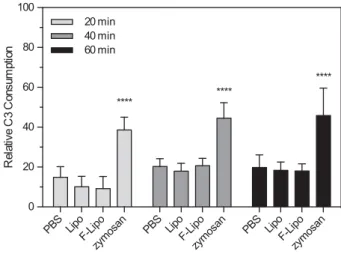
To support the link between the in vitro and in vivo experiments complement activation was also measured via C3 consumption in pig plasma. The data, presented inFigure 4, reveal the lack of complement activation.
Comparing the results of the in vivo study with data published on Doxil and AmBisome, we observed the lack of reactivity for Pad-PC-Pad liposomes. Even at one order of magnitude higher concentrations the non-aggregated Pad-PC-Pad liposomes do not trigger any reactivity in vivo.
Pulmonary artery pressure PAP [kPa]
Pulmonary artery pressure PAP [mmHg]
0 5 10 75 80 85 115 120 125 255 260 265 0 5 10 187 192 197 395 400 405 0 5 10 29 34 39 2 4 6 0 10 20 30 40 0 10 20 30 40 0 10 20 30 40 0 2 4 6 0 2 4 6 0 A1 A2 A3 A4 B2 B3 B4 C2 C3 Time [minutes] 0.1 mg/kg zymosan 0.1 mg/kg zymosan 5.0 mg/kg Pad-PC-Pad 0.5 mg/kg Pad-PC-Pad 0.5 mg/kg Pad-PC-Pad 5.0 mg/kg Pad-PC-Pad 5.0 mg/kg Pad-PC-Pad 5.0 mg/kg Pad-PC-Pad 5.0 mg/kg Pad-PC-Pad
Figure 3. PAP change is normalized to time zero. (A) Subsequent injections of Pad-PC-Pad liposomes at 0.5 and 5 mg/kg, followed by an injection of 0.1 mg/kg zymosan as positive control. The sample was re-suspended and filtered before the first injection. (B) Injection of directly filtered Pad-PC-Pad liposomes to avoid liposome aggregate formation. (C) Injection of non-filtered liposomes at 5 mg/kg lipid concentration.
In conclusion, this pilot study was performed to explore the in vitro complement activation by Pad-PC-Pad and the in vivo CARPA reactivity in a pig model, which is more sensitive for
CARPA than humans. The clinical relevance of the study lies in the increased risk for non-IGE-mediated reactions in patients treated for the first time with liposomal drugs. Although premedication reduces the risk of anaphylactic reactions, the risk for such reactions remains present. The findings indicate that Pad-PC-Pad liposomes do not directly or indirectly cause anaphylactic reactions even with a dose as high as 110 mg/kg. In comparison to approved phospholipid formulations,7,17 Pad-PC-Pad with a ten- or even hundred-fold higher dose reveals much less CARPA reactivity in vivo. To this end, Pad-PC-Pad is regarded as a promising phospholipid for liposomal drug delivery applications in humans.
Appendix A to C. Supplementary data
Supplementary data to this article can be found online at http://dx.doi.org/10.1016/j.nano.2015.12.364.
References
1. Go AS, Mozaffarian D, Roger VL, Benjamin EJ, Berry JD, Borden WB, et al. Heart disease and stroke statistics—2013 update. A report from the American Heart Association. Circulation 2013;127:e6-e245.
2. Crielaard BJ, Lammers T, Schiffelers RM, Storm G. Drug targeting systems for inflammatory disease: one for all, all for one. J Control Release 2012;161:225-34.
Table 1
In vivo hematology analysis of the immune reaction of the pigs towards the artificial liposomal formulations.
Baseline Post injection follow up (6H) Reference
values15,16,a
Pig 1 Pig 2 Pig 3 Pig 1 Pig 2 Pig 3
Glucose [mmol/L] 2.8 5.3 3.7 4.7 3.6–8.3
Creatine kinase [IU/L] 1297.0 1156.5 2107.5 1225.0 1156.5 1470.5 153–5427
GOT [IU/L] 40.5 29.0 29.0 23.5 32–84
GPT [mg/dL] 32.0 26.5 24.5 20.5 13–111
LDH [U/L] 672.5 815.0 1005.5 718.5 380–634
Bilirubin total [mmol/dL] 1.4 1.7 2.3 1.2 0.3–8.2
Na+[mmol/L] 143.0 140.5 127.5 143.0 135.5 130.0 131–150 K+[mmol/L] 4.5 4.8 5.0 5.1 5.5 5.4 3.7–7.1 Cl−[mmol/L] 100.0 101.5 91.0 99.0 98.5 96.0 93–108 Urea [mmol/L] 4.5 5.5 7.4 6.5 3.6–10.7 Creatinine [mmol/dL] 81.0 84.5 78.5 71.0 70–239 Troponin T5 [ng/dL] 14 6 5 9 5 4 b500 CKMB [nmol/dL] b0.3 b0.3 b0.3 b0.3 b0.3 b0.3 b50 PT [s] 11.9 12.5 12.5 11.5 APTI [s] 48.3 27.2 42.9 N240.0 D-dimer [μg/L] 0.02 0.0 0.83 0.18 b500b Hemoglobin [g/L] 94.0 97.5 108.0 94.5 105.5 103.0 88–127
Red blood cells [1012/L] 6.7 6.7 7.9 6.8 7.3 7.3 5.5–9.1
Platelet [109/L] 295.5 361.0 425.5 288.0 394.5 317.0 207–873
White blood cells [109/L] 11.1 22.5 23.2 9.4 21.2 22.2 5.44–25.1
Granulocyte [109/L] 6.7 16.4 16.7 5.2 18.5 17.8 1.1–13.3
Granulocyte [%] 60.2 73.0 72.0 55.2 87.3 80.0 40–75
Leucocyte [109/L] 4.1 5.3 5.9 3.5 2.4 3.3 3.8–15.0
Leucocyte [%] 37.1 23.6 25.3 37.5 11.5 15.1 25–45
GOT: glutamic oxaloacetic transaminase. GPT: glutamic-pyruvate transaminase. LDH: lactate dehydrogenase. CKMB: creatine kinase-myocardial band. PT: prothrombin time. APTI: activated partial thromboplastin time.
a Pig Yorkshire reference values. b
Human reference values because of lack of pig data.
Figure 4. Relative C3 consumption, here Pad-PC-Pad liposomes with the pig plasma labeled P1 to P5, is a standard to in vitro quantify complement activation in animal samples, cf. Figure S3. The statistical analysis, as described forFigure 2, demonstrates that the complement activation of the Pad-PC-Pad liposomes corresponds to the negative control.
3. Ruiz-Esparza GU, Flores-Arredondo JH, Segura-Ibarra V, Torre-Amione G, Ferrari M, Blanco E, et al. The physiology of cardiovascular disease and innovative liposomal platforms for therapy. Int J Nanomedicine 2013;8:629-40.
4. Saxer T, Zumbuehl A, Müller B. The use of shear stress for targeted drug delivery. Cardiovasc Res 2013;99:328-33.
5. Holme MN, Fedotenko IA, Abegg D, Althaus J, Babel L, Favarger F, et al. Shear-stress sensitive lenticular vesicles for targeted drug delivery. Nat Nanotechnol 2012;7:536-43.
6. Nath P, Basher A, Harada M, Sarkar S, Selim S, Maude RJ, et al. Immediate hypersensitivity reaction following liposomal amphotericin-B (Amamphotericin-Bisome) infusion. Trop Dr 2014;44:241-2.
7. Szebeni J. Complement activation-related pseudoallergy: a new class of drug-induced acute immune toxicity. Toxicology 2005;216:106-21.
8. Chanan-Khan A, Szebeni J, Savay S, Liebes L, Rafique NM, Alving CR, et al. Complement activation following first exposure to pegylated liposomal doxorubicin (Doxil®): possible role in hypersensitivity reactions. Ann Oncol 2003;14:1430-7.
9. Lyass O, Uziely B, Ben-Yosef R, Tzemach D, Heshing NI, Lotem M, et al. Correlation of toxicity with pharmacokinetics of pegylated liposomal doxorubicin (Doxil) in metastatic breast carcinoma. Cancer 2000;89:1037-47.
10. Northfelt DW, Dezube BJ, Thommes JA, Miller BJ, Fischl MA, Friedman-Kien A, et al. Pegylated-liposomal doxorubicin versus doxorubicin, bleomycin, and vincristine in the treatment of
AIDS-related Kaposi's sarcoma: results of a randomized phase III clinical trial. J Clin Oncol 1998;16:2445-51.
11.Barratt G, Bretagne S. Optimizing efficacy of Amphotericin B through nanomodification. Int J Nanomedicine 2007;2:301-13.
12.Olson F, Hunt CA, Szoka FC, Vail WJ, Papahadjopoulos D. Preparation of liposomes of defined size distribution by extrusion through polycarbonate membranes. Biochim Biophys Acta Biomembr 1979;557:9-23.
13.Szebeni J, Bedőcs P, Urbanics R, Bünger R, Rosivall L, Tóth M, et al.
Prevention of infusion reactions to PEGylated liposomal doxorubicin via tachyphylaxis induction by placebo vesicles: a porcine model. J Control Release 2012;160:382-7.
14.Müller-Eberhard HJ. The membrane attack complex; 1984.
15.Cooper CA, Moraes LE, Murray JD, Owens SD. Hematologic and biochemical reference intervals for specific pathogen free 6-week-old Hampshire-Yorkshire crossbred pigs. J Anim Sci Biotechnol 2014;5:5.
16.Kaneko JJ, Harvey JW, Bruss ML, editors. Clinical Biochemistry of Domestic Animals. 6th ed. San Diego: Academic Press; 2008.
17.Szebeni J, Fontana JL, Wassef NM, Mongan PD, Morse DS, Dobbins DE, et al. Hemodynamic changes induced by liposomes and liposome-encapsulated hemoglobin in pigs: a model for pseudoallergic cardiopulmonary reactions to liposomes: role of complement and inhibition by soluble CR1 and anti-C5a antibody. Circulation 1999;99:2302-9.