UNIVERSITY OF FRIBOURG, SWITZERLAND
FACULTY OF SCIENCES
DEPARTEMENT OF MEDICINE
In collaboration with the
HAUTE ECOLE FEDERALE DE SPORTS DE MACOLIN
LONG-TERM TRAINING-INDUCED BEHAVIOURAL AND STUCTURAL
PLASTICITY OF INHIBITORY CONTROL IN ELITE FENCERS
Master’s thesis for the title of Masters of Science in
Movement and Sport Sciences
Option Health and Research
Director: Prof. Wolfgang TAUBE
Co-Director: Dr Lucas SPIERER, PhD & Camille CHAVAN, Msc
Marie SIMONET
Fribourg, July 2014
2
Table of contents
Abstract ... 4
1. Introduction ... 5
1.1 Inhibitory control ... 5
1.2 Neural basis of inhibitory control ... 6
1.2.1 Neural network of motor action ... 6
1.2.2 Neural network of stopping network ... 8
1.3 Plasticity ... 12
1.3.1 Motor skills learning ... 12
1.3.2 Structural changes ... 12
1.3.4 Functional changes and neural efficiency ... 14
1.4 The effects of sport practice on inhibitory control ... 15
1.5 Intensive practice of fencing, a way to improve inhibitory control? ... 17
1.5.1 Action inhibition ... 18 1.5.2 Hypothesis ... 18 2. Methods ... 19 2.1 Participants ... 19 2.2 Procedure ... 19 2.3 Behavioural tasks ... 20
2.4 MRI image acquisition ... 22
3. Results ... 23 3.1 Behavioural results ... 23 3.2 MRI results ... 26 4. Discussion ... 30 5. Conclusion ... 38 Acknowledgments ... 39 References ... 40
Declaration of Academic integrity ... 49
Copyright ... 50
Appendices ... 51
Appendix 1: Information for participants (French version) ... 51
Appendix 2: Written consent (French version) ... 55
3 Appendix 4: Laterality test (French version) ... 58 Appendix 5 : Fencing level questionnaire (French version) ... 59
4
Abstract
Inhibitory control (IC) refers to the ability to suppress planed or ongoing actions, thoughts or emotions and has been identified as relying on a fronto-basal network including the right inferior frontal gyrus, the subthalamic nucleus and the pre-SMA. Functional and structural plastic reorganizations of this network have been evidenced, following short to medium term training on simple tasks requiring IC. The aim of the present study is to investigate the behavioural and neuroplastic modifications taking place in the IC network of elite athletes. Sport expertise has been identified as inducing structural reorganizations in networks underlying the sport of interest. However, neuroimaging investigations in a complex sport requiring strong IC abilities have never been conducted. We therefore explored the differences between a group of fencing experts and controls to investigate whether several years of practice might lead to structural modifications of the brain networks underlying inhibitory control in athletes. We addressed this question by using structural magnetic-resonance imaging (MRI) and inhibitory control behavioural tasks (stop-signal task Go/NoGo). Behavioural results revealed that fencers were better than controls on the GNG, and presented significant more GMV the left superior temporal gyrus, the left insula, the left cerebellumand left supramarginal gyrus.
Our results suggest that intensive practice of fencing lead to behavioural improvement, supported by extended GMV in brain’s structures relevant for the control of movement, including reprogramming, preparation and execution of voluntary actions, which are essential for the performance of fencing.
5
1. Introduction
1.1 Inhibitory control
For more than 150 years, it has been known that frontal lobe damages are responsible for executive functions impairments (Kramer et al., 2011; Rapport et al., 2001; Etkin et al., 2013). Executive functions refer to high-level processes playing a role in complex cognition, providing social skills and adapted environmental behaviour, especially in non-routine situations. They comprise a large range of different functions, such as strategies planning, cognitive shifting, working memory, problem solving, cognitive flexibility, initiation of action, multi-tasking or inhibitory control (Chan et al., 2008; Miyake et al., 2000).
The present master thesis will focus on inhibitory control (IC) in particular, which refers to the ability to suppress unwanted actions, thoughts or emotions (Dillon et Pizzagalli, 2007). In a clinical context, deficits within the IC network are associated with psychiatric disorders, such as attention deficit hyperactivity disorder (Paloscia et al., 2013) or developmental coordination disorder in children (Mandich et al., 2002), impulsive-violent behaviour (Chen et al., 2005), obsessive-compulsive disorders (Chamberlain et al., 2006), drug abuse (Fillmore et Rush, 2002), eating disorders (Wu et al., 2013) caused by impulsivity (Guerrieri et al., 2012), or excessive alcohol use during adolescence (López-Caneda et al., 2014).
Although tasks measuring IC (Logan et al., 1984; Verbruggen et Logan, 2008) and the neuronal networks involved in IC (Aron, 2007; Chambers et al., 2009; Aron, 2011) have already been widely studied, plastic modifications after long-term training of tasks involving IC remain unresolved. In neurosciences, the term “plasticity” refers to a property of the brain which can be defined as a reorganization of the nervous system due to environmental and physiological changes, as well as behavioural experience (Pascual-Leone et al., 2005). For instance, some studies revealed positive effects on IC performance following short-term practice of GNG (Manuel et al., 2010; Houben et al., 2011) and of SST (Berkman et al., 2014; Manuel et al., 2013). The aim of the present study is to demonstrate the plastic changes and the behavioural adaptations induced by several years of intense practice of an open-skills sport requiring principally speed ability, quick reaction time and high attentional processes such as fencing. Previous behavioural and electrophysiological investigations of IC in fencers
6
revealed that experts in this sport show faster reaction time (Rossi et al., 1992; Taddei et al., 1991) and earlier inhibition-related event-related potentials components (Di Russo et al., 2006) in laboratory IC tasks. These findings revealed that fencing practice increases the speed of the IC mechanism and reduces the amount of information necessary to decide initiating or withholding a movement. However, while sport expertise has repeatedly been shown to induce functional and structural brain modifications of the underlying brain networks (Roberts et al., 2013), such investigations have never been conducted in sports with a strong IC component such as fencing. We addressed this question by using brain magnetic-resonance imaging (MRI) and behavioural tasks (SST and GNG).
1.2 Neural basis of inhibitory control
Neuroimaging studies are relevant to provide a specific understanding of the neural bases of IC and the development of the brain network dedicated to stopping in order to develop clinical intervention for the pathologies associated with deficits of this cognitive ability (see
the section “inhibitory control”). The neural network underlying IC has already been largely
documented and well understood, but the literature still lacks evidence about the precise organization and localization of the “stopping network” (Aron et al., 2003; Aron, 2011). The present master thesis focus on the inhibition of motor actions, but it is important to mention that literature highlighted different other kinds of inhibition, such as inhibition of thoughts or of emotions (Dillon et Pizzagalli, 2007). So far, the inhibition of motor actions has been more studied than others, because its measure is easier to implement and gives direct and reliable result of the inhibition processes.
1.2.1 Neural network of motor action
For a better understanding of the neural network of IC, it is necessary to expose the neural network of both the perception of environmental stimuli and of the motor voluntary action. In terms of perception, the environmental stimuli are transmitted via afferent interneurons to the posterior horn of the spinal cord, where a second neuron is activated. The fibers then cross the median line and activate a third neuron in the thalamus, that sends the information to the primary somatosensory cortex located in the postcentral gyrus (Garcia et Stippich, 2013, taken from Bähr and Frotscher, 2003; Zilles and Rehkämpfer, 1998). According to the athletes of our study, this stage of “environmental perception” is
7
crucial for the fencing performance. Indeed, athletes face spatial information which has to be discriminated and processed as fast as possible. In this context, the role of the occipito-parietal premotor dorsal network (Husain et Nachev, 2007) involved in the visuo-motor coordination (Desmurget et al., 1999) is crucial. More particularly, the inferior parietal lobe (IPL) seems to play an important role in spatial processing, in visual guidance of action, as well as in attentional control (Husain et Nachev, 2007). Whereas sub-structures of the superior parietal lobe and the intraparietal sulcus may affect spatial functions and vision-for-action, more inferior parietal structures might contribute in non-spatial functions, such as the maintaining and the control of attention or the detection of important cues (Husain et Nachev, 2007).
The “motor circuit”, one of the five basal ganglia-thalamo-cortical loops proposed by (Alexander et al., 1986), depends on a cortico-subcortical network involving more specifically the striatum, the substantia nigra, the pallidum and the thalamus. The supplementary motor area (SMA), the primary motor cortex (M1), the premotor area and the somatosensory cortex send excitatory projections towards the subthalamic nucleus (STN) (hyperdirect pathway), the putamen and, to a lesser extent, the caudate nucleus, both of which form the striatum (direct and indirect pathway). The main role of the SMA and M1 is respectively the programming and control of movement, and the implementation of the motor voluntary action (Garcia et Stippich, 2013).
1) In the hyperdirect pathway, the STN excites the globus pallidus internus (GPi) and the substantia nigra pars reticulate (SNpr), inhibiting the ventrolateral nucleus of the thalamus and leading to a suppression of the thalamo-cortical projections.
2) In the direct pathway, inputs from the putamen are sent to the globus pallidus internus (GPi) and to the caudolateral portions of the SNpr. GPi and SNpr project inhibitory inputs to the ventrolateral nucleus of the thalamus, which sends excitatory projections to the SMA.
3) In the slower indirect pathway, inputs from the putamen are sent to the globus pallidus externus (GPe). Inhibitory inputs received by GPe regulate the inhibitory connections between GPe and GPi-SNpr / GPe and STN, which increases the activity in GPi-SNpr. They lead to a suppression of thalamo-cortical projections.
8
The direct pathway comprises 2 inhibitory synapses, which lead to global excitation, while the indirect pathway comprises 3 inhibitory synapses, inducing inhibitory overall influence. Indeed, the precise balance of the direct and indirect pathway is required for a fine control of movement (Alexander et al., 1986; DeLong et Wichmann, 2007; Chambers et al., 2009). For an illustration, see the figure 2.
1.2.2 Neural network of stopping network
Functional neuroimaging studies revealed that IC depends on a fronto-basal network, mainly right-lateralized, including two structures of the prefrontal cortex – the right inferior frontal cortex (rIFC) and the dorsomedial frontal cortex (especially the presupplementary motor area (preSMA)) - and the basal ganglia (Aron 2011; Chambers et al., 2009). Firstly, the stopping command seems to be initiated in the rIFC and the dorsomedial frontal cortex. This command is then transmitted to the basal ganglia to suppress the unwanted action. Afterward, the basal ganglia output appear to be suppressed, involving inhibitory effects on the primary motor cortex (M1) (Aron, 2011).
To elucidate plastic modifications following IC training, Manuel et al. (2010) demonstrated that changes manifested within temporo-parietal cortices after around 35 min of GNG training, at an early sensory processing stage (61-104 ms), consistent with behavioural improvement. The implication of the parietal cortex in intentional and stimulus-driven inhibition was exposed by Schel et al. (2014) in a marble task and a SST during an fMRI recording. While the marble task measures the intentional inhibition (internal
9
decision), the SST measures the stimulus-driven inhibition (external stimulus). The marble task used in this study was a computer based speed task where participants could choose between stopping as fast as possible a piece of marble rolling down a ramp from falling by pressing on a response button or inhibiting their action on a voluntary basis (intentional inhibition). They found activation in the lateral prefrontal cortex, the preSMA and the parietal cortex for both types of inhibition: intentional and stimulus-driven inhibition. Accordingly, a study with primates demonstrated the implication of the lateral parietal cortex in response inhibition when performing a Go/Nogo task (Kalaska et Crammond, 1995).
The right inferior frontal cortex
Two structures within the rIFC appear to be crucial in the “stopping network”: the inferior frontal junction (IFJ) and the inferior frontal gyrus (IFG), both participate at the early stages of the preparatory phase of response inhibition (Aron, 2007; Cai et al., 2013; Forstmann et al., 2008). Although the IFJ and the IFG appear to be involved in target detection and IC, each structure seems to have its own functional role as exposed by Aron (2011).
The inferior frontal gyrus
The IFG initiates the IC process, which is implemented approximately 150-300 msec after the presentation of the stop signal (Swann et al., 2009). According to this finding, Cai et Leung (2011) suggested that the IFG played a more important role in the triggering of the stopping process rather than in stopping itself. Beside its role in the implementation of the stopping network,especially the dorsal-posterior (Cai et Leung, 2011), the IFG is crucial for the detection of salient cues, necessary for the stopping process, and seem to play a more “general role” in executive functions (Hampshire et al., 2010). However, the role of the rIFG does not seem to be only limited to the implementation of IC or target detection. Effectively, recent evidences revealed an extension of the role of the rIFG as a “brake” which could play a role by pausing or slowing actions or suppressing “urges” (Aron, 2011; Chambers et al., 2009; Rae et al., 2014; Aron et al., 2014).
10
The inferior frontal junction
In contrast to the IFG which seems to be principally relevant for the triggering of the stopping process, the mainly role of the IFJ appears in attentional processing. Indeed, it has been shown that the IFJ is activated in attention detection (Aron, 2011). Additionally, Cai et Leung (2011) demonstrated that the IFJ may play a role in the detection of relevant targets.
The preSMA
Besides the crucial role of the IFG in IC, the presupplementary motor area (preSMA) appears to be an important part of the “stopping network” (Aron, 2011; Chambers et al., 2009; Rae et al., 2014). However, its role in the IC process seems to be slight different. Whereas the rIFC may be responsible for the implementation of IC, the preSMA could be in charge of producing a control signal (Aron, 2011), especially in decision-making among multiple responses (Forstmann et al., 2008). This function is sustained by the fact that the preSMA is involved in the motor action (as explained before), such as planning and updating (Chambers et al., 2009). However, the specific role of the preSMA in the stopping process explained by Aron (2011) appeared to be debated, as exposed by Cai et al. (2012). By performing transcranial magnetic stimulation (TMS) over the right preSMA, Cai et al. (2012) provided evidence of the implication of the preSMA in the implementation of the stopping process.
The basal ganglia
The third critical cerebral structure implicated for stopping behaviour is the basal ganglia (Aron, 2011). The basal ganglia are composed of different subcortical structures in the forebrain including the striatum, the STN, the globus pallidus and the substantia nigra (Graybiel, 2000). Excitatory and inhibitory projections interconnect these different subcortical structures, reaching the cerebral cortex via the thalamus (Alexander et al., 1986). Two structures of the basal ganglia are relevant in the IC process: the STN, which excites the globus pallidus to supress the thalamocortical projections (Aron et Poldrack, 2006), and the striatum (Chambers et al., 2009; Aron, 2011), although the activity and the importance of the STN in selective inhibition is debated (Smittenaar et al., 2013).
11
As previously stated (see the section on neural networks of motor action), response execution is controlled by three different ways: hyperdirect, indirect and direct. Hence, when an action has to be suppressed, the three pathways involved for the motor action proceed identically. Then, via an additional hyperdirect pathway, the IFG excites the STN, involving excitatory inputs to GPi and SNpr and leading to a suppression of the thalamo-cortical projection. If this stop hyperdirect pathway is activated early enough, the motor action triggered by the direct pathway will be suppressed. Otherwise, the motor response will be executed (Nambu et al., 2002). Additionally, the indirect pathway (detailed in the
section neural pathway of motor action) induces inhibitory overall influence on the motor
action. Thus, when the cortico-striato-GPe-STN-GPi/SNr indirect pathway is more activated, the inhibition of the motor action surpasses the direct motor action pathway (Chambers et al., 2009; Nambu et al., 2002). To summarize, the indirect pathway and the direct pathway work simultaneously. Adapted and flexible movements are due to the right balance between these two pathways, as well as the additional hyperdirect pathway whose task is to inhibit inappropriate action.
Fig. 2: The different pathways for response control (inspired by Nambu et al., 2002; Chambers et al., 2009). The arrows represent excitatory projections, while circles represent inhibitory projections.
12
1.3 Plasticity
In their field of practice and due to their proficiency, it is easy to notice the level difference between expert athletes and novices. However, the perceptual-cognitive functions underlying superior performance are more complex to perceive. In a recent review, Mann et al. (2007) exposed the difference between experts and non-experts in perceptual-cognitive performance, such as accuracy in decision-making, fast anticipation in opponents’ intention, quick response time or prolonged quiet eye period. Hence, brain plasticity induced by the improvements of perceptual-cognitive functions has been at the centre of several researches. The studies conducted in this field focused on the effects of short term training, leaving the effects of plastic reorganisations following longer training sessions unresolved.
1.3.1 Motor skills learning
Although we are often not aware to learn, we acquire a lot of new motor skills in everyday activities. It is therefore interesting to know which brain structures are implicated in the process of motor learning. In his review, Hikosaka et al. (2002) exposed the importance of the basal ganglia and the cerebellum for motor sequence learning and described a model in which two parallel loop circuits operate: the spatial sequence and the motor sequence. Spatial learning involves the frontoparietal cortices with the associative area of the basal ganglia and the cerebellum, whereas motor learning involves the motor area of the basal ganglia and the cerebellum. Consistent with these findings, Doyon et Benali (2005) suggested that new motor sequence learning involved brain plasticity within a cortico-striatal and cortico-cerebellar network, including prefrontal, parietal and limbic areas, the striatum, the cerebellum and the cortical motor areas. Because specific brain structures are involved in motor learning, neuroimaging methods provide a useful tool to investigate the structural and functional changes in the brain induced by motor skills learning or training.
1.3.2 Structural changes
Due to the age-related alterations in human brain, cortical plasticity occurs inalienably (Jellinger et Attems, 2013). However, these age-related alterations are not the only cause of structural changes in the brain. Indeed, long-term or short-term practice of motor skills in
13
music or sports (Bengtsson et al., 2005; Roberts et al., 2013), as well as learning new tasks (Draganski et al., 2004; Driemeyer et al., 2008) induce structural changes.
White matter changes
Using diffusion tensor imaging (DTI), Roberts et al. (2013) investigated the association between rapid and complex movements trained by karate experts for several years and the changes in white matter. They found lower fractional anisotropy in the karate expert than in the non-athlete group. In fact, the relation between fractional anisotropy and performance is controversial. So far, there is no evidence that higher fractional anisotropy correlates with superior performance. They also revealed a positive correlation between the age at the beginning of the practice and the fractional anisotropy. These findings are consistent with a previous study investigating extensive piano practice, which revealed that white matter plasticity is dependant of the age at which training began (Bengtsson et al., 2005). For positive correlation, the practice should begin before the fibre tracts involved have been completely matured (Bengtsson et al., 2005). In accordance with these findings, an animal study suggested that new motor skills learning leads to structural changes in white matter, especially an increase in myelination (Sampaio-Baptista et al., 2013). In comparison with the study of Draganski et al. (2004) who showed changes in GMV after juggling practice, Scholz et al. (2009) investigated the white matter changes by using the same visuo-motor task. The trained group showed significant increases in fractional anisotropy in white matter.
Grey matter changes
Long-term training of already-learned motor skills (Huang et al., 2013; Jacini et al., 2009), as well as learning a new task (Draganski et al., 2004) can alter grey matter (GM). In his review, Chang (2014) found that the volume and the density of the GM increased in the brain areas involved in the practice. For example, long-term intensive practice of gymnastics may induce an increased GM density in brain areas involved in gymnastic-related motor or cognitive skills, such as motor and decision making abilities or visuomotor and visuospatial abilities (Huang et al., 2013). Moreover, by studying experts in judo practice, Jacini et al. (2009) found more GMV in frontal lobes, involved in motor planning and execution, and in structures of the prefrontal lobe, related to working memory and cognitive process. In contrast with long-term motor practice, some studies investigated the GM changes in the
14
learning of new task. Indeed, Draganski et al. (2004) investigated the structural brain changes after the practice of a three-ball cascade juggling routine over three months. Using magnetic-resonance imaging, they found an expansion in GMV in visual motor area, precisely in the middle temporal visual area 5 (MT/V5) and in the left posterior intra-parietal sulcus, but three months after the end of training GM previously expanded decreased. These findings suggested that the structural plasticity could occur in both directions and that a motor skill has to be practiced to maintain the volume of GM acquired by a previous training (Chang, 2014). Despite the fact that long-term training showed structural brain plasticity, learning a new task appears to be more critical for the structural changes (Driemeyer et al., 2008).
1.3.4 Functional changes and neural efficiency
Advances in neuroimaging offer multiple possibilities to investigate the functional brain changes occurring after short-term or long-term training. The plasticity of the neural activity is modulated by long-term motor training (Nakata et al., 2010), which increases the selectivity and the efficiency of neural activity (Kelly et Garavan, 2005; Schiltz et al., 1999).
In his review, Chang (2014) compared several studies using TMS, fMRI or EEG and suggested a functional reorganization in the motor areas linked to the skill investigated, in the hippocampus due to an aural training (two semester) or in the fronto-temporal network because of the modulation of working memory and attention during sport or music training. This functional reorganization represents changes found in the activation and connectivity between brain’s areas. A decreased activation in some areas seems to be linked to an increased connectivity between the areas involved, which indicates a more efficient neural network between these areas (Kelly et Garavan 2005).
Sport field
As an illustration regarding sensorimotor cortex reorganization, it has been shown that elite badminton players presented larger cortical representation of the playing hand than novices (Pearce et al., 2000). According to these findings, Karni et al. (1995) found more extensive cortical representation in the motor area following training of a finger tapping task. In another sport field, a study conducted with expert golfers showed more efficient neural network for specialized motor programs. The authors did not find any activation in
15
limbic areas and basal ganglia, structures that were activated in novice players and are known to play a role in the filtering of relevant sensory and cognitive information required to plan out a precise golf shoot (Milton et al., 2007).
Music field and the notion of “efficacy synaptic inputs”
Studies with professional musicians found an enlargement of the motor and sensory representation of the fingers of the left hand in string players (Elbert et al., 1995; Schwenkreis et al., 2007). By investigating piano players, two study showed that long-term motor practice induced for the same movement less activation of the neurons in cortical motor areas, especially in premotor areas and M1 (Krings et al., 2000; Jäncke et al., 2000). Consistent with these findings, a study conducted with monkeys suggested a reduction in the synaptic activity required to produce motor skills trained during 1 to 6 years, which led to more efficacy of the M1 processing (Picard et al., 2013). This notion of “efficacy synaptic inputs” was further reiterated by Miall (2013) who suggested that 10’000 hours of motor skills practice could lead to greater efficiency in the activation of M1 and therefore could perhaps decrease the mental effort during the practice.
1.4 The effects of sport practice on inhibitory control
The benefits of regular physical activity on the metabolism, the cardiovascular system (Knight, 2012),social skills (Eime et al., 2013), physical well-being (Warburton et al., 2006) or even quality of life (Perales et al., 2014) are well known. Additionally, several studies investigated the beneficial effects of physical activity and sport practice on the brain, especially on the cognitive processes (Hillman et al., 2008; Kramer et al., 2007; Mann et al., 2007; Yarrow et al., 2009). Effectively, daily extensive aerobic physical training impacts attention processing, decision making and fast stimulus discrimination (Nakata et al., 2010). Among the multiple facets of open loop sports, two are beneficial for IC: aerobic fitness and cognitive demand. IC plays an important role in sport situations where the athletes have to suddenly suppress an inappropriate prepared action (Coxon et al., 2007).
Aerobic training
Kramer et al. (2005) revealed that regular aerobic fitness training produced positive effects on cognitive processes, particularly on executive control. Additionally, a review
16
demonstrated a relationship between aerobic fitness training and the improvement of several cognitive processes in older adults, especially executive functions. They also found a link between aerobic fitness training and the reduction of age-related deterioration of cognitive processes, including neurodegenerative disorders, such as Alzheimer disease or vascular dementia (Hillman et al., 2008). More particularly, aerobic training activates the prefrontal and parietal cortices which are implicated in inhibitory control (Colcombe et al., 2004). This finding is consistent with the study of Kramer et al. (1999) who demonstrated the sensitivity of executive processes to an aerobic exercise intervention (walking) in previously sedentary adults. In terms of vascular changes, an experiment with monkeys showed that 5 months running practice increased the vascular volume in the motor cortex, as well as an improvement in cognitive tests and increased fitness (Rhyu et al. 2010).
Open loop sports
In addition to the benefits of aerobic training, the practice of open loop sports has a positive influence on cognitive ability. Vestberg and colleagues (2012) have shown that the practice of soccer improved IC and working memory. Open loop sports, in contrast to close loop sports practiced in stable environment (e.g. swimming, artistic gymnastics, high jump or running), require to assess quickly the situation on the field, to pick up as fast as possible relevant information in a constant changing and unpredictable situation with time constraints, to organize the following actions and to inhibit planned motor process (Nuri et al., 2013). Cognitive abilities are therefore widely trained during the practice of open loop sports. IC performance in open loop sports athletes compared to non-athletes has already been studied by performing discriminative reaction task (Go/Nogo task), simple reaction task or stop-signal task. Studies revealed fewer errors committed and shorter reaction time with basketball and baseball players compared to non-athletes in Go/Nogo task and simple reaction task (Kida et al., 2005; Nakamoto et Mori 2008b; Nakamoto et Mori 2008a). In a stop-signal task, Wang et al. (2013) found shorter stop signal reaction time (SSRT) with tennis players compared to swimmers and non-athletes, which demonstrated better IC performance for the athletes from the open-loop sport.
To assess discrimination time, Ozel et al. (2004) used mental rotation task in athletes taken from different disciplines and non-athletes. Whereas they showed that athletes
17
discriminated the objects and mentally rotated them faster than non-athletes, they found a tendency showing that athletes from open loop sports responded slightly faster than closed-loop athletes. Accordingly, by recording event-related potentials during discriminative reaction task and simple reaction task in elite fencers and non-athlete group, Di Russo et al. (2006) demonstrated that elite fencers presented faster stimulus discrimination due to higher attention in early visual processing and earlier neural activity of the posterior cingulate gyrus, relevant for visuospatial transformations. Still in the field of fencing, it has been shown that fencers were faster than controls in response speed because they terminated sooner the information processing stage. Furthermore, even if the task became more difficult, the fencers could sustain the same timing, while the control group delayed the response execution (Rossi et al., 1992). Finally, Taddei et al. (1991) found that fencers presented shorter latencies than controls during the processing of a visual field. In other words, due to their uncertain spatial environment, fencers seem to develop fast stimulus discrimination as well as reaction time, two crucial components for the performance of fencing. Indeed, detecting quickly the opponents’ intention and coping as fast as possible by preparing quick and appropriated responses are crucial abilities for the performance of fencing.
1.5 Intensive practice of fencing, a way to improve
inhibitory control?
Fencing is an open loop combat sport requiring high physical capacities (Tsolakis et Vagenas, 2010), important perceptual and psychological demands (Roi et Bianchedi, 2008), as well as fast response stopping (Di Russo et al., 2006), involving aerobic and executive control demands. Perceptual-cognitive abilities are a core component for the expertise of several sports, as has been shown by Williams et Ericsson (2005). By reacting to the opponents’ actions as fast as possible by planning motor response and inhibiting some prepared intentions to execute the most appropriate movement at the right time, we assume that fencers would train indirectly IC and therefore could improve IC performance. So far, it has been proven that fencers showed faster reaction time (Rossi et al., 1992; Taddei et al., 1991) and earlier inhibition-related event-related potentials components (Di Russo et al., 2006) in laboratory IC tasks.
18
1.5.1 Action inhibition
As exposed in the section “the effect of sport practice on inhibitory control”, aerobic fitness training and the cognitive demand of open-loop sports might enhance IC performance. Because fencing requires important aerobic capacity and high perceptual and psychological demands (Roi et Bianchedi, 2008), we assume that fencers who underwent several years of extensive practice may show better IC performance than non-experts. According to this hypothesis, Di Russo et al. (2006) showed that the response inhibition process was the most relevant difference between fencers and non-fencers. In the fencers group, they found stronger activity in the neural network involved in response inhibition process, especially in the anterior cingulate gyrus. Additionally, Chan (2011) proposed a study dissociating high-fit fencers, fit fencers, high-fit non-fencers and averagely-fit non-fencers. They found that averagely-fit fencers showed superiority in IC relative to the high-fit non fencers, whereas averagely-high-fit fencers presented similar number of errors than averagely-fit non-fencers. These findings indicate the importance of the physical skills, beside the cognitive component, for an improvement in IC. According to these two studies, the aerobic and the cognitive components of the fencing practice seem to play a positive role in IC performance.
1.5.2 Hypothesis
It has been shown that extensive training induced superior performance in cognitive functions in elite athletes (Yarrow et al., 2009), as well as changes in brain structure (Draganski et al., 2004) and in efficacy of synaptic activity (Picard et al., 2013). According to these findings, the aim of this master thesis was first to identify behavioural differences between fencing experts and non-athletes on a stop-signal task and a Go-Nogo task, and then, using structural brain magnetic-resonance imaging, investigate whether an extensive training might lead to plastic reorganizations of the brain networks underlying inhibitory control in athletes. Based on the previous literature exposed in the different chapters of this introduction, we expected top-level athletes to show faster reaction time and a lower error rate on IC tasks, accompanied by GMV increase in the brain network underlying inhibitory control, yielded by extensive IC training.
19
2. Methods
2.1 Participants
The study included 10 male sword fencing experts aged between 18 and 25 years old (mean 21.3 ; SD 2.3); 8 right-handed as determined by the Edinburgh handedness Inventory (Oldfield, 1971). Six of them were classified in the top 14 of the swiss senior selection tournament, three in the swiss senior national tournament and one classified 2nd in the swiss junior selection tournament. All athletes have begun the practice before thirteen years old (mean years of experience 12.7; SD 3.7) with an average of 5.15 training sessions per week (SD 1.6), for a total of 11.5 hours average (SD 4.5) practice per week. They all participated at least once at an international championship. The athlete population was compared with 18 healthy right-handed participants (8 male, mean age 25.1; SD 2.8). The participants did not report any psychological problems or any illness at the time of the experience. They signed an informed consent to participate at the experiment and were fully instructed of all procedures of the study. The informed consent and the procedures of the experiment were approved by the local Ethics Committee. Participants received a compensation to take part in the study.
2.2 Procedure
The experiment included an MRI recording and three behavioural tasks: a stop-signal task (SST) (Logan et al., 1984) and a Go/No Go task (GNG) (Donders, 1969) designed to investigate response inhibition; as well as a control N-back task assessing working memory. Before starting the experiment, participants were asked to fill a health questionnaire; an MRI safety questionnaire, a laterality test and a sport level questionnaire (see the section
“Appendix”). The SST was performed before the MRI recording, while the GNG and the
N-back occurred during the MRI data acquisition. After performing the SST, participants lied on their back in the MRI and were fitted with a 32-channels standard imaging head coil. Sound attenuating memory foam was used to stabilize the head and minimize head movements. A mirror was placed in front of their eyes in a way that they could see the visual stimuli presented at 60 Hz on a LCD screen (32’’ NNL LCD Monitor, NordicNeuoLab, Bergen, Norway) using the E-Prime 2.0 software (Psychology Software Tools, Inc., www.pstnet.com/
20
PA, USA), placed at their feet. The stimuli appeared on the screen with visual angle of 1.34° (height) and 1.2° (length). The index finger and the middle finger were placed on a four-buttons MR-compatible response box to execute the tasks.
2.3 Behavioural tasks
The Stop-signal task
The participant was placed in front of a black computer screen and had to discriminate as fast as possible two different visual stimuli, a square and a circle, displayed at the centre of the screen by pressing on a left or right button. After some trials, a sound was played, in which case participants had to inhibit their motor response and refrain from pressing the key. Each trial started with the presentation of a white cross for 250 msec, after which either the square or the circle appeared for 1.250 msec (the maximal RT) or until participants responded. Participants had to react by pressing on two different buttons of the keyboard of the computer as quickly as possible (square: left button press with the left index; circle: right button press with the right index), unless an auditory tone (the stop signal) was presented through headphones after a short delay (SSD, stop signal delay). The SSD represent the time between the onset of the go stimulus and the stop-signal. The longer the SSD, the more difficult it is to withhold the response. The stop-signal was presented after 25% of the trials. Participants were thus required to respond (press on button) on 75 % of the trials. Initially, the SSD was set at 250 msec, but was manipulated by varying the delay between the onset of the go stimulus and the stop-signal to get a probability of successfully stopping of .50. According to this probability, a failed inhibition induced a decrease of the SSD by 50 msec, while a successful inhibition induced an increase by 50 msec. The experiment started with a short practice block of 32 trials followed by three blocks of 64 trials each. Between the blocks, feedback about the performance (the number of incorrect response on no-signal trials; the number of missed responses on signal trials; the mean reaction time on no-signal trials) was displayed during the resting period. The whole experiment included 224 trials in total.
The Go/Nogo task
To elicit response inhibition, we used a GNG task. A trial started with a black fixation cross displayed in the centre of the screen on a white background for a duration that randomly
21
varied between values ranging from 1.2 to 2.2 msec. Then, 9 letters were presented (A, E, H, I, M, O, S, T, X), one after the other, in a pseudo randomized order for 50 msec. In the Go trials, participants had to press on the response button with the index finger (right index finger for the right-handed and left index finger for the left-handed) as quickly as possible when one of eight letters was presented (A, E, I, M, O, S, T); In the NoGo trials, participants had to withhold the response when the letter X was displayed. Participants completed a total of 5 blocks of 80 stimuli each. One block lasted 176 sec with a resting period of 24 sec between the blocks. The whole task lasted a total of 17 min. Each block started with the presentation of the instructions of the task, although the participants were already informed about the task before the MRI recording. To establish prepotent response tendency, the probability for the conditions was 70 % Go condition (56 Go stimuli per block) and 30 % NoGo stimuli (24 NoGo stimuli per block). Due to this probability, the participants performed a total of 280 Go stimuli and 120 NoGo stimuli.
The tapping task
The tapping task was used to isolate brain activity elicited by the index motor action (button press) during the behavioural tasks, to subtract it from the Go trials. It consisted of 1 block of 30 stimuli, presented at the end of the GNG and N-back tasks. The stimulus, a picture of a hand, was presented for 0.5 sec on the screen and the participant had to press on the response button with the index finger (right index finger for the right-handed and left index finger for the left-handed). The participant was instructed to do the pressing movementwith big amplitude without time pressure. The ISI (inter-stimulus interval) varied between 1.2 and 2.2 sec. The whole task lasted a total of 67 sec.
The N-back task
The N-back task was used as a control task. A trial started with a black fixation cross displayed in the centre of the screen on a white background for a duration that randomly varied between values ranging from 1.2 to 2.2 msec. Then, 10 letters were presented (A, E, H, I, M, O, S, T, U, X), one after the other, in a pseudo randomized order for 50 msec.When the letter X was displayed, participants were instructed to remember the penultimate letter and press on one button if the target was a consonant and on another button it was a vowel. Participants completed 4 blocks, each containing 72 non-target stimuli and 8 target stimuli.
22
Thus, 32 targets stimuli were presented during the whole experiment. The whole task lasted a total of 13min 36sec. Each block started with the presentation of the instructions of the task, although the participants were already informed about the task before the MRI recording.
2.4 MRI image acquisition
Structural MRI measurement was performed with a 3T MRI scanner (Discovery MR750; GE Healthcare, Waukesha, Wisconsin USA) at the Fribourg’s hospital in Switzerland (www.h-fr.ch). High resolution anatomical T1-weighted scans were acquired for each participant with a posterior to anterior direction in the coronal plane. We used the following image acquisition parameters: FSPGR BRAVO sequence, voxel size: 0.86 x 0.86 x 1 mm, number of slice: 276, repetition time (TR) = 7300 msec, echo time (TE) = 2.8 msec, flip angle = 9°), parallel imaging with an acceleration factor of 1.5 with an intensity correction (SCIC).
Analyses of MRI data
Pre-processing of T1-weighted images was performed with Statistical Parametric Mapping software (SPM8) running under MATLAB (MathWorks, Natick, MA, USA). Voxel based morphometry (VBM) (Ashburner et Friston 2000) executed with the VBM8 toolbox running on SPM8. Each scan was first segmented into white and grey matter volumes and cerebrospinal fluid. The images were spatially normalized into Montreal Neurological Institute (MNI) space by means of the diffeomorphic registration algorithm (DARTEL, Ashburner 2007). Then, each map was modulated by the Jacobian deformation determinants. Grey and white matters were finally smoothed using an 8 mm FWHM isotropic Gaussian kernel.
23
3. Results
3.1 Behavioural results
All participants’ data were described using means and standard deviation. Microsoft Office Excel was used to create tables and graphs. For group difference analysis, independent sample t-tests were performed with IBM SPSS Statistics Version 20.
The Stop-signal task
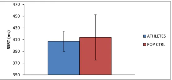
Stop signal reaction time (SSRT) was chosen as the critical variable to estimate IC performance. SSRT is a measure of the time required to stop the response that has already been initiated (Logan et al., 1984). An independent sample t-test showed no significant difference (p>0.05) on SSRT [t(22,3)=(- 0.59); p=0.56], indicating that the athletes group (mean ± SD = 407.26 ± 17.23 msec) presented no significantly superior motor inhibition than the control group (mean ± SD = 413.78 ± 38.67 msec).
Fig. 3: SSRT in milliseconds. SSRT was calculated by substracting average stop-signal delay (SSD) from the mean of response time in go trials. Means and standard deviations.
350 370 390 410 430 450 470 SS R T (m s) ATHLETES POP CTRL
24
The Go/Nogo task
The GNG task provided four types of results:
Go trial (A, E, H, I, M, O, S, T) Nogo trial (X)
Participant pressed on button HIT False Alarms (FA)
Participant did not press on button MISS Correct Rejection
We indexed the behavioural performance by the percentage of false alarms and the average of the reaction time in the HIT. Group difference analysis was performed using independent sample t-test.
False alarms rate
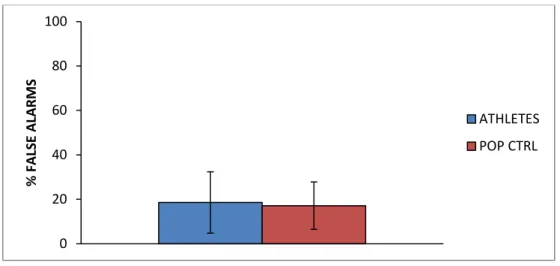
An independent sample t-test compared the performance of the athletes group (FA mean ± SD = 18.58 ± 13.79 %) with the control group (17.13 ± 10.7 %) and showed that the false alarms rate did not significantly (p>0.05) differ between athletes and non-athletes [t(15,1)=0.29; p=0.78].
Fig. 4: Percentage of false alarms. Means and standard deviations.
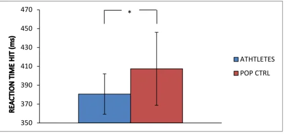
Reaction time HIT
Independent sample t-test revealed that reaction time (RT) to the HITs (a correct response to Go stimuli) differed significantly (p<0.05) between the athletes group and the control group [t(26)=(-2.36); p=0.026]. 0 20 40 60 80 100 % FAL SE A LA R M S ATHLETES POP CTRL
25 350 370 390 410 430 450 470 ATHTLETES POP CTRL
These results revealed that athletes (RT mean ± SD = 380.63 ± 24.44 msec) responded significantly faster than the control group (407.39 ± 38.67 msec) without showing an increase of false alarms, which prove a better ability of IC.
Fig. 5: Reaction time in the HIT condition in milliseconds. Means and standard deviations.
The N-back task
In the N-back task, four types of results were provided:
Target ("X") No Target
Correct response given Correct Response (CR) False Alarms
Wrong response given Wrong Response
No press MISS
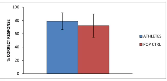
We indexed the behavioural performance by the percentage of correct response. An unpaired t-test compared the performance of the athletes group (CR mean ± SD = 78.87 ± 12.69 %) with the control group (72.09 ± 17.59 %) and showed that the correct response rate did not significantly (p>0.05) differ between athletes and non-athletes [t(24)=1.18; p=0.25].
26 0 20 40 60 80 100 % C ORR ECT R ES PO N SE ATHLETES POP CTRL
Fig. 6: Percentage of correct response. Means and standard deviations.
3.2 MRI results
In our study, we focused on structural measurements, more particularly on differences in grey matter volume (GMV) between experts and controls. To compare the groups, we carried out a VBM analysis on the T1-weighted structural images to identify the brain
structures showing a significant difference in GMV. Two sample t-test analyses were used to compare both groups.
VBM results of expert group and non-athlete group
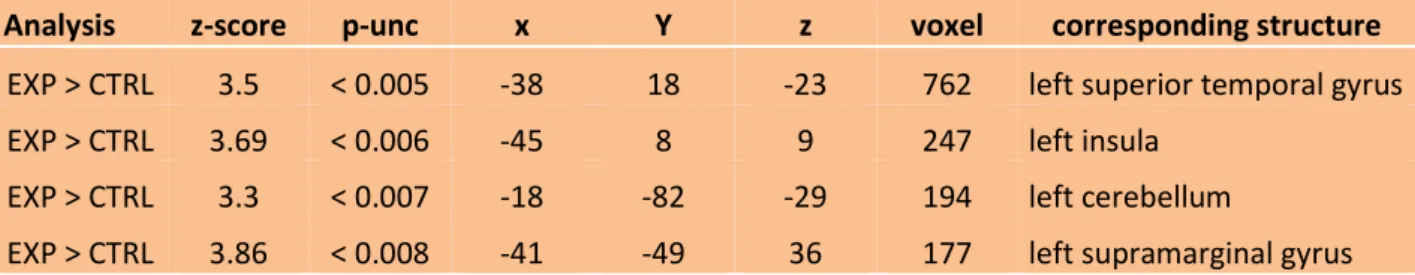
The two sample t-test on the GMV revealed significant regional volume difference between expert group and non-athlete group in four cortical structures. The expert group demonstrated significantly more GMV than the non-athlete group in the left superior temporal gyrus, the left insula, the left cerebellum and left supramarginal gyrus (all results are displayed at p<0.005, uncorrected). Table 1 provides the size of the clusters and their exact coordinates. In Fig. 7-10, the images provide a 2D visualization of the four clusters.
27 Table 1: VBM results of GMV comparison between the expert group (EXP) and the non-athlete group (CTRL) showing statistically significant difference. Coordinates [x, y, z] allude to the Montreal Neurological Institute of space. They matched the brain structure according to automated anatomical atlas (Tzourio-Mazoyer et al., 2002).
MRI results: Expert group and non-athlete group comparison:
Fig. 7: MRI results: GMV comparison EXP > CTRL obtained by the VBM method (p<0.005, uncorrected). The cursors are centred on the left superior temporal gyrus [-38, 18, -23], cluster-level: 762 voxels, with Z = 3.5. Colour scale indicates the z-score, which correlate with the significance of the between-group GMV difference.
Analysis z-score p-unc x Y z voxel corresponding structure
EXP > CTRL 3.5 < 0.005 -38 18 -23 762 left superior temporal gyrus
EXP > CTRL 3.69 < 0.006 -45 8 9 247 left insula
EXP > CTRL 3.3 < 0.007 -18 -82 -29 194 left cerebellum
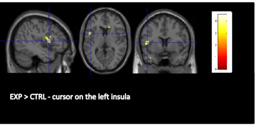
28 Fig. 8: MRI results: GMV comparison EXP > CTRL obtained by the VBM method (p<0.005, uncorrected). The cursors are centred on the left insula [-45, 8, 9], cluster-level: 247 voxels, with Z = 3.69. Colour scale indicates the z-score, which correlate with the significance of the between-group GMV difference.
Fig. 9: MRI results: GMV comparison EXP > CTRL obtained by the VBM method (p<0.005, uncorrected). The cursors are centred on the left cerebellum [-18, -82, -29], cluster-level: 194 voxels, with Z = 3.3. Colour scale indicates the z-score, which correlate with the significance of the between-group GMV difference.
29 Fig. 10: MRI results: GMV comparison EXP > CTRL obtained by the VBM method (p<0.005, uncorrected). The cursors are centred on the left supramarginal gyrus [-41, -49, 36], cluster-level: 177 voxels, with Z = 3.86. Colour scale indicates the z-score, which correlate with the significance of the between-group GMV difference.
30
4. Discussion
In their field of practice, fencers have to react as fast as possible to the opponents’ actions by planning motor response and inhibiting some prepared intentions to execute the most appropriate movement at the right time. In line with previous studies, we expected that many years of intensive fencing practice would lead at the behavioural level to a better performance in IC and, in terms of structural imaging, to plastic reorganizations of the athletes’ inhibition related brain network. The present study includes 10 elite fencers and provides evidence of structural changes in IC underlying structures, through computational anatomy. Behavioural performance in the GNG task illustrates that fencers react faster than controls without committing more false alarms, indicating better IC abilities. This result is congruent with our hypothesis based on previous literature. However, the way brain structures underlying inhibition respond to extensive IC training had never been tested. This thesis brings evidence to practice related structural plasticity, through an increase in the GMV of the experts, in specific brain structures crucial for IC.
Behavioural performance
We examined IC performance by carrying out two behavioural tests: the stop-signal task (SST) and the Go/No go task. These two tests have already been used in many studies to train and assess IC performance (Spierer, et al., 2013), their reliability is thus not debated. In the SST, we observed no significant difference between the experts and the non-athletes group. These results do not correspond to the expected findings. Indeed, we assumed that fencers would have better performance than non-athletes in SST, because after years of practice, they are expected to handle situations requiring high IC abilities. Performance on the GNG task showed in contrast that the expert group presented better IC performance. Indeed, the experts responded faster than the control group without showing an increase of the false alarm rate. This finding is consistent with the results of Rossi et al. (1992), who demonstrated that high level fencers were faster than non-athletes in a GNG task, even if the task became more complex. Moreover, as already showed by Taddei et al. (1991) and Di Russo et al. (2006), we bring one more evidence of faster stimulus discrimination when testing fencers.
31
The question why these two tasks did not bring the same results is hard to answer. Although these tasks both assess motor response inhibition, it seems that they did not assess precisely the same cognitive processes nor involve the same neural networks (Swick et al., 2011). On one hand, these two tasks involve admittedly overlapping but distinct neural systems. While more precisely the right middle frontal gyrus and the right inferior parietal lobule are activated in GNG tasks, left anterior insula and bilateral thalamus are activated in SST (Swick et al., 2011). On the other hand, although they both assess global inhibition, it is unclear if the results can be interpreted in the same way. In the SST, the participants have to respond to an auditory signal, while in the GNG they only respond to visual stimuli. This difference could bring one potential explanation concerning the question why these two tasks did not involve the same cognitive processes. Further investigations, combining both the GNG and the SST, need to be conducted to understand which cognitive processes are precisely involved and which are the related-neural systems.
The SST and the GNG task used in our study assessed global inhibition, which refers to the ability to suppress an initiated inappropriate response as quickly as possible (Aron et Verbruggen, 2008). On the other hand, selective inhibition refers to the ability to suppress a global stop mechanism while concurrently executing another movement or re-initiating a new response (Aron, 2011; see also Coxon et al., 2007). In the context of fencing, the athletes always have to stop a prepared motor response and re-initiate another one to face to the opponent’s attacks as well as to counterattack very fast. In selective inhibition, the goal is not only to inhibit response, but also to keep maintaining the attention and controlling others particular parameters (Aron et Verbruggen, 2008). According to these remarks, fencers seem to train more particularly selective inhibition rather than the global inhibition in their field of practice. In this regard, it would probably be more appropriated to use tasks assessing the selective inhibition. Considering the fact that only few studies investigated the selective inhibition mechanism, further experiments need to be conducted to determine whether selective inhibition tasks could provide evidences that open-loop sport practice enhances selective IC performance.
The N-back task, used to assess the working memory, was chosen as control task. The results showed that the expert population did not present significant higher performance
32
than the non-athletes population, compatible with our hypothesis that fencing extensively trains IC.
Inhibitory control training and structural changes
Cortical and subcortical GMV develops since childhood, through adolescence, until early adulthood (Durston et al. 2001; Wierenga et al. 2014). It is therefore interesting to see how these GMV changes can be modulated by long-term intense sport practice that started during childhood. In our study, we investigated whether intense fencing practice would lead to structural changes of the IC underlying network. In line with previous neuroimaging data on training and cortical plasticity, particularly on GMV changes (Draganski et al., 2004; Huang et al., 2013; Driemeyer et al., 2008; Wenzel et al., 2014), our study demonstrated significant GMV difference between experts and non-athletes in four structures of the brain. However, because the current sample size is small, the neuroimaging result should be interpreted with caution. While we expected to find differences in the right lateralized fronto-basal ganglia stopping network (Aron, 2011; Chambers et al., 2009), significant GMV difference was found in the left superior temporal gyrus, the left insula, the left cerebellum and the left supramarginal gyrus. Although we previously exposed that IC depends on a fronto-basal network mainly right-lateralized (Chambers et al., 2009; Aron, 2011), our results showed in contrast a left-lateralized GMV extend in four structures. Hirose et al. (2012) demonstrated a significant correlation between brain activations in the left inferior frontal gyrus, the left superior frontal gyrus, the left precentral gyrus/middle frontal gyrus, the left temporoparietal junction and the efficiency index in a GNG task. They suggested that the involvement of the left hemisphere could play an additional role in IC, if the right-lateralized structures were already fully engaged, which could be the case when response inhibition is more difficult (Swick et al., 2008).
Left insula
While the left insula seems to be involved principally in language, working memory, perception, socio-emotional and sensorimotor processes (Chang, 2014; Clos et al., 2014), it also appears to play a role in the IC network (Swick et al., 2011; Chambers et al., 2009). Using SST, Boehler et al. (2010) showed a significant correlation between shorter SSRT and stronger activity in the left anterior insula, associated with stopping efficiency. According to
33
this finding, Swick and colleagues (2011) exposed in their meta-analyses the clusters of activation in GNG and SST and found that the left anterior insula is activated to a greater extent in the STT than in GNG. Consistently, in a recent experiment using SST, Albrecht et al. (2014) found that dopamine is released in some cortical structures, including the insula. It is also interesting to notice that insula is admittedly involved in IC, but its role is broader. Some studies presented the insula as being relevant to awareness (Craig, 2009), for the detection of salient event and for the generation of appropriate behaviours in response to these salient stimuli (Menon et Uddin, 2010), as well as for attentional control (Nelson et al. 2010). This being said, the insula has been proven as playing a role in IC, but it has also been demonstrated as having a more general function in attentional control, awareness and detection of salient stimuli.
Left supramarginal gyrus
The left supramarginal gyrus plays an important role for motor action reprogramming, more particularly for switching motor plans (Rushworth et al., 2001), after spatial information is processed. Moreover, by using TMS, Tunik et al. (2008) demonstrated that the supramarginal gyrus as well as the inferior frontal gyrus pars opercularis is involved in planning goal-oriented actions without disturbing the motor plan. By stimulating these two structures, the authors found that movement execution remained the same, but motor action was delayed. Furthermore, Hartwigsen et al. (2012) presented evidence that the left supramarginal gyrus prevents inappropriate motor actions, and thus is important for the control of motor actions. This role in action reprogramming and suppression of incorrect response is highly relevant for the performance of fencing. To summarize, this structure appears to be involved in the suppression or the slowing down of inappropriate actions.
Left superior temporal gyrus
The superior temporal gyrus, a structure of the temporal lobe where the auditory cortex is situated, is mainly involved in the cortical language circuit to produce or interpret language (for a model, see Friederici, 2012). Regarding our subject, Criaud et Boulinguez (2013) suggested that the superior temporal gyrus is connected with the motor system and is involved in an indirect way with response inhibition. However, its role in inhibitory control remains unclear and is poorly studied. This structure seems to rather have a role in
34
visuospatial attention (Karnath et al., 2001) and in attentional modulations related to decisional mechanisms (Lim et al., 2011). These roles in attentional processing can give an explanation on why we found an extent of GMV in the left superior temporal gyrus. Indeed, fencing requires high attentional demand (Roi et Bianchedi, 2008) and we therefore assume that the superior temporal gyrus is highly activated during the practice of fencing.
More collectively, the superior temporal gyrus was studied as a crucial structure in schizophrenia. Indeed, many studies showed abnormalities in the superior temporal gyrus in patients with schizophrenia (Honea et al., 2005; Shenton et al., 2001; Sun et al., 2009). Because of its connections with the temporal limbic brain areas, dysfunctions within the superior temporal gyrus or its connections can lead to hallucinations or thought disorder (Allen et al. 2008). In their review, Honea and colleagues (2005) showed that schizophrenia patients demonstrated a reduced volume in the left superior temporal gyrus. More precisely, volume deficits in the left superior temporal gyrus were correlated with auditory hallucinations (Barta et al., 1990). These auditory hallucinations appear to be associated with less activity in temporal cortical areas that overlap with areas that are involved for external speech processing and therefore lead to a competition for common resources of the auditory cortex (Woodruff et al. 1997). With regards to this finding, Kim et al. (2003) hypothesized that damages inauditory perception could be due to an impairment of normal language discriminating inhibition.
Furthermore, some studies reported that patients with schizophrenia presented impairment in cognitive control (Eich et al., 2014; Tully et al., 2014). In a recent research, Eich et al. (2014) studied the mechanisms underlying working memory deficits in schizophrenia patients. They revealed that impaired inhibitory control in working memory had negative effects regarding the behavioural results. In a review, Barch et Ceaser (2012) argued that deficits in inhibition in schizophrenia could be explained as deficits in a more general context-processing, which was recently reconceptualised as impairment in proactive control. Therefore, because patients with schizophrenia present abnormalities in the superior temporal gyrus and also present impairments in cognitive control, we can suggest that the superior temporal gyrus may have an indirect role in the underlying network of IC.
35
Left cerebellum
The cerebellum is involved in the preparation and execution of movement, as well as in mental representations, cognitive functions, such as language (Buckner, 2013; Leiner, 2010; Ito, 2008) and to a lesser extent IC (Rubia et al., 2001). Although it is hard to draw any clear explanations from the extent of the superior temporal gyrus in the expert group, apart from its role in visuospatial attention (Karnath et al., 2001) and attentional modulations related to decisional mechanisms (Lim et al., 2011), it is easier to understand the involvement of the cerebellum during sport training, respectively during fencing practice. Indeed, the model for voluntary movement and mental activity, developed by Ito (2008), which describes the importance of the cerebellum in these two processes, could explain why GMV difference was found in this structure. In this model, it is seen that the cerebellum is involved in voluntary movement and serves more particularly as a location for the copy of mental model. From then on, this structure is constantly activated during fencing practice for the control of voluntary actions. When it comes to its implication in inhibition, only a few studies reported that the cerebellum actually plays a role in IC (Rubia et al., 2001; Mulder et al., 2008). Therefore, the literature suggests that the cerebellum is involved in voluntary movement, but its role in IC remains unclear.
Sport expertise: how long do the structural effects last?
In neurosciences, the term “plasticity” refers to a property of the brain defined as a reorganization of the nervous system due to environmental and physiological changes, as well as behavioural experience (Pascual-Leone et al., 2005). Many studies have already investigated whether sport training or leaning new tasks could induce white matter or grey matter changes and how this brain plasticity occurred (Roberts et al., 2013; Huang et al., 2013; Jacini et al., 2009; Draganski et al., 2004). This reorganization of the nervous system is at the centre of our study.
In line with Debarnot et al. (2014), our study demonstrated a relationship between intensive sport practice and modifications in GMV, as well as behavioural improvements. The authors highlighted an interesting question: at what time do structural changes start to be detected and how long do they last. As a consequence, further research would be interesting. Firstly, it would make sense to carry out a study with young fencers (i.e., children