The structural differences between patient-derived α-synuclein strains dictate characteristics of Parkinson’s disease, multiple system atrophy and dementia with Lewy bodies
Texte intégral
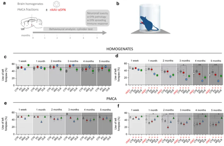
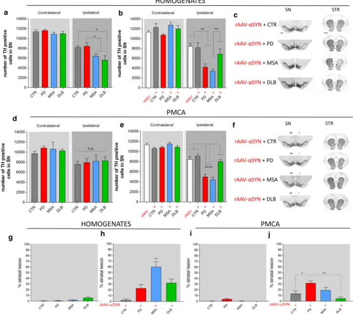
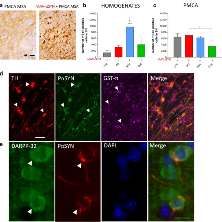
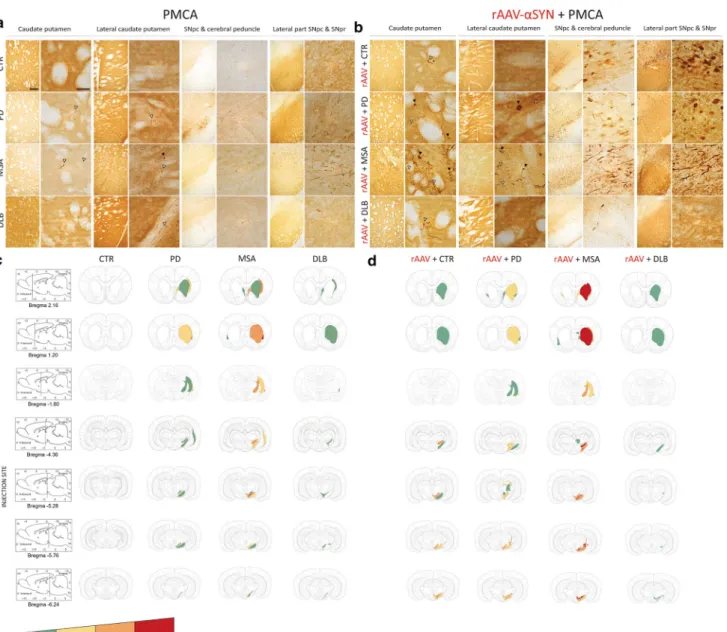
Figure
Documents relatifs
Abbreviations: AhR, aryl hydrocarbon receptor; B[a]P, benzo[a]pyrene; ECSIM, environmental control system for intestinal microbiota; FM, fecal microbiota; GIT, gastrointestinal
diagrams afterwards) that these muscles contain overlapping arrays of protein filaments spaced apart in an aqueous environment ; that these filaments, in turn,
Coupez et coller le prochain object qui devrait apparaitre dans la suite1. `
En 2006, le MELS crée la tâche d’enseignant-ressource (ER) pour soutenir les enseignants dans l’intégration des élèves à risque ou HDAA en classe ordinaire..
The inhibition of c-Abl activity in cells either by downregulating directly its expression and protein level using siRNA or by inhibiting its kinase activity using specific
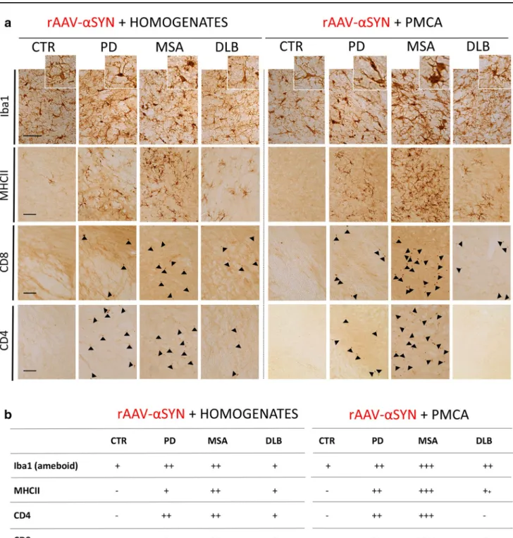
The severity of neuronal and oligodendroglial aggrega- tion pathology in representative tissue sections through the deep cerebellar nuclei, the surrounding white matter, and
The conserved 7 DQAXXLR motif (X, any residue; Fig. 2b) of YlxH is found at the N terminus of the activator helix, with DQA forming a 3 10 -helical turn that locates at the
doi:10.1371/journal.pone.0116641.g004.. CathD activity was not significantly modified by the presence of GAGs extracted from stressed and unstressed cells at doses up to 1 μg/mL in
