HAL Id: cea-03123519
https://hal-cea.archives-ouvertes.fr/cea-03123519
Submitted on 28 Jan 2021HAL is a multi-disciplinary open access
archive for the deposit and dissemination of sci-entific research documents, whether they are pub-lished or not. The documents may come from teaching and research institutions in France or abroad, or from public or private research centers.
L’archive ouverte pluridisciplinaire HAL, est destinée au dépôt et à la diffusion de documents scientifiques de niveau recherche, publiés ou non, émanant des établissements d’enseignement et de recherche français ou étrangers, des laboratoires publics ou privés.
Utility of Scanning Transmission X-ray Microscopy to
Investigate Chemical Composition in Perovskite Thin
Films
Haeyon Jun, Denis Tondelier, Bernard Geffroy, Jean-Eric Bourée, Sufal
Swaraj, Yvan Bonnassieux
To cite this version:
Haeyon Jun, Denis Tondelier, Bernard Geffroy, Jean-Eric Bourée, Sufal Swaraj, et al.. Utility of Scanning Transmission X-ray Microscopy to Investigate Chemical Composition in Perovskite Thin Films. Journées Nationales du Photovoltaïque, Jan 2021, Dourdan, France. 2021. �cea-03123519�
Utility of Scanning Transmission X-ray Microscopy
to Investigate Chemical Composition in Perovskite Thin Films
Haeyeon Jun1,2, Denis Tondlier1, Bernard Geffroy1,3, Jean-Eric Bourée1, Sufal Swaraj2 and Yvan Bonnassieux11LPICM-CNRS (UMR7647), Ecole polytechnique, IP Paris, 91128 Palaiseau, France 2Synchrotron SOLEIL, L'Orme des Merisiers Saint-Aubin, BP 48 91192 Gif-sur-Yvette Cedex
3 Université Paris-Saclay, CEA, CNRS, NIMBE, LICSEN, 91191 Gif-sur-Yvette, France
Organic–inorganic lead halide perovskites with remarkable photoelectric properties have achieved power conversion efficiencies of 25.2 %, making them a promising candidate as an emerging solar cell technology [1]–[4]. Formation of high crystalline perovskite film is necessary for stable and high efficient solar cells. Among them, vacuum-based deposition processes are profitable because highly uniform and smooth thin films can be obtained [5], [6]. Especially, characterization of evaporated perovskite thin films using various technique contribute to the accurate analysis in the physical or chemical point of view [7].
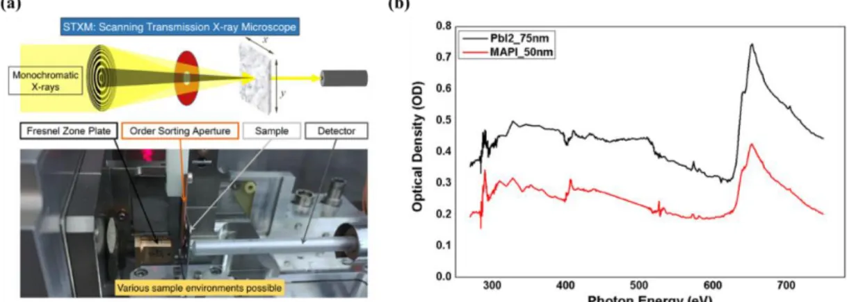
Scanning transmission X-ray microscopy (STXM) is an experimental technique based on near edge X-ray absorption fine structure (NEXAFS) spectroscopy [8]. This spectro-microscopy technique allows us to obtain quantitative chemical composition maps in bulk of materials with high spatial resolution (~ 50 nm). We have been exploring the utility of this technique in order to characterize perovskite films synthesized with evaporation techniques. In this poster, we present some strategies developed to study these materials. In particular, we have shown how the soft x-ray energy range from 270 to 750 eV, that includes the C K-edge, N K-edge, Pb N-edge and I M-edge, can be used to quantify elemental and molecular concentration[9]–[11].
Keywords
Organic–inorganic halide perovskite, Co-evaporation, Scanning transmission X-ray microscopy Reference
[1] D. B. Mitzi, C. A. Feild, Z. Schlesinger, and R. B. Laibowitz, J. Solide State chem., 114, 1. 159–163, 1995.
[2] S. D. Stranks et al., Science, 342, 10, 341–345, 2013.
[3] J. H. Noh, S. H. Im, J. H. Heo, T. N. Mandal, and S. Il Seok, Nano Lett., 13, 4, 1764–1769, 2013. [4] “NREL efficiency chart.” [Online]. Available: https://www.nrel.gov/pv/cell-efficiency.html. [5] M. Liu, M. B. Johnston, and H. J. Snaith, Nature, 501, 7467, 395–398, 2013.
[6] L. K. Ono, S. Wang, Y. Kato, S. R. Raga, and Y. Qi, Energy Environ. Sci., 7, 12, 3989–3993, 2014. [7] M. Sowinska, C. Das, K. Wojciechowski, and Z. Rouissi, ISSRNS, 15, 1, 1500962, 2016.
[8] H. Ade, Academic Press, 32, 225-262. 1998.
[9] J. Stöhr, R. Jaeger, J. Feldhaus, S. Brennan, D. Norman, and G. Apai, Appl. Opt., 19, 23, 3911, 1980. [10] C. Reverdin et al., AIP Conf. Proc., 207, 5, 207–212, 2008.
[11] M. W. Lin et al., Adv. Mater. Interfaces, 3, 17, 1–12, 2016.
Figure 1. (a) Schematic of scanning transmission X-ray microscope (b) Carbon K-edge, Nitrogen N-edge and Iodide M-edge NEXAFS spectroscopy of PbI2 and MAPbI3 perovskite film.