HAL Id: cea-02400209
https://hal-cea.archives-ouvertes.fr/cea-02400209
Submitted on 23 Mar 2020
HAL is a multi-disciplinary open access
archive for the deposit and dissemination of
sci-entific research documents, whether they are
pub-lished or not. The documents may come from
teaching and research institutions in France or
abroad, or from public or private research centers.
L’archive ouverte pluridisciplinaire HAL, est
destinée au dépôt et à la diffusion de documents
scientifiques de niveau recherche, publiés ou non,
émanant des établissements d’enseignement et de
recherche français ou étrangers, des laboratoires
publics ou privés.
Perspectives in using Raman spectroscopy for
characterizing the microstructure of plutonium-bearing
materials
Laeticia Medyk, Patrick Simon, Aurélien Canizarès, Dario Manara, Rudy
Konings, Jean-Yves Colle, Romain Vauchy, Christophe Valot, Gilles
Montagnac, Philippe Martin
To cite this version:
Laeticia Medyk, Patrick Simon, Aurélien Canizarès, Dario Manara, Rudy Konings, et al.. Perspectives
in using Raman spectroscopy for characterizing the microstructure of plutonium-bearing materials.
Plutonium Futures 2018, Sep 2018, San Diego, United States. �cea-02400209�
Perspectives in using Raman spectroscopy for characterizing the microstructure of plutonium-bearing materials
Laetitia Medyk1, Patrick Simon2, Aurélien Canizares2, Dario Manara3, Rudy J. M. Konings3, Jean-Yves Colle3, Romain Vauchy1, Christophe Valot1, Gilles Montagnac4, Philippe M. Martin1
1
CEA, Nuclear Energy Division, Research Department on Mining and Fuel Recycling Processes, SFMA/LCC, 30207 Bagnols-sur-Ceze, France
2Conditions Extremes et Materiaux : Haute Temperature et Irradiation (CEMHTI) - CNRS :UPR3079 - CS 90055 45071,
45071 Orleans, France
3European Commission, Joint Research Centre (JRC), Postfach 2340, 76125 Karlsruhe, Germany 4
Laboratoire de Géologie de Lyon, CNRS, ENS de Lyon, 69364 Lyon, France
INTRODUCTION
In the frame of the development of uranium-plutonium mixed oxide fuels for Sodium-cooled Fast Reactors (SFRs), characterizing nuclear materials by various techniques is paramount.
These fast neutron reactors imply the use of a (U,Pu)O2-x
ceramic fuel with a Pu/(U+Pu) content between 19 and 30 mol.%. Furthermore, the physico-chemical and microstructural properties of such fuels, such as chemical homogeneity, oxygen stoichiometry (O/(U+Pu) ratio) and crystallographic structure, have to meet precise criteria for being introduced in the reactor core.
As evidenced in numerous studies by various experimental techniques, a supplementary difficulty with such high plutonium content is the existence of a miscibility gap comprised within the UO2-PuO2-Pu2O3 region1,2. Its
presence is conditioned by the difference in the possible oxidation states adoptable by the two constituting cations. The miscibility gap itself is composed of three sub-domains, consisting in closely related cubic-type phases and its extent is a function of Pu content and temperature. The temperature of phase separation, i.e. the temperature at which this bi- (or tri-) phasic domain appears, depends on the oxygen to metal ratio (hereinafter O/M ratio) of the material. Recent high temperature X-ray diffraction (HT-XRD) studies allowed observing in situ the phase separation occurring in oxygen-hypostoichiometric uranium-plutonium mixed oxides with high plutonium contents3–5.
Nevertheless, HT-XRD characterizations on (U,Pu)O2-x
were performed on powder and thus did not allow observing
in situ the microstructure changes induced by the phase
separation phenomenon3,4.
As highlighted by recent studies by Talip et al6 and Elorrietta et al7, Raman microscopy is a promising tool for characterizing the physico-chemical properties such as, among many others, the cation distribution homogeneity, the grain size, the crystal defects that are of main interest for the production of nuclear fuels.
The development of a new in situ Raman device dedicated to handling transplutonium-bearing materials is currently in
progress in our laboratory (ATALANTE facility, CEA Marcoule, France).
We propose to present at the Plutonium Futures 2018 conference our first results obtained on U0.75La0.25O2-x and in
situ high temperature measurements on CeO2-x as the
authorization of handling plutonium-bearing materials is not obtained yet. By the time of the conference and thanks to the fruitful collaboration existing between JRC Karlsruhe and our laboratory, we will be able to present our first results on (U,Pu)O2-x samples as well.
RESULTS
The results bellow were obtained on a U0.75La0.25O2-x
sintered pellet. The sample was prepared by gel-supported precipitation, also referred as sol−gel external gelation8
. The whole preparation route is presented elsewhere9.
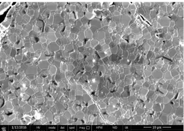
The following SEM picture (Fig. 1.) shows the microstructure of the polished U0.75La0.25O2-x sintered pellet.
The estimated mean grain size is <5 µm.
Fig. 1. SEM picture of the U0.75La0.25O2-x sintered pellet
The following Raman imaging measurements were performed at CEMHTI, Orléans, France using an InVia Reflex Renishaw system with a 514 nm LASER source (5mW and x50 objective). A 1 µm step size was used to
obtain 100x100 µm Raman maps. The Figure 2. represents a fake colored intensity map of the T2g line superimposed with
the optical image.
Fig. 2. Map of the Raman T2g intensity line (fake colors)
obtained on U0.75La0.25O2-x (100x100 µm)
As shown by Maslova et al.10 on UO2 sample, a part of the
T2g intensity inhomogeneities is due to different orientations
of the ceramic grains that allows observing the microstructure itself (grain boundaries, microstructural defects, etc). Thus, as revealed here, the methodology established on UO2 can applied to mixed oxides.
In a second part, we will present our results obtained by in
situ Raman (532 nm laser source at LGL of ENS Lyon,
France) on CeO2-x samples using a nuclearized version of
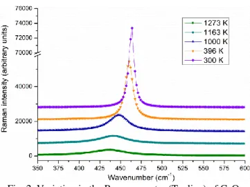
Raman micro furnace developed by Montagnac et al.11. Furthermore, this unique device is associated with a setup allowing measuring, imposing and monitoring the variations in the oxygen partial pressure during a given heat treatment. As shown in Figure 3, a significant shift in the T2g line is
observed as a function of temperature related to the lattice thermal expansion. Moreover, both the intensity loss and the line broadening are induced by thermal agitation of anionic sublattice.
Fig. 2. Variation in the Raman spectra (T2g line) of CeO2
with temperature in air
The same experiments were performed on a similar sample in reducing conditions (pO2 = 1.10-15 bar) up to 1420 K. the
magnitude of the shift previously observed on the T2g line
position was larger under these conditions. This phenomenon is explained by the in situ reduction of stoichiometric CeO2 to oxygen-hypostoichiometric CeO2-x.
We hope this promising result could be useful for determining the O/M ratio of the materials studied as a function of the thermodynamic conditions (T and pO2).
Finally, uranium-plutonium mixed oxide samples with 19 mol.% Pu are being characterized at JRC Karsruhe with the experimental setup described in ref12. Experiments are performed on sintered pellets, manufactured by powder metallurgy, and show variations in the T2g line position as a
function of the local plutonium concentration. Raman mapping of such samples are planned in the near future.
REFERENCES
1. T. L. MARKIN and R. S. STREET, “The uranium-plutonium-oxygen ternary phase diagram,” J. Inorg. Nucl. Chem. 29 9, 2265 (1967);
https://doi.org/10.1016/0022-1902(67)80281-1. 2. C. SARI, U. BENEDICT, and H. BLANK, “A study of
the ternary system UO2-PuO2-Pu2O3,” J. Nucl. Mater.
35 3, 267 (1970);
https://doi.org/10.1016/0022-3115(70)90211-4.
3. T. TRUPHÉMUS et al., “Structural studies of the phase separation in the UO2–PuO2–Pu2O3 ternary system,” J. Nucl. Mater. \b432 1, 378 (2013)
4. R. VAUCHY et al., “High temperature X-ray diffraction study of the kinetics of phase separation in
hypostoichiometric uranium–plutonium mixed oxides,” J. Eur. Ceram. Soc. 34 10, 2543 (2014);
https://doi.org/10.1016/j.jeurceramsoc.2014.02.028. 5. R. C. BELIN et al., “In situ high temperature X-Ray diffraction study of the phase equilibria in the UO2–
PuO2–Pu2O3 system,” J. Nucl. Mater. 465, 407 (2015); https://doi.org/10.1016/j.jnucmat.2015.06.034.
6. Z. TALIP et al., “Characterization of un-irradiated MIMAS MOX fuel by Raman spectroscopy and EPMA,” J. Nucl. Mater. \b499, 88 (2018)
7. J. M. ELORRIETA et al., “Raman study of the oxidation in (U, Pu)O2 as a function of Pu content,” J. Nucl. Mater. \b495 Supplement C, 484 (2017)
8. U. CARVAJAL-NUNEZ et al., “Charge Distribution and Local Structure of Americium-Bearing Thorium Oxide Solid Solutions,” Inorg. Chem. 51 21, 11762 (2012); https://doi.org/10.1021/ic301709d.
9. D. PRIEUR et al., “Aliovalent cation Substitution in UO2: Electronic and Local Structures of U1–yLayO2±x Solid Solutions,” Inorg. Chem. (2018);
https://doi.org/10.1021/acs.inorgchem.7b02839. 10. O. A. MASLOVA et al., “Raman imaging and principal
component analysis-based data processing on uranium oxide ceramics,” Mater. Charact. 129, 260 (2017); https://doi.org/10.1016/j.matchar.2017.05.015. 11. G. MONTAGNAC et al., “Anharmonicity of graphite
from UV Raman spectroscopy to 2700K,” Carbon 54, 68 (2013); https://doi.org/10.1016/j.carbon.2012.11.004. 12. M. NAJI et al., “An original approach for Raman
spectroscopy analysis of radioactive materials and its application to americium-containing samples,” J. Raman Spectrosc., n/a (2015); https://doi.org/10.1002/jrs.4716.