I"
DNA Adducts as Molecular Biomarkers
of Exposure to Benzo(a)pyrene
by
Deirdre Michelle Lawrence
B. S., BiologySpelman College, 1989
Submitted to the Division of Toxicology in Partial Fulfillment of the Requirements for the Degree of
Doctor of Philosophy
in Toxicologyat the
Massachusetts Institute of Technology
September 1997
01997 Massachusetts Institute of Technology All Rights Reserved
Signature of Author
Division of Toxicology June 30, 1997 Certified by /
6 6 r. Gerald N. Wogan, Thesis Supervisor
Accepted by
LIN"ARIES
Dr. Peter C. Dedon, Chairman Committee on Graduate Studies, Division of Toxicology
This doctoral thesis has been examined by a committee of the Division of Toxicology as follows:
Professor Peter C.
Dedo..--Chairman
r osseforP Gerald
N W an
6
Thesis SupervisorProfessor Steven R~ Tannenbaum
-Professor William G. Thilly I
i R •JA•LIL•IIB --- • ]•E IAJLA I ! -~~~ ---- WW II•i
---DNA Adducts as Molecular Biomarkers of
Exposure to Benzo(a)pyrene
by
Deirdre Michelle Lawrence
Submitted to the Division of Toxicology on June 30, 1997 in partial fulfillment of the requirements for the degree of Doctor of Philosophy in Toxicology
Abstract
It is believed that the formation of DNA adducts is an essential initial step in carcinogenesis. To better understand this initial step and its relationship to cancer, this study primarily focuses on benzo(a)pyrene (BaP), a highly carcinogenic model polycyclic aromatic hydrocarbon (PAH) and an ubiquitous environmental contaminant. The objective of this dissertation was to identify and quantify carcinogen-DNA adducts as biomarkers of exposure to BaP.
The BaP-DNA adduct levels were measured by Adduct Detection by Acylation with Methionine (ADAM), a new postlabelling method that was recently developed by our group. Nucleoside adducts of fluoranthene, 4-aminobiphenyl and 8-hydroxy-2'-deoxyguanosine (8-OHdG) were acylated with 3SS-methionine and detected in femtomolar quantities. This method was
adapted for analysis of BaP-DNA adducts and optimized with 3H-BaP using unlabeled methionine. Acylated products of benzo(a)pyrene-deoxyguanosine (BaP-dGuo) from different sources,
including 3H-BaP-dGuo isolated from calf thymus DNA modified enzymatically in vitro or by reaction with BPDE, have shown identical RP-HPLC retention times. The 35S content of acylated products was linearly correlated (r = 0.860) with the nucleoside adduct concentration over the
range from 20-1000 femtomoles.
BaP-DNA adduct levels in random human lung samples were found to range from 240-51,120 adducts per cell. Similarly, paraffin-embedded breast tissues contained BaP-DNA adduct levels from 35-1764 adducts in 10' normal nucleotides (0.05-14.7 femtomole BaP/pg DNA) with an average of 44,000 adducts/cell.
Adult CD-1 mice were injected i.p. with [3H]-benzo(a)pyrene ([3H]-BaP) (2mg, 117 mCi/mmol) and sacrificed 24 hr later. Liver DNA was isolated, enzymatically hydrolyzed to nucleosides, purified by immunoaffinity chromatography and analyzed with the ADAM procedure
for BaP-DNA adducts. BaP-DNA adduct levels were quantified by both radioactivity (3H in BaP) and acylation with 35S-methionine with the ADAM procedure. An excellent correlation was found between the adduct levels determined independently by 3H and '3S. A newborn mouse lung adenoma assay was used to generate information about BaP-DNA adducts formed in vivo. Newborn CD-1 mice were injected i.p. with a tumorigenic dose of 210 p.g of BaP (0.83mmole) three times over a two week dosing regimen starting at birth. Control mice were given DMSO as
5.5 months. Both target (lung) and nontarget (liver, kidney) tissues were collected. Tumors were
induced by this regimen in the male mice. DNA was isolated, enzymatically hydrolyzed to nucleosides, purified and analyzed with the ADAM procedure for BaP-DNA adducts. The RP-HPLC profiles of treated mice were compared with their prospective controls. Relationships of adduct levels to tumorigenesis were summarized.
Successful acylation conditions were used to analyze benzo(a)pyrene adducts in DNA extracted from samples of tissues of CD-1 mice injected with benzo(a)pyrene and samples of human lung and embedded breast tissues. RP-HPLC profiles of the acylation products of adducts isolated from mouse tissues and human lung samples eluted at the same retention time as that of an authentic BaP-dGuo standard.
The postlabelling method combined with a subsequent RP-HPLC profile allows characterization of the carcinogen-DNA adducts present in the tissues. A data base will be generated to analyze DNA as a dosimeter of exposure. This data base will be used to correlate molecular biomarkers, specifically BaP-DNA adducts with tumor frequency. A direct relationship will verify the use for carcinogen-DNA adducts as dosimeters of exposure to genotoxic chemicals and will be imperative in understanding the role carcinogens play in initiating cancer.
Thesis Supervisor: Dr. Gerald N. Wogan Title: Professor of Toxicology
Dedication
To the members of my family...
Let me take a few minutes to brag about important family members in my life
Mom
Andrea W. Lawrence, Ph.D., 1993 Georgia Institute of Technology
Grandad
Emory Williams, World traveler, Retired Co-op Program Director Northwestern University
Allegra/Muffin
Allegra J. Lawrence, J.D. 1996, Yale Law School
Valerie, "The Baby"
Valerie Lawrence, B.A. expected 1998, Atlanta Metropolitan College
mother
of
nieceAlexandra "Tigger"
Dad
President & CEO, Inchworm Industries, Inc.
Uncle Ronald
Ron M. Lawrence, Attorney at Law, J.D., LL.M
Acknowledgements
I would like to sincerely thank the members of my thesis committee: Dr. Peter C. Dedon, chairman, for his pragmatic outlook, Dr. Steven R. Tannenbaum for his contagious enthusiasm about science and Dr. William G. Thilly for his constant encouragement and support. I especially appreciate my thesis advisor, Professor Gerald N. Wogan for his continuous guidance and confidence in my ability to succeed.
I owe my life to GOD and my major support systems:
My parents for loving me unconditionally. You set the example of leadership as you strive for greatness. Allegra for constantly doing my editing over the phone and for wanting us to be friends in addition to being sisters. Valerie for making sure people remember that I'm human. To Uncle Ronald, my favorite Uncle who is my godfather that has his own law firm and lives in Atlanta...ha ha ha! Thank you for making me laugh when things got rough. Grandad Emory for always coming to the rescue. Samuel M. Clemons, Jr. my soul-mate, fellow movie buff and future partner for life. Thank you for spoiling me.
I owe my success to the help of Laura Trudel--for the large quantities of antibody and necessary extra pair of hands; Faye Sayedi-for the tremendous amount of work on the standard and TBM synthesis; Fayad Sheabar, William Boadi and Sarah Moody Robinson-for the
development and application of the ADAM method and the animal studies. Wang-for showing me not to be afraid to repair an HPLC myself. Now I know how to take them apart and put them back together...no problem, Xia- for her animal expertise. Your work is a piece of art. The Headquarters Staff of Division of Toxicology have looked out for me well. Thank you Debra, Roni, Jack, Blair, Terri, Janet, Mary, Alice and Carrie. Thank you, Denise for all of the computer work/desktop publishing.
Other MIT checkpoints: Office of Graduate Education Dean Isaac (Ike) Colbert and Margaret (Margot) Daniels Tyler. Thank you for your faith in me. My second moms at MIT: Mary Ni and Carla Kirmani-you helped me to love myself and not take life so seriously. My allies: Tony Gray for much needed ice cream breaks; Lynn Roberson-you saved my life. Thank you for helping me ward off the energy vampires; Jennifer Thomas-you make the world's best brownies...don't tell Don. Our giggling sessions are addictive. I fully appreciate the helpful hands and encouraging words of Paul Chodak, Karl Reid, Robert Selders, Jeff Thomas, the other members of "The Crew": Carsie Hall, Charles Clarke, BGSA and Color Creations -my family away from home.
Special thanks to the people who sent in reinforcements when I became battle weary. My thesis group Cathryn Shaw and Araba Lamouse'-Smith -you got me to the other side. Cathryn: I am inspired by your bright smile, perserverance and courage. Araba: I would not have been able to make 94-95 as BGSA chair a successful year without you. Uncle Johnny---got me interested in this whole research thing beginning with UMARC. Dawn Hilliard (Williams)--MY HUMAN DIARY. I feel like we are connected beyond words. I have enjoyed our deep
things at a "bargain price." Thank you for the gospel music and Mikki Howard. Robbin-you've shown me how to ask for what I want and say no to the things I don't. Don Brunson-When are we going to start our catering business? Thank you for everything. You have helped me more than you know. Carla Ferguson taught me how to put aside my pride and accept help from others: "Dee, if you don't let people do for you...you are stopping them from being blessed." Cassandra Clay for helping me find my inner voice. Ken and Alan--I cannot believe I let you get away with petnames like deedree, diadora, kikuyu, lulu, kooloo...thanks for the getaways to Roxbury, Dorchester & New Hampshire.
I owe a huge amount of gratitude to my friends "back home" who have stuck by me despite my inability to return calls. Lateefah Cabey, Zabrina Furlow, Debbie Hinkson, Kim Johnson, and Kirkpatrick Terry...or should I say "captain kirk"?
TO THE PEOPLE THAT I HAVE LOST DURING MY TIME AT MIT--Grandpa Ed, Grandma Julia, Uncle Maurice and Uncle Gillis. I miss you dearly. 'Til we meet again". Please say Hello to my Gramma Jeanne.
Table of Contents ABSTRACT 3 DEDICATION 5 ACKNOWLEDGMENTS 6 TABLE OF CONTENTS 8 LIST OF FIGURES 11 LIST OF TABLES 13 LIST OF ABBREVIATIONS 14 1. INTRODUCTION 15 2. LITERATURE SURVEY 18
2.1 Polycyclic aromatic hydrocarbons 18
2.1.1 Source and composition 18
2.1.2 Metabolism and biological effects ofbenzo(a)pyrene 19
2.1.3 Selection of BaP 22
2.2 Dosimetry of exposure to genotoxic compounds 23
2.2.1 Covalent adducts as dosimeters 23
2.2.2 Relevance of covalent modification of DNA to carcinogenesis 25
2.2.3 Methods for detecting carcinogen-DNA adducts 26
2.2.4 Use of DNA adducts as molecular biomarkers of exposure 30
3. SYNTHESIS OF BENZO(A)PYRENE-DEOXYGUANOSINE ADDUCT 34
3.1 Introduction 34
3.2 Methodology 35
3.2.1 Chemicals and enzymes 35
3.2.2 Preparations of modified DNA 36
3.3 Results and discussion 41
3.3.1 HPLC analysis 43
3.3.2 Electrospray-MS analysis of BaP-dGuo 44
3.4 Conclusions 45
4. BaP-DNA ADDUCT DETECTION BY ACYLATION WITH METHIONINE 63
4.1 Introduction 63
4.2 Materials 64
4.2.1 Chemicals 64
4.2.2 Chromatography 65
4.2.3 ADAM method 65
4.3 Acylation of benzo(a)pyrene-deoxyguanosine with NHS-TBM 66
4.3.1 Acylation with unlabelled NHS-TBM 66
4.3.2 Acylation with 35S-NHS-TBM 67
4.3.3 Results and discussion 68
4.4 Acylation of benzo(a)pyrene-deoxyguanosine with TBM 70
4.4.1 Synthesis of 35S-TBM 70
4.4.2 Acylation of benzo(a)pyrene-deoxyguanosine with 35S-TBM 71
4.5 Results and discussion 72
4.6 Conclusions 73
5. DNA ADDUCTS OF BENZO(A)PYRENE: ANIMAL STUDIES 88
5.1 Introduction 88
5.2 Short term dosing/Acute Exposure 91
5.2.1 Animal treatment 91
5.2.2 Fixation and embedding tissues 91
5.2.3 Removal ofparraffin 91
5.2.4 Isolation of DNA 92
5.2.5 Enzymatic hydrolysis of DNA 93
5.2.7 HPLC analysis 93
5.2.8 Acylation with 35S-TBM-NHS 94
5.3 Results and Discussion 95
5.3.1 Chromatography 95
5.3.2 Adduct levels 96
5.4 Newborn mouse lung adenoma assay 97
5.4.1 Treatment of Preweanling Mice with benzo(a)pyrene 97
5.4.2 Isolation of DNA 98
5.4.3 Enzymatic hydrolysis of DNA 99
5.4.4 Immunoaffinity chromatography 99
5.4.5 Manufacturing of 35S-TBM 100
5.4.6 Acylation with 35S-TBM 100
5.5 Results and Discussion 101
5.5.1 Tumor incidence 101
5.5.2 Adduct levels 101
5.6 Conclusions 103
6. DNA ADDUCTS OF BENZO(A)PYRENE: HUMAN STUDIES 114
6.1 Introduction 114
6.2 Materials and Methods 115
6.2.1 Preparation of acylation reagents 116
6.2.2 Removal of parraffin 116
6.2.3 DNA isolation 117
6.2.4 Enzymatic hydrolysis 117
6.2.5 Immunoaffinity chromatography 118
6.2.6 Acylation with 35S-TBM 118
6.2.7 Quantitation of BaP-DNA adducts 119
6.3 Results and Discussion 119
7. CONCLUSIONS and RECOMMENDATIONS FOR FUTURE RESEARCH 129
List of Figures Figure
Number Title Page
Figure 2.1 Microsomal activation of benzo(a)pyrene 33
Figure 3.1 Scheme to make BaP-modified deoxyguanosine 47
Figure 3.2 Organic synthesis of protected BaP-dGuo 48
Figure 3.3 Spectral scan of BaP-modified calf thymus DNA 49
Figure 3.4 HPLC chromatogram of BaP-modified DNA digested to
nucleosides 50
Figure 3.5 HPLC Chromatograms of 3H-BaP-dGuo after enzymatic
hydrolysis. 51
Figure 3.6 Co-injection of unlabelled BaP-dGuo and 3H-BaP-dGuo 52
Figure 3.7 HPLC radio-chromatogram of 3H-BaP-dGuo after extraction and
purification with Sep-pak C18 column 53
Figure 3.8 HPLC chromatogram of BaP-dGuo 54
Figure 3.9 Electrospray-MS of Fraction I: deoxyguanosine 55
Figure 3.10 Electrospray-MS of Fraction II: benzo(a)pyrene-deoxyguanosine 56 Figure 3.11 Electrospray-MS fragmentation of m/z 570 from Fraction II 57
Figure 3.12 Electrospray-MS/RP-HPLC analysis of HPLC BaP-dGuo 58
Figure 3.13 HPLC chromatogram of an oligonucleotide with BPDE 59
Figure 4.1 Description of Adduct Detection by Acylation with Methionine
(ADAM) 75
Figure 4.2 Flow diagram for carcinogen-nucleoside labelling with ADAM. 76 Figure 4.3 Flow diagram of solvent extraction after acylation reaction. 77
Figure 4.4 Acylation of BaP with unlabelled TBM-NHS 78
Figure 4.5 Co-elution of reaction products after acylation of BaP-dGuo with
TBM-NHS 79
Figure 4.6 Acylation of 3H-BaP-dGuo with 3"S-TBM-NHS
Figure
Number Title Page
Figure 4.7 HPLC chromatogram of3H-BaP-dGuo acylated with 35
S-TBM-NHS after toluene extraction 81
Figure 4.8 HPLC chromatogram of3H-BaP-dGuo acylated with 35
S-TBM-NHS after hexane extraction/Sep-pak purification 82
Figure 4.9 In situ synthesis of "S-TBM 83
Figure 4.10 HPLC chromatogram of BaP-dGuo acylated with 35S-TBM-NHS 84
Figure 4.11 Correlation between amount of 3H-BaP-dGuo acylated and the
35S-acylation products 85
Figure 5.1 Protocol for 24 hr exposure of mice to 3H-BaP 104
Figure 5.2 Radiochromatogram of 3H-BaP treated mouse sample after
enzymatic hydrolysis and sep-pak purification 105
Figure 5.3 HPLC profiles of mouse DNA acylated with 35S-TBM-NHS. The
mice were treated with BaP for 24 hours. 106
Figure 5.4 Protocol for the Newborn Mouse Lung Adenoma Assay. 107
Figure 5.5 HPLC chromatogram of acylated mouse sample from newborn
mouse assay 108
Figure 5.6 Persistence of 3H-BaP in DNA from male mouse tissues in the
newborn mouse lung adenoma assay 109
Figure 6.1 Scheme for the detection of BaP-DNA adducts from human tissue 123 Figure 6.2 HPLC Chromatograms of the acylation of 3H-BaP-dGuo with
35S-TBM. 124
Figure 6.3. Radiochromatograms of acylation products of DNA modified with
List of Tables Table
Number Title Page
Table 3.1 Summary of HPLC solvent gradients 60
Table 3.2 Summary of microsomal activation of BaP in the presence of calf 61 thymus DNA.
Table 3.3 Modification with BaP 62
Table 4.1 Recovery ofadducts during solvent extraction after acylation with
35S-TM-NHS
86
Table 4.2 Signal to noise ratio 87
Table 5.1 Modification levels in BaP treated mouse liver determined on the basis of the 3H-BaP internal standard and the 35S acylation products
110 Table 5.2 Summary of the percentages of animals with at least one tumor 111 Table 5.3 Lung and liver tumor frequency after 5.5 months in male mice
dosed with 210 jtg BaP in newborn mouse lung adenoma assay 112 Table 5.4 Lung and liver tumor frequency after 5.5 months in female mice
dosed with 210 jtg BaP in newborn mouse lung adenoma assay 113 Table 6.1 BaP-DNA modification levels in human lung samples determined
by the ADAM procedure 126
Table 6.2 BaP-DNA modification levels in paraffin embedded human breast
samples determined by the ADAM procedure 127
List of Abbreviations ADAM AP BaP BaP-dGuo BPDE BPDE-dGuo Dnase I Glucose-6-P 3H-BaP NP, SPD SVPD TBM-NHS TEA THF DIC DTBDC DMF DMSO 35S-TBM-NHS
Adduct Detection by Acylation with Methionine alkaline phosphatase
Benzo(a)pyrene
Benzo(a)pyrene-deoxyguanosine adduct
anti-benzo(a)pyrene-r-7,t-8-dihydrodiol-t-9,1 0-epoxide (+)
BaP-dGuo adduct made using anti-benzo(a)pyrene-r-7,t-8-dihydrodiol-t-9,10-epoxide (+) and 2'-deoxyguanosine
deoxyribonuclease I glucose-6-phosphate
3H-Benzo(a)pyrene
Nuclease P,
spleen phosphodiesterase snake venom phosphodiesterase
t-butoxycarbonyl-L-methionine,N-hydroxysuccinimidyl ester Trielthylamine tetrahydrafuran N',N'-diisopropylcarbodiimide Di-tert-butoxy-dicarbonate Dimethylformamide Dimethylsulfoxide 35
Chapter 1. Introduction
Polycyclic aromatic hydrocarbons (PAHs) are released into the environment by the burning of organic materials such as coal, fuel oils, petrol, wood, refuse, and various plant materials. The main sources of exposure for human populations include coke plants, which account for 30% of airborne PAHs, coal-fired power plants and domestic coal- and wood-burning stoves. Other contributions to air pollution are industrial activity, automobile traffic and the burning of coal for domestic use. Higher concentrations of atmospheric PAHs are found in urban areas in western countries (up to 10 ng/m3 of benzo(a)pyrene (BaP)). These
carcinogens are metabolized to reactive intermediates which covalently bind to cellular molecules, resulting in DNA adducts and protein adducts. Traditionally, measurements of contaminants in the environment (e.g. air, soil, water, food) have been used for assessment of exposure. These measurements make it difficult to assess human exposure to polycyclic aromatic hydrocarbons, since exposure to PAHs occurs mainly through inhalation, ingestion and skin absorption (Oesch, 1983). Accurate measurements of individual exposure, such as DNA damage as a result of exposure to environmental carcinogens may be used to
appropriately assess human health risk factors for cancer.
It is believed that the formation of DNA adducts is an essential initial step in
carcinogenesis. Concentrating investigation efforts towards DNA-adducts in the target cells will help to decipher the relationship between dose and exposure in human populations. The
objective is to identify and quantify carcinogen-DNA adducts as biomarkers of exposure to genotoxic chemicals. The need for validated sensitive methods to determine carcinogen exposure in an individual is imperative. The BaP-DNA adduct is a form of genetic damage and may lead to mutations. The appearance of a stable adduct may give way to the loss of a base or the insertion of an incorrect base during DNA repair. A mistake in the coding could lead to errors in transcription to mRNA or in translation to protein. The resulting faulty protein may could be a regulatory protein that is normally responsible for growth or the halting of growth. The error may result in over-expression or under-expression of a regulatory growth protein, thereby resulting in tumor formation.
To better understand how DNA adducts are related to cancer, this research focuses on benzo(a)pyrene (BaP) since it is a highly carcinogenic PAH and a ubiquitous environmental contaminant (Zaleski, 1991). Chapter 2 reviews the literature discussing the carcinogenicity of BaP demonstrated in animals and the advantages and disadvantages of techniques that measure BaP in humans. The major objective of this body of work was to further develop the ADAM method created in the Wogan laboratory and apply it to the identification and
quantification of carcinogen-DNA adducts. The ability to measure BaP-DNA adducts will make it possible to examine the relationship between BaP-DNA adducts and exposure to BaP. The synthesis of an internal benzo(a)pyrene-nucleoside adduct is described in Chapter 3. Chapter 4 outlines the optimal digestion and purification conditions obtained through the use of in vitro-modified DNA. Chapter 5 describes the application of the method to quantify DNA adduct levels in animals treated with BaP. The method was applied to mouse tissues to determine the level of BaP-DNA adducts formed in vivo. This includes the newborn mouse
lung adenoma assay in which newborn CD-i mice were injected i.p. with a tumorigenic dose of 210 pg BaP/35 pl DMSO three times over a two week period. Both target (lungs, livers) and nontarget (kidneys) tissues were collected and analyzed via the ADAM procedure for BaP-DNA adducts. The postlabelling method combined with a subsequent HPLC profile allowed the characterization of the carcinogen-DNA adducts present in the tissues.
The final goal is to analyze BaP-DNA adducts in human tissues. Chapter 6 describes the successful application of the ADAM method to analyze BaP-DNA adducts in human lungs and embedded breast tissues. The conclusions are summarized in Chapter 7 which also
outlines ideas for future work. A data base will be generated to analyze DNA as a dosimeter of exposure. This data base will be used to correlate molecular biomarkers, specifically BaP-DNA adducts with tumor frequency. A direct relationship will verify the use for carcinogen-DNA adducts as dosimeters of exposure and will be imperative in understanding the role carcinogens play in initiating cancer.
Chapter 2 Literature Survey 2.1 Polycyclic aromatic hydrocarbons
2.1.1 Source and composition
Polycyclic aromatic hydrocarbons (PAHs) are released into the environment by the burning of organic materials such as coal, fuel oils, petrol, wood, refuse, and various plant materials. The main sources of exposure for human populations include coke plants, accounting for 30% of airborne PAHs (Harris, 1989), coal-fired power plants and domestic coal-burning and wood-burning stoves. Other contributions to air pollution are industrial activity, automobile traffic and the burning of coal for domestic use. Higher concentrations of atmospheric PAHs are found in urban areas in western countries (up to 10ng/m' of benzo(a)pyrene) (Hemminki, 1990).
BaP is a highly carcinogenic PAH and an ubiquitous environmental contaminant. Human exposure to BaP can be as high as 100 ng/m3 in heavily polluted air, 23 ng/m3 in drinking water,
and 100 gtg/kg in smoked foods. Exposures are particularly high for coke oven workers, gas works operators and asphalters, whose occupations have been associated with increased cancer risk. It is important to better understand the relationship between an increase exposure to BaP and an increase in cancer risk.
2.1.2 Metabolism and biological effects of BaP
PAHs are metabolized to reactive intermediates which covalently bind to cellular
molecules, resulting in DNA adducts and protein adducts. The metabolism of BaP takes place in two phases. Phase I involves the conversion, by oxidative enzymes and epoxide hydrolases attached to the microsomal membrane, to oxidized derivatives (epoxides, phenols, quinones) is performed. The oxygenase is also know as "aryl hydrocarbon hydroxylase' (AHH). In phase 2, these polar metabolites are conjugated with glutathionine by enzymes mainly present in the cytosol which make the substances soluble enough to be excreted (Osborne and Crosby, 1987).
Figure 2.1 illustrates the microsomal activation of BaP which covalently binds
deoxyguanosine. The biologically active important metabolites of benzo(a)pyrene are the diol epoxides 7,8-dihydroxy-9,10-epoxybenzo(a)pyrene (BPDE) in which the epoxide group is situated in the bay region of the molecule (C'0-C1 ). The most abundant reaction products of the
(+) and (-) anti-BPDE enatiomers with native DNA are adducts with a covalent linkage between the C'O position of BPDE and the exocyclic amino group of deoxyguanosine residues (Geacintov
et al., 1997). There are two diasterereomeirc forms of each diol epoxide that are distinguished
from one another because the most distant hydroxyl group can either be anti or syn relative to the orientation of the epoxide group. In the case of the bay region BaP diol epoxides, each of the two diasteromers, 7r, 8t-dihydroxy-t9,epoxy and 7r,8t-dihydroxy-c9, epoxy-7,8,9,
10-tetrahydrobenzo(a)pyrene (called anti- and syn-BPDE, respectively), can be resolved into two optically active (+)- and (-)-enantiomers. For anti-BPDE, the absolute configurations of the two
respectively). The 4 adduct conformations for the DNA lesion are stereoisomers but chemically identical. They are (+)-trans-, (-) trans-, (+)-cis-, and (-)-cis-anti-BP-N2-dG. The major adduct
formed by the metabolism of BaP arose from the trans addition of 7R-anti-BPDE to the amino group of deoxyguanosine (Osborne and Crosby, 1987). The newly formed compound,
BaP-DNA adduct, is a form of genetic damage and may lead to mutations. It is believed that DNA adducts are an essential initial step in carcinogenesis since these mutations could activate an oncogene or inactivate a tumor suppressor gene resulting in uncontrollable growth.
In experiments where the binding of BaP to DNA was examined (Meehan et al., 1977), two adducts were obtained for both deoxyguanosine and deoxyadenosine. The formation of the two adducts could have resulted from the reaction with both stereoisomers of (±) BaP diol epoxide or alternatively, a single isomer forming cis and trans addition products. High-resolution mass spectroscopy has quantitatively determined that the exocyclic amine of guanine is the preferred binding site. The excision repair of adenine carcinogen adducts in a number of cases is known to occur at a greater rate than those of deoxyguanosine (Dipple, 1977). The N2-exocyclic amine adduct of deoxyguanosine may therefore have a relatively long biological half life (Meehan,
1977).
The exocyclic amino groups of purine residues in native DNA are the primary targets of the covalent binding reactions of PAH diol epoxides. The attachment of PAH diol epoxides to the N2 groups of guanine or N' of adenine residues can occur by either trans or cis addition. Thus
each PAH diol epoxide entiomer can give rise to two adducts with different absolute
configurations about the PAH-exocyclic amino group linkage site. The spectrum of adducts fromed when (+)- or (-)-anti-BPDE reacts with nucleic acids in an aqueous environment depends
on the nucleic acid composition, secondary structure of the DNA, solution composition, etc. Cheng et al., (1989) reported typical results on the binding of these two entionmers to native DNA. In the case of the (+)anti-BPDE enantiomer, the dominant adduct (>90%) is the
(+)-trans-anti-BPDE-N2-dG. Whereas in the case of of (-) anti-BPDE, a smaller proportion of
(-)-trans-adducts (63%) but greater proportions of (-)-cis-anti-BPDE-N2-dG (22%) and
(-)-trans-anti-BPDE-N6-dA adducts (15%) were found.
There are several things known about the properties of DNA containing BPDE adducts. The bound BPDE can be detected by fluorescence and the spectrum is similar to that of BPDE (Osborne and Crosby, 1987). The temperature at which the double stranded structure breaks up ("melting point") is lowered by treatment with BPDE. This implies that the modified bases do not properly fit into the normal DNA structure. When anti-BPDE or syn-BPDE was incubated at neutral pH, apurinic sites developed which gave rise to single strand breaks in alkaline conditions (Shooter et al., 1977). These sites resulted mainly from the loss of BPDE-7-guanine adduct from the DNA (Osborne and Merrifield, 1985).
There are two types of anti-BPDE adducts that have been characterized on the basis of how the BaP molecule is aligned with the DNA. Type I adducts have the pyrene chromophore intercalated into the DNA structure. This minimizes the contact of the hydrophobic flat aromatic ring systmes with the aqueous solvent environment. This binding is noncovalent. Type II adducts describe the external binding of the BaP molecule, lying outside of the DNA bases. These react to form covalent adducts. Based on UV optical characteristics of anti-BPDE-polynucloetide and stereochemically defined, site specific oligonucleotide adducts, it was shown that trans-anti-BPDE-N2-dG adducts have type II conformations, while the isomeric cis-adducts have type I
conformations (Geacintov, 1991; Geacintov, 1997).
The biological activities of racemic anti-BPDE have been investigated more extensively that those of the syn diastereomer. Racemic anti-BPDE is known to cause mutations in the critical codons 12 and 61 of the human c-Ha-rasl protoncogene (Vousden et al.,1986). Since the
(+)-anti-BPDE is highly tumorigenic while the (-)-anti-BPDE is not (Buening et al., 1978, Slaga
et al., 1979) and the (+)-anti-BPDE is more mutagenic in mammalian cell systems (Brookes 1982), it seems that the chirality exerts a influence on the biological activities ofanti-BPDE (Geacintov et al., 1997).
Bulky PAH-DNA lesions are known to stall DNA polymerases at or near the sites of the adducts which could allow for the formation of bulged intermediates in which the modified nucleic acid residues are no longer paired with their partner bases on the complementary strands
(Geacintov, et al, 1997). Such slipped, misaligned framshift intermediates are believed to give rise to deletion and point mutations (Shibutani et al., 1993).
2.1.3 Selection of benzo(a)pyrene
Benzo(a)pyrene was isolated from coal tar in 1930. Upon application of BaP synthesized by Cook et al. (1933) to the backs of 10 mice, tumors developed in all five of the surviving animals. Since then, many researchers have tried to understand the mechanism by which BaP causes cancer. To better understand how DNA adducts are related to cancer, this study proposes to focus on benzo(a)pyrene. Benzo(a)pyrene was chosen in this study because it is a highly
cancer.
2.2 Dosimetry of exposure to genotoxic compounds 2.2.1 Covalent adducts as dosimeters
DNA adducts
The level of binding of BaP to DNA applied to mouse skin has been determined many
times (Phillips et al., 1978; Pereira et al., 1979; Ashurst et al., 1983). When the mice of three
different strains were treated with 1 pmole BaP each, DNA isolated from the skin 19 hours later contained about 29 nmole BaP, whether the strain was resistent or not. Tthis level was
approximately linear with dose. The extent to which BaP binds internally is less well known. Dunn et al (1983) demonstrated that the extent of binding to DNA was proportional to the dose over a wide dose range (10-7 to 10'3 g/mouse). A linear relationship was similarly found at low
dose for binding to liver, lung or stomach DNA (Adriaenssens et al., 1983)
Approaches for using DNA lesions, to elucidate dose-response relationships in carcinogen-exposed humans, have been developed through molecular dosimetry. Molecular dosimetry can measure an individual's exposure after taking into account differences in absorption, distribution, biotransformation and DNA repair. This is more accurate for dose-response relationships than determining external exposure. The challenge in measuring DNA lesions in humans is that they occur in relatively low quantities. The upper limit in humans is
about 100 fmole/mg DNA (3 DNA lesions per 105 normal nucleotides) (Lohman, 1992). The disadvantage is that samples of human target tissues cannot be collected routinely. However, surrogates like DNA from nucleated blood cells must be used if the level of DNA adducts in target and surrogate tissues is parallel.
Protein adducts
Adducts to serum albumin and to hemoglobin (Hb) are the protein adducts used for molecular dosimetry in humans. Protein adducts in hemoglobin and serum albumin can be analyzed by physicochemical methods. These include gas chromatography (GC)-MS or by immunoassay (Wogan, 1992). Hb adducts have been used to monitor occupational exposures. HPLC combined with synchronous scanning fluorescence spectroscopy allowed for the detectin of covalently bound BaP residues in Hb and DNA (Weston et al., 1989).
Protein adducts have several advantages. They are generally easier to collect than DNA adducts and therefore can be obtained in large quantities. Since they have a relatively low turnover rate they are useful in determining accumulated exposure.
DNA adduct and protein adduct analyes have been useful in the detection and quantification of exposure through occupation, smoking and dietary contamination. The measurement of DNA and protein adducts is designed to serve as a marker of exposure and biologically effective dose. The pattern of DNA adducts formed can be altered by repair
mechanisms and by different tissues and cell types. The replication of carcinogen-modified DNA can result in the fixation of a mutation which could be the initial carcinogenic step (Wogan, 1992).
2.2.2 Relevance of covalent modification of DNA to carcinogenesis
Evidence suggests that there is a relationship between adduct levels and the promotion of tumors. A linear dose-response relationship up to the dose of 1mg/kg was found when BaP was administered to rats by single injection over the dose range of 4 gg/kg to 4 mg/kg body weight (Lutz, 1978). Animals were sacrificed 50 hours later and total carcinogen binding to liver DNA determined. The dose response relationship was nonlinear at higher doses. This direct
relationship between binding level and dose over a wide dose range (10-' to 10-3g/mouse) was
found when BaP-DNA adduct levels were measured in target (stomach) and nontarget (liver) organs of mice (Dunn, 1983; Wogan and Gorelick, 1985) A significant consideration in determining the carcinogenicity of BaP is the extent to which the DNA adducts are repaired and/or persist in the target organ. For example, DNA adducts were measured in skin epidermis (Albert, et al ) by ELISA method in mice given single weekly application of BaP (8 gg-64 pg).
The linear increase in DNA adducts was not as steep above the 32 gtg/week, yet there was a sharp rise in tumor response above the 32 gg/week dose rate. All of the tumors were initially
papillomas, which converted to carcinomas after an average of 8 weeks.
32P-Postlabelling analysis revealed the presence of multiple adducts in the WBC DNA of
smokers with a total concentration of 1 adduct in 108-10o' normal nucleotides (Jahnke et al., 1990).
2.2.3 Methods for detecting carcinogen-DNA adducts
To study the interaction of BaP with DNA, sensitive methods are necessary to detect DNA-bound BaP in experimental models or in exposed individuals.
Immunoassays
Using antibodies to detect BaP-DNA adducts has the advantage of having high specificity for BaP or for one particular BaP-nucleoside adduct. In the immunological approach of detecting carcinogens, monoclonal or polyclonal antibodies are raised against either carcinogen-modified DNA or carcinogen-nucleoside DNA adduct coupled to protein carriers. The antibodies are then
used to quantify specific adducts in the DNA of exposed cells. The antiserum of the animal can be used in several ways (Osborne and Crosby, 1987): (1) Radioimmunoassay: Antiserum was raised in rabbits by injecting an (+)-anti-BPDE-DNA+methylated bovine serum albumin comples. Samples were tested by determining how much they competitively inhibited the reaction between this antiserum and tritiated (+)-anti-BPDE-DNA (Poirier et al., 1980). (2) Enzyme-linked immunosorbent assay (ELISA): The immunoprecipitate was detected by linking it to an enzyme, e.g. with a complex consisting of goat antibody to rabbit immunoglobin G and alkaline
phosphatase, and measureing the enzyme activity. This assay was reported to detect 0.1-0.4 fmole BaP adducts in 1tg of DNA (Hsu et al., 1981, Perera et al., 1982). (3) Visualization of bound antibody. The antibody bound to BaP-DNA can be observed under a microscope after labelling it with a fluorescein derivative, or individual molecules observed with an electron microscope after labelling with a ferritin complex (Slor et al., 1981). (4) Monoclonal antibodies:
Cell lines created by fusing myeloma cells with spleen cells from mice treated with a BaP-protein conjugate can be cloned to produce highly specific antibodies which will detect as little as 3 fmole BPDE (Santella et al., 1984).
Fluorescence
In 1975, fluorescent spectra like that of BaP were obtained (Kodama and Nagata, et al., 1975; Jerntrom et al.,1978). Up until then solutions of DNA from mouse skin or hamster embryo
cells treated with BaP were weakly flourescent (Daudel et al., 1975; Ivanovic et al., 1976). The better fluorescence spectra have been obtained a number of ways: (1) Hydrolysis of the DNA: The free bases, released from the polymer with mild acid gave better results because the
nucleosides have a higher fluorescence intensity and a clearer spectrum than the intact nucleic acid (Osborne and Crosby 1987). Good results were obtained by treating DNA containing BPDE adducts with acid at 80°C to release BaP-tetrahydro-tetrols which were seperated by HPLC and quantified by their fluorescence (Rahn, et al., 1982; Shugart, et al., 1983). (2) Synchronous fluorescence: By simultaneously scanning the excitation and emission wavelengths with a
difference of 34 nm, BaP-tetrahydrotetrols liberated from BaP-DNA with acid gave a sharp peak at 379nm. This allowed the detection of adducts down to 1 in 107 DNA nucleotides (Vahakangas
et al., 1985). (3) Cooling to 77K. The fluoresence intensity is increased by a factor of 40
(Ivanovic et al., 1982). (4) Cooling to 4K and exciting with laser light at 377 nm. The emission spectrum consisted of many sharp peaks (Heisig et al., 1984; Osborne and Crosby, 1987). The fluorescence assays are sensitive but can only be used to quantitiate adducts that have fluorescent characteristics.
Postlabelling
Aromatic carcinogen-DNA adducts have been analyzed via 2P-postlabelling techniques (Randerath, 1989). This method involves enzymatic digestion of DNA to deoxynucleoside 3'monophosphates. A T4 polynucleotide kinase-catalyzed transfer of 2P from adenosine
(gamma-a2P)triphosphate converts the 3'monophosphates to deoxynucleoside
3',5'-(5'-32P)diphosphates. In previous adaptations of the approach, the DNAs were first digested
with a mixture of micrococcal nuclease (MN) and spleen phosphodiesterase (SPE), yielding 3'-monophosphates of normal (Np) and adducted nucleosides (Xp). The nucleotides were subsequently converted to 5'-32P-labelled nucleoside 3',5'-bisphosphates by incubation with (gamma-32P)-ATP and T4 polynucleotide kinase (PNK) (Randerath, 1981). The labelled adducts
are then purified and analyzed by thin-layer chromatography, autoradiography and scintillation counting. This procedure detected 1 adduct in 10'-10lo nucleotides. This method was modified for fluoranthene-DNA adducts with the use HPLC-liquid scintillation counting (Gorelick, 1989). With both 32P-postlabelling techniques, the sensitivity was increased with the use of higher
amounts of DNA.
An alternative 32P-postlabelling procedure (Randerath, 1989b) introduced the use of nuclease P 1 prostastic acid phosphatase (PAP) to generate a mixture of adducted dinucleoside monophosphates (XpN) and normal nucleosides (N). The enzymatically enriched dinucleotides, not the nucleosides are labelled at the 5'termini to yield 32P-labelled dinucleoside diphosphates
([32P]pXpN). The dinucleotides were either directly mapped by polyethyleneimine (PEI)-cellulose TLC or cleaved with snake venom phosphodiesterase to produce 5'-monophosphates. Distinct carcinogen-specific chromatographic profiles were generated by the adduct of the pX type and
differed from those of the corresponding dinucleotide (pbpN) and nucleoside bisphosphate (pXp) adducts.
32P-postlabelling is very sensitive. It is reported to be able to measure 1 bulky adduct in
1010 normal nucleotides. The disadvantage is the cumbersome nature of the sheilding necessary
with the use of 32P. With regards to the applications that use TLC, the adduct spots are
sometimes not readily identifiedIn addition, there dependence on an enzymatic reaction for the postlabelling of the adduct. The use of enzymes introduces complicated substrate kinetic factors that can be difficult to account for. The use of 35S removes the need to use protective sheilding,
although proper procedures for the use of radioactive substances must be adhered to. Two (35S)phosphorothioate postlabelling procedures for the HPLC analysis of PAH-DNA adducts have been developed using 35S (Lau, 1991). BaP-modified DNA was digested to nucleotide
3'-phosphates by micrococcal nuclease and spleen phosphodiesterase and the adducted nucleosides were extracted with 1-butanol. The adducted nucleoside-3'-phosphates were 5'-thiophosphorylated by T4 polynucleotide kinase (T4PNK) and adenosine
5'-O-(3-(sS)thiotriphosphate) to yield (35S)BaP-nucleoside-5'-phosphorothioate-3'-phosphate
adducts. (35S)BaP-nucleoside-5'-phosphorothioate adducts were also prepared using a nuclease
Pl/prostatic acid phosphatase DNA degradation method. There were no significant differences in the HPLC profiles of the (35S)phosphorothioate-postlabelled adducts obtained from these two
procedures. This method sensitively measures PAH-DNA adduct but not non-PAH adducts. In the Wogan laboratory, another postlabeling method (Sheabar, 1994) was developed which subjects nucleoside adducts to acylation with 35S-methionine. This published method has detected femtomolar (1042 " molar) quantities of dGuo, 4-ABP-dGuo and fluoranthene adducts. A
chromatographically pure adduct is incubated in the presence of t-butoxycarbonyl-L-methionine, N-hydroxysuccinimidyl ester (TBM-NHS), tetrahydrafuran (THF) and pyridine for 2 hours at 37°C. The reaction mixture was dried, solvent extracted and analyzed via reversed phase HPLC.
The chemical reaction resulted in products that are more nonpolar and therefore appear later in the chromatogram than the expected retention time of the adduct. Chromatograms generated in the presence of TBM-NHS distinctly differ from reaction mixtures in the absence of TBM-NHS. "3S has a half life 88 days which is more than 6 times longer than the half life of 32p (14 days) The
new method takes advantage of the long half life and the high specific activity at which the product is available. The research in this thesis describes the further development of the method, its validation to measure BaP-dGuo adducts and some recent improvements in the method.
2.2.4 Use of DNA adducts as molecular biomarkers of exposure
Smoking
Although many carcinogens have been identified in cigarette smoke, there is still
controversy about which carcinogens are predominately responsible for the development of lung cancer. Smoking is a major source of BaP exposure at 20-50 ng/cigarette (Poirier, 1980) and is considered to contribute to human lung cancer risk. The aryl hydrocarbon hydroxylase (AHH) activity is believed to reflect the cytochrome P450IAl-mediated reactions which are expected to control the rate at which aromatic carcinogen-derived electrophiles react with DNA in target cells of the lung. An HPLC/fluorescent assay allowed the measurement of low levels of BaP-DNA adducts in human lung (Alexandrov, 1992) and showed the formation of anti- and syn-BaP-DNA
adducts in lung samples of smokers. A positive correlation between the lung microsomal AHH activity and the formation ofBaP-DNA adduct was shown. This finding is supported by Geneste et al (Geneste, 1991), whose laboratory compared the pulmonary DNA adducts and AHH
activity. Non-tumorous lung specimens were collected from smokers and ex-smokers who were undergoing thoracic surgery for malignant lung diseases. The four ex-smokers tested had significantly lower amounts of DNA adducts in comparison to smokers. The levels ranged from 1-13 adducts /10' unmodified nucleotides in smokers and were of the same order of magnitude as those reported in human bronchial epithelia of smokers (Phillips, 1990). It was found that the AHH activity was significantly higher in smokers who had smoked until 1 week before surgery. In addition a positive linear correlation was observed between DNA adduct levels and AHH activity. This relationship could explain why AHH activity or inducibility appears to be a marker for lung cancer risk in smokers (Petruzzeli, 1988). These findings are supported by another case (Izzotti, 1991) where the presence of BaP-DNA adducts were investigated via synchronous fluorescence spectrophotometry in cells, mostly pulmonary alveolar macrophages, recovered by bronchoalveolar lavage. Out of the 39 individuals no adduct was detected in samples from
nonsmokers or ex-smokers, whereas 84.6% of samples from current smokers exhibited typical fluorescent peaks The samples from 4 subjects that quit smoking during the last 6 months were positive. It appears that when assessing the internal dose of inhaled BaP near target cells of the respiratory tract, the detection of BaP-DNA adducts in alveolar macrophage represents a sensitive biomonitoring tool.
Emissions from coke-ovens pose a significant risk of cancer to exposed coke workers when monitoring human exposure. In a biomonitoring study (Haugen, 1986), in which the BaP-DNA adducts were analyzed in coke oven workers, analysis of the topside coke-oven
samples from the work atmosphere showed that the exposure to PAH was high. Silesia is a highly industrialized region in southern Poland and is at present one of the most polluted areas in the world. Sensitive methods such as enzyme-linked immunoassays, synchronous fluorescence spectrophotometry, and 3 2P-postlabelling assay, are used to detect carcinogen-DNA adducts in
exposed individuals. Enzyme-linked immunosorbent assay (ELISA) was used to analyze peripheral while blood cells from Polish residents for the presence of PAH covalently bound to DNA (Perera, 1992) in order to evaluate the biologic dose and effects of ambient pollution. The 32P-postlabelling method and sister chromatid exchange (SCE) measurement were performed on
the same blood samples to compare results. The two measures of adducts (ELISA and 32P-postlabelling) and SCE all demonstrated significant association with exposure as well as
seasonal variation. Adduct levels in industrialized residents were found to be 30.4 adducts/108 Jan-March and 4.2/10' in Sept-Oct whereas rural residents had levels at 11.01/108 and 3.0/108 respectively. This experiment was a follow up to a previous study which showed that residents of industrialized towns in Poland had adduct levels and patterns similar to those of coke-oven
workers, but two to three times higher than those of residents in rural areas (Hemminki, 1990). The large interindividual variation revealed by the biomarkers indicates individual differences in exposure, metabolic activation of PAHs and/or repair of DNA. It is still unclear as to how the
variations in DNA repair rates influence in determining tissue site and risk of cancer in the general population (Gorelick, 1989).
Bay 12 1 resson 9 W4 7 '6 5 Benzo[a]pyrne (BP) OH BP-7.8-dioi 0 BP-7.8-eoWide BP-7.8-diol-9. IO-cpoxide
I oKY)c
Figure 2.1 Metabolism of BaP
Chapter 3. Synthesis of Benzo(a)pyrene-deoxyguanosine 3.1 Introduction
As discussed in Chapter 2, the metabolism of BaP has been extensively studied. Both in
vitro and in vivo studies have demonstrated that the major detectable BaP bound DNA adduct is
formed as a result of the BPDEI metabolite binding though the exocyclic amino group of
deoxyguanosine (Garner et al., 1985). In order to measure BaP-DNA adducts in human samples, it is most important to have a method that has been validated. Since the goal is to measure
BaP-DNA adducts it is necessary to have a BaP-dGuo adduct standard to validate the method. A
standard had to be synthesized to determine the recovery ofBaP adducts and the efficiency of the
ADAM method.
There are numerous ways to generate a benzo(a)pyrene-deoxyguanosine adduct. The early process incubated DNA (calf-thymus or salmon sperm) with benzo(a)pyrene and 3-methylcholanthrene-induced rat liver microsomes. When radioactive BaP was used to generate
3H-BaP-dGuo, usually <
1% BaP was bound to the DNA. When commercially available BPDE
was reacted with calf-thymus DNA, polyG or oligonucleotides it resulted in 1-2% of the bases being modified (Pulkrabek, 1977) or a modification level of 1.3% 3H-BaPDE-DNA (Tierney,
1977; Manchester, 1990).
standard be in the nucleoside form so that the ADAM method could be used. In order to mimic
in vivo conditions the method using rat liver microsomes and 3H-BaP was chosen first. This was
important for producing the HPLC chromatograms. The modified DNA was digested to nucleosides with a published hydrolysis procedure (Park et al, 1989) and an experimental one used with the ADAM procedure. Once the recovery throughout the modification, digestion, purification, and acylation process was determined, the goal was to scale up the production of the standard. A large quantity was necessary to obtain structural information.
Since the efficiency of the microsomal reaction was low, the next step was to use the procedure involving the commercially available anti-benzo(a)pyrene-diol-epoxide (anti-BPDE). BPDE was reacted with 2'-dGuo and synthesized oligonucleotides. Several micrograms of BaP-dGuo were produced to yield an electrospray-MS demonstrating the correct molecular weight. The scale-up of this method produced the larger quantities (1 mg) of material needed but was not pure enough for the NMR analysis of the acylation reaction products. Finally, it was possible to obtain purified milligram quantities of the BaP-dGuo standard through an organic synthesis procedure. The strategies and results of synthesizing the BaP-dGuo standard are reported here in Chapter 3.
3.2 Methodology
Benzo(a)pyrene was obtained from Aldrich Chemical Company (Milwaukee, WI) and 3
H-BaP from Amersham. Anti-benzo(a)pyrene-r-7,t-8-dihydrodiol-t-9, 10-epoxide (+) is a product of Midwest Research Institute (Kansas City, MO) and Chemsyn Science Laboratories (Lenexa, KS) of NCI Chemical Carcinogen Reference Standard Repository (Frederick, MD). Rat liver cytosol was prepared from postmitochondrial supernatant fraction (SL-9) supplied by Molecular
Toxicology (Annapolis, MD). All chemicals and solvents used were reagent grade. Calf thymus DNA, glucose-6-phosphate, glucose 6-phosphate dehydrogenase and Nuclease P, (from
Penicillium citrinum) were purchased from Sigma Chemical Co. (St. Louis, MO). Snake venom
phosphodiesterase from Crotalus adamanteus was a product of ICN (Aurora, OH) and alkaline phosphatase from bovine intestinal mucosa was purchased from Boehringer Mannheim
(Indianapolis, IN). Sep-pak C18 environmental cartridge was purchased from Waters (Milford, MA);. Methanol was purchased from Baxter (McGaw, IL). Chloroform, benzene,
dichloromethane, ether, ethanol, were purchased from Mallinckrodt (Phillipsburg, NJ).
3.2.2 Preparation of modified DNA
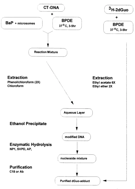
Figure 3.1 summarizes the different ways to synthesize the BaP-dGuo adduct. Each method is described below. Briefly, BaP modified DNA was made by incubating calf-thymus DNA with BaP and rat liver microsomes or with BPDE. The modified DNA was isolated via phenol:chloroform extraction and ethanol precipitation, followed by enzymatic hydrolysis to nucleosides. The use of 2'-deoxyguanosine (2'-dGuo) was also used so that the DNA isolation and enzymatic hydrolysis steps could be omitted.
Microsomal Activation of Benzo(a)pyrene
The method of synthesis of benzo(a)pyrene-deoxyguanosine was similar to the procedure previously described (Gorelick & Wogan, 1989). To prepare in vitro modified DNA, calf thymus DNA (2mg/ml) was shaken in HEPES pH 7.1 with 2.5-3.5 mg/ml microsomal protein, 0.8 mM NADP+, 5 mM glucose-6-phosphate, 0.5 mM MgCl
2, 0.4 units/ml glucose-6-phosphate
dehydrogenase, and 100 mM 3H-BaP (0.5-3 Ci/mmol) or 83 mM BaP in DMSO (1% total
volume). After 90 minutes, the reaction was terminated by the addition of SDS (to 1%) and NaCl (to 250 mM). DNA was isolated by two extractions with an equal volume of phenol/chloroform (1:1), followed by an extraction with chloroform alone until a clear interface was observed. DNA was precipitated in cold ethanol and rinsed sequentially with benzene, chloroform,
dichloromethane, ethanol and ether before drying under nitrogen. The isolated DNA was dissolved in water. Recoveries and binding levels were determined from absorbance at 254 nm (20 OD units/mg DNA/ml) and the liquid scintillation counting of known amounts of DNA. The BaP-DNA adduct was calculated by using the molar extinction coefficient of c=29,000 at 347nm (Weinstein, et al., 1976).
Enzymatic Hydrolysis
Initially a published method (Park, et al.,1989) known to produce nucleosides from PAH-modified DNA was used. This method of enzymatic hydrolysis used SVPD, AP, DNAse, and spleen phosphodiesterase I (37°C, 2 hours). A single digestion procedure that could be used to analyze more than one type of adduct would greatly enhance the flexibility of the ADAM method. The method that was used in the Wogan laboratory for the optimization of the ADAM
method with 2-d-Guo and for the analysis of 4-ABP adducts is as follows: microsomally modified calf thymus DNA (<1.7 mg/ml) was digested to nucleosides with nuclease P1 (76.5 units/mg),
0.450 ml 2 mM ZnCl2, 0.060 ml IM NaAc. The volume was brought up to 3 ml with distilled H
20
and incubated for 1 hour at 370C with agitation, covered with foil. An equal volume of IM Tris
buffer or IM carbonate buffer was added. Note: The carbonate buffer was later substituted for the Tris buffer because the Tris was found to have contributed to the 35S background in the
radio-chromatogram. Snake venom phosphodiesterase (0.879 units/mg) and alkaline phosphatase (16.9 units/mg) were added and the reaction was incubated another hour at 370C, with agitation. The
reaction mixture was stored at -200C until ready for HPLC analysis. The RP-HPLC gradient used
was Gradient A. In order to establish that the digestion yielded nucleosides, a different HPLC gradient (Gradient C as described in Table 3.1) was used and used acetonitrile and H20, pH 4.4.
Reaction of isomeric benzo(a)pyrene-diol-epoxide with DNA or 2'-Deoxyguanosine
Modification of several methods (Weinstein et al., 1976, Vahakangas et al., 1985, Jeffrey et al., 1976) were used for the reaction of BPDE with calf thymus DNA, as follows: 2'-deoxyguanosine (20 mg) dissolved in 20 ml H20 was reacted with 5 mg
benzo(a)pyrene-diol-epoxide in 10 ml THF. The reaction mixture was incubated for 3-6 hours at 37°C in a shaking H20 bath, followed by extractions with 6 x 15ml H20-saturated ethyl acetate and
2 x 15ml H20-saturated diethyl ether. The aqueous phase was recovered. The amount of DNA or deoxyguanosine was determined with absorbance at 254 nm. BaP was calculated by using the molar extinction coefficient of 29,000 at 347 nm. The modified samples were scanned from 200-700 nm. Simultaneously, 3H-2dGuo (10 nmol, 31 Ci/mmol) was reacted with BPDE (0.81
mg) and taken through the same procedure. The rest of the 3H-dGuo-BaP was extracted and
evaporated. The pear shaped flask was washed 3 times with methanol and 3 times with H20.
The amount of 2-dGuo was determined from absorbance at 254 nm (20 OD units/mg DNA/ml) or by scintillation counting for the 3H-dGuo (31 Ci/mmole). BaP was calculated by
using the molar extinction coefficient of e=29,000 at 347 nm. The modified samples were scanned from 200-700 nm. The cold BPDE-dGuo was concentrated by evaporation unit and the pear shaped flask was washed 3 times with methanol and 3 times with H20. The volume was
brought to 1.5 ml and stored until ready for HPLC analysis. At this time a microcolumn (1 gm) was attached to the electrospray-MS. Gradient F was used at a flow rate of 0.1 ml/min.
Reaction of BPDE with oligonucleotides
Several researchers (Cosman, et al., 1990 ;Mao et al., 1995) found that the reaction of
BPDE with an oligonucleotide gave a 30% yield of BaP-dGuo. Oligonucleotides (IM and 10M solutions) were purchased from MIT Biopolymers laboratory. A small scale reaction was performed to determine whether the reaction would produce results similar to those in published literature. The first oligonucleotide used was a 9mer d(ATATGTATA). A quantity of 1.5 pmol, dissolved in 20 mM sodium phosphate, was incubated with 3.0 gmol BPDE (ratio 2 BPDE:1 oligonucleotide) dissolved in THF. All procedures were done under yellow lights. The mixture was incubated overnight at room temperature, covered with foil and in the dark. The reaction was analyzed by RP-HPLC with a Zorbax ODS column 4.6 x 250 mm, at room temperature with a flow rate of 1 ml/min using Gradient D: A = 20 mM NaPhos pH 7.0, B = methanol, linear gradient 0-90%B in 60 minutes. The peak at 20-25 minutes was collected and digested to
nucleosides as described previously, and analyzed by HPLC. A large scale reaction used 3 reaction vials (15 ml polypropylene tubes) each containing 10 pimol of oligonucleotide in 5 ml NaPhos, pH 7.0 and 20 ýtmol of BPDE (6 mg) dissolved in THF and 1.5% TEA. Mao et al.
(1995) found that the addition of TEA increased the modification level. The HPLC anaylsis of the reaction products used a C18 semi-preparative column at room temperature with a flow rate of
3 ml/min. Another oligonucleotide was found to yield a high percentage of BaP-dGuo (Liu, 1996). This 1 lmer d(CTAGGGTATC) 1.3 ptmole was reacted with BPDE (2.6 pimole).
Organic Synthesis of BaP-dGuo
A protocol was established (Seyedi, unpublished) for synthesis of the BaP-dGuo adduct. The chemical strategy is shown in Figure 3.2. The BaP-dGuo was first protected with benzyl groups, then incubated with BPDE overnight at 370 C, in the dark. The mixture was
deprotected and purified. The molecular weight was verified with electrospray-MS.
3.2.3 Chromatography
The samples were analyzed by reverse phase HPLC (model 126, Beckman Instruments, Inc, Columbia, MD) equipped with diode array detector (model 168, Beckman Instruments, Inc, Columbia, MD). A Zorbax-ODS 5 jtm column (4.6 mm x 250 mm, MacMod Chadds Ford, PA) or an Microsorb 5 ipm C18 column (4.6 mm x 250 mm, Rainin, Woburn, MA)
was routinely used with the HPLC gradients are summarized in Table 3.1. The radioactivity was determined on-line with a Radio-Chromatography Detector (Radiomatic Series A-280). Several
co-injections of the BaP-dGuo adduct generated with either microsomal activation or with BPDE were analyzed via HPLC with the diode array detector fixed at 347 nm and 254 nm. The
oligonucleosides would have an absorbance at 254 nm and BPDE would have an absorbance at 347 nm. A radioactive chromatogram was also generated when possible. The 3H label on the
benzo(a)pyrene, when reacted with calf thymus DNA or on the deoxyguanosine, when reacted with BPDE, would give a peak when present.
3.3 Results and Discussion
The reaction involving the modification of calf thymus DNA with 3H-BaP and microsomes
was repeated several times. Typically it resulted in a modification level of 1 BaP in 105 bases. Table 3.2 summarizes several such reactions. The goal was to produce enough adduct to
generate HPLC chromatograms while validating the ADAM method. The specific activity had to be high enough to be able to detect a signal. For example, a sample had to contain approximately 100,000 cpm to produce an acceptable chromatogram. The specific activity of the 3H-BaP ranged
from 125 mCi/mmole to 12,500 mCi/mmole. As the table shows, the higher the specific activity of the 3H-BaP, the lower the modification level of the DNA. For example, the reaction using a specific activity of 12,500 mCi/mmole resulted in a modification level of 1 BaP per 700,000 bases, while a modification of 1 BaP in 11,000 was obtained with 3H-BaP at 500 mC/mmole. Therefore,
the specific activity of 3000 mCi/mmole was used for the rest of the microsomal modification experiments. It consistently yielded a modification level of 1 BaP in 12,000-14,000 bases. The
recovery of the 3H-BaP-dGuo was monitored during the enzymatic hydrolysis and Sep-pak C 18
cartridge purification step. Fifty percent of the original adduct in the DNA was determined to be recovered up to the step when it was ready for acylation.
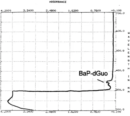
Once the recovery of the processing was determined, the next step was to scale-up the synthesis of the adduct. The goal was to make milligram quantities for the validation of the ADAM method. This would allow structural characterization of the acylation products. A comparison of modification procedures is shown on Table 3.3. The modification level was increased by using the commercially available BPDE. BPDE reacted with 2'-deoxyguanosine resulted in the highest modification level (1:793). The modified samples were scanned from 200 nm to 700 nm revealing a doublet peak from 200 nm to 300 nm (DNA or deoxyguanosine) and between 320 nm to 350 nm (BaP). A typical scan is shown in Figure 3.3. The molecular weight was confirmed by electrospray-MS. The molecular weight of the adduct is 569 yielding a peak at m/z 570 in a protonated spectrum.
When the reaction of BPDE incubated with 2'-deoxyguanosine was scaled up to 100 mg of DNA and 25 mg of BPDE, the amount of adduct recovered (0.1 mg) in the aqueous portion after solvent extraction was considerably lower than expected (2 mg). The solvent extracts were found to contain the adduct as revealed by HPLC analysis. The adduct was HPLC purified and the molecular weight was determined by electrospray-MS. Even after HPLC purification, the adduct was not pure. At this time a microcolumn (1 pm) was attached to the electrospray-MS. Gradient F was used at a flow rate of 0.1 ml/min. There were 4 peaks detected with the same molecular weight (m/z 570) and these peaks correspond to the 4 seen in the HPLC profile (Figure 3.12). The timing on the gradient of the electrospray was different because of periodic machine failure;
however, the profile is the same. The two major peaks most likely correspond to the trans isomers and the two minor peaks correspond to the two cis isomers. All four stereoisomers have the same molecular weight.
3.3.1 HPLC analysis
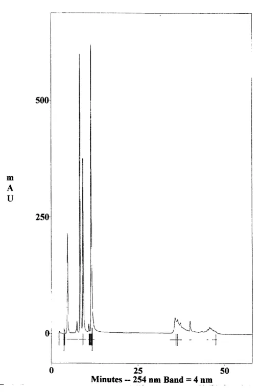
In order to determine if the hydrolysis of the DNA yielded nucleosides, the hydrolysate containing 50 pg of DNA was injected on the HPLC using gradient C. The peaks early in the chromatogram correspond to dC, dG, dT and dA (Figure 3.4). The HPLC profiles/radio-chromatograms generated by the two different enzymatic hydolysis procedures are illustrated in Figure 3.5. The 3H label on the benzo(a)pyrene give the signal designated as cpm on the Y-axis.
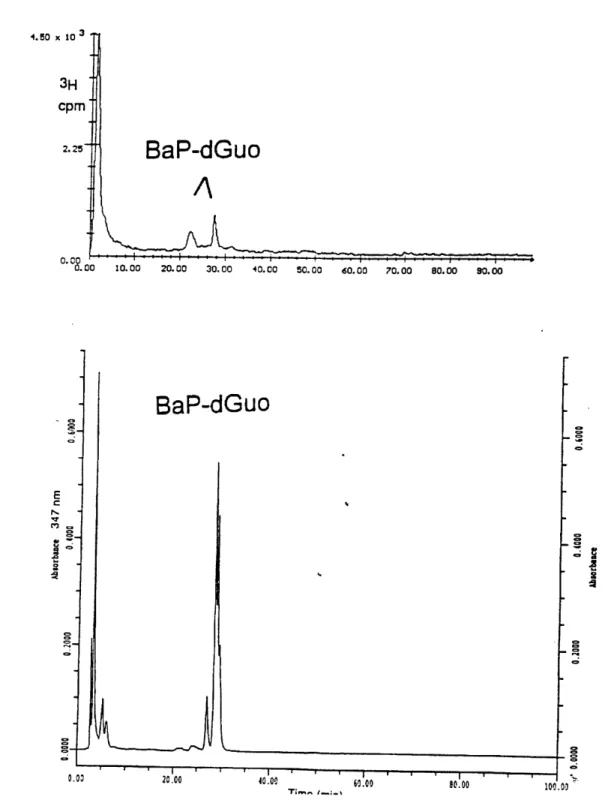
A co-injection of the cold BPDE-dGuo and the 3H-BaP-dGuo, generated with microsomes, gave
the same UV profile at 347 nm and 254 nm and was similar to the radioactive chromatogram (Figure 3.6) 3H-BaP-dGuo. There is a 2 minute delay between the UV-HPLC chromatogram and the start of the radio-chromatogram. The 1s' peak at around 3.5 minutes was identified as
deoxyguanosine or undigested material. The doublet peak at 24 minutes (26 minutes on the radio-chromatogram) is considered to be the BaP-tetrol. The doublet peak at 30 minutes (28 minutes on the radio-chromatogram) is the BaP-dGuo-adduct. These chromatograms are reproducible. Figure 3.7 is an HPLC radio-chromatogram of 3H-dGuo-BaP after extraction and
purification via Sep-pak C, column. The peak at 35 minutes is considered the 8 3H-BPDE-dGuo
adduct. The change in retention time from 26-28 minutes to 35 minutes is due to the change in the column temperature. The column temperature was room temperature instead of 35oC which