HAL Id: hal-01772605
https://hal-amu.archives-ouvertes.fr/hal-01772605
Submitted on 20 Apr 2018
HAL is a multi-disciplinary open access
archive for the deposit and dissemination of
sci-entific research documents, whether they are
pub-lished or not. The documents may come from
teaching and research institutions in France or
abroad, or from public or private research centers.
L’archive ouverte pluridisciplinaire HAL, est
destinée au dépôt et à la diffusion de documents
scientifiques de niveau recherche, publiés ou non,
émanant des établissements d’enseignement et de
recherche français ou étrangers, des laboratoires
publics ou privés.
Distributed under a Creative Commons Attribution| 4.0 International License
The Gene Expression Analysis of Blood Reveals
S100A11 and AQP9 as Potential Biomarkers of Infective
Endocarditis
Franck Thuny, Julien Textoris, Amira Ben Amara, Adil El Filali, Christian
Capo, Gilbert Habib, Didier Raoult, Jean-Louis Mege
To cite this version:
Franck Thuny, Julien Textoris, Amira Ben Amara, Adil El Filali, Christian Capo, et al.. The Gene
Ex-pression Analysis of Blood Reveals S100A11 and AQP9 as Potential Biomarkers of Infective
Endocardi-tis. PLoS ONE, Public Library of Science, 2012, 7 (2), pp.e31490. �10.1371/journal.pone.0031490�.
�hal-01772605�
and
AQP9
as Potential Biomarkers of Infective
Endocarditis
Franck Thuny1,2, Julien Textoris2, Amira Ben Amara2, Adil El Filali2, Christian Capo2, Gilbert Habib1,2, Didier Raoult2, Jean-Louis Mege2*
1 De´partement de Cardiologie, Hoˆpital de la Timone, Aix-Marseille University, Marseille, France, 2 Unite´ de Recherche sur les Maladies Infectieuses Transmissibles et Emergentes (URMITE), Centre National de la Recherche Scientifique (CNRS) Unite´ Mixte de Recherche 6236, Aix-Marseille University, Marseille, France
Abstract
Background:The diagnostic and prognostic assessments of infective endocarditis (IE) are challenging. To investigate the host response during IE and to identify potential biomarkers, we determined the circulating gene expression profile using whole genome microarray analysis.
Methods and Results:A transcriptomic case-control study was performed on blood samples from patients with native valve IE (n = 39), excluded IE after an initial suspicion (n = 10) at patient’s admission, and age-matched healthy controls (n = 10). Whole genome microarray analysis showed that patients with IE exhibited a specific transcriptional program with a predominance of gene categories associated with cell activation as well as innate immune and inflammatory responses. Quantitative real-time RT-PCR performed on a selection of highly modulated genes showed that the expression of the gene encoding S100 calcium binding protein A11 (S100A11) was significantly increased in patients with IE in comparison with controls (P,0.001) and patients with excluded IE (P,0.05). Interestingly, the upregulated expression of the S100A11 gene was more pronounced in staphylococcal IE than in streptococcal IE (P,0.01). These results were confirmed by serum concentrations of the S100A11 protein. Finally, we showed that in patients with IE, the upregulation of the aquaporin-9 gene (AQP9) was significantly associated with the occurrence of acute heart failure (P = 0.02).
Conclusions:Using transcriptional signatures of blood samples, we identified S100A11 as a potential diagnostic marker of IE, and AQP9 as a potential prognostic factor.
Citation: Thuny F, Textoris J, Ben Amara A, El Filali A, Capo C, et al. (2012) The Gene Expression Analysis of Blood Reveals S100A11 and AQP9 as Potential Biomarkers of Infective Endocarditis. PLoS ONE 7(2): e31490. doi:10.1371/journal.pone.0031490
Editor: Roger Le Grand, Commissariat a l’Energie Atomique(cea), France Received June 30, 2011; Accepted January 9, 2012; Published February 3, 2012
Copyright: ß 2012 Thuny et al. This is an open-access article distributed under the terms of the Creative Commons Attribution License, which permits unrestricted use, distribution, and reproduction in any medium, provided the original author and source are credited.
Funding: This work was supported by a grant from the Fe´de´ration Franc¸aise de Cardiologie, to Dr. Franck Thuny. The funders had no role in study design, data collection and analysis, decision to publish, or preparation of the manuscript.
Competing Interests: The authors have declared that no competing interests exist. * E-mail: jean-louis.mege@univmed.fr
Introduction
Infective endocarditis (IE) is a severe disease with an incidence ranging from 30 to 100 episodes per million person-years [1,2]. Mortality is high, and more than one-third of the patients die within the first year of their diagnosis [3,4,5,6]. Despite the development of microbiological and imaging techniques, the diagnosis of IE remains challenging. Indeed, etiologic diagnoses may not be obtained in 2.5% to 31% of cases [1,7], and echocardiography is negative for approximately 10% of cases [1]. Thus, morbidity and mortality remain high, and strategies fail to detect and predict a substantial number of events, such as heart failure and embolic events. One of the reasons for these issues is the complex pathogenesis of the disease that involves many host-pathogen interactions. Indeed, previous endocardial lesions can lead to the exposure of the underlying extracellular matrix proteins, local inflammation with interleukin-1 release, and then thrombus formation, which is termed ‘‘non-bacterial vegetation’’. In the case of bacteremia, valves with pre-existing sterile
vegetations or tissues with minimal lesions can be colonized because of crucial interactions between the bacteria, platelets and endothelial cells via several bacterial surface proteins or plasma-bridging molecules. This process leads to the recruitment of circulating cells, including neutrophils and monocytes, the release of cytokines and procoagulant factors that contribute to the enlargement of vegetations and the protection of bacterial pathogens from host defenses [2].
This complex situation highlights the need for methods that improve the management of IE. Specifically, the identification of new biomarkers for diagnosis and risk stratification will be useful, as well as biological indicators for a rapid surgical management. Molecular signatures of IE represent promising means for addressing these challenges. In particular, microarray-based transcriptomes can identify specific signatures of the disease that might ultimately be translated into clinically useful molecular biomarkers [3]. Recently, we identified a transcriptional signature of IE from valvular tissue [4], but this approach cannot be used by physicians in their daily practice. Peripheral blood is an alternative
to a tissue sample for the molecular profiling of human diseases [3] because it interacts with every tissue in the body and plays a crucial role in many IE pathophysiological processes, such as immunity, inflammation and coagulation.
To investigate the host response during IE and identify potential biomarkers, we determined the circulating gene expression profile through a unique whole genome microarray analysis. We show that patients with IE exhibited a specific transcriptional program with a predominance of gene categories associated with cell activation as well as innate immune and inflammatory responses. We demonstrate that the gene encoding the S100 calcium binding protein A11 (S100A11) and the serum concentration of S100A11 protein are both significantly increased in IE patients, especially in the case of staphylococcal etiology. Moreover, in IE patients, the upregulation of the aquaporin-9 gene (AQP9) is associated with the occurrence of acute heart failure.
Methods Study Population
All participants in the study were prospectively enrolled from January 2009 to December 2010 at the Cardiology Department of La Timone Hospital (Marseille, France), which is an adult tertiary care teaching hospital. The patients were eligible for the study if they had a clinical suspicion of native valve IE. The exclusion criteria were age ,18 years, pregnancy, history of previous IE, intracardiac material (prosthetic valve, pacemaker or implantable cardioverter defibrillator), failure in the RNA extraction proce-dure, and an antibiotic treatment for more than one week. Among the 71 patients eligible for the study, 22 were excluded because of the presence of at least one exclusion criterion. Their baseline characteristics are summarized in Table 1.
The transcriptomic case-control study was performed in: i. 39 consecutive patients with native valve IE (IE group)
diagnosed by a multidisciplinary team who applied the modified Duke criteria [5] (two major criteria in 37 patients and 1 major with 3 minor criteria in 2 patients),
ii. 10 patients with a previous valvular heart disease (VHD) admitted for a suspicion of IE because of persistent fever or new abnormalities detected by echocardiography, but with a final excluded IE diagnosis (excluded IE group). Of these, the final diagnosis was an acute degenerative mitral chordae rupture (n = 4), a respiratory tract infection (n = 2), an urinary tract infection (n = 1), and no diagnosis after a 3-month follow-up (n = 3). Among these 3 patients, fever disappeared spontaneously without antibiotics.
iii. 10 age-matched healthy volunteers (control group).
Initially, 10 IE patients and 5 controls were arbitrarily selected, and their samples were analyzed with microarrays; however, samples from all of the patients were eventually analyzed by quantitative real-time reverse transcriptase-polymerase chain reaction (qRT-PCR).
For each case, the following data were collected: age, sex, comorbidity [6], signs and symptoms, duration of symptoms, biological results, history of antimicrobial therapy for the current illness that prompted the patient to seek medical attention, antecedent disease, predisposing factors for IE including systemic disease, intravenous drug use, treatment received during the course of hospitalization and complications (acute heart failure, embolic event, and cardiac abscess). Echocardiography was performed by a systematic transthoracic and trans-esophageal approach [5]. For the microbiological diagnosis, a diagnostic kit,
including blood cultures and serologies, was used as previously described [7]. When cardiac surgery was required, valvular surgical samples were analyzed using cultures and PCR amplifi-cation.
The study was performed according to the principles of the Declaration of Helsinki. Informed and written consent was obtained from each subject, and the Ethics Committee of the Universite´ de la Me´diterrane´e approved the study.
RNA Preparation and Microarrays
At the admission of patients, peripheral blood was drawn into PAXgene tubes (Qiagen). The RNA was extracted according to the manufacturer’s recommendations, which included a DNase step. The quality and quantity of isolated RNAs were assessed using the Nanodrop (Thermo Scientific) and 2100 Bioanalyzer (Agilent Technologies), and the RNAs were eluted in 80mL of water and stored at 220uC. Subsequently, the RNAs were analyzed using microarray chips (Agilent Technologies), including 45,000 probes (4644 K Whole Human Genome, Agilent Technologies) and one-color microarray-based gene expression analysis, as recently described [8]. Briefly, 400 ng of RNA was labeled with cyanine-3 CTP using a commercial kit (Low RNA Table 1. Characteristics of the patients according to the final diagnosis. IE group (N = 39) Excluded IE group (N = 10) P Value Age, mean 6SD, years 58618 63615 0.42
Sex ratio (M/F) 30/9 7/3 0.65
Intravenous drug user 5 (13%) 0 (0%) 0.57
Cancer 3 (8%) 1 (10%) 1.0
Diabetes 5 (13%) 1 (10%) 1.0
Comorbidity index .2 15 (38%) 3 (30%) 0.72 Fever, temperature .38uC 38 (97%) 7 (70%) 0.02 Vascular phenomena* 14 (36%) 1 (10%) 0.15 Immunologic phenomena{ 0 (0%) 0 (0%) NA Heart failure 16 (41%) 5 (50%) 0.72 Serum creatinine .20 mg/L 3 (8%) 1 (10%) 1.0 Valve localization of abnormality 1.0
Aortic 17 (44%) 4 (40%)
Mitral 23 (59%) 6 (60%)
Right valves 3 (8%) 0 (0%)
Severe valvular regurgitation 32 (82%) 7 (70%) 0.41
Vegetation 32 (82%) 0 (0%) ,0.001 Abscess 10 (26%) 0 (0%) ,0.001 Blood cultures ,0.001 Streptococci{ 27 (69%) 0 (0%) Staphylococci1 12 (31%) 1 (10%) Negative 0 (0%) 9 (90%) NA = not applicable.
*Including arterial emboli, septic pulmonary infarcts, mycotic aneurysms, intracranial hemorrhages, conjunctival hemorrhages, and Janeway’s lesions.
{
Including glomerulonephritis, Osler’s nodes, Roth’s spots, and rheumatoid factor.
{
Including three enterococci.
1
Including nine Staphylococcus aureus. doi:10.1371/journal.pone.0031490.t001
Input Fluorescent Amplification Kit, Perkin Elmer). The samples were deposited on a slide, and hybridization was performed at 65uC using the In situ Hybridization Plus kit (Agilent Technologies) for 17 hours. The arrays were scanned with a pixel size of five microns using the DNA Microarray Scanner G2505B (Agilent Technologies). The image analysis and correction of intra-array signals were performed with the Feature Extraction Software A.9.1.3 (Agilent Technologies). Microarray data analysis was performed using the R and the Bioconductor software suite. Raw data were filtered and normalized using the Agi4x44PreProcess library. Unsupervised and supervised analysis were done using hierarchical clustering, principal component analysis (made4 library) [9], and Significance Analysis of Microarray (SAM) algorithm (siggenes library). Genes were considered to be differentially expressed if False Discovery Rate (FDR, Benjamini-Hochberg [10]) was below 1% and absolute fold change was above 1.5. Functional enrichment analysis was performed on selected genes with DAVID Tools [11], using the following ontologies: Gene Ontology (GO) [12], INTERPRO [13] and KEGG pathways [14]. The figures were designed using Cytoscape [15] and Inkscape softwares. The data were generated according to the Minimum Information About a Microarray Experiment guidelines and were deposited in the National Center for Biotechnology Information’s Gene Expression Omnibus [16]. The data are accessible using the following accession number: GSE29161.
qRT-PCR Analyses
Reverse transcription of 150 ng of total RNA per reaction was performed with the MMLV-RT kit (Invitrogen), as previously described [8]. Quantitative real-time PCR was performed using gene-specific primers designed using Primer3 [17]. The list of primers with their sequences is provided in the Table S1. The
transcriptional profiles of the selected genes were screened in homemade 384-well plates using the 7900HT Fast Real Time PCR System, and the qRT-PCR data were extracted using the SDS 2.2 software (Applied Biosystems). The results were normalized using the housekeeping gene b-actin and are expressed as FC = 2–DDCt, where DDCt = (CtTarget2CtActin)assay
2(CtTar-get2CtActin)control, as previously described [8]. S100A11 Assay
The serum concentrations of S100A11 protein were assessed by enzyme-linked immuno-sorbent assay kit (USCN, Life Science Inc.) according to the manufacturer’s protocol. The sensitivity of the test is 0.37 ng/mL.
Statistical Analysis
The categorical data were reported as frequencies and percentages and were compared using the Fischer exact two-tailed test. The continuous data are expressed as the mean 6 SEM or the median and interquartile range. Comparisons between two groups were performed using the Mann-Whitney U test, and comparisons between the three groups of individuals were performed using the Kruskal-Wallis test with post-hoc Dunn’s multiple comparison test. This statistical analysis was conducted using SPSS for Windows, version 16.0 (SPSS Inc., Chicago, Illinois). Values of P,0.05 were considered statistically significant.
Results
Microarray Analysis of Patients with IE
A whole-genome microarray approach was used to define the peripheral transcriptional signature of IE. For that purpose, we
Figure 1. Gene expression signature between endocarditis and control patients. Differential gene expression between infective endocarditis (IE) and control patients was analyzed by Principal Component Analysis (A) and Hierarchical clustering (B). In panel A, IE patients (red) are clearly separated from control patients (blue) on the first component (x-axis) of the PCA. In panel B, selected genes with FDR,1% and absolute fold change above 1.5 were represented as a heatmap, with genes in rows and samples in columns. Gene expression level was color-coded from blue to red.
arbitrarily selected ten IE patients and five controls. The principal components for the analysis of the overall gene expression showed that the IE patients and controls were organized in two different groups (Figure 1, Panel A). Supervised analysis using the SAM algorithm identified 1,782 probes differentially expressed with an absolute fold change above 1.5, and a FDR,1% (Figure 1, Panel B). Modulated genes in the IE patients and controls were distributed in two different clusters. Finally, we found that 1,274 probes (912 genes) were upregulated and 508 probes (339 genes) were downregulated in IE patients compared with controls. Taken together, these different analyses of the gene expression demon-strated that the patients with IE exhibited a specific transcriptional program.
Functional Analysis of Modulated Genes in Patients with IE
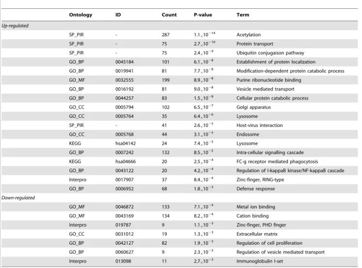
The genes that were differentially expressed in IE patients were classified into different categories based on GO, SP_PIR key-words, Interpro and KEGG ontologies (Table 2). In the upregulated genes, there was an enrichment of the biological processes related to cell activation, post-translational modification of proteins, intra-cellular transport and localization, phagocytosis and immune response. Post-translational modification of proteins was supported by the following terms: ‘‘acetylation’’, ‘‘ubiquitin
conjugation pathway’’ and ‘‘modification-dependant protein catabolic process’’. Intra-cellular transport and localization, as well as phagocytosis were supported by the following terms: ‘‘establishment of protein localization’’, ‘‘protein transport’’, ‘‘vesicle mediated transport’’, ‘‘golgi apparatus’’, ‘‘lysosome’’, ‘‘endosome’’ and FC-c receptor mediated phagocytosis’’. These latter terms, together with the terms ‘‘host-virus interaction’’, ‘‘regulation of I-kappaB kinase/NF-kappaB cascade’’, ‘‘Zing finger, RING-type’’ and ‘‘defense response’’ support the involve-ment of the up-regulated genes in the immune response. In the downregulated genes, the GO terms corresponding to cation binding, extracellular matrix and regulation of vesicle-mediated transport were enriched. Two KEGG pathway linked to the immune response were enriched in the upregulated signature: ‘‘lysosome’’ and ‘‘FC-c receptor mediated phagocytosis’’.
Validation of Modulated Genes in Patients with IE
Because of the large number of modulated genes and the density of transcriptomic networks, the following procedure was used to validate the microarray data. Seven upregulated genes were selected from a list of the 300 most modulated genes according to their putative role in IE pathophysiology. Their expression was determined by qRT-PCR in an enlarged cohort of IE patients (n = 39) and controls (n = 10). The genes encoding aquaporin-9
Table 2. Ontology enrichement analysis.
Ontology ID Count P-value Term
Up-regulated
SP_PIR - 287 1.1610
214
Acetylation
SP_PIR - 75 2.7610210 Protein transport
SP_PIR - 75 2.461029 Ubiquitin conjugaison pathway
GO_BP 0045184 101 6.1610
28
Establishment of protein localization
GO_BP 0019941 81 7.761028 Modification-dependent protein catabolic process
GO_MF 0032555 199 8.961028 Purine ribonucleotide binding
GO_BP 0016192 81 9.0610
28
Vesicle mediated transport
GO_BP 0044257 83 1.5610
28
Cellular protein catabolic process GO_CC 0005794 102 6.561027 Golgi apparatus
GO_CC 0005764 35 6.461026 Lysosome
SP_PIR - 41 2.661025 Host-virus interaction
GO_CC 0005768 44 3.161025 Endosome
KEGG hsa04142 24 7.461025 Lysosome
GO_BP 0007242 132 8.5610
25
Intra-cellular signalling cascade KEGG hsa04666 20 2.561024 FC-g receptor mediated phagocytosis
GO_BP 0043122 20 4.261024 Regulation of I-kappaB kinase/NF-kappaB cascade
Interpro 0017907 37 8.4610
24
Zinc-finger, RING-type GO_BP 0006952 68 1.861023 Defense response
Down-regulated
GO_MF 0046872 133 7.1610
24
Metal ion binding GO_MF 0043169 134 8.261024 Cation binding
Interpro 019787 9 1.161023 Zinc-finger, PHD finger
GO_CC 0031012 19 1.3610
23
Extracellular matrix
GO_BP 0042127 82 1.961023 Regulation of cell proliferation
GO_BP 0060627 9 2.361023 Regulation of vesicle mediated transport
Interpro 013098 11 2.7610
23
Immunoglobulin I-set doi:10.1371/journal.pone.0031490.t002
(AQP9), S100A11, plasminogen activator urokinase receptor (PLAUR), solute carrier family 16, member 3 (SLC16A3), transforming growth factor beta 1 (TGFb1), interferon gamma-inducible protein 16 (IFI16), apolipoprotein B mRNA editing enzyme, and catalytic polypeptide-like 3B (APOBEC3B) were significantly upregulated in IE patients (P,0.001 for all, Figure 2). Because these results may be due to the underlying valvular abnormality or the global inflammatory response, they were compared with those obtained in the group of patients with excluded IE after an initial suspicion (n = 10). The S100A11 gene was significantly upregulated in the IE patients as compared with the patients with excluded IE (P,0.05, Figure 2). The upregulated expression of the S100A11 gene had functional consequences. Indeed, the serum concentration of S100A11 protein was higher in patients with IE than in patients with excluded IE (5.061.0 versus 2.160.5 ng/mL, P,0.05).
Finally, we tested whether the bacterial etiology of IE plays a role in the modulated expression of the S100A11 gene. In our series, 23 patients were infected with staphylococci (Staphylococcus aureus in nine patients) and 16 patients with streptococci. The expression of the S100A11 gene was significantly higher in staphylococcal IE than in streptococcal IE and patients with excluded IE (P,0.01 and P,0.001, respectively) (Figure 3, Panel A). Similarly, the serum concentration of S100A11 protein was slightly increased in staphylococcal IE when compared with streptococcal IE and was significantly (P,0.05) increased when compared with patients with excluded IE (Figure 3, Panel B). Taken together, these results suggested that the determination of S100A11 at both the transcriptional and the protein levels was potentially predictive of IE. The S100A11 determination also discriminated between staphylococcal and streptococcal IE.
IE Transcriptional Signature and Major IE-Related Complications
Finally, we analyzed the modulation of the seven genes by qRT-PCR at admission according to IE-related complications, including acute heart failure (n = 17), embolic events (n = 14), and cardiac abscess (n = 10). The expression of the AQP9 gene was increased only in patients who experienced acute heart failure (P,0.05). The expression of the S100A11, PLAUR, SLC16A3, TGFb1, IFI16, and APOBEC3B genes was not related to acute heart failure, embolic events, or abscess (Figure 4). Taken together, these results suggested that the AQP9 gene might be a marker of prognosis in IE.
Discussion
The aim of the present study was to characterize the peripheral transcriptional profile of patients with IE and to identify potential biomarkers. The clinical history of IE is dependent on the causative microorganism, the presence or absence of pre-existing cardiac disease, and the mode of presentation. The modified Duke criteria for IE diagnosis have been validated [5]; but they have clear limitations, especially when blood cultures and/or echocar-diography are negative. Other parameters have been proposed to increase the yield of the diagnostic process, but none have been definitely implemented into the diagnostic criteria. A small study suggested that serum procalcitonin may be a valuable diagnostic
marker in patients with suspected IE [18]. New imaging techniques, such as computed tomography (CT) [19], positron emission tomography/CT scan [20], three-dimensional echocar-diography [21], or systematic cerebral MRI [22], are emerging to improve IE diagnosis; their final place has yet to be defined. The transcriptional analysis of a disease is a promising method to identify novel candidate biomarkers that might ultimately be translated into clinical practice [3]. In the present study, we found that blood transcriptomics may be used to characterize IE. We
Figure 2. Analysis of genes modulation using qRT-PCR. Blood samples from 39 patients with IE, 10 patients with excluded IE after an initial suspicion, and 10 controls were analyzed by qRT-PCR for the expression of seven genes that were found to be upregulated by microarray. The results were normalized with the b-actin gene. *P,0.001 for the comparison between IE patients and controls. {P,0.05 for the comparison between IE patients and patients with excluded IE.
doi:10.1371/journal.pone.0031490.g002
Figure 3. Expression ofS100A11gene and S100A11 protein. A, The S100A11 gene was significantly overexpressed in IE patients as compared with excluded IE patients (P,0.05). *P,0.001 for the comparison between staphylococcal IE and excluded IE. {P,0.01 for the comparison between staphylococcal IE and streptococcal IE.B, The S100A11 serum level was higher in staphylococcal IE (6.962.0 ng/mL) than in excluded IE (2.160.5 mg/mL) and staphylococcal IE (4.261.2 ng/ mL). {P,0.05 for the comparison between staphylococcal IE and excluded IE. 1P = 0.17 for the comparison between staphylococcal IE and streptococcal IE.
doi:10.1371/journal.pone.0031490.g003
have recently described the valvular transcriptional signature of IE [4]; to our knowledge, this is the first time that the host response in IE was studied at the blood level, which is more accessible and relevant than valvular tissue in the clinical practice.
The present analysis of the peripheral signature of IE allowed the identification of potential candidates for the diagnostic and prognostic assessment of IE. We showed that the expression of the S100A11 gene was increased in patients with a final diagnosis of IE as compared with patients with an excluded IE diagnosis after an initial suspicion. The increased expression of the S100A11 gene was essentially related to Staphylococcus infection. These transcrip-tional results were consistent with higher serum concentrations of the S100A11 protein in IE patients. S100A11 is a member of the family of S100 proteins that are localized in the cytoplasm and/or nucleus of a wide range of cells. S100A11 proteins are involved in endocytosis and exocytosis [23], regulation of enzyme activity, cell growth regulation, apoptosis and inflammation [24]. Note that endocytosis and inflammation were two hallmarks of the peripheral signature of IE determined in this study. Regarding
the role of S100A11 in the pathophysiology of IE, our analysis extracted from the litterature a pathway that involves receptor for advanced glycation end-products (RAGE) and S100A11, which is one of its ligand. RAGE ligation causes cellular activation via signalling cascades including nuclear factor NF-kB, MAP kinases leading to induction of inflammatory cytokines, proteases ans oxydative stress [25]. NF-kB and the presence of RAGE ligand also up-regulates RAGE expression, ensuiring maintenance and amplification of the initial signal (Figure 5). RAGE and their ligands are strongly involved in the pathogenesis of systemic inflammation and represent a potential therapeutic target in sepsis and several acute infectious diseases [26]. Although S100A protein family can be associated with several human diseases, such as neurological diseases, cardiomyopathy, cancers, and inflammatory diseases, only few data exist on the specific implication of the S100A11 in pathology. In our microarray analysis, we compared IE patients with healthy subjects in order to bring out great significant differences with a small sample size. Thereafter, we analyzed S100A11 gene expression in a population of patients with
Figure 4. Gene expression according to the major IE-related complications. A, The expression of the AQP9 gene was significantly increased only in patients who experienced acute heart failure.B and C, The expression of the S100A11, PLAUR, SLC16A3, TGFb1, IFI16, and APOBEC3B genes was not related to acute heart failure, embolic events, or abscess. *P,0.05.
a suspicion of IE, i.e. in the setting of the clinical practice. Obviously, our study did not demonstrate that a single dosage of S100A11 is appropriate for the definite diagnosis of IE whatever the situation. Indeed, it does not make sense to use S100A11 without a specific clinical context. Our study showed that, in the case of a clinical suspicion of IE (i.e. a specific population of patients), S100A11 might be a marker of the IE diagnosis in association with other parameters. Moreover, the primers used for RT-PCR were specific and did not amplify the other S100A proteins. Finally, our results were significant both by RT-PCR (S100A11 gene expression) and enzyme-linked immuno-sorbent assay (S100A11 protein in the serum). Thus, our results are probably not due to chance. Since our work was an exploratory study (first step to determine new candidate biomarkers for IE), we cannot definitely consider S100A11 as a biomarker for the diagnosis of IE, but this investigation open new perspectives for future studies that will test its sensibility and specificity in larger populations of patients with a clinical suspicion of IE.
In addition, the expression of the AQP9 gene was clearly increased in IE patients who experienced acute heart failure, suggesting that its determination may be useful to the prognosis of IE. The detection of predictors of acute heart failure is of crucial
importance because it can indicate the need for valvular surgery at an early stage of the disease [27]. Despite the identification by echocardiography and biological factors that are associated with acute heart failure [2], this complication remains the first cause of death during IE [5]. This fact highlights the need for additional markers. The AQPs are cell membrane-embedded proteins that facilitate water movements by increasing membrane water permeability and water flux in response to osmotic gradients [28]. The AQPs are involved in multiple different pathological processes, including abnormalities of renal function [28], myocar-dial [29], and brain [30] edema. Interestingly, we previously reported that the AQP9 gene is overexpressed in the valvular tissue of IE patients and that the AQP9 protein is expressed in endothelial cells lining the lumen of neo-vessels [4]. The association between the modulation of the expression of the AQP9 gene and acute heart failure may be due to the upregulation of the AQP9 gene in patients with the most severe valvular damages. Patients with fluid retention may also differently express the AQP9 gene in organs, such as the lungs and kidneys. These hypotheses offer new perspectives in the prognostic assessment of patients with IE and could have therapeutic implications because patients at high risk for hemodynamic instability are candidates for
Figure 5. S100A11 relationship with RAGE pathway. RAGE pathway and its relationship with S100A11 were extracted from the literature and shematically represented on top of a virtual cells. Activation of inflammatory molecules induce the release of S100A11. S100A11 can homodimerize by interacting with its ‘‘EF-hand’’ domains. Transglutaminase TGM2 creates covalent bounds and a stable S100A11 homodimer. This S100A11 dimer interact with RAGE receptor, which in turn activates the MAP kinase pathway, and transduce membrane signals to the nucleus. The key transcription factors NF-kB, SP1, FOS and JUN, and ELK1 then up-regulate the transcription of their target genes.
doi:10.1371/journal.pone.0031490.g005
an early surgical management [27]. We are aware that the relative small sample size restricts the statistical power of our study, for exemple, confounding factors other than age have not been addressed in the analysis. Thus, it may be useful to extend our results to a larger cohort of patients to confirm the clinical relevance of the use of both biomarkers for the diagnosis and prognosis of IE.
In summary, we are the first to report the circulating gene expression profile of IE patients. This peripheral signature enabled us to address, at least in part, the complex pathogenesis of IE. Our analyses revealed an enrichment of processes related to cell activation and the inflammatory and immune responses. The identification of novel candidates, such as S100A11 and AQP9, for
diagnostic and prognostic assessment has likely provided new perspectives for future clinical studies.
Supporting Information
Table S1 List of primers. (DOC)
Author Contributions
Conceived and designed the experiments: JLM DR CC. Performed the experiments: FT ABA AEF. Analyzed the data: FT JT AEF. Contributed reagents/materials/analysis tools: FT JT ABA AEF CC GH JLM DR. Wrote the paper: FT CC JLM.
References
1. Evangelista A, Gonzalez-Alujas MT (2004) Echocardiography in infective endocarditis. Heart 90: 614–617.
2. Widmer E, Que YA, Entenza JM, Moreillon P (2006) New concepts in the pathophysiology of infective endocarditis. Curr Infect Dis Rep 8: 271–279. 3. Mohr S, Liew CC (2007) The peripheral-blood transcriptome: new insights into
disease and risk assessment. Trends Mol Med 13: 422–432.
4. Benoit M, Thuny F, Le Priol Y, Lepidi H, Bastonero S, et al. (2010) The transcriptional programme of human heart valves reveals the natural history of infective endocarditis. PLoS One 5: e8939.
5. Li JS, Sexton DJ, Mick N, Nettles R, Fowler VG, Jr., et al. (2000) Proposed modifications to the Duke criteria for the diagnosis of infective endocarditis. Clin Infect Dis 30: 633–638.
6. Charlson ME, Pompei P, Ales KL, MacKenzie CR (1987) A new method of classifying prognostic comorbidity in longitudinal studies: development and validation. J Chronic Dis 40: 373–383.
7. Raoult D, Casalta JP, Richet H, Khan M, Bernit E, et al. (2005) Contribution of systematic serological testing in diagnosis of infective endocarditis. J Clin Microbiol 43: 5238–5242.
8. Ben Amara A, Ghigo E, Le Priol Y, Lepolard C, Salcedo SP, et al. Coxiella burnetii, the agent of Q fever, replicates within trophoblasts and induces a unique transcriptional response. PLoS One 5: e15315.
9. Culhane AC, Thioulouse J, Perriere G, Higgins DG (2005) MADE4: an R package for multivariate analysis of gene expression data. Bioinformatics 21: 2789–2790.
10. Benjamini Y, Hochberg Y (1995) Controlling the false discovery rate: a practical and powerful approach to multiple testing. J R Statist Soc B 57: 289–300. 11. Dennis G, Jr., Sherman BT, Hosack DA, Yang J, Gao W, et al. (2003) DAVID:
Database for Annotation, Visualization, and Integrated Discovery. Genome Biol 4: P3.
12. Ashburner M, Ball CA, Blake JA, Botstein D, Butler H, et al. (2000) Gene ontology: tool for the unification of biology. The Gene Ontology Consortium. Nat Genet 25: 25–29.
13. Apweiler R, Attwood TK, Bairoch A, Bateman A, Birney E, et al. (2000) InterPro–an integrated documentation resource for protein families, domains and functional sites. Bioinformatics 16: 1145–1150.
14. Kanehisa M, Goto S, Kawashima S, Nakaya A (2002) The KEGG databases at GenomeNet. Nucleic Acids Res 30: 42–46.
15. Smoot ME, Ono K, Ruscheinski J, Wang PL, Ideker T (2011) Cytoscape 2.8: new features for data integration and network visualization. Bioinformatics 27: 431–432.
16. Barrett T, Edgar R (2006) Gene expression omnibus: microarray data storage, submission, retrieval, and analysis. Methods Enzymol 411: 352–369.
17. Untergasser A, Nijveen H, Rao X, Bisseling T, Geurts R, et al. (2007) Primer3Plus, an enhanced web interface to Primer3. Nucleic Acids Res 35: W71–74.
18. Mueller C, Huber P, Laifer G, Mueller B, Perruchoud AP (2004) Procalcitonin and the early diagnosis of infective endocarditis. Circulation 109: 1707–1710. 19. Feuchtner GM, Stolzmann P, Dichtl W, Schertler T, Bonatti J, et al. (2009)
Multislice computed tomography in infective endocarditis: comparison with transesophageal echocardiography and intraoperative findings. J Am Coll Cardiol 53: 436–444.
20. Van Riet J, Hill EE, Gheysens O, Dymarkowski S, Herregods MC, et al. (2010) F-FDG PET/CT for early detection of embolism and metastatic infection in patients with infective endocarditis. Eur J Nucl Med Mol Imaging 37: 1189–1197.
21. Liu YW, Tsai WC, Lin CC, Hsu CH, Li WT, et al. (2009) Usefulness of real-time three-dimensional echocardiography for diagnosis of infective endocarditis. Scand Cardiovasc J 43: 318–323.
22. Duval X, Iung B, Klein I, Brochet E, Thabut G, et al. (2010) Effect of early cerebral magnetic resonance imaging on clinical decisions in infective endocarditis: a prospective study. Ann Intern Med 152: 497–504, W175. 23. Seemann J, Weber K, Gerke V (1997) Annexin I targets S100C to early
endosomes. FEBS Lett 413: 185–190.
24. He H, Li J, Weng S, Li M, Yu Y (2009) S100A11: diverse function and pathology corresponding to different target proteins. Cell Biochem Biophys 55: 117–126.
25. Ghavami S, Rashedi I, Dattilo BM, Eshraghi M, Chazin WJ, et al. (2008) S100A8/A9 at low concentration promotes tumor cell growth via RAGE ligation and MAP kinase-dependent pathway. J Leukoc Biol 83: 1484–1492. 26. Creagh-Brown BC, Quinlan GJ, Evans TW, Burke-Gaffney A (2010) The
RAGE axis in systemic inflammation, acute lung injury and myocardial dysfunction: an important therapeutic target? Intensive Care Med 36: 1644–1656.
27. Thuny F, Beurtheret S, Mancini J, Gariboldi V, Casalta JP, et al. (2009) The timing of surgery influences mortality and morbidity in adults with severe complicated infective endocarditis: a propensity analysis. Eur Heart J. 28. Agre P, Kozono D (2003) Aquaporin water channels: molecular mechanisms for
human diseases. FEBS Lett 555: 72–78.
29. King LS, Kozono D, Agre P (2004) From structure to disease: the evolving tale of aquaporin biology. Nat Rev Mol Cell Biol 5: 687–698.
30. Badaut J (2010) Aquaglyceroporin 9 in brain pathologies. Neuroscience 168: 1047–1057.