HAL Id: hal-01326507
https://hal.inria.fr/hal-01326507
Submitted on 26 Oct 2016HAL is a multi-disciplinary open access
archive for the deposit and dissemination of sci-entific research documents, whether they are pub-lished or not. The documents may come from teaching and research institutions in France or abroad, or from public or private research centers.
L’archive ouverte pluridisciplinaire HAL, est destinée au dépôt et à la diffusion de documents scientifiques de niveau recherche, publiés ou non, émanant des établissements d’enseignement et de recherche français ou étrangers, des laboratoires publics ou privés.
Optimal electrode diameter in relation to volume of the
cochlea
Dan Gnansia, Thomas Demarcy, Clair Vandersteen, Charles Raffaelli, Nicolas
Guevara, Hervé Delingette, Nicholas Ayache
To cite this version:
Dan Gnansia, Thomas Demarcy, Clair Vandersteen, Charles Raffaelli, Nicolas Guevara, et al.. Optimal electrode diameter in relation to volume of the cochlea. European Annals of Otorhi-nolaryngology, Head and Neck Diseases, Elsevier Masson, 2016, 133, Supplement 1, pp.S66-S67. �10.1016/j.anorl.2016.04.013�. �hal-01326507�
Optimal electrode diameter in relation to volume of the cochlea
Dan Gnansia1, Thomas Demarcy2, Clair Vandersteen3, Charles Raffaelli4, Nicolas Guevara3, Hervé Delingette2, Nicolas Ayache2
1. Oticon Medical CI Scientific Research, Vallauris, France
2. Asclepios Project Team, Inria Sophia Antipolis-Mediterranee Research Centre, Sophia Antipolis, France
3. Head and Neck Surgery University Institute, Nice University Hospital, Nice, France 4. Department of Radiology, Nice University Hospital, Nice, France
Optimal electrode diameter in relation to volume of the cochlea
Abstract
Volume of the cochlea is a key parameter for electrode-array design. Indeed, it constraints the diameter of the electrode-array for low-traumatic positioning in the scala timpani.
The present report shows a model of scala timpani volume extraction from temporal bones images in order to estimate a maximum diameter of an electrode-array. Nine temporal bones were used, and passed to high-resolution computed tomography scan. Using image processing techniques, scala timpani were extracted from images, and cross-section areas were estimated along cochlear turns. Cochlear implant electrode-array was fitted in these cross-sections.
Results show that the electrode-array diameter is small enough to fit in the scala timpani, however the diameter is restricted at the apical part.
Introduction
Most cochlear implant (CI) users can today achieve a good speech recognition score in quiet, however these performances show a large interindividual variability. Several factors can indeed influence the outcomes, nevertheless, speech performances after cochlear implantation have increased during the last 2 decades thanks to technical improvements. New speech coding strategies and optimized electrode-array designs were the most significant evolutions accounting for speech performances (Zeng, 2004). Emphasis was also given to surgical trauma minimization to the inner ear structures, the surgical technique has then been modified to decrease the array-related trauma (Lehnhardt, 1993).
Electrode-array design is also a key factor in this evolution. CI manufacturers now propose electrode-arrays with softer mechanical profile and smaller diameter to minimize insertion trauma and ensure perfect placement. In order to define this optimal length and diameter for electrode-arrays, it is then essential to evaluate the volume of the cochlea, which will define the useful location to place the array.
The goal of the present report is therefore to propose an evaluation of the scala timpani volume from several human specimens, in order to define the maximum possible diameter for the electrode-array along the cochlea (as a function of insertion length). The Evo electrode-array (Oticon Medical, Vallauris, France) will finally be considered with regards to its diameter in this useful volume.
Material and method
The morphological variability of the scala tympani was analyzed using high-resolution computed tomography (CT) scans of nine human temporal bones harvested with a nondestructive preparation method. Using image processing and semi-automated segmentation methods, scala timpani 3D shapes were extracted from all images and cross-section areas were evaluated in radial planes along cochlear turns.
In order to evaluate the maximum diameter for an electrode-array, a model of elliptic shape was fitted on extracted cross-section areas, with estimation of width W and height H, as shown in figure 1. In this proposed model, the height gives an estimation of the maximum possible diameter for the electrode-array in the scala timpani.
*Manuscript (without Author Identification)
Figure 1: Schematics of cochlear cross-section extracted from a temporal bone, with fit of elliptic model on the scala timpani showing width W and height H. Electrode-array with
maximum possible diameter is shown in dotted line.
The Evo electrode-array was fitted into this model. This electrode-array has been designed to minimize insertion trauma (0.4 to 0.5mm diameter) with 20 electrodes on a single insertion length (25mm) offering long insertion adapted to several anatomical configurations and atraumatic surgeries. It was shown that insertion forces in fresh human cochleas (<5mN) was significantly reduced when compared to previous electrode-array generation, with a longer atraumatic insertion (Nguyen et. al., 2012).
Results and conclusions
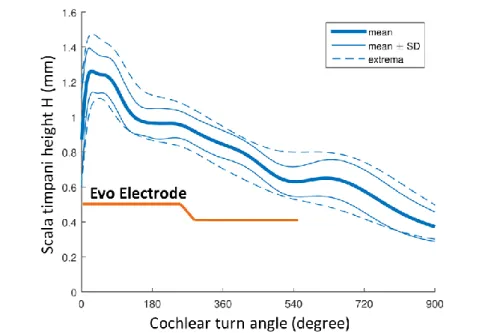
The height H of the elliptic model fitted into cross sections averaged over the nine analyzed temporal bones as a function of cochlear turn is shown in figure 2. Value of interest for usable space for a CI electrode-array is the minimal value for this height along cochlear turns. Particularly, it is observed here a minimum height of 0.7mm after 1 turn (360°), 0.55mm after 1.5 turns (540°) and 0.4mm after 2 turns (720°).
Figure 2: Mean, standard deviations and maximum height of the elliptic model fitted in scala timpani cross sections as a function of cochlear turn angle.
Evo electrode-array diameter as a function of cochlear turn angle (i.e. insertion angle) is presented on figure 2 as well, demonstrating that the useful volume of the cochlea is enough to fit this electrode. More generally, the challenging part for electrode-array design is the tip in the case of long insertion above 1.5 turns, with diameter below 0.5mm is required.
This model is a first approximation of cochlear usable volume with regards to electrode-array diameter, however the measures are based on 9 specimens only. It could be improved with more temporal bone observations. Moreover the elliptic fit to estimate the maximum diameter for electrode-array is an approximation of the scala timpani volume which could be refined, especially in case of uncommon anatomical configurations. Finally, this elliptic fit does not take into account the electrode array position into the scala timpani (perimodiolar, mid scalar or lateral wall electrode-array), which may affect the maximum possible diameter of the electrode-array.
Acknowledgements
This work was supported by the French National Association for Research in Technology (ANRT) through the CIFRE Grant 2013-1165 and Oticon Medical.
Thank you to Clair Vandersteen and Nicolas Guevara (Head and Neck Surgery University Institute, Nice University Hospital, Nice, France) for temporal bone extraction and ; and to Thomas Demarcy, Hervé Delingette and Nicolas Ayache (Asclepios Project Team, Inria Sophia Antipolis-Mediterranee Research Centre, Sophia Antipolis, France) for temporal bone analysis and image processing.
References
Zeng, F.G. 2004. Trends in cochlear implants. Trends Amplif. 8, 1-34.
Lehnhardt E. 1993. Intracochlear placement of cochlear implant electrodes in soft surgery technique (in German). HNO 41: 356–359.
Nguyen Y, Miroir M, Kazmitcheff G, Sutter J, Bensidhoum M, Ferrary E, Sterkers O, Bozorg Grayeli A. 2012. Cochlear implant insertion forces in microdissected human cochlea to evaluate a prototype array. Audiol Neurootol. 17(5):290-8.