HAL Id: hal-02347287
https://hal.archives-ouvertes.fr/hal-02347287
Submitted on 5 Nov 2019
HAL is a multi-disciplinary open access
archive for the deposit and dissemination of
sci-entific research documents, whether they are
pub-lished or not. The documents may come from
teaching and research institutions in France or
abroad, or from public or private research centers.
L’archive ouverte pluridisciplinaire HAL, est
destinée au dépôt et à la diffusion de documents
scientifiques de niveau recherche, publiés ou non,
émanant des établissements d’enseignement et de
recherche français ou étrangers, des laboratoires
publics ou privés.
Distributed under a Creative Commons Attribution| 4.0 International License
cynomolgi erythrocytic stages
Adeline Chua, Jessica Jie Ying Ong, Benoit Malleret, Rossarin Suwanarusk,
Varakorn Kosaisavee, Anne-Marie Zeeman, Caitlin Cooper, Kevin Tan, Rou
Zhang, Bee Tan, et al.
To cite this version:
Adeline Chua, Jessica Jie Ying Ong, Benoit Malleret, Rossarin Suwanarusk, Varakorn Kosaisavee, et
al.. Robust continuous in vitro culture of the Plasmodium cynomolgi erythrocytic stages. Nature
Com-munications, Nature Publishing Group, 2019, 10 (1), �10.1038/s41467-019-11332-4�. �hal-02347287�
Robust continuous in vitro culture of the
Plasmodium cynomolgi erythrocytic stages
Adeline C.Y. Chua
1,2,3,18
, Jessica Jie Ying Ong
2,3,18
, Benoit Malleret
1,4
, Rossarin Suwanarusk
1,2
,
Varakorn Kosaisavee
4,5
, Anne-Marie Zeeman
6
, Caitlin A. Cooper
7
, Kevin S.W. Tan
4
, Rou Zhang
4
,
Bee Huat Tan
3
, Siti Nurdiana Abas
3
, Andy Yip
3
, Anne Elliot
7
, Chester J. Joyner
8,9
, Jee Sun Cho
4
, Kate Breyer
10
,
Szczepan Baran
10
, Amber Lange
10
, Steven P. Maher
7
, François Nosten
11,12
, Christophe Bodenreider
3
,
Bryan K.S. Yeung
3
, Dominique Mazier
13,14
, Mary R. Galinski
9,15
, Nathalie Dereuddre-Bosquet
16
,
Roger Le Grand
16
, Clemens H.M. Kocken
6
, Laurent Rénia
1,4
, Dennis E. Kyle
7
, Thierry T. Diagana
3
,
Georges Snounou
13,14,16
, Bruce Russell
2
& Pablo Bifani
1,3,4,17
The ability to culture pathogenic organisms substantially enhances the quest for fundamental
knowledge and the development of vaccines and drugs. Thus, the elaboration of a protocol for
the in vitro cultivation of the erythrocytic stages of Plasmodium falciparum revolutionized
research on this important parasite. However, for P. vivax, the most widely distributed and
dif
ficult to treat malaria parasite, a strict preference for reticulocytes thwarts efforts to
maintain it in vitro. Cultivation of P. cynomolgi, a macaque-infecting species phylogenetically
close to P. vivax, was brie
fly reported in the early 1980s, but not pursued further. Here, we
define the conditions under which P. cynomolgi can be adapted to long term in vitro culture to
yield parasites that share many of the morphological and phenotypic features of P. vivax. We
further validate the potential of this culture system for high-throughput screening to prime
and accelerate anti-P. vivax drug discovery efforts.
https://doi.org/10.1038/s41467-019-11332-4
OPEN
1Singapore Immunology Network, A*STAR, Singapore 138648, Singapore.2Department of Microbiology and Immunology, University of Otago, Dunedin
9054, New Zealand.3Novartis Institute for Tropical Diseases, Singapore 138670, Singapore.4Department of Microbiology and Immunology, Yong Loo Lin School of Medicine, National University of Singapore, Singapore 119077, Singapore.5Department of Parasitology and Entomology, Faculty of Public Health, Mahidol University, Bangkok 10400, Thailand.6Department of Parasitology, Biomedical Primate Research Centre, Rijswijk 2288, The Netherlands.7Center for Tropical and Emerging Global Diseases, University of Georgia, Athens 30602, USA.8Division of Pulmonary, Allergy, Critical Care & Sleep Medicine, Emory University, Atlanta 30322, USA.9Emory Vaccine Center, Emory University, Atlanta 30317, USA.10Laboratory Animal Services, Scientific Operations, Novartis Institutes for Biomedical Research, East Hanover 07936-1080, USA.11Shoklo Malaria Research Unit, Mahidol-Oxford Tropical Medicine Research Unit, Faculty of Tropical Medicine, Mahidol University, Mae Sot 63110, Thailand.12Centre for Tropical Medicine and Global Health, Nuffield Department of Medicine Research Building, University of Oxford Old Road Campus, Oxford OX3 7FZ, UK.13Sorbonne Universités, UPMC Univ Paris 06, CR7, Centre d’Immunologie et des Maladies Infectieuses (CIMI-Paris), Paris F-75013, France.14CIMI-Paris, INSERM, U1135, CNRS, Paris F-75013, France.15Division of Infectious Diseases, Department of Medicine, Emory University, Atlanta 30322, USA.16CEA-Université Paris Sud 11-INSERM U1184, Immunology of Viral
Infections and Autoimmune Diseases (IMVA), IDMIT Department, IBJF, DRF, Fontenay-aux-Roses 92265, France.17Faculty of Infectious and Tropical
Diseases, London School of Hygiene & Tropical Medicine, London WC1E 7HT, UK.18These authors contributed equally: Adeline C.Y. Chua, Jessica Jie Ying Ong. Correspondence and requests for materials should be addressed to B.R. (email:b.russell@otago.ac.nz) or to P.B. (email:micpb@nus.edu.sg)
123456789
T
he development of a protocol for the routine continuous
in vitro culture of Plasmodium falciparum in 1976
1released malaria researcher from the reliance on in vivo
observations. This led to the major fundamental and translational
advances in all aspects of the life cycle of this parasite that is
responsible for the highest mortality rates globally. Recent
recognition that the widespread species P. vivax causes substantial
morbidity
2made it imperative to devise efficient means to control
it. Furthermore, effective measures that could lead to its
ination would require the development of novel drugs to
elim-inate the hypnozoite, the dormant liver form responsible for
relapses that also characterise P. vivax infections, as well as
strategies to thwart transmission. However, research on P. vivax
remains severely hampered because, to date, attempts to maintain
this parasite in routine in vitro blood cultures have been hindered
by the strict restriction to invasion of reticulocytes, a minor
short-lived fraction of peripheral blood. Historically, a parasite of
Southeast Asian macaques, P. cynomolgi, has been used as the
favoured model for P. vivax. The remarkable morphological and
biological similarities between these two parasite species are now
known to extend to their genetic make-up
3,4. The hepatic stages
of malaria parasites were
first discovered using P. cynomolgi
5as
was the hypnozoite
5–7. Indeed, P. cynomolgi was central to
the development of primaquine
8and remains so in the search
for novel anti-relapse compounds both in experimental
infec-tions
9,10and as a basis for the recently developed in
vitro-cultured hypnozoite model
11–13.
In the last decade, high-throughput screens based on cultured
P. falciparum or on those of the hepatic stages of the rodent
parasites P. berghei or P. yoelii, as reviewed in Hovlid and
Win-zeler
14, have enriched the drug discovery pipeline with a wealth of
promising novel lead compounds. However, such screening
strategies are precluded for P. vivax because of the limited
availability of infected blood from patients, an obstacle that would
be circumvented should in vitro-cultured P. cynomolgi be
avail-able. There were two reports of continuous in vitro culture of the
blood stages of two strains of P. cynomolgi (Berok in one, and
Vietnam in the other) in the early 1980’s
15,16. Given the regular
and onerous use of P. cynomolgi in macaques for biological and
drug discovery programmes, it was surprising that these early
observations were not exploited further.
Following careful assessment of various culture conditions, we
describe here the robust continuous cultivation of the blood
stages of P. cynomolgi lines derived from the Berok strain. We
show that the in vitro-cultured P. cynomolgi (from ex vivo or
cryopreserved stocks) retain the key characteristics that these
parasites share with P. vivax. We further demonstrate that these
parasites are suitable for high-throughput screening for
anti-malarial compounds.
Results
Propagation of P. cynomolgi strains. Three P. cynomolgi lines
were available to us: the Berok strain cryopreserved in 2003 from
Aotus trivirgatus (initially isolated in 1964), the B strain
(P. cynomolgi bastianelli, initially isolated in 1959) and the M
strain (P. cynomolgi Mulligan strain
first isolated in the 1930s).
Separate Macaca fascicularis monkeys were successfully infected
with one or other of these lines (see the Methods section),
and blood samples were collected to initiate cultures. We
con-ducted preliminary experiments using the Berok strain, to test
various culture conditions, media and materials in short-term
cultures initiated from cryopreserved stocks prepared from
infected monkey blood (Fig.
1
a). This allowed us to define an
optimal working protocol that was then tested on Berok strain
parasites from different animals, and on B and M strain parasites
freshly collected from infected macaques. Blood was collected
when most parasites were mature (late trophozoites and
schi-zonts), and depleted of leucocytes before enrichment of the mature
parasites on a Percoll gradient. In vitro cultures were initiated at
parasitaemia > 1% (50,000 parasites/µL) and monitored daily by
microscopic examination of Giemsa-stained smears. In repeated
independent experiments, the parasitaemia of the B and M strain
cultures declined to become undetectable within a few days
(Fig.
1
b). By contrast, erythrocyte invasion was observed in the
cultures initiated with the Berok strain obtained from different
monkeys (K2, K3 and K4). Nonetheless, the multiplication rates of
the K2-initiated and K3-initiated cultures were low by comparison
with that of the K4-derived parasites. Thus, we opted to continue
subsequent work with the K4 culture. This initial Berok K4 line
culture, had a multiplication rate ranging from twofold to fourfold
over more than
five cycles, such that regular dilution of the culture
became necessary. The K4 line was used to constitute working
cryopreserved stocks to conduct the studies described below.
Biological characteristics of in vitro-cultured Berok K4.
Suc-cessful propagation of the Berok K4 line was maintained for up to
180 days in cultures initiated with thawed parasites. Culture
conditions were further refined, with robust growth best observed
under reduced oxygen environment (5% CO
2, 5% O
2and 90%
N
2) to reach parasitaemias beyond 10% (Fig.
1
c). Growth rates
were unaffected by
flask configuration (24-well plate, six-well
plate, T25 or T75
flasks). Successful in vitro maintenance of the
Berok K4 stabilates was obtained independently in six additional
laboratories (the National University of Singapore, the University
of Otago in New Zealand, the University of Malaya in Malaysia,
the Shoklo Malaria Research Unit in Thailand, the Novartis
Institute for Tropical Diseases, Emeryville, California and at the
Université Pierre et Marie Curie in France). Genotyping of the
reticulocyte binding protein gene complex, whose types and copy
number vary between strains
3,17–19, confirmed that the K4 line
parasites were indeed derived from the original Berok stock
(Supplementary Fig. 1). The morphology of the parasites sampled
at 2 hourly intervals for 48 h (Fig.
1
d) was indistinguishable from
that observed or previously described from infected animals, with
eight to sixteen merozoites observed in fully matured schizonts
around 46–48 h. Although macrogametocytes and
micro-gametocytes were intermittently observed from day 6 on, their
levels remained low and did not exceed 0.01% gametocytaemia.
Culture condition modifications that would lead to the higher
sexual stage production in vitro are currently being tested.
In vitro-cultured Berok K4 line retains transmission ability. In
vitro-cultured Berok K4 line parasites were used successfully to
infect a naive intact M. mulatta monkey, where asexual and
sexual stages were observed 6 days post infection (dpi) and
thereafter. Anopheles stephensi mosquitoes fed on infected blood
collected at 11 and 12 dpi (0.85% and 1.4% parasitaemia,
respectively) showed oocysts a week later (feed 11 dpi: in 40% of
mosquitoes, average of 3.5 oocysts/mosquito; feed 12 dpi: in 90%
of mosquitoes, average of 37.6 oocysts/mosquito). Eighteen days
after feeding, lots of 100,000 sporozoites isolated from salivary
glands (feed 12 dpi: ca. 31,000 sporozoites per mosquito) were
successfully used to infect two naive M. mulatta monkeys by
intravenous inoculation (Fig.
2
a), and the course of the infections
was monitored by microscopy. Both monkeys became patent
11 dpi, and were later administered a 5-days chloroquine regimen
to eliminate blood-stage parasites in order to monitor for relapses
as previously described
20. Monkey 1 relapsed 31 dpi and 52 dpi,
after which radical cure was administered (chloroquine and
pri-maquine), while the second remained negative for the whole
1,000,000
Cryopreserved stock from
Macaca fascicularis
Cryopreserved
Maintenance in
Aotus trivirgartus
(1980s–2000)
Initial isolation from Malaysian Macaca fascicularis (1960s) Molecular studies Rhelogical studies Drug studies 150 100 50 % Gro w th –50 0 0 1 Log concentration (nM) 2 3 4 0.5 1.0 1.5 µm Morphological studies Transmission studies K1 K4 K2/K3 Slow growth K4 in vitro Continuous in vitro culture 1,000,000 100,000 100,000 10,000 10,000 1000 100
d
b
a
c
0 0 14 16 18 32 30 28 42 44 46 48 34 20 6 8 10 12 26 40 22 24 36 G 5 µm G 38 2 2 4 4 Days (post thaw)Berok B M
Days (post thaw)
0 5 10 15 20 25 30 6 8 P a rasites/ µ L P a rasites/ µ L 100.000 µ m
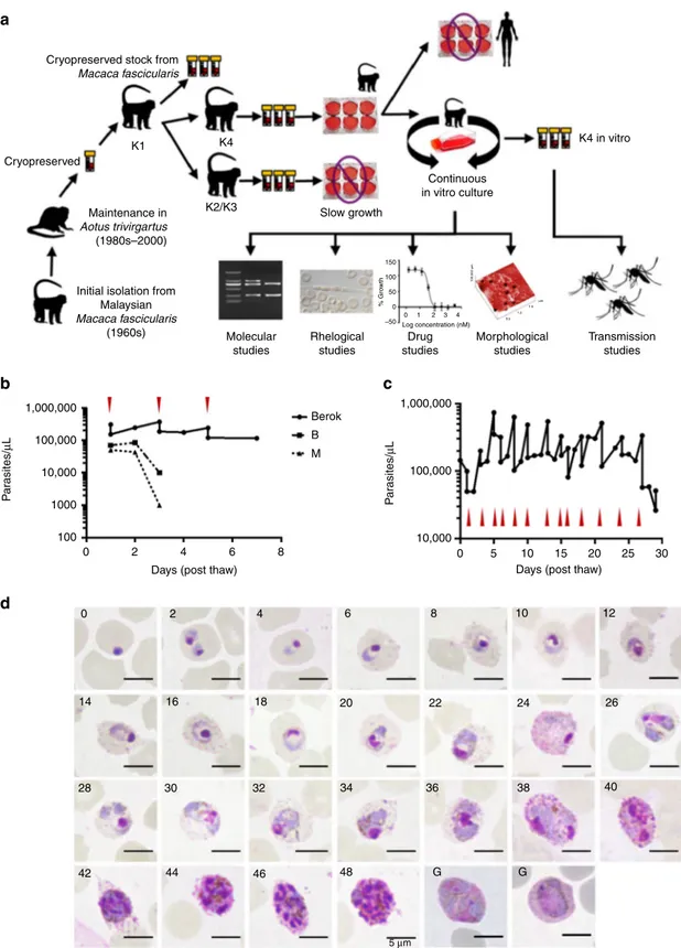
Fig. 1 Adaptation of P. cynomolgi Berok K4 line to continuous culture. a Schematic diagram of the successful adaptation of P. cynomolgi (Berok) from in vivo to in vitro culture.b Pilot ex vivo culture of P. cynomolgi Berok, B and M strain. The in vitro-cultured Berok K4 had to be sub-cultured at days 1, 3 and 5 due to robust growth, in contrast to the M and B strains where parasitaemias decreased to undetectable levels after day 3.c P. cynomolgi (Berok) in vitro culturing was further optimised to enable substantial multiplication (up to tenfold increase) that necessitated frequent dilution of the cultures when high parasitaemias were reached (red arrowheads).d Mature schizonts of culture adapted P. cynomolgi Berok K4 were enriched and allowed to re-invade fresh red blood cells that were then monitored every 2 h to document the complete asexual erythrocytic cycle in vitro. Scale bar represents 5μm
duration of follow-up (102 dpi). The time to patency was similar
to that noted for P. cynomolgi M strain infections (using the same
sporozoites inoculum and infection route), as was that for the
first
relapse (around day 27.5 ± 3.25 dpi in eight monkeys infected by
the M strain
20). Usually, monkeys infected with P. cynomolgi M
strain relapse at least once, regardless of inoculation dose, which
contrasted with the relapse episodes noted in only one of two
rhesus monkeys inoculated with the P. cynomolgi Berok
K4 sporozoites. Sporozoites were also used to infect in
vitro-cultured M. mulatta primary hepatocytes, as previously
descri-bed
11, and 6 days later schizonts and uninucleate parasites
(possibly hypnozoites) were detectable (Fig.
2
b). The average
infectivity of the sporozoites derived from the P. cynomolgi Berok
K4 line was lower, around 16 pre-erythrocytic (PE) forms per
10,000 inoculated sporozoites, than that of those derived from the
P. cynomolgi M strain, around 23 PE forms per 10,000 inoculated
sporozoites (Fig.
2
c). Enumeration of the two hepatic forms
suggested that the P. cynomolgi Berok K4 line generated half the
10
a
b
c
d
Monkey 1 Hypnozoite 150 100 80 60 40 20 Hypnoz oites (% of total PE f o rms) 0 100 P . cynomolgi PE f o rms per w ell 50 0 Donor 1Rhesus hepatocytes and strains
Rhesus hepatocytes and strains M K4 M K4 M K4 Donor 2 Donor 3 Donor 1 M K4 M K4 M K4 Donor 2 Donor 3 p < 0.05 p < 0.05 p < 0.05 p < 0.05 p < 0.05 n.s 50 µm 50 µm P a rasitemia (%) Liver schizont Monkey 2 1 0.1 0.001 0.01 0.0001 0.00001 0 10 20 30 40 50 dpi 60 70 80 90 100 0 10 20 30 40 50 dpi 60 70 80 90 100
number of hypnozoites (around 30%) compared with that
pro-duced by the P. cynomolgi M strain (around 60%) (Fig.
2
d).
Further studies are required to ascertain whether the failure of
one of the monkeys infected with the Berok K4 line to relapse
within the 102-day observation period reflects the lower numbers
of uninucleate forms observed in vitro, or to a delayed relapse
pattern where the
first relapse occurs many months after
spor-ozoite inoculation, or to residual chloroquine that might have
suppressed a
first relapse.
Nanostructure and rheology of Berok K4 line-infected RBCs.
P. cynomolgi Berok K4 dramatically alters the nanostructural and
rheological properties of the infected red blood cell (iRBC) in a
manner similar to that observed for P. vivax. Caveolae (~90
nm-diameter openings) appear on the surface of the infected RBC’s
(Supplementary Fig. 2). While these caveolae, which are generally
associated with vesicle complexes, have the same dimensions as
those noted in P. vivax, these are present in significantly lesser
densities (Fig.
3
a–d), our data on caveolae agree with previous
in-depth analyses conducted on P. cynomolgi Berok
21. The
P. cynomolgi Berok K4-infected RBC increases in size with
maturation (rings ~5 µm and schizonts ~8 µm) with rosettes
forming ~20 h post invasion (Fig.
3
e, f), a timeline similar to that
previously noted for P. vivax
22. It is interesting that rosettes had
not been observed in earlier studies on the B and M strains
23. The
P. cynomolgi Berok K4 rosettes formed highly stable adhesive
bonds requiring around 400 pN (piconewton) to affect a
separation (Fig.
3
g, h), an adhesive force similar to that recorded
for P. vivax
24.
High-throughput drug susceptibility assay using the Berok K4
line. We opted to validate the SYBR green I proliferation assay
14,25for use with the P. cynomolgi Berok K4 line, because it is used
routinely for anti-P. falciparum drug screening, to establish dose
response and single-point screens based on 96- and 384-well-plate
assay formats (Fig.
4
a–c). Using the BioTek Synergy™ 4 hybrid
microplate reader, the range of the 72 h
fluorescent read-out in the
SYBR green I proliferation assay showed a linear correlation with
parasitaemia in the range of 0.3–2%. Culture medium was
sup-plemented with 20% macaque (Mf) serum for the Berok K4 line,
whereas the routine 0.5% AlbuMAX was used for P. falciparum.
Nonetheless, Z’ prime values from the average of 16 DMSO wells
(negative control) and 16 mefloquine wells (positive control)
ran-ged from 0.8 to 0.9 (Supplementary Fig. 3). In order to minimise
the use of materials from naive macaques for culture, initial
parasitaemia of 0.5 and 2% were chosen. A SYBR green I
pro-liferation assay was performed in a 384-well-plate format using a
set of reference compounds and selected compounds from the
Malaria Box (Medicines for Malaria Venture, Switzerland) (Fig.
4
a,
b). The reference compounds included the licensed antimalarials
chloroquine, lumefantrine, pyrimethamine, artemisinin,
atova-quone and artesunate, as well as three drug candidates: the
phosphatidylinositol-4-OH kinase (PI(4)K) inhibitor KDU691, the
imidazolopiperazines KAF179 and the spiroindolone KAF246
(KAF179 and KAF246 are analogues to KAF156 and KAE609
currently in phase 2b clinical trials
26). The IC
50values measured for
both species were broadly concordant, though P. cynomolgi proved
more sensitive to artesunate, artemisinin and atovaquone (Fig.
4
a).
The assay was also conducted for 38 in-house synthesised
com-pounds from the Malaria Box (Fig.
4
b), and identified compounds
that had differential inhibitory activity against the two parasites
species.
For
example,
MMV000563,
MMV007839
and
MMV008294 were highly active against P. falciparum, but not P.
cynomolgi.
In order to evaluate whether the type of culture supplement
(serum or AlbuMAX) influences inhibitory activity, the Pathogen
Box chemical library (Medicines for Malaria Venture,
Switzer-land) was screened in a 384-well format as a single-point assay at
10 µM (Supplementary Fig. 4) using P. cynomolgi (20% Mf serum)
or P. falciparum (20% human serum or 0.5% AlbuMAX). The
Pathogen Box comprises 400 compounds, of which 125 are
antimalarial tool–compounds, 26 are reference compounds while
the rest include compounds active against tuberculosis (n
= 116),
kinetoplastids (n
= 70), helminths (n = 32), cryptosporidiosis
(n
= 11), toxoplasmosis (n = 15) and dengue (n = 5) (
http://
www.pathogenbox.org/
). Assay data for P. cynomolgi in 20% Mf
serum (Column A) proved to be highly comparable with that
obtained for P. falciparum in 20% human serum (Column B), but
both assays differed significantly from the P. falciparum assay
performed in 0.5% AlbuMAX (Column C). This observation was
not surprising given the high protein content of 20%
serum-supplemented media with the likely consequent effect on protein
binding. As above, the rate of inhibition of nine antimalarial
compounds
(MMV676380,
MMV023388,
MMV026550,
MMV007625,
MMV023949,
MMV007638,
MMV676442,
MMV006833 and MMV020289) were significantly different for
both parasite species in the presence of 20% serum (Fig.
4
c).
Overall, the SYBR green I proliferation assay demonstrated robust
reproducibility, at single point and for dose response, for both
Plasmodium species in serum-supplemented cultures.
Schizont maturation assays in the Berok K4 line and P. vivax.
Cultured P. cynomolgi is to be ultimately used as a surrogate for
P. vivax in drug sensitivity assays. Therefore, we carried out the
standard schizont maturation assay for P. vivax, using tightly
syn-chronised Berok K4 ring-stage cultures as well as several clinical
P. vivax isolates. The parasites were seeded on plates containing
serially diluted chloroquine and parasite development was assessed
about 44 h later by
flow cytometry (Fig.
5
a). In parallel, the SYBR
green I proliferation assay was carried out for the same drug using
Fig. 2 Transmission study from P. cynomolgi Berok K4 continuous culture. a In vivo blood-stage parasitaemia in two rhesus monkeys infected with 100,000 P. cynomolgi Berok K4 sporozoites (because of the use of a log scale for parasitaemia, negative smears are shown as 0.0001% parasitaemia). Both monkeys became blood-stage patent on day 11 post infection (dpi). Arrows indicate drug treatment (black arrows: 5-day chloroquine treatment, red arrow: 7-day primaquine treatment). Monkey 1 was bled for stocks on day 15, and relapsed (measured as thin smear positivity) on days 31 and 52, after which it was treated with chloroquine and primaquine. Monkey 2 was bled on day 19 dpi, and did not relapse during the follow-up period of 102 dpi, after which it was treated with primaquine.b In vitro infection of primary rhesus hepatocytes with P. cynomolgi Berok K4 sporozoites produced both hypnozoites (left panel) and developing liver-stage schizonts (right panel). Cultures werefixed at day 6 dpi, and stained with anti-PcHsp70 and a secondary antibody labelled with Alexa 568fluorescent dye. Nuclei were stained with DAPI. An average of 16 PE forms per 10,000 inoculated P. cynomolgi Berok K4 sporozoites were observed. Scale bar represents 50μm. c The total PE forms of in vitro infection rate of various primary rhesus hepatocytes with P. cynomolgi M strain sporozoites and P. cynomolgi Berok K4 line sporozoites.d The percentage of hypnozoites observed in vitro using primary rhesus hepatocytes from different donors infected with P. cynomolgi M strain or P. cynomolgi Berok K4 line sporozoites. The data (c and d) were analysed using the Welch’s t test with the significance level set at P < 0.05. The histograms represent means (n = 3), and the error bars the standard error of the mean (SEM) of replicatesthe Berok K4 line. Each dot represents individual P. vivax clinical
isolates and replicates for the Berok K4 line, respectively. The IC
50values for chloroquine were similar for P. cynomolgi in both assays
(Fig.
5
b, c), and equally comparable with the IC
50values (~50 nM)
obtained from the P. vivax clinical isolates (Fig.
5
d).
Discussion
The control of the globally distributed Plasmodium vivax became,
over the last decade, a priority for the malaria community.
Indeed, the biological characteristics that distinguish this species
from P. falciparum, in particular the propensity to relapse and its
5
b
d
f
e
g
h
c
a
P. vivax P. vivax P. vivax P. vivax P. vivax P. cynomolgi P. cynomolgi P. cynomolgi P. cynomolgi P. cynomolgi p < 0.05 4 3 Ca v e olae per µ m 2 2 200 n.s n.s 84 96 150 100 50 Ca v e olae diameter (nm) 10 Ch04 Ch01 Ring Schizont Trophozoite 0 800 i ii iii iv 600 400 200 Adhesion f o rce (pN) 0 Ring 4.5 384.0 343.2 5 Trophozoite Schizont 8 6 4 Rosetting (%) 2 0 1 0 100.000 µ m 100.000 µ m 0.5 1.0 1.5 µm 0.5 1.0 1.5 µmconsequences and the increased vectorial capacity, are a
sig-nificant challenge to current elimination efforts
27. Moreover, the
increasing reports of resistance to chloroquine and the lack of an
anti-hypnozoite drug that could be mass-deployed have made it a
priority to seek novel compounds active against this parasite.
Furthermore, efforts to develop a vaccine against P. vivax
sig-nificantly lag behind those devoted against P. falciparum. The
inability to culture P. vivax in the laboratory has in equal
mea-sures thwarted efforts to conduct functional studies and to screen
for novel lead compounds targeting P. vivax. Thus, investigations
of all aspects of P. vivax and the infection it causes are restricted
to
field and clinical observations, and to the few samples that can
be obtained from patients prior to treatment. The high cost and
ethical limitations inherent to the use of primate models, P. vivax
lines adapted to New World non-human primates, or P.
cyno-molgi in macaques, further constrain the scope of any
investiga-tions. The availability of in vitro-cultured P. cynomolgi makes this
suitable model for P. vivax available to a broad range of
researchers and opens the way to apply genetic manipulation
technologies that are as yet precluded for P. vivax.
The
first protocol for in vitro cultivation of the erythrocytic
stages of malaria parasite was made for P. falciparum and P. vivax
in 1912
28, though growth was restricted to a few cycles. Sustained
multiplication over extended periods (months), i.e., continuous
cultures, proved elusive and was
finally achieved only in 1976 for
P. falciparum
1,29after sustained efforts spanning three decades.
Within a few years, the continuous cultivation of four macaque
parasite species, P. knowlesi, P. cynomolgi, P. inui and P. fragile
were reported
30. Whereas cultures of P. knowlesi were later
exploited to conduct genetic and biological studies
31,32, none
appear to have been similarly employed for P. cynomolgi, despite
the evident benefits that this could have provided over the years
to researchers investigating P. vivax. The fastidious testing of
culture conditions that we carried out and our subsequent
observations on the inability of some strains to grow in culture
might in part explain this gap. Human serum to supplement the
culture medium (successfully used in the initial publication from
1981) proved unsuitable in our hands, despite testing of
numer-ous batches. Moreover, though collected from naive animals, then
prepared and stored in a standard manner, the serum and in
some cases the red blood cells collected from only some but not
all naive M. fascicularis proved suitable for sustained parasite
multiplication.
It is not clear at present why establishment in vitro under the
adopted standard conditions appears to be limited to the Berok
line (the P. cynomolgi M or B strains failed to thrive, despite
numerous attempts). This phenomenon is not novel or peculiar
to P. cynomolgi, as similar variable success in adapting
field-collected P. falciparum isolates has long been observed. The
successful adaptation to in vitro culture remains elusive and
multifaceted. It is well documented that the rbp genes are
dif-ferent in the various strains of P. cynomolgi
3,17. It will be useful to
explore whether the fact that only the Berok strain, but neither
the B nor M strain, possesses the rbp1b gene played a role. This
gene is present in P. vivax
3though this species remains
unculti-vatable in vitro. Clearly, the mechanisms of merozoite invasion
are complex, and will require detailed investigations. Moreover,
red blood cell tropism might have played a role. In a recent study
by Kosaisavee et al.
33, the P. cynomolgi B strain equally invaded
all types of monkey erythrocytes, while it was restricted to
Duffy-positive human reticulocytes.
The Berok strain, initially derived from a wild M. nemestrina
collected in peninsular Malaysia in the early 1960s, was
main-tained thereafter by blood and/or sporozoite inoculation
pri-marily in M. mulatta and later also in Aotus monkeys
34. This
strain has not been cloned, and it is possible that it might harbour
diverse parasite genotypes which could vary in their proportion
over the course of infection and or in different hosts. It is at
present unknown whether the derived Berok parasites collected
from the various monkeys (K2–K4) differed in the proportion of
parasites that can rapidly adapt to thrive in culture, possibly as
genetically distinct lines from an otherwise heterogeneous
P. cynomolgi population, or as phenotypic variants selected from
an otherwise homogeneous population. Elucidation of the
molecular basis for this differential growth could provide
fun-damental insights on the biology of blood-stage parasites. It is
likely that lines from other P. cynomolgi isolates and strains, or
indeed from those of other macaque parasite species, would also
be amenable to blood-stage cultivation, provided that the initial
failures are met with perseverance.
It is our intention to facilitate the dissemination of the Berok
K4 line from P. cynomolgi, a species proven as an excellent
sur-rogate for P. vivax, so as to broaden the scope of investigations
that in vitro-cultured parasites would allow. Routine cultivation is
easily initiated from cryopreserved stocks using the conditions
and methodology that were optimised to enable robust growth
and large-scale production, should this be needed. We have
shown that in vitro-produced Berok K4 parasites are
indis-tinguishable morphologically from in vivo and ex vivo parasites,
and that they retained their infectivity to monkeys and
subse-quently to mosquitoes to generate infective sporozoites. The
presence of caveolae, and the occurrence of rosettes, provides
additional evidence that this parasite species also shares the
characteristic
morphological, phenotypical
and rheological
alterations observed for P. vivax-infected red blood cells.
The last 10 years have witnessed significant successes in the
search for new antimalarial compounds, with 17 new drug
can-didates developed since 2010
35,36. The key contribution was the
development of a standard P. falciparum asexual blood-stage
SYBR green I proliferation assay adapted to automated screening
technologies
14. Such strategies are not possible for P. vivax, and
high-throughput screening of compounds would require a
pro-hibitively large number of P. cynomolgi-infected macaques. We
validated the potential of the P. cynomolgi Berok K4 line cultures
to extend high-throughput screens to identify lead compounds
active against P. vivax. It was interesting to note that some of the
compounds from the Malaria Box and the Pathogen Box used for
the validation were inhibitory to one, but not the other of the
Fig. 3 Phenotypic and rheological characterisation of the Berok K4 line from in vitro culture. a P. cynomolgi Berok K4-infected RBCs exhibit caveolae structures (yellow arrows) that are similar to those in P. vivax-infected RBCs (scanning electron microscopy, scale bars represent 1µm and 100 nm for area shown at higher magnification in white box). b An atomic force microscope scan of trophozoite-infected human blood cells revealed caveolae occurred at lower frequency when compared with P. vivax.c, d The median (+ /- IQR) dimensions of these caveolae were similar (P. vivax n = 177, P. cynomolgi n = 91). e, f Amnisflow imaging clearly shows that the mature erythrocytic stages P. cynomolgi Berok K4; readily formed rosettes with uninfected red blood cells, which are also a key feature P. vivax (n= 5). g, h A dual micropipette aspiration method was used to demonstrate the rheological stability of the P. cynomolgi Berok K4 rosettes (n= 5). As observed in P. vivax, P. cynomolgi rosettes are tightly attached and the cells require around 400 pN to disrupt the adhesion. The non-parametric data resented inb, d, f and h were analysed using the Mann–Whitney U test with the significance level set at P < 0.05. The histograms and lines on box plots and scatter plots represent medians, and the error bars the interquartile range (IQR)parasite species (P. cynomolgi and P. falciparum) used for
the validation. Thus, it is likely that activity data from P.
cyno-molgi-based screens would be a more reliable predictor of
activity against P. vivax than those that rely on P. falciparum or
on the two most often used parasites of rodents P. berghei and
P. yoelii
14. Of late, various screening assays for P. vivax and
P. falciparum liver stages has been developed to evaluate novel
compounds
13,37–39. Indeed, hypnozoites responsible for relapses,
a major obstacle to successful control of P. vivax, are formed by
P. cynomolgi, but not by any of the rodent parasite species.
The urgent need to
find an alternative to the anti-hypnozoitocidal
8-aminoquinoline compounds, whose potential toxicity impedes
10
a
b
c
1 0.1 0.01 0.001 0.0001 0.0001 Atovaquone KAF246 Pyrimethamine Artemisinin Artesunate KAF179 Lumefantrine Mefloquine Chloroquine KDU691 Proguanil P . f a lcipar um IC 50 ( µ M) in 0.5% Alb uMAX 0.001 P. cynomolgi IC50 (µM) in 20% Mf serum 0.01 0.1 10 MMV000563 A B 10 A B C APc/Pf (in 20% serum) > 4 fold
Pf/Pc (in 20% serum) > 4 fold B C A B C % inhibition (%) 100 60 40 20 Anti-malar ial compounds 0 80 9 8 7 6 5 4 3 2 A: P. cynomolgi in 20% Mf serum B: P. falciparum in 0.5% AlbuMAX 1 IC50 (µM) MMV006250 MMV007839 MMV008127 MMV008270 MMV008294 MMV665987 MMV666022 MMV008160 MMV007041 MMV007384 MMV666607 MMV007978 MMV666101 MMV665943 MMV665850 MMV665948 MMV396663 MMV666102 MMV007020 MMV007374 MMV008416 MMV665814 MMV009063 MMV000986 MMV006913 MMV006820 MMV006429 MMV011944 MMV665994 MMV666081 MMV000963 MMV006319 MMV006169 MMV006825 MMV665977 MMV665941 MMV011256 MMV006087 MMV676380 MMV023388 MMV026550 MMV007625 MMV023949 MMV007638 MMV676442 MMV006833 MMV020289 A: P. cynomolgi in 20% Mf serum 1
B: P. falciparum in 20% human serum C: P. falciparum in 0.5% AlbuMAX
widespread use, led to the development of an in vitro-based
P. cynomolgi hepatic-stage assay
11,12. One of the major logistical
challenges that precludes high-throughput screening is the
availability of infective sporozoites that can only be obtained from
mosquitoes fed on blood collected from P. cynomolgi-infected
macaques. The fact that the in vitro-maintained Berok K4 line
parasites retained infectiousness to mosquitoes subsequent to
cultivation augurs well for the eventual use of in vitro cultures to
support mosquito infections. We are currently exploring diverse
modifications to our culture protocols aimed at promoting
gametocytogenesis, a process known to be highly sensitive to
culture conditions, as was shown for P. falciparum
40. This would
minimise non-human primate use, and substantially increase
opportunities for routine and regular sporozoite production.
Finally, it is important to point out that the P. cynomolgi model
uniquely provides the possibility to confront fundamental
biolo-gical, immunological and pathological insights, as well as
ther-apeutic approaches derived from in vitro observations with the
realities of experimental in vivo infections in the natural host. The
ability to refine, or indeed to discard, a candidate compound(s) or
vaccine formulation(s) through an in vitro–in vivo ‘to-and-fro'
significantly enhances the value of this pre-clinical model.
It is hoped that the availability of easily maintained in vitro
erythrocytic P. cynomolgi parasites could now be exploited to
conduct critical fundamental and translational research to
develop drugs and vaccines against P. vivax, a widespread species
whose control will determine the success of current efforts to
eradicate malaria.
Methods
Ethical committees and animal welfare. Macaca fascicularis (cynomolgus monkeys) were maintained at the Novartis Laboratory Animal Services, New Jer-sey, USA, (Novartis-LAS) and SingHealth Experimental Medicine Center, Singa-pore. Both institutions were audited and approved by the Novartis Animal Welfare Compliance. All animals were housed in accordance with the Guide for the Care and Use of Laboratory Animals and the Association for the Assessment and Accreditation of Laboratory Animal Care (AAALAC) Standards. All studies were approved by the Novartis Ethical Review Council and Novartis Institutional Ani-mal Care and Use Committees prior (IACUC) to study initiation. In addition, work at SingHealth was approved by the SingHealth IACUC. The BPRC is an AAALAC-certified institute. Several rhesus macaques (M. mulatta) used in this study were captive bred for research purposes, and were housed at the BPRC facilities under compliance with the Dutch law on animal experiments, European directive 2010/ 63/EU and with the‘Standard for humane care and use of Laboratory Animals by Foreign institutions’ identification number A5539–01, provided by the Department of Health and Human Services of the USA National Institutes of Health (NIH). Prior to the start of experiments at BPRC, all protocols were approved by the local independent ethical committee, according to Dutch law. Rhesus macaques were infected with 1 × 10⁶P. cynomolgi Berok K4 blood-stage parasites and bled at peak parasitaemia. About 300 female Anopheles stephensi mosquitoes Sind–Kasur strain Nijmegen (Nijmegen University Medical Centre St Radboud, Department of Medical Microbiology) were fed with this blood. Clinical samples utilised in this study were collected from P. vivax-infected malaria patients attending the clinics of the Shoklo Malaria Research Unit (SMRU), Mae Sot, Thailand, under the following ethical guidelines in the approved protocol: OXTREC 45–09 (University of Oxford, Centre for Clinical Vaccinology and Tropical Medicine, UK) and MUTM 2008–215 from the Ethics committee of Faculty of Tropical Medicine, Mahidol University.
P. cynomolgi B, M and Berok strains. Three P. cynomolgi strains were used in this study, the Berok strain41, the B strain (P. cynomolgi bastianelli)42and M strain
(P. cynomolgi Mulligan strain)43,44. The“B” strain or P. cynomolgi bastianelli was
isolated by Garnham in 1959 from an infected M. fascicularis (previously named M. irus) near Kuantan, in the East Coast of Malaysia42. The P. cynomolgi Berok
strain originated from Perak, Malaysia, where it was isolated from an infected M. nemestrina monkey41. The Berok strain sample that was used to derive the
Berok K4 line consisted of a single cryopreserved 1 -mL sample passage through Aotus monkeys and dated as 2003. The sample was thawed out as described below, and washed prior to a tail–vein administration to a splenectomised M. fascicularis monkey. The P. cynomolgi‘M' strain was first described by Mulligan in 193544, and
later shown to be transmitted to man 196143. It has been suggested that the B and
M strain isolates have been mixed-up at some time in the past, and that some of the B and M strain aliquots in use might actually have the same parasite line3. All
strains of P. cynomolgi ultimately originated from stock collections at the CDC, which were then since propagated in monkeys at the recipient laboratories. In this study, the B and M strain were provided by B.R., the Berok strain cryovial used to generate the K4 line was provided by D.E.K.
P. cynomolgi base medium composition for continuous culture. The RPMI-1640 (Roswell Park Memorial Institute) supplemented with GlutaMAX (Gibco # 61870–036) containing 30 mM HEPES (Sigma-Aldrich), 0.2% (w/v) D-glucose and 200μM hypoxanthine (Calbiochem). In all, 20% (v/v) M. fascicularis serum (heat inactivated) was added prior to use. The medium wasfilter-sterilised over 0.22 -µm filter.
Macaque blood and serum extraction and preparation. M. fascicularis ery-throcytes and serum were obtained from Singapore Health Services Pte Ltd. Blood was collected by venous puncture into lithium heparin vacutainers (Becton-Dickinson). The blood collected was adjusted to 50% haematocrit with the RPMI-1640 supplemented with 30 mM HEPES before passing through pre-equilibrated non-wovenfilters (Antoshin) to deplete leucocytes. The erythrocytes were pelleted by centrifugation at 1800 rpm (650 rcf) for 5 min at room temperature (RT), washed thrice in the RPMI-1640 with 30 mM HEPES and stored at 4 °C in the same medium at 50% haematocrit until use. For collection of M. fascicularis serum, the blood was collected by venous puncture into SST serum separation vacutainers (Becton-Dickinson), inverted gentlyfive times and allowed to clot in a vertical position. The tubes were then centrifuged at 3000 rpm (1810 rcf) for 10 min at RT. The M. fascicularis serum supernatant was collected and heat inactivated for 1 h at 56 °C and stored at−20 °C until use.
In vitro parasite growth in M. fascicularis RBCs and serum. P. cynomolgi parasite cultures were started from 1 mL of stabilate cryopreserved samples. The frozen samples were thawed-out using the sodium chloride method45. Briefly,
sequentially decreasing concentrations of NaCl were added starting with 0.2 mL of 12% (w/v) NaCl added slowly drop-wise with gentle mixing, incubated at RT for 5 min without shaking, and further diluted in 10 mL of 1.6% (w/v) NaCl added drop-wise with gentle mixing. The samples were pelleted by centrifugation at 1800 rpm (650 rcf) for 5 min at RT, the supernatant discarded and samples were sub-jected to afinal wash in 10 mL of 0.9% NaCl added drop-wise with gentle mixing, pelleted by centrifugation at 1800 rpm (650 rcf) for 5 min at RT and re-suspended in complete medium comprising P. cynomolgi base medium supplemented with 20% (v/v) macaque serum. Macaque blood was added to a 5% haematocrit. The parasites were cultured in plates in a modified candle jar (Stemcell technologies 27310) at 37 °C comprising about 3–5% CO2, and 8–10% O246,47. Cultures were monitored daily by Giemsa-stained thick and thin smears by microscopy and parasitaemia adjusted between 0.5 and 5% for routine culture. The medium was changed daily. In order to eliminate the use of the candle jar, the cultures were adapted to trimix gas (5% CO2, 5% O2, 90% N2). All other conditions were the same as for the modified candle jar. For culture in trimix gas, the parasites were gassed and cultured in non-ventedflasks.
Parasite synchronisation. The infected RBCs from in vitro P. cynomolgi Berok K4 werefirst synchronised with 5% sorbitol, and then matured to schizonts which Fig. 4 Drug susceptibility testing using P. cynomolgi Berok K4 in vitro culture. a Correlation of P. cynomolgi Berok K4 and P. falciparum IC50values of common antimalarial reference compounds in a SYBR green I proliferation assay. The potency of the compounds was comparable between the two species, except for artemisinin, atovaquone and artesunate which were more potent in P. cynomolgi as compared with P. falciparum. A X= Y line indicates equal inhibition towards compound.b Heatmap showing IC50(μM) of a representative set of compounds from the Malaria Box. Majority of the compounds showed activity against both P. falciparum and P. cynomolgi, except for six compounds—MMV008127, MMV006250, MMV008270, MMV000563, MMV007839 and MMV008294 which displayed an IC50> 10µM for P. cynomolgi, and < 5 µM for P. falciparum. c Heatmap showing percentage of inhibition of the 125 antimalarial compounds from the Pathogen Box in P. cynomolgi Berok K4 and P. falciparum in different serums and concentrations. Nine compounds (MMV676380, MMV023388, MMV026550, MMV007625, MMV023949, MMV007638, MMV676442, MMV006833 and MMV020289) showed more than fourfold difference in inhibition between P. cynomolgi Berok K4 and P. falciparum in their equivalent serums
Control Hoechst CQ (nM)
a
b
c
d
0.00 0.13% 0.18% Ethidium 0.20 0.15 0.10 0.05 0.00 100 80 60 40 20 0 0 1 2 3 0 1 2 3 4 –0.05 –50 n.s 49.1 48.7 P. vivax P. cynomolgi 0 50 IC50: 48 nM IC50: 42 nM % g ro wth % schiz ont IC 50 (nM) 100 150Log concentration (nM) Log concentration (nM)
0.00% 0.04% 0.15%
0.00% 0.03% 0.12%
598.42 74.80 18.70 Low High
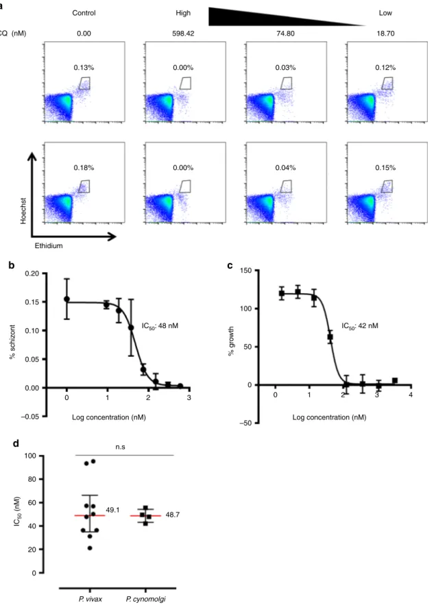
Fig. 5 Correlation of chloroquine IC50of P. cynomolgi Berok K4 continuous culture with P. vivax clinical isolates.a Flow cytometry dot plots (Ethidium/ Hoechst) of chloroquine-treated P. cynomolgi Berok K4 continuous culture gated for schizonts population.b IC50determination of chloroquine in P. cynomolgi Berok K4 continuous culture using the schizont maturation assay (n= 3) with error bars representing standard deviation (SD). c IC50 determination of chloroquine in P. cynomolgi Berok K4 continuous culture using SYBR green I proliferation assay (n= 3) with error bars representing standard deviation (SD)d IC50of chloroquine in P. cynomolgi Berok K4 continuous culture and P. vivax clinical isolates using the schizont maturation assay. The median (+ /− IQR) values for the IC50of P. vivax clinical isolates and of P. cynomolgi Berok K4 continuous culture were similar. The non-parametric data ind was analysed using the Mann–Whitney U-Test with the significance level set at P < 0.05. The histograms and lines on box plots and scatter plots represent medians (P. vivax n= 10, P. cynomolgi n = 4) and the error bars the interquartile range (IQR)
were enriched using MACS LD columns (Miltenyi Biotech Asia Pacific Pte Ltd.) on magnetic sorter, where theflow through containing rings and early trophozoites were discarded. The retained late-stage parasites were eluted from the column, washed three times with the RPMI-1640 supplemented with 30 mM HEPES and then returned to culture.
Determination of parasitaemia via light microscopy. Thick and thinfilm smears were prepared with 5 µL of packed red blood cells for Giemsa staining, para-sitaemia was determined from the thinfilm under a light microscope with ×1000 magnifications.
Transmission study from Berok K4 continuous culture. Monkey infections and mosquito feedings were performed as previously described48. Briefly, cryopreserved
in vitro-adapted P. cynomolgi Berok K4 parasites were thawed as described above, and one million parasites were used to infect one M. mulatta monkey via intra-venous injection, while the monkey was under ketamine sedation. Parasitaemia was monitored through Giemsa-stained thin smears. Female An. stephensi mosquitoes were fed with infected monkey blood using a glass feeder system on days 12 and 13 post infection, after which the monkey was cured from blood-stage parasites with chloroquine (three daily intramuscular injections 7.5 mg/kg). Oocysts were counted in at least ten mosquitoes at day 7 after the infected blood meal. The remaining mosquitoes were given a second uninfected blood meal to promote sporozoite invasion of the salivary glands.
Sporozoites from Berok K4 continuous culture. As previously described48,
sporozoites were isolated from salivary glands of infected An. stephensi mosquitoes 18 days post-infected blood meal.
Infectivity of Berok K4 sporozoites from continuous culture. M. mulatta hepatocytes from different donors were isolated as previously described48, and then
seeded onto collagen-coated 384-well plates at a density of 28,000 hepatocytes/well in William’s B medium as previously described48. Two days later, 20,000
spor-ozoites were added to each well. Infections were carried out in six replicates per experimental condition. Daily medium refreshments were performed, and the infected cells werefixed in 4% PFA after 6 days of sporozoite infection. Parasites were immunostained with anti-PcHsp70 antibodies and goat-anti-rabbit Alexa 568 red secondary antibodies (Invitrogen A11011), while the nuclei were stained with DAPI before they were visualised using the Operetta high content screening automated microscope. Image analysis was done using the analysis algorithm as designed in previous study48with a minor adaptation for the change influorescent
label of the secondary antibody.
Micropipette aspiration and RBC sphericity measurement. The micropipette aspiration technique was modified from Hochmuth, 200049. Briefly, 1 µL of packed
red blood cells containing ~1% parasitaemia and suspended in 1 mL of PBS (1% BSA). The samples were mounted onto the Olympus IX71 Inverted Micro-scope. A borosilicate glass micropipette (~1.5 -µm inner diameter) was used to extract the cell membrane under a negative pressure at a pressure drop rate of 0.5 pa/s. The corresponding cell membrane deformation was monitored using a ×100 oil immersion lens. The cell membrane deformation was recorded using the QColor5 High Resolution Color CCD Digital Fire Wire Camera (Olympus) and processed by QCapture Pro 6.0 (Olympus). The cell membrane shear modulus was calculated using the hemispherical cap model49. To quantify the binding
force between uninfected red cells and a iRBC (rosetting cells), a double-pipette aspiration method was used50. A rosette was held by a micropipette (diameter=
2.0 ± 0.2 µm). A second micropipette was used to aspirate the uninfected red cell at increased levels of pressure. The force (F) to detach the red cell from the iRBC was calculated as F= πr2× P, where r is the inner diameter of the second micropipette, and P is the pressure required for cell detachment. The aspiration pressure was measured by a pressure transducer (P61 model, Validyne Engineering), and recorded by USB-COM Data logger (Validyne Engineering). The process was recorded using a Dual CCD Digital Camera DP80 (Olympus®) at one frame/s. Recorded images were analysed with CellSens Dimension (Olympus®). Atomic force microscopy. Infected RBCs (iRBCs) were harvested at the tropho-zoite stage and processed as follows: 200 µl of blood media mixture were supra-vitally stained with 1 µL of DAPI for 15 min in an incubator and prepared as smears (unfixed and air dried) for Atomic Force Microscopy (AFM). At least 20 iRBCs from each isolate and cell type was scanned using AFM. The total area analysed per parasite was 15 µm2(n= 15 for each respective group, of 1 µm2area/ infected red cell). We were able to conduct serial measurements (with AFM, then and with Giemsa) by using a copper microdisk grid (H7finder grid, SPI Supplies, PA) attached underneath the glass slide allowing us to locate and image the same cell. These thin smears werefirst AFM scanned by a Dimension 3100 model with a Nanoscope IIIa controller (Veeco, Santa Barbara, CA) using the tapping mode. The probes used for imaging were 125 -µm long by 30 -µm wide single-beam shaped cantilevers (Model PPP-NCHR-50, Nanosensors) with tip radius of curvature of
5–7 nm. Images were processed using the Nanoscope 5.30 software (Veeco, Santa Barbara, CA).
Electron microscopy. Sorted cells coated on poly-lysine (Sigma) glass coverslips werefixed in 2.5% glutaraldehyde, washed and treated with 1% osmium tetroxide (Ted Pella Inc.) before critical point drying (CPD 030, Bal-Tec). Glass coverslips were sputter coated with gold in a high vacuum (SCD005 sputter coater, Bal-Tec) and imaged with afield-emission scanning electron microscope (JSM-6701F, JEOL) at an acceleration voltage of 8 kV51.
Image stream analysis of resetting. We determined the percentage of rosetting of P. cynomolgi iRBC by image stream analysis using a method adapted from Lee et al.52. Briefly, iRBCs stained with Hoechst and dihydroethidium were suspended
in PBS to a haematocrit of 2% were assayed with the ImageStream 100 (Amnis, Seattle, WA)fitted with a ×60 objective. At least 200 untreated parasites for each single-stain condition were used to create a compensation matrix. During screening, 10,000 parasites were acquired and gated using the technique adopted from Malleret et al.53along with the added selection of cells that were not singlets
(one or more cells adhering to an infected cell staining positive for Hoechst). Analysis was performed with the IDEAS software (version 4.0).
DNA extraction, PCR amplifications and sequencing. As described previously54,
the DNA was extracted, and the genes rbp1b, rbp2a and rbp2b were compared between the P. cynomolgi Berok, P. cynomolgi B strain and P. cynomolgi M strain. Compound libraries. The Pathogen Box (http://www.pathogenbox.org/), modelled after the Malaria Box55, is an source library comprising of 400 diverse
open-source compounds targeting against neglected tropical diseases. The Malaria Box was an open-source library (until December 2015), which comprised 400 diverse compounds with antimalarial activity. In addition, the three drug candidates (the phosphatidylinositol-4-OH kinase (PI(4)K) inhibitor KDU69156, the
imidazolo-piperazines KAF17957and the spiroindolone KAF24658) were included as
addi-tional controls.
Compound plate preparation. Test compounds were prepared by serially diluting a 10 mM compound stock threefold with DMSO for eight concentration points in the master compound plate. In all, 100% DMSO was used as the negative control, and 10 mM mefloquine was used as the positive control. A 1000-fold dilution (either 100 nL or 50 nL) of compound from the master plate were spotted onto a 96-well or 384-well assay plate, respectively using Mosquito®nanoliter dispenser (Cambridge, UK). The plates were then sealed with a removable foil seal using PlateLoc Thermal Microplate Sealer (Agilent) until use.
In vitro high-throughput IC50drug susceptibility assay. There are various high-throughput screens based on cultured P. falciparum25,59–63or on the hepatic stages
of the rodent parasites P. berghei or P. yoelii57,61described. Here, we adapt the
SYBR green I proliferation assay as described by Plouffe et al. Briefly, 50 µL of the P. cynomolgi culture were dispensed manually into the 384-well plate at bothfinal parasitaemia of 0.5% (for dose response assay) or 1% (for single point assay) and the haematocrit adjusted to 2.5%. The 384-well assay plates were incubated at 37 °C for 72 h in 5% CO2, 5% O2and 90% N2. After a 72 h incubation, 10 µL of lysis buffer, consisting of 5 mM EDTA, 20 mM Tris-HCI pH 7.5, 1.6% Triton X-100 and 0.16% Saponin, were added to each well, and the plate was incubated in the dark for 24 h at RT. The plate was read forfluorescence using BioTek Synergy™ 4 hybrid microplate reader (Vermont, USA) using a bottom read mode at excitation wavelength of 485 nm and emission wavelength of 528 nm.
Robustness of in vitro IC50drug susceptibility assay. The quality of the assay was determined with the Z’ value64, which takes into account the signal dynamic
range and the data variation associated with the signal measurements and is cal-culated as follow: Z’ = 1 – ((3 SD of positive control + 3 SD of negative control)/ (average of positive control–average of negative control)). An assay with Z’ ≥ 0.5 < 1 is considered robust. Three repeats were carried out from which the Z’ values showed the robustness of the assay. Readfluorescence (bottom read), λex = 485 nm.λem = 528 nm. Fluorescence values were normalised based on maximum fluorescence signal values for DMSO-treated wells and the minimum fluorescence signal values for wells containing the highest concentration of negative control compound, 10 µM mefloquine.
Schizont maturation assay usingflow-cytometry analysis. The maturation assay for P. vivax was carried out as described previously65,66, while parasite
growth was assessed byflow cytometry53(Fig.5a). In all, 200μL of tightly
syn-chronised ring-stage culture of P. cynomolgi was dispensed manually in the 96-well compound plate at afinal parasitaemia of 0.5% and haematocrit adjusted to 2%. The assay plates were incubated at 37 °C for around 44 h in 5% CO2, 5% O2and 90% N2until mid-late schizonts stage (> 5 merozoites) was observed in the drug-free control wells via Giemsa staining. Each well was well mixed, and 20 µL was
harvested into a small curved-bottom tube (Micronic) before 0.5μL of 1 mg/mL dihydroethidium (Sigma) and 1μL of 800 μM of Hoechst 33342 (Sigma) was added and made up to 100μL with PBS. The tubes were incubated at RT for 20 min, and 100,000 events were acquired with an Accuri C6 (BD Biosciences, USA). The data were analysed using FlowJo software (Tree Star Inc.).
Statistical analysis. The parametric data presented in Fig.2c, d were analysed using the Welch’s t test with the significance level set at P < 0.05. The histograms represent means, and the error bars the standard deviation (SD). The non-parametric data (all data sets failed the D’Agostino & Pearson normality test) presented in Fig.3b, d, f and h and Fig.5d were analysed using the Mann–Whitney
U test with the significance level set at P < 0.05. The histograms and lines on box plots and scatter plots represent medians and the error bars the interquartile range (IQR). All analyses were carried out using GraphPad Prism™ 7 for windows (GraphPad Software Inc, USA).
Reporting Summary. Further information on research design is available in the Nature Research Reporting Summary linked to this Article.
Data availability
The source data underlying Figs. 1b, 1c, 2c, 2d, 3a-h, 4a-c, 5b-d, Supplementary Figs. 1 and 4 are provided as a Source Datafile. Any additional data that support the findings of this study may be requested from the corresponding authors.
Received: 22 October 2018 Accepted: 28 June 2019
References
1. Trager, W. & Jensen, J. B. Human malaria parasites in continuous culture. Science 193, 673–675 (1976).
2. World Health Organization. World Malaria Report 2016 (World Health Organization, Geneva, 2016).
3. Tachibana, S. et al. Plasmodium cynomolgi genome sequences provide insight into Plasmodium vivax and the monkey malaria clade. Nat. Genet. 44, 1051–1055 (2012).
4. Pasini, E. M. et al. An improved Plasmodium cynomolgi genome assembly reveals an unexpected methyltransferase gene expansion. Wellcome Open Res. 2, 42 (2017).
5. Shortt, H. E. & Garnham, P. C. Demonstration of a persisting exo-erythrocytic cycle in Plasmodium cynomolgi and its bearing on the production of relapses. Br. Med J. 1, 1225–1228 (1948).
6. Krotoski, W. A. et al. Observations on early and late post-sporozoite tissue stages in primate malaria. II. The hypnozoite of Plasmodium cynomolgi bastianellii from 3 to 105 days after infection, and detection of 36- to 40-hour pre-erythrocytic forms. Am. J. Trop. Med. Hyg. 31, 211–225 (1982). 7. Krotoski, W. A. et al. Observations on early and late post-sporozoite tissue
stages in primate malaria. I. Discovery of a new latent form of Plasmodium cynomolgi (the hypnozoite), and failure to detect hepatic forms within thefirst 24 hours after infection. Am. J. Trop. Med. Hyg. 31, 24–35 (1982). 8. Schmidt, L. H. et al. Plasmodium cynomolgi infections in the rhesus monkey.
Am. J. Trop. Med. Hyg. 31, 609–703 (1982).
9. Deye, G. A. et al. Use of a rhesus Plasmodium cynomolgi model to screen for anti-hypnozoite activity of pharmaceutical substances. Am. J. Trop. Med. Hyg. 86, 931–935 (2012).
10. DiTusa, C. et al. Causal prophylactic efficacy of primaquine, tafenoquine, and atovaquone-proguanil against Plasmodium cynomolgi in a rhesus monkey model. J. Parasitol. 100, 671–673 (2014).
11. Dembele, L. et al. Towards an in vitro model of Plasmodium hypnozoites suitable for drug discovery. PloS ONE 6, e18162 (2011).
12. Dembele, L. et al. Persistence and activation of malaria hypnozoites in long-term primary hepatocyte cultures. Nat. Med. 20, 307–312 (2014).
13. March, S. et al. A microscale human liver platform that supports the hepatic stages of Plasmodium falciparum and vivax. Cell Host Microbe. 14, 104–115 (2013).
14. Hovlid, M. L. & Winzeler, E. A. Phenotypic screens in antimalarial drug discovery. Trends Parasitol. 32, 697–707 (2016).
15. Nguyen-Dinh, P., Gardner, A. L., Campbell, C. C., Skinner, J. C. & Collins, W. E. Cultivation in vitro of the vivax-type malaria parasite Plasmodium cynomolgi. Science 212, 1146–1148 (1981).
16. Zhou, Z. X., Li, G. R., Ye, J. S., Xi, Y. H. & Huang, R. Z. Continuous in vitro cultivation of erythrocytic Plasmodium cynomolgi. Chin. Med. J 97, 84–88 (1984).
17. Sutton, P. L. et al. Characterizing the genetic diversity of the monkey malaria parasite Plasmodium cynomolgi. Infect., Genet. Evol.: J. Mol. Epidemiol. Evolut. Genet. Infect. Dis. 40, 243–252 (2016).
18. Okenu, D. M. et al. The reticulocyte binding proteins of Plasmodium cynomolgi: a model system for studies of P. vivax. Mol. Biochem. Parasitol. 143, 116–120 (2005).
19. Galinski, M. R., Medina, C. C., Ingravallo, P. & Barnwell, J. W. A reticulocyte-binding protein complex of Plasmodium vivax merozoites. Cell 69, 1213–1226 (1992).
20. Zeeman, A. M. et al. PI4 kinase is a prophylactic but not radical curative target in Plasmodium vivax-type malaria parasites. Antimicrob. Agents Chemother. 60, 2858–2863 (2016).
21. Akinyi, S. et al. A 95 kDa protein of Plasmodium vivax and P. cynomolgi visualized by three-dimensional tomography in the caveola-vesicle complexes (Schuffner’s dots) of infected erythrocytes is a member of the PHIST family. Mol. Microbiol. 84, 816–831 (2012).
22. Lee, W. C. et al. Glycophorin C (CD236R) mediates vivax malaria parasite rosetting to normocytes. Blood 123, e100–e109 (2014).
23. Russell, B. M. & Cooke, B. M. The rheopathobiology of Plasmodium vivax and other important primate malaria parasites. Trends Parasitol. 33, 321–334 (2017).
24. Zhang, R. et al. A basis for rapid clearance of circulating ring-stage malaria parasites by the spiroindolone KAE609. J. Infect. Dis. 213, 100–104 (2016). 25. Plouffe, D. et al. In silico activity profiling reveals the mechanism of action of
antimalarials discovered in a high-throughput screen. Proc. Natl Acad. Sci. USA 105, 9059–9064 (2008).
26. White, N. J. et al. Antimalarial activity of KAF156 in falciparum and vivax malaria. New Engl. J. Med. 375, 1152–1160 (2016).
27. Olliaro, P. L. et al. Implications of Plasmodium vivax biology for control, elimination, and research. Am. J. Trop. Med. Hyg. 95, 4–14 (2016). 28. Bass, C. C. & Johns, F. M. The cultivation of malarial Plasmodia (Plasmodium
vivax and Plasmodium falciparum) in vitro. J. Exp. Med. 16, 567–579 (1912). 29. Haynes, J. D., Diggs, C. L., Hines, F. A. & Desjardins, R. E. Culture of human
malaria parasites Plasmodium falciparum. Nature 263, 767–769 (1976). 30. Trigg, P. I. Recent advances in malaria parasite cultivation and their
application to studies on host-parasite relationships: a review. Bull. World Health Organ. 63, 387–398 (1985).
31. Kocken, C. H. et al. Plasmodium knowlesi provides a rapid in vitro and transfection system that enables double-crossover gene knockout studies. Infect. Immun. 70, 655–660 (2002).
32. Moon, R. W. et al. Adaptation of the genetically tractable malaria pathogen Plasmodium knowlesi to continuous culture in human erythrocytes. Proc. Natl Acad. Sci. USA 110, 531–536 (2013).
33. Kosaisavee, V. et al. Strict tropism for CD71(+)/CD234(+) human reticulocytes limits the zoonotic potential of Plasmodium cynomolgi. Blood 130, 1357–1363 (2017).
34. Collins, W. E., Warren, M. & Galland, G. G. Studies on infections with the Berok strain of Plasmodium cynomolgi in monkeys and mosquitoes. J. Parasitol. 85, 268–272 (1999).
35. Wells, T. N., Hooft van Huijsduijnen, R. & Van Voorhis, W. C. Malaria medicines: a glass half full? Nat. Rev. Drug Discov. 14, 424–442 (2015). 36. Phillips, M. A. et al. Malaria. Nat. Rev. Dis. Prim. 3, 17050 (2017). 37. Roth, A. et al. A comprehensive model for assessment of liver stage therapies
targeting Plasmodium vivax and Plasmodium falciparum. Nat. Commun. 9, 1837 (2018).
38. Khetani, S. R. & Bhatia, S. N. Microscale culture of human liver cells for drug development. Nat. Biotechnol. 26, 120–126 (2008).
39. March, S. et al. Micropatterned coculture of primary human hepatocytes and supportive cells for the study of hepatotropic pathogens. Nat. Protoc. 10, 2027–2053 (2015).
40. Ponnudurai, T. et al. Infectivity of cultured Plasmodium falciparum gametocytes to mosquitoes. Parasitology 98(Pt 2), 165–173 (1989). 41. Bennett, G. F., Warren, M. & Cheong, W. H. Biology of the simian malarias of
southeast Asia. II. The susceptibility of some Malaysian mosquitoes to infection withfive strains of Plasmodium cynomolgi. J. Parasitol. 52, 625–631 (1966).
42. Garnham, P. C. A new subspecies of Plasmodium cynomolgi. Rivista Di Parassitologia 20, 273–278 (1959).
43. Coatney, G. R. et al. Transmission of the M strain of Plasmodium cynomolgi to man. Am. J. Trop. Med. Hyg. 10, 673–678 (1961).
44. Mulligan, H. W. Descriptions of two species of monkey Plasmodium isolated from Silenus irus. Arch. f. Protist 84, 285–314 (1935).
45. Moll, K., Ljungström, I., Perlmann, H., Scherf, A. & Wahlgren M. Methods In Malaria Research 5th edn (BioMalPar, Manassas, VA, 2008).
46. Emerson R. & Held, A. A. Aqualinderella fermentans gen. et sp. n., a phycomycete adapted to stagnant waters. II. isolation, cultural characteristics, and gas relations. Am. J. Bot. 56, 1103-1120 (1969).