APPROACHES FOR ASSESSING THE PRESENCE AND IMPACT OF THYROID HORMONE DISRUPTING CHEMICALS
IN DELPHINID CETACEANS By
Eric Wilson Montie
B.S. Zoology, University of Rhode Island, 1993 M.S. Environmental Toxicology, Clemson University, 1999
Submitted in partial fulfillment of the requirements for the degree of
Doctor of Philosophy
at the
MASSACHUSETTS INSTITUTE OF TECHNOLOGY
and the
WOODS HOLE OCEANOGRAPHIC INSTITUTION
September, 2006
©2006 Eric Montie All rights reserved.
The author herby grants to MIT and WHOI permission to reproduce paper and electronic copies of this thesis in whole or in part and to distribute them publicly.
Signature of Author________________________________________________________
Joint Program in Oceanography/Applied Ocean Science and Engineering Massachusetts Institute of Technology and Woods Hole Oceanographic Institution September 2006
Certified by______________________________________________________________
Mark E. Hahn Thesis Supervisor
Accepted by_____________________________________________________________
Edward F. DeLong, Chair Joint Committee for Biological Oceanography Massachusetts Institute of Technology and Woods Hole Oceanographic Institution
APPROACHES FOR ASSESSING THE PRESENCE AND IMPACT OF THYROID HORMONE DISRUPTING CHEMICALS
IN DELPHINID CETACEANS by
Eric Wilson Montie
Submitted in partial fulfillment of the requirements for the degree of Doctor of Philosophy
ABSTRACT
Cetacean blubber is a primary site for lipid storage, which the animal utilizes during periods of energetic stress. It is important to understand how the blubber responds to factors such as ontogeny, water temperature, reproductive status, and nutritional state because blubber is also the primary bioaccumulation site for persistent organic pollutants (POPs) such as polychlorinated biphenyls (PCBs). During periods of lipid mobilization such as lactation, PCBs from the blubber are mobilized into the circulatory system and may cause toxic effects. One particular toxic mechanism may include the induction of cytochrome P450 enzymes in the integument and liver, which could enhance the
biotransformation of PCBs to hydroxylated metabolites (OH-PCBs). OH-PCBs may then interfere with thyroid hormone dependent neurodevelopment. The goals of these studies were to investigate the relationships between lipid dynamics and PCB effects and to devise a quantitative approach to assess neurodevelopment in delphinid cetaceans. Blubber morphology, cytochrome P450 1A1 (CYP1A1) expression in the skin-blubber biopsy, blubber and plasma PCBs, and plasma OH-PCBs were assessed in bottlenose dolphins (Tursiops truncatus). In addition, magnetic resonance (MR) images of the post-mortem brain in situ were obtained from Atlantic white-sided dolphin (Lagenorhynchus
acutus) specimens.
These results showed that: 1) Factors such as ontogeny, water temperature, and reproductive status affected blubber morphology in bottlenose dolphins. In response to warmer water, the lipid content of the blubber decreased and this appeared to involve loss of lipids from adipocytes in the middle blubber layer. Similar to the effects of starvation on blubber morphology, lactation decreased adipocyte size predominantly in the deeper blubber, 2) CYP1A1 levels in the deep blubber were significantly related to the total plasma TEQ98 concentrations, adipocyte shrinkage, and plasma OH-PCB levels, 3) Through in situ MR imaging of stranded, Atlantic white-sided dolphin specimens, the size of brain structures that depend on thyroid hormones for maturation could be measured accurately. Future studies can use this technique, coupled with chemical analysis of brain regions, to determine if thyroid hormone disrupting chemicals in delphinid cetaceans are associated with changes in the size of brain structures.
Thesis Supervisor: Mark E. Hahn
Title: Senior Scientist, Biology Department, WHOI
ACKNOWLEDGEMENTS
I would like to thank Dr. Patricia Fair and Dr. Gregory Bossart for initiating The Bottlenose Dolphin Health and Risk Assessment (HERA) Project, a collaborative effort between the National Ocean Service, Center for Coastal Environmental and Biomolecular Research, National Oceanic and Atmospheric Administration (NOAA) and Harbor
Branch Oceanographic Institution. The HERA Project was conducted under National Marine Fisheries Permit No. 998-1678-00, issued to Dr. Gregory Bossart, of Harbor Branch Oceanographic Institution in March 2003. I would like to thank the numerous researchers who participated in the capture and release field study of bottlenose dolphins in South Carolina and Florida. I am especially thankful to Larry Hansen, Eric Zolman, Dr. Forrest Townsend, Mr. Larry Fulford, Steven McCulloch, the NOAA and HBOI staff and all of the veterinarians who provided their expertise, and all the volunteers whose help made the health assessment studies possible. In addition, I am greatly indebted to the hard work of Greg Mitchum who graciously provided the blubber lipid content and PCB data; Dr. Magali Houde and Dr. Derek Muir who provided the plasma PCB and OH-PCB concentrations; Wayne McFee who provided the age data; Todd Speakman and Eric Zolman who through their valiant efforts provided important life history data; Dr. Vicke Starczak and Dr. Andy Solow for all their statistical expertise. I would like to thank Scott Garvin (my intern), Dr. Joanna Wilson, Dr. Jim Staruk, and Bruce Woodin for assistance in histology and immunohistochemistry. I would also like to thank Jeff Adams, Dr. Carolyn Angell, Dr. Julie Goldstein, Maggie Holbrook, Dr. Matt Jenny, Dr. Tin Klanjscek, Elizabeth Murdoch, Melissa Recks, Asha Samuels, and Dr. Gloria Seaborn for helpful discussions. I would also like to thank Dr. Ross Norstrom for insightful discussions on PCB toxicokinetics.
I would like to thank Katie Touhey and the following past and present members of the Cape Cod Stranding Network for coordination and collection of Atlantic white-sided dolphin, common dolphin, harbor seal, and grey seal specimens: Kristen Patchett, Andrea Bogomolni, Betty Lentell, Brian Sharp, Kate Swails, Sarah Herzig, and Trish O’Callaghan. The possession of marine mammal parts was allowed under an
authorization letter from Dana Hartley and the National Marine Fisheries Service Northeast Region. I would like to thank Dr. David Rotstein and Dr. Roger Williams for their assistance in histopathology and parasite identifications. I would like to thank Dr. Lori Marino for cetacean neuro-anatomy consultation. I am especially thankful to Scott Garvin, Rick Rupan, Dr. Tin Klanjscek, Dr. Gareth Lawson, Regina Campbell-Malone, Joy Lapseritis, Paul Ryan Craddock, Tim Cole, Brendan Hurley, Misty Nelson, Brenda Rone, and Misty Niemeyer for assistance during specimen preparation and necropsies. I am indebted to Julie Arruda, Scott Cramer, Iris Fischer, Bill Perrault, Dr. Steven
Sweriduk, Terri Plifka, Cheryl Loring, and Rose Pearson for assistance during MR imaging of specimens and data processing. I would also like to thank Greg Early and Dr. Mark Baumgartner for helpful discussions. I would like to thank all the individuals who assisted in preliminary chemical analysis of brain samples including Wouter Gebbink, Dr. Chris Reddy’s lab, and Dr. Robert Letcher’s lab.
I would especially like to thank my committee members. First and foremost, I would like to thank my advisor Dr. Mark Hahn for giving me the freedom to pursue my research interests. I admire him as a brilliant scientist and a family man, and look forward to having him as a lifelong friend and colleague. I am so thankful to Dr. Gerald Schneider for his expertise in neuro-anatomy and driving down to the Cape from MIT to help in segmentation of MR images. I would like to thank Dr. Michael Moore for his encouragement, passion, and necropsy expertise. To Dr. Darlene Ketten, I am so thankful to you for making a large portion of this thesis possible by making things happen. I thank Dr. Robert Letcher for inviting me to Ottawa to learn chemical analyses and all his patience. To Dr. John Stegeman, I am thankful for your constructive
comments on my work and inspirational discussions in pursuing a career in science. I would like to thank Dr. Chris Reddy for all your encouragement and taking me into your lab to perform chemical analyses. I thank Dr. Peter Tyack for chairing both my thesis proposal defense and thesis defense.
Support networks were vital in finishing this thesis. The MIT/WHOI Joint Program offers incredible support to their students. In particular, I would like to thank Julie Westwater, Marsha Gomes, Laishona Vitelli, and Ronnie Schwartz. Marsha, I thank you for all the talks. I thank the Hahn lab for good science and good times: Dr. Sibel Karchner, Diana Franks, Dr. Maria Hansson, Dr. Matthew Jenny, Dr. Ann Tarrant, Dr. Rebeka Merson, Dr. Brad Evans, Joy Lapseritis, and Kristen Whalen. To WHOI Class 2000: Amanda McDonald, Dr. Welkin Pope, Dr. Tin Klanjscek, Dr. Sheri
Simmons, Dr. Gareth Lawson, Dr. Kristen Gribble, and Joy Lapseritis. I thank you for all the good times and support, even though sometimes it was not all that fun. Joy thanks for helping out so much in the end – I owe you one. To my housemates and good friends, I would like to thank Rick, Paulie, and Scott for your friendship and listening to my problems. It means a lot. I would especially like to thank Scott Garvin – my intern and friend.
I would like to thank surfing and my bros Tim, Murro, Phil, John, Ryan, Kerry, Juan, Guillermo, Duncan, and Steven. We’ve shared a lot of waves and incredible times together – experiencing the fury and calm of the ocean, learning about passion, fear, humility, anger, frustration, joy, friendship, and dedication. We have a bond that will keep us together throughout our lives.
To Rewa and Misty, thank you for the love you offered. I wish you the best in life. Misty, I thank you for sticking around this past year, helping with the edits, and supporting me through a difficult time period in life.
I thank God for my family. Pickens and Liberty, the best and most devoted dogs a man could have. Thank you Liberty for holding on so long – I know you tried sweetie. Mom and Dad, thanks for always being there and believing in me. Mom, your strength in life is an inspiration to me. I only hope I can be as strong as you. Dad, thank you for instilling in me a love for the earth and all the critters in it. I love you both so much.
To the dolphins and the earth, I hope that the human race, for its sake, will realize the value of the forest, the oceans, the rivers, and all the wild creatures and lands. Thank you for sharing yourself with me. In life, I will try my best to be a steward of the land and sea.
Funding for this research was provided by an Environmental Protection Agency STAR fellowship (U-91616101-2) awarded to Eric Montie, NOAA contract #WC1330-02SE0257, NOAA contract #JHT04P1226, NOAA Fisheries Marine Mammal Health and Stranding Response Program, the Florida Protect Wild Dolphins License Plate Fund, the National Woman’s Farm and Garden Association Scholarship awarded to Eric Montie, Shields MRI and CT of Cape Cod, the Quebec Labrador Fund/Atlantic Center for the Environment, Woods Hole Oceanographic Institution Academic Programs Office, Office of Naval Research, and NOAA Fisheries Marine Mammal Health and Stranding
Response Program.
TABLE OF CONTENTS Abstract 3 Acknowledgements 5 Table of Contents 9 List of Figures 11 List of Tables 16 Chapter 1: Introduction 19
Chapter 2: Blubber morphology in wild bottlenose dolphins 59 (Tursiops truncatus) from the Southeast United States: influence of geographic location, age class, and reproductive state
Abstract 60 Introduction 61 Methods 64 Results 72 Discussion 77 Conclusion 85 References 85
Chapter 3: The interrelationships among cytochrome P4501A1 expression, 105 PCBs and hydroxylated metabolites, and blubber dynamics of
Bottlenose dolphins (Tursiops truncatus) from the Southeast United States Abstract 106 Introduction 107 Methods 110 Results 121 Discussion 129 References 143 Chapter 4: Neuroanatomy and brain volumes of the Atlantic white-sided 181
Dolphin (Lagenorhynchus acutus) from magnetic resonance images
Abstract 182
Introduction 183
Methods 186
Results 195
Discussion 203 References 208 Chapter 5: Conclusions and Future Directions 253 Appendix 1: Brain pathologies in common dolphins (Delphinus delphis) and 273
Atlantic white-sided dolphins (Lagenorhynchus acutus) from the Northwest Atlantic discovered by magnetic resonance imaging
Appendix 2: Magnetic resonance images and volumes of the hippocampus 279 in a California sea-lion (Zalophus californianus) exhibiting signs of domoic acid toxicity
Appendix 3: Exposure of bottlenose dolphin (Tursiops truncatus) skin-blubber 283 Biopsies to PCB126: CYP1A1 response and identification of novel biomarkers.
Appendix 4: Type II iodothyronine deiodinase (D2) identification in the 295 skin-blubber biopsy of a bottlenose dolphin (Tursiops truncatus)
Appendix 5: Identification of transthyretin (TTR) in the Atlantic white-sided 297 Dolphin (Lagenorhynchus acutus)
Appendix 6: A comparison of PCBs and PBDEs in winter flounder from 301 Cape Cod Bay, Massachusetts
Appendix 7: PCBs, PBDEs, and hydroxylated metabolites in cerebellum 303 grey matter of the Atlantic white-sided dolphin
LIST OF FIGURES Chapter 1:
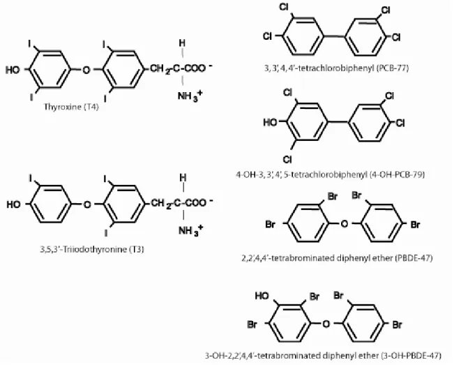
Figure 1. A comparison of thyroid hormones, polychlorinated biphenyls 51 (e.g. PCB-77), and emerging contaminants such as the polybrominated diphenyl ethers (e.g. PBDE-47) and halogenated phenolics (e.g. 4-OH- PCB-79 and 3-OH-PBDE-47).
Figure 2. Interacting mechanisms that may explain the ability of PCBs 52 (and other related compounds) to reduce circulating and tissue levels of thyroid hormones.
Chapter 2:
Figure 1. Map showing the sampling locations of bottlenose dolphins 90 along the Southeast United States Atlantic Coast.
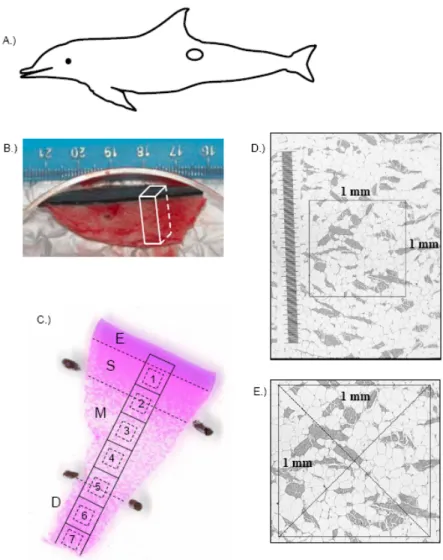
Figure 2. Skin-blubber biopsy sampling site and histological subsampling 91 for analysis of blubber cellular characteristics in bottlenose dolphins.
Figure 3. Light micrograph images of the blubber from bottlenose 92 dolphins. Black scale bars represent 2 mm.
Figure 4. Structural fiber areas (mm2), adipocyte cell counts, and 94 adipocyte cross-sectional areas (um2) in CHS subadult, CHS adult, IRL subadult, and IRL adult dolphins.
Figure 5. Structural fiber areas (mm2), adipocyte cell counts, and 96 adipocyte cross-sectional areas (um2) in CHS females.
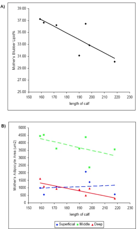
Figure 6. Total blubber lipid % and adipocyte cross-sectional areas 98 versus age in CHS females.
Figure 7. Total blubber lipid % and adipocyte areas in CHS females 99 captured with calves.
Figure 8. Water temperature (oC) at CHS (Charleston Harbor) and IRL 100 (St. Lucie) locations from January through August 2003.
Figure 9. A schematic illustration depicting how the blubber may 101 respond to different factors such as water temperature, ontogeny,
reproductive status, and nutritional state in bottlenose dolphins.
Chapter 3:
Figure 1. Map showing the sampling locations of bottlenose dolphins 152 along the Southeast United States Atlantic Coast.
Figure 2. Skin-blubber biopsy sampling site and subsampling for 153 histology and immunohistochemical (IHC) analysis of cytochrome
P4501A1 (CYP1A1) in bottlenose dolphins.
Figure 3. Light micrograph images of the blubber and CYP1A1 154 staining in the superficial, middle, and deep layers.
Figure 4. Correlation between “original” and “modified” CYP1A1 156 staining scores in vascular endothelial cells averaged over
blubber layers.
Figure 5. Depth specific CYP1A1 expression in vascular endothelial 158 cells in the skin-blubber biopsy of male and female bottlenose
dolphins captured and released at Charleston, SC (CHS) and Indian River Lagoon, FL (IRL).
Figure 6. CYP1A1 expression and Total Toxic Equivalents 160 (TEQ98 ng/g wet wt) in male and female bottlenose dolphins
captured and released in CHS and IRL locations.
Figure 7. Relationships between CYP1A1 expression of vascular 162 endothelial cells and TEQ98 levels in male and female
bottlenose dolphins captured and released at CHS and IRL locations.
Figure 8. Relationships among CYP1A1 expression of vascular 164 endothelial cells, TEQ98 concentrations, and adipocyte areas in
male bottlenose dolphins captured and released at CHS (N = 19) and IRL (N = 21) locations.
Figure 9. Relationships of total blubber PCB concentrations and 166 TEQ98 levels with age in male and female dolphins from CHS and
IRL locations (CHS males N = 19, females N = 12; IRL males N = 21, females N = 6).
Figure 10. A.) Depth specific CYP1A1 expression in vascular 167 endothelial cells in the skin-blubber biopsy of subadult (N = 3),
adults captured with calves (N = 2), pregnant (N = 2),
female dolphins captured and released at CHS location.
Figure 11. Relationships among CYP1A1 expression of vascular 168 endothelial cells, TEQ98 concentrations, and adipocyte
cross-sectional areas in subadult (N = 3), adult (N = 2),
pregnant (N = 2), lactating (N = 3), and simultaneously pregnant and lactating (N = 2) CHS female dolphins.
Figure 12. TEQ98 levels and CYP1A1 expression of vascular 170 endothelial cells in CHS female dolphins captured
with calves (N = 7). Chapter 4:
Figure 1. A comparison between manual and threshold segmentation 213 of native and processed images.
Figure 2. Total brain weights and total brain volumes for Atlantic 214 white-sided dolphins that stranded along the beaches of
Cape Cod, MA between 2002 and 2005.
Figure 3. Three-dimensional reconstruction of the brain of specimen 216 CCSN05-084-La from magnetic resonance (MR) images.
Figures 4-11. Anterior-to-posterior, post-mortem MRI sequence of a 218 subadult male brain (CCSN05-084-La) intact within the skull.
Figures 12-19. Midline-to-lateral, post-mortem MRI sequence of a 224 subadult male brain (CCSN05-084-La) intact within the skull.
Figure 20. Three-dimensional reconstruction of the brain of 229 Specimen CCSN05-040-Fetus-La from magnetic resonance
(MR) images.
Figures 21-27. Anterior-to-posterior, post-mortem MRI sequence 230 of a male fetus brain (CCSN05-040-Fetus-La) intact within the skull.
Figures 28-33. Midline-to-lateral, post-mortem MRI sequence of 234 a male fetus brain (CCSN05-040-Fetus-La) intact within the skull.
Figure 34. A.) Three-dimensional reconstruction of fetal brain surface 238 (CCSN05-039-fetus-La). B.) Measured brain volume (cm3) versus
actual brain weight (g).
Figure 35. A visual comparison of the degreee of myelination 239
(i.e white matter tracts) during ontogeny. Figure 36. A quantitative comparison of the degreee of 240
myelination (i.e white matter tracts) during ontogeny. Figure 37. Volumes (cm3) of the cerebellum grey matter versus 241
length (cm). Figure 38. Segmentation label maps and mid-sagittal areas of 242
the corpus callosum. Figure 39. Segmentation label maps and volumes of the 243
hippocampus. Figure 40. Three-dimensional reconstruction of the adult specimen 244
brain CCSN05-040-La illustrating the spatial relationship of the hippocampus with the rest of the brain. Chapter 5: Figure 1. An integration of major findings of this thesis and working 255
hypotheses. Appendix 1: Figure 1. Brain lesion in CCSN04-177-Dd. 274
Figure 2. Brain lesion in CCSN04-191-Dd. 275
Figure 3. Brain lesion in CCSN05-038-La. 276
Figure 4. Brain lesion in CCSN05-232-La. 277
Appendix 2: Figure 1. Label maps of the hippocampus and surrounding fluid 280 structures used to determine the volumes of these brain structures for the California sea lion “Shelouba”.
Figure 1. Bottlenose dolphin CYP1A1 amino acid sequence and 288 comparison to the striped dolphin (S. coeruleoalba), the pig (S. scrofa), the house mouse (M. musculus), and the human (H.
sapiens) CYP1A1 amino acid sequences.
Figure 2. Control versus PCB126 treated biopsies. 290 Appendix 4:
Figure 1. Bottlenose dolphin Type II 5’-deiodinase (D2) partial 296 amino acid sequence and its comparison to the pig (S. scrofa), the human (H. sapiens), the house mouse (M. musculus), and the rat (R. norvegicus) D2 amino acid sequences.
Appendix 5:
Figure 1. A comparison of the Atlantic white-sided dolphin TTR to 299 other species.
Appendix 6:
Figure 1. A comparison of PCBs, PBDEs, and organochlorine 302 pesticides in flounder at the east bay and outfall sites of Cape Cod Bay. Appendix 7:
Figure 1. A comparison of PCBs, organochlorine pesticides (OCs), 303 PBDEs, OH-PBDEs, OH-PCBs, and methyl sulphone PCBs in
cerebellum grey matter samples collected from CCSN05-037-La, CCSN05-039-La, and CCSN05-040-La.
LIST OF TABLES Chapter 1:
Table 1. Examples of PCB concentrations found in the blubber of marine 53 mammals.
Table 2. Research studies that have focused on distribution of POPs in rat 54 and human brains.
Table 3. Chemical analysis performed on brains of marine mammals. 56 Table 4. Pathologies in the brain, inner ear, and thyroid gland in fetal and 57 neonatal hypothyroidism.
Table 5. Percent adult brain weight at birth. 58 Chapter 2:
Table 1. Blubber morphological data reported as means and standard 102 errors for each location and age class category for bottlenose dolphins
captured and released in Charleston, SC and Indian River Lagoon, FL during July and August 2003.
Table 2. Blubber morphological data reported as means and standard 104 errors for female bottlenose dolphins captured and released in
Charleston, SC during August 2003. Chapter 3:
Table 1. Objectives and statistical tests to determine the interrelationships 171 among CYP1a1 expression, PCBs and OH-PCBs, and blubber dynamics
of bottlenose dolphins live-captured and released in Charelston, SC and Indian River Lagoon, FL during July and August 2003.
Table 2. Cytochrome P450 1A1 expression of vascular endothelial cells 173 in the blubber of bottlenose dolphins live-captured and released in
Charleston, SCand Indian River Lagoon, FL during July and August 2003.
Table 3. Cytochrome P450 1A1 expression of vascular endothelial cells in 175 the blubber and PCB concentrations of bottlenose dolphins live-captured
and released in Charleston, SC and Indian River Lagoon, FL during July and August 2003.
Table 4. Slope β1, r2, and p-values of the simple linear regression 176 equations for cytochrome P450 1A1 expression of vascular endothelial
cells in bottlenose dolphins live-captured and released in Charleston, SC and Indian River Lagoon, FL during July and August 2003.
Table 5. Slope β1, r2, and p-values of the linear regression equations for 177 TEQ98, total PCB, and OH-PCB concentrations versus age in male
bottlenose dolphinslive-captured and released in Charleston, SC and Indian River Lagoon, FL duringJuly and August 2003.
Table 6. Slope β1, r2, and p-values of the non-linear regression equations 178 for TEQ98, total PCB, and OH-PCB concentrations versus agein female
bottlenose dolphins live-captured and released in Charleston, SCand Indian River Lagoon, FL during July and August 2003.
Table 7. Slope β1, r2, and p-values of the simple linear regression 179 equations for cytochrome P450 1A1 expression of vascular endothelial
cellsin female bottlenose dolphins live-captured and released in Charleston, SC and Indian River Lagoon, FL during July and August 2003.
Table 8. Relationship between OH-PCB concentrations and cytochrome 180 P450 1A1 expression of vascular endothelial cellsof the deep blubber
layer in bottlenose dolphins live-captured and released in Charleston, SC and Indian River Lagoon, FL during July and August 2003.
Chapter 4:
Table 1. Stranding and life history information of Atlantic white-sided 245 dolphin specimens in which magnetic resonance imaging (MRI) was
performed.
Table 2. Comparisons of expected and segmented volumes of water. 246 Table 3. Comparisons of expected and segmented volumes of brain tissue. 247 Table 4. A comparison of manual segmentation volumes and threshold 248 segmentation volumes of white matter, grey matter, and cerebrospinal
fluid from nativeproton density (PD) and processed PD images.
Table 5. Brain and cerebellum volume data of Atlantic white-sided 249 dolphins.
Table 6. Corpus callosum area and hippocampus volume 250 measurements of Atlantic white-sided dolphins.
Appendix 1:
Table 1. Stranding and life history information of common dolphins 278 and Atlantic white-sided dolphins exhibiting brain lesions.
Appendix 2:
Table 1. Hippocampus and surrounding fluid structure volumes for the 281 California sea lion “Shelouba”.
Appendix 3:
Table 1. Degenerate primer sequences used in RT-PCR to identify 291 CYP1A1 and actin in bottlenose dolphin skin-blubber biopsy samples.
Table 2. Biopsies processed for SSH experiments. 292
Table 3. Total RNA concentrations isolated from all untreated and treated 293 biopsy samples.
CHAPTER I: INTRODUCTION
POPs and Emerging Contaminants in Marine Mammals
Marine mammals bioaccumulate persistent organic pollutants (POPs) such as organochlorine pesticides like dichlorodiphenylethanes (i.e. DDTs), dieldrin, chlordanes, and hexachlorocyclohexanes (HCHs), as well as industrial solvents and their byproducts such as chlorinated dibenzo-p-dioxins, dibenzofurans, and polychlorinated biphenyls (PCBs) (Blomkvist et al., 1992; DeLong et al., 1973; Hansen et al., 2004; Kannan et al., 1993; Muir et al., 1996; Ross et al., 2000; Tuerk et al., 2005). In some populations of marine mammals, the levels of POPs in blubber are extremely high (Table 1.). Exposure of marine mammals to these compounds has been associated with mass mortalities and health effects, including reproductive abnormalities and immune dysfunction (DeLong et al., 1973; Kannan et al., 1993; Ross et al., 1996).
Emerging environmental contaminants may pose a new threat to the health of marine mammals. The flame-retardants are one class of emerging contaminants
(Birnbaum and Staskal, 2004; de Boer et al., 1998; Hooper and McDonald, 2000). These compounds include polybrominated biphenyls (PBBs), polybrominated diphenyl ethers (PBDEs), tetra-bromobisphenol A (TBBPA), and hexabromocyclododecane (HBCD). Although these compounds are similar in structure and behavior to well-known
environmental contaminants such as PCBs, they have not been banned domestically or internationally, except for a voluntary phase-out of pentabromodiphenyl ether
(pentaBDE) by the sole manufacturer on December 31, 2004. These chemicals are produced globally at an estimated 150,000 tonnes a year (de Boer et al., 1998). Like PCBs and DDT, PBDEs have lipophilic and metabolically resistant properties that make them long-lived, bioaccumulating environmental pollutants (de Boer et al., 1998). In a study that has alarmed both the scientific and political community, Meironyte et al. (1999) showed that the sum of the concentrations of PBDE congeners in Swedish human milk from 1972 to 1997 had increased from 0.07 to 4.02 ng/g lipids; over the same time period, the total toxic equivalents (TEQ) from PCBs in human milk in Sweden decreased. PBDEs and other brominated flame-retardants may be the “new PCB problem”.
Halogenated phenolics have also emerged as important environmental
contaminants in wildlife and humans (Letcher et al., 2000; Sandau, 2000). These include such pollutants as the hydroxylated metabolites of PCBs (OH-PCBs), the hydroxylated metabolites of PBDEs (OH-PBDEs), and pentachlorophenol. These compounds interact with the thyroid hormone system and have been recently recognized as a group of contaminants that may pose a threat to human and marine mammal health (Brouwer et al., 1998; Letcher et al., 2000). Research has shown that these compounds are retained in the plasma of humans and marine mammals(Houde et al., 2006; Letcher et al., 2000; McKinney et al., 2006; Sandala et al., 2004). Most species have plasma OH-PCB
concentrations ranging from 5-30% of the total PCBs (Sandau, 2000). However, in some marine mammals like polar bears, OH-PCB levels are generally higher than PCB
concentrations (Sandau, 2000).
Marine mammals accumulate and retain a mixture of PCB and PBDE congeners, as well as OH-PCB and OH-PBDE congeners. The mixture of these chemicals in marine mammals and their additive impacts on the thyroid hormone system are a cause for concern, particularly for the fetus and neonate that depend on a functional thyroid hormone system for proper neuro-development (Figure 1).
Induction of Xenobiotic Metabolizing Enzymes and Formation of Hydroxylated Metabolites
PCBs induce cytochrome P450 monooxygenases (CYP) and are metabolized by these enzymes. These enzymes biotransform PCBs to OH-PCB metabolites. To better understand hydroxylated metabolites of PCBs and their effects, it is important to explain the processes that lead to their formation. CYP enzymes biotransform PCBs by inserting an oxygen into these compounds. Oxygen insertion can eventually lead to the formation of hydroxylated metabolites. One mechanism involves epoxidation followed by epoxide ring opening. In the epoxide ring intermediate, a chlorine atom can shift its position to another carbon. This shift has been given the name National Institute of Health Shift or “NIH Shift” (Guroff et al., 1967). PCB metabolism studies have shown that several
isomers of hydroxylated metabolites can be formed from one PCB congener through an NIH Shift mechanism (Ishida et al., 1991).
The superfamilies of CYPs exist in a wide range of species from bacteria to mammals, exhibiting an enormous diversity in genetic structure. According to their amino acid sequences, the CYP genes are classified into over 74 families. OH-PCBs are derived from phase I metabolism of parent PCB congeners by enzymes belonging to the CYP1A and CYP2B (and possibly CYP isoforms) enzyme families (Letcher et al., 2000; Yoshimura et al., 1987). The hydroxylation of PCBs by either CYP1A or CYP2B isozymes is dependent upon the chlorine substitution pattern of the PCB congener on each of the biphenyl rings. In rat liver, CYP1A is important in phase I oxidative metabolism of PCB congeners with chlorine substituents at one or both para positions, and with adjacent non-halogenated ortho and meta carbons on at least one ring
(Kaminsky et al., 1981; Mills et al., 1985). CYP2B is important in phase I oxidative metabolism of PCB congeners that have two ortho-chlorines and meta-, para-vicinal hydrogens.
Historically, PCBs have been divided into three different groups based on their induction of CYPs (Safe, 1984). PCB congeners that cause CYP1A induction contain chlorines in both para and at least two meta positions with no substitution in the ortho position. These congeners are termed the coplanar PCBs because the rings can achieve a planar configuration. The induction mechanism of CYP1A type enzymes involves the activation of the aryl hydrocarbon receptor (AHR) signaling pathway (Hahn, 1998). AHR ligands include planar halogenated aromatic hydrocarbons (PHAHs) (i.e. non-ortho and some mono-ortho substituted PCBs and 2,3,7,8-tetrachlorodibenzo-p-dioxin (TCDD or dioxin)) and polycyclic aromatic hydrocarbons (PAHs).
PCBs that contain at least one chlorine in the ortho position of the biphenyl ring cause CYP2B induction (Honkakoski and Negishi, 1998). However, the most active phenobarbitol-type inducers are PCBs that contain at least two ortho and two para chlorine substituents (Denomme et al., 1983). This chlorine pattern reduces free rotation of the biphenyl rings, which hinders a planar biphenyl configuration. The induction
mechanism of CYP2B type enzymes involves the constitutive androstane receptor (CAR) (reviewed by Waxman (1999)). “Mixed” inducers are chemicals that can induce both CYP1A and CYP2B enzymes.
In summary, these enzymes (and possibly other CYPs) are induced by PCB substrates and act on the substrate, introducing a hydroxyl group into the aromatic ring. At this point, the hydroxylated metabolite can be converted into a more water-soluble product and excreted, or retained in plasma or perhaps other tissues.
CYP1A1 Induction as a Biomarker in Delphinid Cetaceans
CYP1A1 induction is a valuable biomarker of exposure to PHAHs and has been used extensively in fish, birds, and marine mammals (Stegeman and Hahn, 1994). Its advantages include the extensive database demonstrating its relationship to PHAH exposure (Moore et al., 1998). In delphinid cetaceans, CYP1A1 has been shown to be a valuable biomarker of exposure to PHAHs (White et al., 1994; Wilson et al., 2005). Its advantages include the relatively robust methods that exist for its detection (formalin preservation followed by immunohistochemistry) and the fact that it can be measured in skin-blubber biopsy samples (Angell et al., 2004). In vitro assays have demonstrated CYP1A1 induction in sperm whale (Physeter macrocephalus) skin biopsy slices exposed to β-napthoflavone (BNF), a prototypical CYP1A1 inducer (Godard et al., 2004).
In the integument, CYP1A1 expression is strongest and most frequent in vascular endothelial cells of the arterial system and capillaries within the blubber of cetaceans (Angell et al., 2004). This is consistent with earlier observations that CYP1A is highly inducible in vertebrate endothelial cells (Stegeman et al., 1989). It has been suggested that the movement of AHR agonists from the blubber across the endothelial cells and into the bloodstream (i.e. as occurs during blubber lipid mobilization) could induce CYP1A1 in vascular endothelial cells (Angell et al., 2004). In other vertebrate species, PCBs and DDTs have been shown to move out of adipose tissue during lipid mobilization (Dale et al., 1962; Findlay and De Freitas, 1971; Sodergren and Ulfstrand, 1972). Hence,
understanding blubber morphology and lipid dynamics may be important factors in
understanding CYP1A1 expression in the blubber biopsy, its relationship to AHR
agonists (e.g. non-ortho and mono-ortho PCBs), and its involvement in the production of HO-PCBs. Understanding these processes in delphinids is a major goal of this thesis.
Blubber Morphology and Dynamics
Blubber is dynamic and multifunctional, serving many roles: it functions biomechanically to provide support during locomotion and increases efficiency by streamlining the body surface (Hamilton et al., 2004; Pabst, 2000); it contributes to buoyancy (Dearolf et al., 2000; Kipps et al., 2002; McLellan et al., 2002); it is a primary site for lipid storage, which the animal utilizes during periods of energetic stress (Aguilar and Borrel, 1991; Koopman et al., 1996; Koopman et al., 2002; Struntz et al., 2004). The high lipid content also provides insulation, decreasing the heat loss from the body core to the external environment (Dunkin et al., 2005; Worthy and Edwards, 1990).
Histological and biochemical evidence from stranded specimens suggest that cetacean blubber is stratified (Aguilar and Borrell, 1990; Koopman et al., 1996; Koopman et al., 2002; Struntz et al., 2004). For example, in bottlenose dolphins
(Tursiops truncatus) that either stranded or were killed incidentally in fishing operations in North Carolina and Virginia, Struntz et al. (2004) showed dramatic blubber
stratification based in adipocyte number, adipocyte area, and structural fiber density. At the mid-thoracic site, adipocyte areas and numbers varied significantly across the blubber depth, with smaller and fewer adipocytes near the epidermis or “superficial” layer. Adipocyte numbers and size increased in the “middle” blubber and then decreased again in the “deep” layer near the border of the sub-dermal connective tissue sheath and muscle layer. These data, as well as the impacts of emaciation on blubber morphology, have brought forth the hypothesis that the “inner” or “middle” and “deep” blubber layers are more dynamic with regards to lipid mobilization, while the “outer” or “superficial” blubber is more static (Aguilar and Borrell, 1990; Koopman et al., 2002; Struntz et al., 2004)
Blubber is the primary bioaccumulation site for POPs such as organochlorine pesticides and PCBs (Marsili and Focardi, 1997; Schantz et al., 1993; Tirpenou et al., 1998). During periods of lipid mobilization such as lactation, POPs are mobilized into the circulatory system (Norstrom and Muir, 1994; Ridgway and Reddy, 1995; Wolkers et al., 2004). Thus, knowledge of the structure and dynamics of blubber is important in marine mammal toxicology because this information is essential for understanding the mobilization of pollutants from the blubber into the bloodstream and investigating associated health effects on the animal and its offspring.
Maternal Transfer of POPs and Halogenated Phenolics
In many marine mammal species, there is a strong correlation between increasing POP blubber residue levels and age, until animals reach sexual maturity (Borrell et al., 1995; Cockcroft et al., 1989; Ross et al., 2000). At this time and thereafter, females experience a pronounced decrease in contaminant burdens in the blubber, while males continue to accumulate POPs throughout their lives. This reduction in contaminant burdens in sexually mature females has been best explained by the transfer of these burdens from the maternal blubber to offspring during pregnancy and lactation. It has been predicted that first-born dolphin calves receive a fourfold higher initial burden of PCBs than subsequent calves, with 90% of this load being transferred through lactation (Cockcroft et al., 1989). It was estimated that almost 80% of the contaminant burden of a lactating female is passed to a first-born calf and that this transfer would take
approximately seven weeks after birth (Cockcroft et al., 1989).
Researchers conducting a thirty-year study in Sarasota Bay, Florida have
discovered that first-born bottlenose dolphin calves rarely survive (Wells, 2000). In the same population of bottlenose dolphins (i.e. from Sarasota, Florida), first-born calves also have higher PCB concentrations than subsequent calves of similar age (Wells et al., 2005). The mechanism for this high mortality is unknown and could involve mother inexperience or possibly contaminant transfer to the calf. The relative high and acute exposure of the first-born calf to environmental chemicals is a subject of concern.
There is no information about the maternal transfer of halogenated phenolics in marine mammals. In rat studies, maternal exposure to Aroclor 1254 from gestation days (GD) 10 to 16 resulted in accumulation of the metabolite
4-OH-2,3,3’,4’,5-pentachlorobiphenyl (4-OH-CB107) in fetal plasma and brain (Morse et al., 1996). Recent work has shown that prenatal exposure of radiolabeled 4-OH-CB107 resulted in the accumulation of this compound in the fetus (Meerts et al., 2002). In fact, the fetal/maternal ratios at GD 20 in liver, cerebellum, and plasma were all greater than 1 (11.0, 2.6, and 1.2, respectively). Transthyretin (TTR), a thyroid hormone binding protein, is thought to be responsible for maternal to fetal transport of thyroxine (T4) across the placenta (Achen et al., 1992). The high binding affinity of xenobiotics such as OH-PCBs and other halogenated phenolics to TTR has been hypothesized to result in facilitated transport of these compounds across the placenta to the fetus (Meerts et al., 2002).
Contaminants in the Brain
Many POPs that are found at high levels in milk and that are maternally transferred in marine mammals are neurotoxic (Vedder, 1996). These include such organochlorine insecticides as the dichlorodiphenylethanes (i.e. DDTs), the cyclodienes (i.e dieldrin, chlordanes), and the cyclohexanes (i.e. hexachlorocyclohexane or HCH) (reviewed by Ecobichon (1996)). DDT poisoning is associated with effects on the central nervous system (CNS) in humans. DDT elicits its effects at the level of the neuronal membrane by reducing potassium transport across the membrane. The cyclodienes are potent neurotoxicants that block the λ-aminobutyric acid (GABA) receptor found in the CNS. The blocking of this ion channel impedes the uptake of chloride ions by neurons and causes a state of uncontrolled excitation. Technical grade HCH used in insecticides contains a mixture of isomers: the λ- and α-isomers are convulsant poisons; the β- and δ-isomers are CNS depressants. PCBs are also neurotoxic but the exact mechanism is unclear and most likely involves multiple mechanisms (Seegal, 2000).
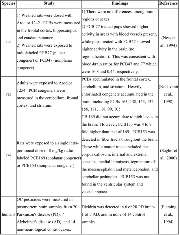
POP distribution in the brain has been studied in rats and humans (Table 2) but there is limited information about the distribution of halogenated phenolics in the brain. Meerts et al., (2002) have shown the accumulation of [14 C]-labeled 4-OH-CB107 in fetal rat cerebellum and forebrain. More detailed regional analysis has not yet been
completed. An important point to consider when hypothesizing the distribution of halogenated phenolics in the brain is the ability of TTR to bind to these compounds and alter their distribution. In humans, the three thyroxine transport proteins (albumin, thyroid binding globulin, and TTR) are synthesized by the liver, but only TTR is
synthesized in the brain in the epithelial cells of the choroid plexus (Dickson et al., 1987). All the newly synthesized TTR is transported towards the brain into the cerebrospinal fluid (CSF). In fact, the ratio of transthyretin to albumin concentration is 30-fold higher in the CSF than in blood plasma. TTR in CSF serves as the main thyroxine transport protein (Schreiber et al., 2001). Since TTR is synthesized in the choroid plexus and is secreted into the CSF, it is possible that these tissues retain higher levels of OH-PCBs, OH-PBDEs, and other halogenated phenolics, specifically compounds that have a high affinity for TTR. Consistent with this, Takasuga et al. (2004) observed that the levels of OH-PCBs in human CSF were higher than the levels of PCBs, opposite of what was found in the serum.
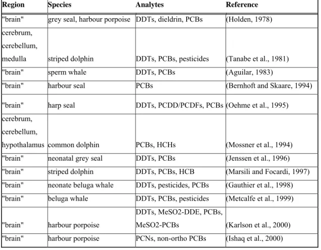
Chemical analysis has been limited in marine mammal brains (Table 3). Studies have not addressed whether contaminants bioaccumulate in specific brain regions. Furthermore, our understanding of the distribution and bioaccumulation of halogenated phenolics in the brain of delphinid cetaceans is non-existent. Above all, there is very limited knowledge on exposure of the brain to environmental chemicals during the fetal and neonatal stage, which is especially important because in the fetus and neonate, the blood-brain barrier is incomplete and the brain is still developing (Eriksson, 1997). POPs and Halogenated Phenolics Decrease Thyroid Hormone Levels
There is much evidence that POPs, including the brominated flame retardants and the halogenated phenolics, can interfere with the thyroid hormone system in rats, humans, and seals. Decreased serum levels of T4 have been correlated with exposure to PCBs
both in rats and in humans (reviewed by Brouwer et al. (1998)). Furthermore, Brouwer et al. (1989) showed that consumption of PCB-contaminated fish caused vitamin A and thyroid hormone deficiencies in the common seal, Phoca vitulina. New classes of halogenated pollutants - the brominated flame retardants – have recently been identified as thyroid hormone disrupters (Birnbaum and Staskal, 2004). Of particular interest are the PBDEs, which have been shown to drastically reduce circulating T4 concentrations (Zhou et al., 2002).
To date, there are at least three independent, but possibly interacting, mechanisms that may explain the ability of PCBs (and other related compounds) to reduce circulating and tissue levels of thyroid hormones. First, PCBs have been shown to change thyroid gland structure, perhaps directly interfering with thyroid gland function (Collins et al., 1977). These findings are consistent with the report ofByrne et al. (1987) that PCB exposure reduces the ability of thyroid stimulating hormone (TSH) to increase serum T4
in vivo. Recently, Pocar et al. (2006) used a primary porcine thyrocyte culture (derived
from pigs) as an experimental model to show that TCDD and PCB126 significantly down-regulate the sodium iodide symporter (NIS) and the cathepsins (Cat B and L). NIS is an important enzyme in thyroid epithelial cells, where it catalyzes the active
accumulation of iodide. Cat B and L help in the proteolysis of thyroglobulin, which allows controlled liberation of T4 and 3,3’,5-triiodothyronine (T3) from the thyroid follicle into the circulatory system. Both NIS and Cat B & L are important in thyroid hormone production. Thus, PCBs may directly interfere with the ability of the thyroid gland to respond to TSH.
Second, PCBs can increase the metabolism of thyroid hormones. Research in the past has shown that PCB exposure increased the bile flow rate, as well as biliary
excretion of 125I-T4 (Bastomsky et al., 1976). PCB exposure also induces the expression and activity of UDP-glucuronosyltransferase (UDP-GT) (Kolaja and Klaassen, 1998) and increases T4 glucuronidation (Visser et al., 1993). UDP-GT induction could explain the increased bile flow rate and excretion of T4. Thus, these actions may facilitate serum T4 clearance by hepatic metabolism, reducing the half-life of T4 in the blood. Finally, as
previously stated, OH-PCBs bind to TTR in the blood, and can potentially displace T4 in vivo (Cheek et al., 1999). These three mechanisms of toxicity may combine to interfere
with the ability of the thyroid gland to respond to TSH and produce thyroid hormones, reduce the half-life of T4 in the serum, and lessen the carrying capacity of the blood for T4.
Thyroid Hormone Action
T4 is the main product released from the normal thyroid. It is considered the inactive prohormone because T3 is the ligand that modulates the thyroid hormone receptor (TR). T4 is transported to target tissues via three transport proteins exhibiting different T4 affinities – thyroid binding globulin (TBG), TTR, and albumin. The distribution of these binding proteins is not universal in the animal kingdom (Schreiber and Richardson, 1997). Currently, there are limited data on how T4 traverses the vascular barrier and reaches the target cell.
Activation of Thyroxine. Type I and Type II 5’-deiodinases (D1 and D2,
respectively) activate the prohormone T4 to form the active hormone T3 (reviewed by Kohrle (1999)). D1 can also inactivate the active hormone T3 to form 3,
diiodothyronine (T2) or iodothyronine sulfates. Another deiodinase isoenzyme, the 5-deiodinase (D3), inactivates the prohormone T4 by eliminating iodine to form the
inactive product rT3 (reverse T3) or T2. T3 homeostasis in tissues is maintained by these three enzymes (D1, D2, and D3). The presence and activity of these enzymes are tissue specific.
D2 is especially important because of its apparent role in the development of the central nervous system and the cochlea, and its reaction to hypothyroidism. D2 is
expressed in the brain, inner ear, the severely hypothyroid anterior pituitary, the placenta, the skin, and brown adipose tissue in rodents (Bates et al., 1999; Campos-Barros et al., 2000; Kohrle, 1999; Schroder-van der Elst et al., 1998; Tu et al., 1997). During
hypothyroidism, D2 activity increases because the protein is stabilized and the half-life is prolonged (as cited in (Kohrle, 1999)). These observations have led to the theory that D2
produces T3 for local cellular demands independent of circulating T3 (Kohrle, 1999). For example, Schroder-van der Elst et al. (1998) investigated deiodinase activities in fetal rat tissues at several levels of iodine deficiency. One of the more important findings was that D2 activity increased in the fetal skin, brain, and placenta as a result of iodine deficiency. Even more interesting was the higher level of D2 in fetal skin compared to the brain and the increased skin D2 activity in even mild iodine deficiency. Based on these findings, the authors concluded that skin D2 is physiologically important in fetal thyroid hormone economy. Skin D2 contributes to the intracellular T3 content of the skin and, possibly, to the plasma T3.
Campos-Barros et al. (2000) investigated deiodinase expression in the mouse cochlea before the onset of hearing. D2 activity increased rapidly in the mouse cochlea to peak around postnatal day 7, after which activity decreased by P10. The peak in activity a few days before the onset of hearing suggests an important role for D2 in increasing local levels of T3. Such a role for D2 activity has been further supported in rats made mildly hypothyroidic by an antithyroid chemical propylthiouracil (PTU) or PCBs (Crofton et al., 2000; Goldey, 1995a; Goldey and Crofton, 1998; Herr et al., 1996). Both of these treatments reduced serum levels of T4 but not T3 because protective
measures maintained serum T3 levels. Nonetheless, auditory deficits were seen. These studies support the view that circulating T3 levels are inadequate for the developing cochlea and increased D2 activity is necessary to convert T4 to T3, in order to increase local T3 levels for normal cochlear development.
Thyroid Hormone Receptor. T3 acts primarily at the nuclear level by regulating
the transcription of thyroid-hormone-responsive genes, as reviewed in Anderson (2001). Thyroid hormones enter the cell, move to the nucleus, and bind to the thyroid hormone receptor (TR), a receptor belonging to the larger family of nuclear receptors. Two isoforms of TR exist, known as TRα and TRβ. T3 binds to the TR with much higher affinity than T4 and is thought to be the active hormone in the nucleus. TR interacts with specific DNA sequences known as thyroid hormone response elements (TREs). TR binds to the TRE as a heterodimer with the retinoid X receptor (RXR). These TREs are
located in the proximal promoter regions of thyroid hormone-responsive genes. The genes give rise to proteins that are very important in development of the inner ear, retina, cerebellum, hippocampus, and cerebral cortex.
Thyroid Hormone Responsive Genes in the CNS. Numerous T3-regulated genes
have been identified in the rodent CNS (reviewed by Anderson (2001)), but roles for these gene products in the brain are not well established. Recently, it has been found that many of these genes encode transcriptional regulatory proteins, one of which is the mammalian basic transcription element-binding protein (BTEB) (Denver et al., 1999). Overexpression of BTEB in neuro-2a cells has been shown to dramatically increase the number and length of neurites, suggesting an important role of BTEB in dendritic growth (Denver et al., 1999). Furthermore, T3 administration was shown to increase BTEB mRNA levels in primary neurons, astrocytes, and oligodendrocytes prepared from E16 (for neurons) and P2 (for astrocytes and oligodendrocytes) rat brain.
It is known that oligodendrocytes express active forms of thyroid hormone receptors and that thyroid hormones are important in myelination, as reviewed by
Anderson (2001). In fact, the most striking effect of neonatal hypothyroidism is the delay in myelinogenesis and a decrease in the number of myelinated axons, without any effect on the total number of axons. It has been shown that thyroid hormones regulate the expression of several key enzymes and proteins of the myelin sheath (Barradas et al., 2001). These include 2’3’-cyclic nucleotide 3’-phosphodiesterase (CNPase), myelin basic protein (MBP), proteolipidic protein (PLP), as well as
myelin-associated/oligodendrocytic basic protein (MOBP). The expression of these genes is reduced in rats made hypothyroid as neonates (Barradas et al, 2001). Interestingly, deficiency of thyroid hormone during the neonate stage induced a permanent down-regulation of MOBP 22 kDa isoform and PLP expression in adulthood.
Hypothyroidism, Environmental Chemicals, and Neurodevelopment
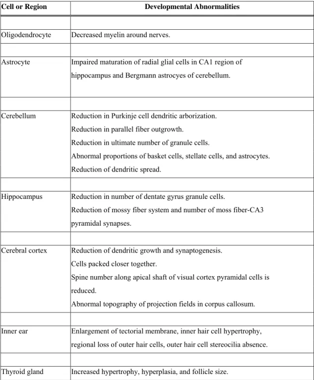
If thyroid hormone deficiencies (hypothyroidism) occur during fetal or neonatal development, severe pathological situations can occur. Hypothyroid effects include
disorders of process outgrowth, synaptogenesis, and myelination in neuron development, as reviewed by Anderson (2001) (Table 4). These disorders manifest themselves as smaller and more tightly packed peripheral and central neuronal cell bodies. The affected areas can be related to the various deficits in learning and motor skills of hypothyroid animals. This is revealed in the disorders known as cretinism (in the case of fetal development) and congenital hypothyroidism (in the case of neonatal development). Cretinism occurs when there is a severe iodine deficieny in the diet of pregnant women and is characterized in the fetus by extreme mental retardation, deaf-mutism, impaired voluntary motor activity and hypertonia (Delange, 2000). Congenital hypothyroidism, if untreated, results in severe intellectual deficits in children (Song et al., 2001).
Hypothyroidism has been shown to cause decreases in brain volume and weight in both clinical and experimental cases. Brain magnetic resonance (MR) imaging in
patients with hypothyroidism before and after treatment showed a significant increase in brain size with thyroid hormone supplement (Oatridge et al., 2002). Furthermore, surgical thyroidectomy of sheep fetus at 98 days causes a significant reduction in brain weight at birth (McLntosh et al., 1982). In rats dosed with propylthiouracil to induce neonatal hypothyroidism, there was a significant decrease in brain and cerebellar weights (Nathaniel et al., 1988). This condition was alleviated with T4 replacement therapy.
PCB exposure in humans is associated with cognitive and behavioral retardation (Gilbert et al., 2000; Schantz, 1996a; Schantz et al., 2001; Schantz et al., 1991; Schantz et al., 1995; Schantz et al., 1996b; Wong et al., 1997). This may be partly explained by the ability of these compounds to affect brain development by interfering with the thyroid hormone system. This hypothesis is supported by a series of significant findings. First, neurological deficits observed in humans associated with PCB exposure are similar to those deficits observed in the offspring born from hypothyroxinemic women (Gilbert et al., 2000; Goldey and Crofton, 1998). Second, exposure of pregnant rats to OH-PCBs (specifically 4-OH-CB107) results in the transfer of this compound to the fetal brain, a decrease in total T4 (TT4) in fetal plasma and brain samples, and concomitant increase of D2 activity in fetal forebrain (Meerts et al., 2002). Third, it has been shown that
exposure of rat offspring to PCBs results in severe hearing loss and motor deficits (Goldey and Crofton, 1998). These deficits are accompanied by a drastic decrease in circulating T4, and the deficits are attenuated by T4 replacement therapy. Cochlear pathologies in these rats reveal outer hair cell losses similar to lesions common in severe hypothyroidism (Crofton et al., 2000; Goldey et al., 1995). Fourth, in mouse cerebellar culture assays, HO-PCBs inhibit thyroid-hormone-dependent arborization of Purkinje cell dendrites (Kimura-Kuroda et al., 2005). Fifth, in fetal rats, Aroclor 1254 (a PCB
mixture) decreases the density of oligodendroglial cells of the corpus callosum (Sharlin et al., 2006).
Brain Development in Marine Mammals
Considering that thyroid hormone is especially important in development and neurological outcome in offspring, environmental pollutants like PCBs, PBDEs, and halogenated phenolics that are maternally transferred and affect thyroid function may affect the development of the brain. Delphinid cetaceans (especially first-borns) may be particularly sensitive to these effects because of the high degree of bioaccumulation and maternal transfer of chemicals during a critical period of brain development.
Odontocetes (toothed whales, dolphins, and porpoises) have undergone unique evolutionary adaptations to live constantly in an aquatic environment. One of the most prominent modifications has been in relative brain size. In fact, several odontocete species have encephalization quotients (a measure of relative brain size) that are second only to modern humans (Marino, 1998b; Ridgway and Brownson, 1984). Several studies have been completed on odontocete neuroanatomy, as reviewed by Morgane et al. (1986) and Ridgway (1990). However, few studies have focused on quantitative measurements of odontocete brain structures (Marino et al., 2000; Tarpley and Ridgway, 1994). Fewer studies have focused on odontocete prenatal neuroanatomy or provided quantitative data on prenatal brain structures (Marino et al., 2001b).
Table 5 lists neonatal brain weights as a percentage of total adult brain weight for a variety of odontocete species (Marino, 1998a, 1999). These values fall between the
rhesus monkey, which has a very high brain weight at birth, and the human, which has a very low brain weight at birth. Compared to any other primate or cetacean, humans are born with the least developed brain (i.e. in terms of percent adult brain weight at birth). The harbor porpoise (Phocoena phocoena) and the La Plata river dolphin (Pontoporia
blainvillei) are born with very mature brains, 85-90% of adult size at birth (Marino,
1998a, 1999). Dolphins belonging to the family Delphinidae (e.g. T. truncatus, D.
delphis, and O. orca) are born with brains between 42% and 60% of adult size. In
delphinid cetaceans, the brain will grow 40% to 60% more with some of that
development occurring during nursing, when exposure to neurodevelopmental toxicants is extremely high.
Rationale and Approach for Thesis Research
Cetacean blubber is a primary site for lipid storage, which the animal utilizes during periods of energetic stress. This process affects the structure of blubber. It is likely that multiple factors affect blubber morphology in delphinid cetaceans. These factors may include ontogeny, geographic location, water temperature, sex, reproductive status, and nutritional state. It is important to understand how the blubber responds to these factors because blubber is also the primary storage site for persistent organic pollutants (POPs). During periods of lipid mobilization such as lactation, POPs from the blubber are mobilized into the circulatory system and may cause toxic effects. One particular toxic mechanism may include the induction of cytochrome P450 enzymes (e.g. CYP1A and CYP2B enzymes) in the integument and liver, which could enhance the production of OH-PCBs. OH-PCBs (as well as parent PCBs that are not hydroxylated) may then interfere with the thyroid hormone system and affect neurodevelopment. The goal of this thesis is to investigate some of these hypotheses and devise a quantitative approach to assess neurodevelopment in delphinid cetaceans.
In delphinid cetaceans, POPs accumulate in the blubber in high quantities. In other vertebrate species, PCBs and DDTs have been shown to move out of adipose tissue during lipid mobilization. Hence, knowledge of the structure and dynamics of blubber is
important because this information is essential for understanding the mobilization of pollutants from the blubber into the bloodstream and investigating associated health effects to the animal and its offspring. Chapter 2 describes an investigation of the factors that influence blubber morphology and blubber dynamics in bottlenose dolphins (Tursiops truncatus) captured and released from the coastal waters of Charleston, SC (CHS) and Indian River Lagoon, FL (IRL). The specific objectives of Chapter 2 are to:
• Determine if the blubber was stratified in these live-captured bottlenose dolphins; • Compare the blubber morphology of dolphins captured at two geographic
locations (CHS vs. IRL);
• Investigate the influence of age class and sex on blubber morphology, while controlling for differences in geographic location;
• Examine how blubber morphology varies with reproductive state.
CYP1A1 has been shown to be a valuable biomarker of exposure and effect to halogenated aromatic hydrocarbons (e.g. non-ortho and mono-ortho PCBs). In the integument, CYP1A1 expression is strongest and most frequent in vascular endothelial cells of the arterial system and capillaries within the blubber of delphinids. It has been suggested that the movement of AHR agonists (e.g. non-ortho and mono-ortho PCBs) from the blubber across the endothelial cells and into the bloodstream (i.e. as occurs during blubber lipid mobilization) could induce CYP1A1 in vascular endothelial cells. Hence, understanding blubber morphology and lipid dynamics may be important factors in understanding CYP1A1 expression in the blubber biopsy, its relationship to AHR agonists, and its involvement in the production of HO-PCBs. In Chapter 3, I report an investigation of the interrelationships among CYP1A1 expression, PCBs and OH-PCBs, and blubber dynamics of the bottlenose dolphins studied in Chapter 2. Specifically, the objectives of Chapter 3 are to:
• Quantitatively test the hypothesis that CYP1A1 expression is stratified in the blubber of these dolphins;
• Compare depth-specific expression in CHS and IRL dolphins;
• Determine if there is a relationship between depth-specific expression and total blubber and plasma 2,3,7,8-TCDD Toxic Equivalents (TEQ);
• Explore the role of blubber dynamics in CYP1A1 induction;
• Investigate the relationship between depth-specific CYP1A1 expression and plasma HO-PCB concentrations.
PCBs and their hydroxylated metabolites (OH-PCBs) can interfere with the thyroid hormone system and normal brain development. Delphinid cetaceans (especially first-borns) may be particularly sensitive to these effects because of the high degree of bioaccumulation and maternal transfer of chemicals during a sensitive time period of brain maturation. It is important to develop approaches to assess the effects of environmental chemicals on neurodevelopment in odontocetes. Presently, suitable methods do not exist. Magnetic resonance imaging (MRI), a common diagnostic tool in human medicine, has recently been used to study the comparative neuroanatomy of the beluga whale (Marino et al., 2001a), the fetal common dolphin (Delphinus
delphis)(Marino et al., 2001b), the bottlenose dolphin (Marino et al., 2001c), the harbor
porpoise (Phocoena phocoena) (Marino et al., 2003b), the dwarf sperm whale (Kogia
simus) (Marino et al., 2003a), the spinner dolphin (Stenella longirostris orientalis)
(Marino et al., 2004b), and the killer whale (Marino et al., 2004a). MR imaging offers a non-invasive and non-destructive method of acquiring a permanent archive of external and internal brain structure data. In addition, MR imaging, coupled with advanced software image analysis, can accurately determine regional brain volumes, while traditional dissection and photography can introduce more error in performing quantitative measurements.
Chapter 4 illustrates a novel, quantitative approach to assess neurodevelopment in a delphinid cetacean, the Atlantic white-sided dolphin (Lagenorhynchus acutus), by determining the volumes of brain structures from MR images of the post-mortem brain intact within the skull with the head still attached to the body (i.e. in situ imaging). In future studies, this approach might be used to understand the potential impacts of
the size of brain regions that depend on thyroid hormones for maturation (such as
cerebellum grey matter, corpus callosum, and hippocampus). Specifically, the objectives of Chapter 4 are to:
• Validate techniques by determining if MR imaging coupled with advanced
software image processing and segmentation could accurately determine volumes; • Provide an anatomically labeled MRI-based atlas of the fetal and subadult
Atlantic white-sided dolphin brain;
• Determine the white matter and grey matter volumes of the total brain and cerebellum along an ontogenetic series from fetus to adult using MR images; • From MR images, determine the mid-sagittal area of the corpus callosum and the
volumes of the left and right hippocampal formation.
REFERENCES
Achen MG, Harms PJ, Thomas T, Richardson SJ, Wettenhall REH, Schreiber G. 1992. Protein synthesis at the blood-brain barrier. The major protein secreted by amphibian choroid plexus is a lipocalin. Journal of Biological Chemistry 267(32):23170-23174.
Aguilar A. 1983. Organochlorine pollution in sperm whales, Physeter macrocephalus, from the temperate waters of the eastern North Atlantic. Marine Pollution Bulletin 14(9):349-352.
Aguilar A, Borrel A. 1991. Heterogeneous distribution of organochlorine contaminants in the blubber of baleen whales: implications for sampling procedures. Marine Environmental Research 31:275-286.
Aguilar A, Borrell A. 1990. Patterns of lipid content and stratification in the blubber of fin whales (Balaenoptera physalus). Journal of Mammalogy 71:544-554. Anderson G. 2001. Thyroid hormones and the brain. Frontiers in Neuroendocrinology
22(1):1-17.
Angell C, Wilson J, Moore M, Stegeman J. 2004. Cytochrome P4501A1 expression in cetacean integument: implications for detecting contaminant exposure and effects. Marine Mammal Science 20:554-566.
Barradas PC, Vieira RS, De Freitas MS. 2001. Selective effect of hypothyroidism on expression of myelin markers during development. Journal of Neuroscience Research 66(2):254-261.
Bastomsky C, Murthy P, Banovac K. 1976. Alterations in thyroxine metabolism produced by cutaneous application of microscope immersion oil: effects due to polychlorinated biphenyls. Endocrinology 98:1309-1314.
Bates JM, St Germain DL, Galton VA. 1999. Expression profiles of the three
iodothyronine deiodinases, D1, D2, and D3, in the developing rat. Endocrinology 140(2):844-851.
Bernhoft A, Skaare JU. 1994. Levels of selected individual polychlorinated biphenyls in different tissues of harbour seals (Phoca vitulina) from the southern coast of Norway. Environmental Pollution 86(1):99-107.
Birnbaum LS, Staskal DF. 2004. Brominated Flame Retardents: Cause for Concern? Environmental Health Perspectives 112(1):9-17.
Blomkvist G, Roos A, Jensen S, Bignert A, Olsson M. 1992. Concentrations of sDDT and PCB in seals from Swedish and Scottish waters. Ambio 21(8):539-545. Borrell A, Bloch D, Desportes G. 1995. Age trends and reproductive transfer of
organochlorine compounds in long-finned pilot whales from the Faroe Islands. Environmental Pollution 88:283-292.
Brouwer A, Morse DC, Lans MC, Schuur AG, Murk AJ, Klasson-Wehler E, Bergman A, Visser TJ. 1998. Interactions of persistent environmental organohalogens with the thyroid hormone system: mechanisms and possible consequences for animal and human health. Toxicology and Industrial Health 14(1-2):59-84.
Brouwer A, Reijnders PJH, Koeman JH. 1989. Polychlorinated biphenyl (PCB)-contaminated fish induces vitamin A and thyroid hormone deficiency in the common seal (Phoca vitulina). Aquatic Toxicology 15(1):99-106.
Byrne JJ, Carbone JP, Hanson EA. 1987. Hypothyroidism and abnormalities in the kinetics of thyroid hormone metabolism in rats treated chronically with polychlorinated biphenyl and polybrominated biphenyl. Endocrinology 121(2):520-527.
Campos-Barros A, Amma LL, Faris JS, Shailam R, Kelley MW, Forrest D. 2000. Type 2 iodothyronine deiodinase expression in the cochlea before the onset of hearing. Proceedings of the National Academy of Sciences, USA 97(3):1287-1292. Cheek AO, Kow K, Chen J, McLachlan JA. 1999. Potential mechanisms of thyroid
disruption in humans: interaction of organochlorine compounds with thyroid receptor, transthyretin, and thyroid-binding globulin. Environmental Health Perspectives 107(4):273-278.
Cockcroft VG, De Kock AC, Lord DA, Ross GJB. 1989. Organochlorines in bottlenose dolphins (Tursiops truncatus) from the East coast of South Africa. South African Journal of Marine Science 8:207-217.
Collins W, Capen C, Kasza L, Carter C, Dailey R. 1977. Effect of polychlorinated biphenyl (PCB) on the thyroid gland of rats. Ultrastructural and biochemical investigations. American Journal of Pathology 89:119-130.
Corrigan FM, Murray L, Wyatt CL, Shore RF. 1998. Diorthosubstituted polychlorinated biphenyls in caudate nucleus in Parkinson's disease. Experimental Neurology 150:339-342.
Corrigan FM, Wienburg CL, Shore RF, Daniel SE, Mann D. 2000. Organochlorine insecticides in substantia nigra in Parkinson's disease. Journal of Toxicology and Environmental Health 59(4):229-234.
Crofton K, Ding D, Padich R, Taylor M, Henderson D. 2000. Hearing loss following exposure during development to polychlorinated biphenyls: a cochlear site of action. Hearing Research 144(1-2):196-204.
Dale EW, Gaines TB, Hayes WJ. 1962. Storage and excretion of DDT in starved rats. Toxicology and Applied Pharmacology 4:89-106.
de Boer J, Wester PG, Klamer HJ, Lewis WE, Boon JP. 1998. Do flame retardants threaten ocean life? Nature 394:28-29.
Dearolf JL, McLellan WA, Dillaman RM, Frierson D, Jr., Pabst DA. 2000. Precocial development of axial locomotor muscle in bottlenose dolphins (Tursiops
truncatus). Journal of Morphology 244:203-215.
Delange F. 2000. Endemic Cretinism. Braverman L, Utiger R, editors. Philadelphia: Lippincott Williams and Wilkins. 743-754 p.
DeLong RL, Gilmartin WG, Simpson JG. 1973. Premature births in California sea lions: association with high organochlorine pollutant residue levels. Science
181(4105):1168-1170.
Denomme M, Bandiera SM, Lambert I, Copp L, Safe L, Safe S. 1983. Polychlorinated biphenyls as phenobarbitone-type inducers of microsomal enzymes. Structure-activity relationships for a series of 2,4-dichloro-substituted congeners. Biochemical Pharmacology 32(19):2955-2963.
Denver RJ, Ouellet L, Furling D, Kobayashi A, Fujii-Kuriyama Y, Puymirat J. 1999. Basic transcription element-binding protein (BTEB) is a thyroid hormone-regulated gene in the developing central nervous system. Evidence for a role in neurite outgrowth. The Journal of Biological Chemistry 274(33):23128-23134. Dickson PW, Aldred AR, Menting JGT, Marley PD, Sawyer WH, Schreiber G. 1987.
Thyroxine transport in choroid plexus. Journal of Biological Chemistry 262(29):13907-13915.
Dunkin RC, McLellan WA, Blum JE, Pabst D. 2005. The ontogenetic changes in the thermal properties of blubber from Atlantic bottlenose dolphin Tursiops
truncatus. The Journal of Experimental Biology 208:1469-1480.
Ecobichon D. 1996. Toxic Effects of Pesticides. In: Klaassen CD, editor. Casarett & Doull's Toxicology - The Basic Science of Poisons. 5th ed. New York: The McGraw-Hill Companies, Inc. p 643-689.
Eriksson P. 1997. Developmental neurotoxicity of environmental agents in the neonate. Neurotoxicology 18(3):719-726.