HAL Id: tel-02506351
https://tel.archives-ouvertes.fr/tel-02506351
Submitted on 12 Mar 2020
HAL is a multi-disciplinary open access archive for the deposit and dissemination of sci-entific research documents, whether they are pub-lished or not. The documents may come from teaching and research institutions in France or abroad, or from public or private research centers.
L’archive ouverte pluridisciplinaire HAL, est destinée au dépôt et à la diffusion de documents scientifiques de niveau recherche, publiés ou non, émanant des établissements d’enseignement et de recherche français ou étrangers, des laboratoires publics ou privés.
Deciphering the biosynthetic pathway of carbon
monoxide dehydrogenase, a key enzyme of the watergas
shift reaction
Marila Alfano
To cite this version:
Marila Alfano. Deciphering the biosynthetic pathway of carbon monoxide dehydrogenase, a key en-zyme of the watergas shift reaction. Inorganic chemistry. Université Grenoble Alpes, 2019. English. �NNT : 2019GREAV036�. �tel-02506351�
THÈSE
Pour obtenir le grade de
DOCTEUR DE LA COMMUNAUTE UNIVERSITE
GRENOBLE ALPES
Spécialité : Chimie inorganique et bio inorganique
Arrêté ministériel : 25 mai 2016
Présentée par
Marila ALFANO
Thèse dirigée par Christine CAVAZZA, Chercheur CEA, CEA
Grenoble
préparée au sein du Laboratoire de Chimie et Biologie des
Métaux, Grenoble
dans l'École Doctorale de Chimie et Science du vivant
Etude de la biosynthèse de la monoxyde de
carbone déshydrogénase, une enzyme-clé
de la réaction du gaz a l’eau
Thèse soutenue publiquement le 17 Octobre 2019, devant le jury composé de :
Dr. Carole Duboc
Directrice de recherche CNRS Grenoble, Présidente
Dr. Christelle Hureau
Directrice de recherche CNRS Toulouse, Rapportrice
Pr. Deborah Zamble
Professeur des universités, Université de Toronto, Rapportrice
Dr. Anne Volbeda
Chercheur CEA, CEA Grenoble, Examinateur
Dr. Laurent Terradot
Directeur de recherche CNRS Lyon, Examinateur
Dr. Christine Cavazza
Acknowledgments
After three years of working on my PhD project, the day of writing the final acknowledgments pages has arrived. It was a period of learning and maturation, from both a personal and scientific point of view. Writing this manuscript has had a strong impact on my personality and on how I currently perceive our society. I would like to say a few words of thanks to all the people who have supported and helped me during this period.
I would like to start thanking my supervisor Dr. Christine Cavazza for her continuous support during my PhD, starting from the first days with the easiest protocols to the last ones with the most hypothetical discussions about the CODH maturation. Your patience, motivation, knowledge and consistent guidance helped me reach the scientific maturity I have now. The results I achieved during this PhD are the clear example of the quality of your scientific approach and I will always be thankful for this and all the knowledges you transmitted me. I also really appreciated the scientific and non-scientific discussions we had during these years and the fact that you have always tried to recognize my efforts when I deserved it. Thanks also for all the opportunities you offered to present my data and increase my scientific knowledge. I could not imagine a better supervisor, and combined to such a successful PhD path, you showed me the best side of science.
Besides Christine, I would like to thank the rest of my thesis committee: Prof. Deborah Zamble, Dr. Christelle Hureau, Dr. Laurent Terradot, Dr. Anne Volbeda, and Dr. Carole Duboc, for their thoughtful comments on this manuscript and most importantly the discussion we had during the defense, which was my proof that science can be done in a constructive and cooperative way. I thank the entire LCBM laboratory and the BIOCAT team for the welcome I received, for the stimulating discussions, for the advices endowed during the presentations and for forcing me into a perpetual intensive French class! Thanks to the chef Stephane Menage for the job he does maintaining the boat in the good direction, the always-targeted questions and the great humanity that characterizes him. Thanks to Julien, for the constant help in the lab and the bio-mol you tried to teach me; Christian, for the endless explanations on the purification-systems; Giulia, for the discussions on amide-ne in CooT; Philippe, for the crystal and non-crystal related chats and for being my office mate; Jaione, for all the chocolate we bought in the last years; Lucile, for the support during the last months and Umberto, to whom I wish good luck with the CODH.
A special thanks to my CSI committee, Isabelle and Irina, for the discussions which incented me to look at my research from various perspectives.
Thanks also to my external collaborators, Stefano Ciurli, Barbara Zambelli, Francesco Musiani, Luca Signor, to always be active and interested about this project.
An acknowledgment also to CEA for the grant that allowed this thesis to happen.
I also want to thank my Grenoble family (Raquel, Tomas, Jorge, Arianna, Marie Laure, Alvaro, Jaione, Xenia, Ana2, Javi, Dani, Andrea, Isa, Giorgos, Eugenia, etc.) for always being there for a laugh and the shoulder to depend on during these three years.
A big thanks to my family, my parents and my sisters, that are always there when we need each other. They are not completely sure about what I am doing in France, but they support me anyhow! Marta, Martina e Silvia, for always being there in the important moments, and Annalisa that has been my first ‘PhD student’ model giving me a preview on the PhD-life style.
Last but not least, I thank Vanni. Your constant support is my strength in the daily life, if I am the person and the scientist I am today, it is also because of you. Your point of view has always been my final confirmation and I am really lucky to share my life with you.
“ The saddest aspect of life right now is that science gathers knowledge faster than society gathers wisdom.”
Table of Contents
Preface ... 5 Préambule ... 9 CHAPTER 1 Introduction ... 15 1.1 Nickel enzymes ... 19 1.1.1 Urease ... 19 1.1.2 Glyoxalase I ... 211.1.3 Acireductone Dioxygenase (ARD) ... 23
1.1.4 Lactate racemase (LarA) ... 24
1.1.5 Superoxide Dismutase (NiSOD) ... 26
1.1.6 Methyl Coenzyme M Reductase (MCR) ... 28
1.1.7 [Ni-Fe] hydrogenase ... 30
1.1.8 Acetyl-coenzyme A synthase/ Carbon Monoxide Dehydrogenase ... 33
1.1.9 Monofunctional Carbon Monoxide Dehydrogenase (CODH) ... 36
1.2 Nickel in the active site: where is it coming from? ... 43
1.2.1 Urease maturation ... 43
1.2.2 [Ni-Fe] hydrogenase maturation ... 50
1.2.3 [NiFe]-Carbon Monoxide Dehydrogenase maturation... 61
1.3 Heterologous production of CODH in E. coli ... 77
1.4 Our study model: Rhodospirillium rubrum ... 83
Statement of purpose ... 89
CHAPTER 2 CooT ... 91
2.1.1 RrCooT paper ... 97
2.1.2 Unpublished data ... 125
2.1.2 Circular Dichroism characterization of CooT WT, CooT-Cys1 and CooT-Gly1 ... 125
2.1.3 pH dependence ... 132
2.1.4 Crystallization assays for holo-RrCooT ... 134
2.1.5 N-terminal amine transamination reaction ... 137
2.2 C. hydrogenoformans CooT ... 141
2.2.1 ChCooT paper ... 143
2.3 Comparison of CooT with HypC ... 159
CHAPTER 3 RrCooJ ... 163
3.1 Crystal structure of RrCooJ-∆ ... 165
3.2 The mystery of the 315 nm absorption peak ... 171
3.3 RrCooJ papers ... 173
3.3.1 Paper I ... 173
3.3.2 Paper II ... 203
CHAPTER 4 Heterologous production of CODH in E. coli ... 215
4.1 RESULTS AND DISCUSSIONS ... 216
4.1.1 Heterologous expression and purification ... 216
4.1.2 In cellulo activity of RrCODH vs RrCODH-CooJ vs RrCODH-CooJΔ ... 220
CHAPTER 5 Conclusions and perspectives for further research ... 225
CHAPTER 6 Methods ... 233
6.1.1 Crystallization ... 233
6.1.2 Synchrotron radiation ... 235
6.1.3 Data Analysis ... 235
6.2 Circular Dichroism ... 239
6.3 Isothermal titration calorimetry ... 241
6.4 Multi angle laser light scattering ... 243
6.5 Nuclear magnetic resonance ... 245
6.6 X-ray absorption spectroscopy (XAS) ... 247
6.7 Biological small-angle scattering (SAXS) ... 249
Annex Materials and methods from chapter 4 ... 253
Table 1 Nickel enzymes comparison ... 259
Abbreviations ... 259
Review ... 261
5
Preface
In the current society, the severe exploitation of fossil fuels to match the constant improvements of living standards is creating major problems on our eco-system. The development of new sustainable strategies to meet the evolving demand of green-energy sources and eco-compatible commodity products is, currently, one of the biggest scientific and social concern. Among the possible renewable sources, biomass conversion has been appointed as one of the feasible alternatives to fossil fuels. Organic wastes are the biomass starting feedstock and can be gasified, producing a synthetic gas called “syngas”, mainly composed in H2, CO and CO2 in a variable ratio. The accurate control of this ratio is the crucial
step to create viable industrial products, and one of the most promising ways to achieve such control is by using the direct conversion of CO according to the reaction CO + H2O H2 + CO2,
known as the water gas shift (WGS) reaction. Nowadays, the WGS reaction is commonly employed in the industries but it requires the use of expensive inorganic catalysts, working at high temperatures and pressures, not compatible with the “green-energy” potential of this feedstock.
A possible renewable alternative comes from nature. In fact, a group of anaerobic microorganisms, survivors of the early anoxic life conditions, is capable to grow using CO as substrate, employing a perfect biological WGS reaction catalyzed at room temperature and pressure. In this process, the conversion of CO into H2 and CO2 requires two key enzymes, the
[NiFe]-CO dehydrogenase (CODH), which oxidizes CO to CO2, coupled to a
[Ni-Fe]-hydrogenase, for the production of H2. The possibility to draw inspiration directly from nature
to solve our issues is becoming more and more a reality. However, in order to use microorganisms as a cost effective and environmentally friendly technology, we first need to acquire a deep understanding of how their enzymatic process occurs.
The key enzymes of the biological WGS reaction, [Ni-Fe]-CODH and [Ni-Fe]-hydrogenase, belong to the family of the metalloenzymes, known to be the most efficient catalysts in term of activity, substrates selectivity and product conversion. In particular, these enzymes harbor peculiar metallocluster-centers constituted by a heteronuclear Fe/S cluster with an additional Ni-ion, which constitute the catalytic site capable of promoting our desired WGS reaction. Given the complexity of such clusters, the need for specific machineries to build, assemble
6
and deliver these centers into the enzymes is not surprising and it requires elaborated maturation pathways. Understanding how the biosynthesis of these complex metal active sites occurs is one of the most challenging topics in the field of bio-inorganic chemistry. Regarding our two enzymes of interest, intense studies have been devoted to decode the reaction mechanism and the maturation pathway of the [Ni-Fe]-hydrogenase, leading to the synthesis of different bio-inspired complexes for bio-technological applications. On the other hand, there is an important lack of knowledge about [Ni-Fe]-CODH, particularly concerning its maturation mechanism. For this reason, in our project we are trying to provide a tangible contribution to unravel this knot.
In the context of my PhD project, titled “Deciphering the biosynthetic pathway of Carbon
Monoxide Dehydrogenase, a key enzyme of the water gas shift reaction”, I worked on the step
concerning the enzyme activation via nickel insertion. While the structure of the enzyme is known and has been deeply characterized to unveil its reaction mechanism, little information is available on the nickel insertion and the activation of its active site.
Historically, the role of the chaperone proteins (CooC, CooT, CooJ) has been poorly investigated, mainly due to the difficulties encountered during their purification from
Rhodospirillum rubrum. In this manuscript, I present the results we accomplished in the
characterization of two out of three of these chaperones, CooT and CooJ, in the hope of providing one of the missing puzzle pieces needed to complete the main picture.
This manuscript is divided in 6 main chapters. The bibliographic introduction provides a general overview of the impact of nickel on the biological systems, to then enter in a brief description of the nine currently discovered nickel enzymes, comprising for each their function, structure and active site. A second section will open with a detailed description of the maturation mechanisms, in particular the nickel insertion step, for the urease, hydrogenase and mono-functional CODH. In this context, I place under the magnifying glass the currently known accessory proteins responsible for the nickel insertion into these enzymes active sites. Their structure, nickel coordination, interaction with other partners and the enzyme itself is described to the best of the present knowledge. The final section is devoted to the description of our study model, R. rubrum. Chapters 2, 3 and 4 are devoted to the presentation of the results obtained during these three years. Chapter 2 presents the results
7
accomplished on the characterization of CooT, Chapter 3 that ofCooJ and Chapter 4 the preliminary study accomplished on the recombinant RrCODH in E. coli. The conclusions and the further perspectives for the project are presented in Chapter 5. The final Chapter 6, is a quick overview of the physics behind the methods I used during the characterizations of these proteins.
9
Préambule
Dans la société actuelle, l'exploitation intensive des combustibles fossiles pour faire face au développement continu du niveau de vie crée des problèmes majeurs dans notre écosystème. L'élaboration de nouvelles stratégies durables pour répondre à l'évolution de la demande de sources d'énergie verte et de produits de commodité éco-compatibles est, à l'heure actuelle, l'une des plus grandes préoccupations scientifiques et sociales. Parmi les sources renouvelables possibles, la conversion de la biomasse a été désignée comme l'une des alternatives possibles aux combustibles fossiles. Les déchets organiques sont la matière première de départ de la biomasse et peuvent être gazéifiés, produisant un gaz synthétique appelé "gaz de synthèse", principalement composé de CO2, H2 et de CO dans un rapport
variable. Le contrôle précis de ce rapport est l'étape cruciale pour créer des matières premières nobles et l'un des moyens les plus prometteurs d'y parvenir est d'utiliser la conversion directe du CO selon la réaction CO + H2O H2 + CO2 connue sous le nom de
réaction du gaz à l'eau (WGS). De nos jours, la réaction WGS est couramment utilisée en industries mais elle nécessite l'utilisation de catalyseurs inorganiques coûteux, fonctionnant à haute température et pression, incompatibles avec la portée "énergie verte" de cette matière première.
Une alternative renouvelable possible vient de la nature. En fait, un groupe de micro-organismes anaérobies est capable de croitre en utilisant le CO comme substrat, via une réaction WGS biologique catalysée à température et pression ambiante. Dans ce processus, la conversion du CO en H2 et CO2 nécessite deux enzymes clés, la [NiFe]-CO déshydrogénase
(CODH), qui oxyde le CO en CO2, couplée à une [NiFe]-hydrogénase, pour produire du H2. La
possibilité de s'inspirer directement de la nature pour résoudre nos problèmes devient de plus en plus une réalité. Cependant, l'utilisation des micro-organismes en tant que technologie rentable et respectueuse de l'environnement est fondamentale et nécessite souvent la compréhension de leur processus métaboliques.
Les enzymes clés de la réaction WGS biologique, la [NiFe]-CODH et la [NiFe]-hydrogénase, appartiennent à la famille des métalloenzymes, connus pour être les catalyseurs les plus efficaces en termes d'activité, de sélectivité des substrats et de conversion des produits. Les deux enzymes d’intérêt possèdent des centres multi métalliques particuliers, constitués d'un
10
cluster Fe/S hétéronucléaire avec un ion Ni supplémentaire et éventuellement des ligands diatomiques tel que du CO. En raison de leur complexité, le besoin de machineries spécifiques pour assembler et délivrer ces centres métalliques au cœur des enzymes n'est pas surprenant et cela exige des voies de maturation élaborées. Comprendre comment se produit la biosynthèse de ces sites actifs multi-métalliques est l'un des sujets les plus complexes en métallo-enzymologie. En ce qui concerne la [NiFe]-hydrogéanse et la CODH, de nombreuses études ont été consacrées au décryptage du mécanisme réactionnel et de la maturation de la l’hydrogénase, qui ont conduit à la synthèse d’une collection des complexes bio-inspirés pour des applications biotechnologiques. En comparaison, beaucoup moins d’études ont été consacrées à la [NiFe]-CODH, en particulier concernant son mécanisme de maturation, et c'est pour cette raison que dans ce projet, nous essayons d'apporter notre contribution pour élargir le champ de connaissance dans ce domaine.
Dans le cadre de mon projet de thèse intitulé "Décrypter la voie de biosynthèse de la monoxyde
de carbone déshydrogénase, une enzyme clé de la réaction de déplacement du gaz de l'eau",
j'ai travaillé sur l'étape d'activation de l’enzyme par insertion du nickel. Bien que la structure de l'enzyme soit connue et que son mécanisme réactionnel ait été étudié en détails, peu d'informations sont disponibles sur la biogenèse et l'activation de son site actif. Historiquement, le rôle des protéines chaperons (CooC, CooT et CooJ) a été peu étudié, principalement en raison des difficultés rencontrées lors de leur purification à partir de la bactérie Rhodospirillum rubrum. Dans ce manuscrit, je présenterai les études que nous avons accomplies dans la caractérisation de deux de ces chaperons, CooT et CooJ, en espérant qu'un jour ils seront l'une des pièces manquantes du puzzle nécessaire pour compléter le schéma de maturation global.
Ce manuscrit est divisé en 6 chapitres principaux. L'introduction bibliographique donne un aperçu général de l'impact du nickel dans les systèmes biologiques, poursuivie par une brève description des neuf enzymes à nickel actuellement connues, décrivant pour chacune leur fonction, structure et site actif. Une deuxième partie débutera par une description détaillée des mécanisme de maturation, en particulier l'étape d'insertion du nickel, de l'uréase, de l'hydrogénase et de la CODH. Dans ce contexte, le focus sera fait sur les protéines accessoires actuellement reconnues comme responsables de l'insertion du nickel dans les sites actifs de
11
ces enzymes. Leur structure, le mode de coordination du nickel, l'interaction avec d'autres partenaires et l'enzyme elle-même sont décrites au mieux des connaissances actuelles. La partie suivante est consacrée à la description de notre modèle d'étude, Rhodospirillum
rubrum. Les chapitres 2, 3 et 4 sont consacrés à la présentation des résultats obtenus au cours
de ces trois années. Le chapitre 2 présentera les résultats obtenus sur la caractérisation de CooT, le chapitre 3 sur celle de CooJ et le chapitre 4 sur l'étude préliminaire réalisée sur la CODH recombinante produite chez E. coli. Les conclusions et les perspectives du projet sont présentées au cours du chapitre 5. Le dernier chapitre 6, est un bref aperçu des méthodes que j'ai utilisées lors de la caractérisation de ces protéines.
13
15
CHAPTER 1
Introduction
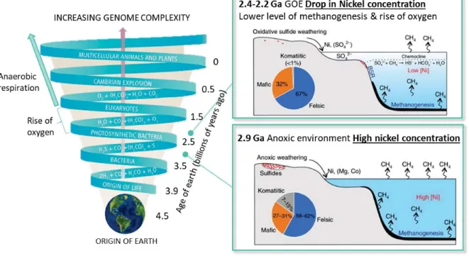
At the beginning of life, the environment is assumed to be electron rich, with an atmosphere abundant in gasses such as H2, CO and CO2 and hot oceans well-supplied in transition metals
as Fe2+ and Ni2+.1 In order to handle these special early living conditions, nickel most likely
featured as a key player in the catalyst development.2 The appearance of oxygen in the
atmosphere 2.4 billion years ago had a dramatic impact on metabolism evolution.3 As oxygen
conquered the ecosystem, many enzymatic reactions pathways, used by anoxic metabolism under reductive conditions, were replaced by aerobic ones. As possible evidence for this change, to date, nickel does not feature in any enzyme active site of higher eukaryotes, with the exception of plants.4,5
The reason why oxygen level increased in the atmosphere, namely the Great Oxidation Event (GOE), remains a mystery and different theories have been suggested. An interesting explanation focuses the attention on nickel and in its role as a metal co-factor in the metabolism of methanogenic archaea. The “nickel famine” theory has been proposed as a possible cause of methane collapse. It correlates the rise of oxygen in the atmosphere with a drastic reduction of nickel flux to archean oceans, a consequence of the cooling of upper-mantle temperatures and decreased eruption of nickel-rich ultramafic rocks, which would have starved the major oceanic microbial community: the methanogens. Their falling left the place to the proliferation of other microorganisms, especially oceanic cyanobacteria, leading to an increased production of O2 via photosynthesis. Such unbalance in the global
methanogen productivity would have caused a reduction of the methane atmospheric levels, contributing to the chain reaction that brought life as we know it. (Figure 1) 1,6
No matter what was the cause for oxygen takeover, the result was a segregation of the microorganisms based on H2 and/or CO oxidation and CO2 reduction pathways to anaerobic
niches, such as ocean’s bottom, animal’s digestive systems and volcanoic mud. In these habitats, they kept their intricate metal-based active sites, the core of the enzyme catalyst, which we are now trying to decrypt. 7
16
Figure 1. How the genomic and the metabolic pathways evolved during Earth’s history. How life started is still unknown but there is evidence of microbial life in the mid-Proterozoic. The exact moment of photosynthesis evolution is not of easy determination, however geochemical data shown that between 2.3-2.2 billion years ago the oxygen amount in the atmosphere was sufficient to form the ozone layer. The GOE event looks like the cause of the increased complexity in both genomes and metabolic pathways. The nickel famine theory is pictured in the boxes, showing a decrease of nickel and consequently methane in the atmosphere during the GOE. Adapted from ref 56.
To date, nine types of enzymes have been identified as reliant on nickel as a catalytic cofactor: [NiFe]-hydrogenase, urease, Ni-superoxide dismutase (NiSOD), [NiFe]-carbon monoxide dehydrogenase (CODH), acetyl-CoA decarbonylase/synthase (ACS), methyl-CoM reductase (MCR), glyoxylase I, acireductone dioxygenase and lactate racemase.8,9 These enzymes,
divided between hydrolases and redox ones, use nickel to catalyze essential reactions for the survival of the organisms, implying the need of a tight regulation of its intracellular concentration. In fact, as for other transition metals, nickel accumulation can be toxic for the cells, causing oxidative stress or replacing essential metals in metalloproteins. At the same time, the natural levels of nickel are generally low in the environment, generally in the nanomolar range with the exception of specific niches, and a constant and sufficient supply is
17
necessary to avoid any deficiency.3,10,11 In responding to this urge, specific proteins are
produced to control nickel homeostasis by transporting and/or storing the metal in the cell, assembling and delivering the metal-centers, modulating its import-efflux or regulating the expression of the other involved factors.12
In the following pages, the nine nickel-dependent enzymes are presented, with a special attention to the anaerobic gas-processing ones. The focus will then switch to the CODH activation mechanism and to the role played by its chaperone proteins in the nickel insertion.
19
1.1 Nickel enzymes
1.1.1 Urease
Urease, an enzyme found in a plethora of organisms including plants, fungi, algae, archaea and bacteria, catalyzes the hydrolysis of urea to produce ammonia and carbamate.12,11,13 The
latter decomposes spontaneously into ammonia and carbonic acid following the reaction: H2N-CO-NH2 + H2O H2N-COOH + NH3
H2N-COOH + H2O H2CO3 + NH3
Considering the profusion of the urea substrate, urease is involved in different biological processes and is an active player in the global nitrogen cycle.
The production of ammonia leads to an overall increase in pH, which can affect both agriculture and human health.13 In fact, urease is used as a virulence factor by many human
pathogens such as Helicobacter pylori, which exploit the increase in pH to colonize the otherwise too acidic stomach environment. In agriculture, the rise of pH causes a decrease of the soil fertility but it also plays a role in the global cycling of nitrogen compounds, used as fertilizers, implying that a right balance is essential to guarantee its positive effect.
Notably, it was the first crystallized enzyme, leading to to Sumner being aworded 1946 the Nobel Prize in chemistry, and it was the first evidence that nickel could be used as an enzymatic co-factor (Dixon, 1975).14,15 Therefore, many years have passed since its discovery
and significant steps have been made in order to understand its structure (49 different urease structures are deposited in the Protein Data Bank), catalytic mechanism, and the activation of this enzyme.16
From the structural point of view, all ureases possess several subunits that share similar tertiary structures. Most of the prokaryotic ureases contain three subunits α, ß and ɤ, respectively corresponding to UreA, UreB and UreC, while they differ in their quaternary structures.11 Considering as a model the urease from Sporosarcina pasteurii, its quaternary
structure is built on a trimer of trimers (α, β,ϒ)3 in which the catalytic center is contained in
the α subunit, making the total count of three active sites per enzyme.17 The α subunit
possesses a flexible helix-turn-helix motif, highly conserved among all ureases, highlighting its suspected importance for the catalytic process. The active site is a di-nuclear center, with the
20
two nickel ions spatially separated by 3.5-3.7 Å, bridged by a carbamylated lysine residue and coordinated by two histidines each. Water molecules fill the remaining coordination positions, yielding one pentacoordinated distorted square-pyramidal based geometry (Ni1) , while the second nickel (Ni2) is additionally bound to an aspartate resulting in a hexacoordinated distorted octahedral geometry.18 (Figure 2)
The assembly of this active site requires different partners and, in most of the bacterial systems, four accessory proteins play this role: UreD, UreF, UreG and UreE. The simplified version of urease maturation sees UreDFG creating a complex that binds to urease apo-enzyme to drive the lysine carbamylation, while UreE is the metallo-chaperone involved in nickel delivery.12 A focus on these accessory proteins will be present in the following section.
Figure 2. X-ray structure of S. pasteurii urease. The three different subunits are highlighted in green for the β subunit, in magenta for the ϒ subunit and in cyan for the α subunit which contains the active site (PDB:4CEU). The zoom shows the di-nuclear active site, the two-nickel ions are bridged by a carbamylated lysine (K220) and a water molecule, and three additional water molecules (W1, W2 and W3) are present. 18
21
1.1.2 Glyoxalase I
Glyoxalase I (Glx I), working in complex with glyoxalase II, is needed to transform the highly cytotoxic metabolic by-product methyglyoxal (CH3-CO-CHO) into non-toxic final ones. It
converts the hemimercaptal substrate (CH3-CO-C(OH)-SG,) formed by the reaction of
methylglyoxal with reduced glutathione (GSH), to S-D-lactoylglutathione (CH3-CH(OH)-CO-SG),
used in turn by Glx II as a substrate to re-generate reduced glutathione and non-toxic D-lactate (CH3-CH(OH)-COOH). 11
CH3-CO-CHO + GSH ↔ CH3-CO-C(OH)-SG Non enzymatic step.
CH3-CO-C(OH)-SG CH3-CH(OH)-CO-SG Detoxification pathway catalyzed by Glx I
CH3-CH(OH)-CO-SG + H2O CH3-CH(OH)-COOH + GSH Hydrolysis catalyzed by Glx II
Glx I from different organisms contains different metals and in Escherichia coli nickel features as the best performing one, maximizing its activity.19 E. coli Glx I can also use Co2+, Cd2+ and
Mn2+ as metal center, presenting a reduced reactivity, whereas Zn2+ substitution results in
enzyme inactivation.19 This change in reactivity is strictly related to the metal site
coordination, which moves form an octahedral geometry for Ni, Co, Cd and Mn to a trigonal bi-pyramidal geometry for Zn.19 The structure of E. coli Glx I is a homodimer, constituted by
two subunits, each of which contributes to the formation of the two active sites. Nickel coordination is composed of a histidine and a glutamic acid from each monomer and is completed by two water molecules. (Figure 3) The potential metallochaperone proteins involved in E. coli Glx I nickel insertion have not yet been identified and it is unclear if they are needed.
Figure 3. Structure of E. coli glyoxalase. The monomers constituting the homodimer are highlighted in green and magenta (PDB:1F9Z). The zoom shows the active site: the nickel ion (green) is coordinated by histidines (H8-56) and glutamic acids (E 56-112) while additional water ligands (red W1-W2) complete the geometry. 19
22
1.1.3 Acireductone Dioxygenase (ARD)
The methionine salvage pathway can be used to generate methionine from 5’-methylthioadenosine (MTA), produced from the S-adenosyl methionine during polyamine biosynthesis. During this pathway, 5’-methylthioadenosine is converted to an acireductone (1,2-dihydroxy-3-keto-5-methylthiopentene) which can be used as a substrate by the ARD enzyme to generate two different products with two different enzymatic reactions.9 One
reaction sees the conversion of 1,2-dihydroxy-3-keto-5-methylthiopentene into formate and 2-keto-4-methylthiobutanoate, then transaminated to methionine (reaction a). The other catalyzes an off-pathway shunt producing CO, 3-methylthiopropionate and formic acid (reaction b).
From the study of the Klebsiella oxytoca (formerly K. pneumoniae) system, it was discovered that the same enzyme catalyzes the two reactions, differing only in the nature of the coordinating metal.20 When iron is present in the binding site, the ARD produce a ketoacid
precursor that can be recycled back to methionine (reaction a). In contrast, when nickel or cobalt is present, ARD catalyzes a non-productive pathway of oxidation, which converts the acireductone into formate, carbon monoxide and methylthiobutyric acid (reaction b). The ARD from this organism is the only currently known enzyme, in the as-isolated state from the native organism, able to perform these two reactions depending on the nature of the metal ion bound to the protein active site.21 The two forms of KoARD have the same sequence, but they
23
different conformations.20 The structure of the Ni-KoARD was solved by NMR22 (Figure 4) and
the coordination of its active site is proposed via a homology model with the X-ray structure of ARD from mammalian Mus musculus (PDB:1VR3) (MmARD).21 The ligand characterization,
by spectroscopic and mutagenesis studies, suggested an octahedral coordination geometry for both metals involving three histidines and one glutamic acid as possible donors supplemented by two additional waters. Interestingly, from NMR spectroscopy studies on KoARD, it was shown that iron binding to the protein provokes an increase in order at the N-terminus and disorder at the C-N-terminus, while nickel binding generates the opposite effect. This re-arrangement in the structure, induced by the different metals, can lead to the active site changing its conformation, triggering a different selectivity to catalyze the two distinct reactions. 23
Advances have been recently reported on understanding the reaction mechanism of the Human Fe-ARD, also thanks to its crystal structure which is supposed to be the native form under normal conditions. In Eukaryotes it is unclear if Ni-ARD has any biological role.24
However, structural information regarding the substrate-ARD complex is poor and it is not evident how microorganisms can choose to insert different metal ions into the same catalytic site. To date, the nickel insertion pathway is unknown.
Figure 4. Structure of K. oxytoca Ni-ADR. The NMR structure is depicted in green (PDB:1ZRR). The zoom shows the active site, the 6-coordinate nickel ion (green) is proposed to be coordinated by three histidines (H98-140-96), one glutamic acid (E102) and two waters. 22
24
1.1.4 Lactate racemase (LarA)
Lactate racemase (LarA), the last characterized nickel enzyme, catalyzes the interconversion between L- and D-lactic acid isomers, as depicted below.
The role of this enzyme is still under discussion and it most likely depends on the nature of the species where is located. The latest information about LarA comes from Lactobacillus
plantarum, which uses L-lactate as a substrate to produce D-lactate.25 This bacteria does not
need lactate to grow, thus the role of LarA has been linked to the rescue pathway for D-lactate production in case of stress (as in the presence of antibiotics).
In L. plantarum, two different operons (larABCDE and larR(MN)QO) have been identified as necessary for the enzyme activity. The enzyme itself is encoded by LarA, LarBCE play a role in the metal trafficking and co-factor biosynthesis, LarD is an aquaporin-like protein and Lar(MN)QO are related to the ABC-transporter for high-affinity nickel uptake. LarR is the transcriptional regulator. L-lactate positively regulates the system, while D-lactate represses the gene expression.26
The structure of this enzyme has been solved, disclosing the peculiar environment of its nickel coordination. In the active site is present a nicotinic acid mononucleotide derivate with two added thiocarboxylate groups, one of which forms a thioamide with lysine 184, namely nickel-pincer nucleotide (NPN) coenzyme (Figure 5). Pincer complexes are not new in chemistry but notably this is the first example of such a molecule in a biological system. The nickel is bound in an almost planar arrangement to the NPN pincer co-factor (using the thioamide sulfur atom, the pyridinium C4 carbon and the thioacid sulfur atom) and the coordination is completed by
a histidine. The stable Ni-C bond in the resting/stable state of this enzyme is surprising; in other Ni-containing enzymes Ni-C bonds have only been observed as reaction intermediates.13 This coordination is essential to guarantee the stability of the metal in the
25
conformations due to a distortion of the NPN pincer coordination and displacement of the His200. From the X-ray structure, it was possible to identify the presence of these two different states in the same asymmetric unit. The closed conformation shields the nickel from the solvent thanks to the close proximity between the N- and C- termini (Figure 5). The open conformation results in a loss of the nickel ion due to the easy accessibility of the active site to the solvent. This implies that: the access of the substrate to the catalytic center has to be rapid to avoid enzyme deactivation and that LarA is most likely able to dynamically switch between open and closed conformations.25The NPN biosynthesis is attributed to the proteins
LarBCE. LarB is responsible for the production of the pre co-factor (P2CMN) from NAD, while LarE catalyzes a two sulfur transfer reactions forming the P2TMN version. LarC is the nickel insertase, which provides the nickel to generate the active form of the pincer cofactor that will bind to LarA (Figure 5).27,2823
Figure 5. Structure of L. plantarum LarA. The nickel containing subunit is highlighted in green (PDB:5HUQ). The zoom shows the active site, the coordinate nickel ion (green) is coordinated by the NPN cofactor ( with C in gray, O in red, N in blue, S in yellow, and P in orange) and His200. The biosynthetic pathway for the NPN cofactor production starts with LarB forming P2CMN from NaAD. LarE catalyzes a sacrificial sulfur transfer reaction to synthesize P2TMN from P2CMN. LarC inserts nickel into P2TMN, forming the NPN cofactor. Adapted from 23.
26
1.1.5 Superoxide Dismutase (NiSOD)
The superoxide dismutase (SOD) role is to protect biological systems from the oxidative damage caused by superoxide radical anions (O2-˙), a byproduct of oxygen metabolism.29 It
catalyzes the dismutation of superoxide anions to hydrogen peroxide and oxygen following the reaction: M(n+1) + O 2-* Mn+ + O2 Mn+ + O 2-* + 2H+ M(n+1) + H2O2 2 O2-* + 2H+ H2O2 + O2
This enzyme can possess different metals in the active site. The most common versions are constituted either by a di-nuclear Cu-Zn center or by a mononuclear Fe or Mn, while the Ni enzyme has some unique characteristics. In fact, whereas the other transition metals can cycle between oxidized and reduced states using the natural midpoint-potential ofO2-˙
disprotonation (0.36 V), Ni cannot catalyze superoxide disproportionation in aqueous solution. The reason why is related to the calculated reduction potential for the species Ni3+/+2,
which is +2.26 V, outside the range required for SOD catalysis.29 It was, therefore, a surprise
when NiSOD was detected and isolated in some Streptomyces bacteria and cyanobacterium. The adaptations required to use this metal as catalytic center (lowering the potential of the Ni3+/+2 couple by over 2 V) resulted in a unique evolution of this enzyme, which has no
sequence homology with the other SODs. Nevertheless, NiSOD redox potential is similar to those of the other SODs and it catalyzes the reaction with the predicted Ni3+/+2 couple, as
confirmed by EPR studies. The answer to how this enzyme is able to lower the Ni3+/+2 potential
must resides within its structure.13 The quaternary structure of NiSOD is exclusive in the SOD
family. It forms an homo-hexamer where all the nickel sites are independent from each other (Figure 6). Interestingly, a posttranslational modification is needed to remove 14 amino acids at the N-terminus in order to promote nickel binding.30 The metal binding site, is in fact,
located by the N-terminus, known as a “Ni-hook”, which coordinates the Ni using the cysteines in position 2 (thiolate and amidate) and 6 (thiolate) and the histidine in position 1 (amine and imidazole). The nickel coordination changes depending on the oxidation state of the metal, switching from a pyramidal geometry for the oxidized state (Ni3+) to a square planar geometry
27
and imidazole ligands are critical for the reduction in potential, the role of the mixed amine-amidate coordination is the subject of different studies and it has been suggested as a possible player in maintaining the right redox potential for the nickel and to protect the active site towards oxygen.32 In fact, in order to access Ni3+, the only way discovered so far in nature is
the use of cysteine thiolate coordination. However, this group can easily be oxidized by the products of the SOD reaction, H2O2 and O2, so cysteines are excluded from other metal SODs
active sites. Interestingly, studies showed that the Cys ligands of NiSOD are unreactive with H2O2 as long as the rate of catalysis is maintained, that the enzyme can be reduced by H2O2
and that the excess peroxide can be removed without loss of activity.33
Concerning the possible accessory proteins involved in the nickel insertion, a putative nickel chelatase (CbiXhp) has been identified. The behavior of this enzyme is fascinating and many efforts have been devoted to understanding its chemical stability, also thanks to peptide-based models.30 Nevertheless, further studies need to be done to understand the maturation
and nickel insertion in the NiSOD production pathway.
Figure 6. Structure of S. seoulensis NiSOD. The homo-hexameric structure shows each subunit in a different color. The active site descripts the oxidized state (Ni3+) coordinated via the amino terminal amine, a backbone
28
1.1.6 Methyl Coenzyme M Reductase (MCR)
Methyl Coenzyme M Reductase (MCR), catalyzing the reduction of methyl-S-coenzyme M by coenzyme B to methane and disulfide bridging CoB and CoM, plays an important role in the global carbon cycle.13
CH3-S-CoM (methyl-S-coenzyme M) + CoB-SH (coenzyme B) CH4 + CoB-S-S-CoM
(heterodisulfide)
The methane formation is the last step of methanogenic archaea metabolism. These strictly anaerobic microorganisms, able to grow on acetate, methanol, formate, CO2 and H2, are
responsible for more than 90 % of the methane present on earth. The only other known source of biological methane comes from the methylphosphonates metabolism (4%),34 so MCR
methanogenic archaea catalysis ia its main producer.35
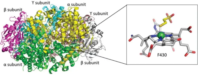
The crystal structure of MCR from Methanothermobacter marburgensis reveals a heterohexamer constituted of three different subunits (α2β2ϒ2) and a Ni-containing co-factor,
namely tetrapyrrole F430, in each α subunit (Figure 7).36
Figure 7. Structure of M. marburgensis MCR. The dimer is shown with each subunit (αβϒ)2 in different color.
Each active site (Ni in green) contains coenzyme F430 with an axial Gln147 ligand, and an additional axially coordinated CoM-SH in this particular structure.36
29
Figure 8. On the left is the representation of the hydrophobic channel in MCR. The F430 cofactor is present at the bottom of it and the Ni is coordinated by the tetrapyrrole ring and Gln147. On the right is the structure of the F430 cofactor. Adapted from 13.
The name of the co-factor derives from its absorption band, precisely at 430 nm, due to the Ni+2 oxidation state. The enzyme also needs a Ni+1 state in order to be active, implying that a
tight control of the metal redox state is required for its functionality. The F430 is not covalently
bound in the active site, but it is deeply buried at the end of a hydrophobic channel that shelters the substrates/products. This arrangement is to prevent solvent accessibility, which could cause a change in the oxidation states. The substrate methyl-S-coenzyme M must enter in this channel before the second substrate, coenzyme B, whose arrival completely impedes the access to the binding site. The ring of the F430 co-factor chelates the nickel ion and its
coordination is completed via an axial glutamine 147 on the opposite side of the ring form the channel.36 The conformational arrangement is shown in Figure 8.
Nickel is present as an inorganic metal complex. The presence of a specific nickel chelatase (CfbA) to promote the insertion of Ni in the sirohydrochlorin has been proven both in vitro and in vivo.37 From this starting intermediate, other Cfb enzymes (CfbEDCB) are needed to
30
1.1.7 [Ni-Fe] hydrogenase
Hydrogenase catalyzes the oxidation of hydrogen into protons and electrons in a reversible way, following the reaction:
H2 ↔ 2H++ 2e-
Hydrogen can have a double role, either as an energy source for the system or as a final product to remove a possible excess of reducing equivalents. 7
Different kinds of hydrogenases are found in nature, present in diverse organisms such as archaea, bacteria and some specific eukaryotes. It is possible to divide them, depending on the metal ions used in their active site, into three not phylogenetically related groups: [Fe-Fe] hydrogenase, [Fe] hydrogenase and [Ni-Fe] hydrogenase.40 [NiFe]-hydrogenase, found in
bacteria and archaea, are divided into four main groups depending on their function: Group 1- Membrane-associated H2 uptake hydrogenases.
Group 2- Soluble uptake hydrogenases and sensory hydrogenases.
Group 3- Heteromultimeric cytoplasmic hydrogenases harboring a reducible cofactor (F420 or NAD(P)).
Group 4- Energy-converting hydrogenases. This group includes the CO-induced hydrogenases involved in our research scope reaction (the WGS reaction see session 1.4). Ech hydrogenase from Methanosarcina barkeri and hydrogenase 3 from E. coli can be considered as the most studied examples of this class.
A common feature of all [Ni-Fe] hydrogenases is their general assembly in at least two subunits (Figure 9).9 A large catalytic subunit hosts a Ni-Fe active site and a small subunit hosts the
Fe-S clusters needed for electron transfer. The number of Fe-Fe-S clusters differs for each organism. Various structures of this enzyme have been solved, unveiling the presence of a di-nuclear active site composed of a nickel ion coordinated to cysteines of two CxxC motifs, of which one of each pair is used to bridge to the iron ion.12 The Fe coordination is completed by one CO
and two CN- ligands. Between Ni and Fe, a third labile binding position is present for substrate
31
Figure 9. Structure of D. fructosivorans [Ni-Fe]-hydrogenase. The large subunit is shown in green, the small one in magenta. The Fe-S clusters in the small subunit are shown in spheres and the [Ni-Fe]-cluster is highlighted in the active site. (x=labile position for substrate binding)
A common feature among all the structures is the highly buried position of the active site, which suggests the presence of a hydrophobic tunnel for dihydrogen access, as well as proton pathways.43 The biosynthesis of [Ni-Fe] hydrogenase is a well-studied process and different
proteins have been identified as responsible for the biogenesis of such a complex enzyme.44
Their role is described in section 1.2.2.
In E. coli, the hycBCDEFG operon encodes for the hydrogenase 3, responsible for hydrogen production from formate thanks to its coupling with formate dehydrogenase-H (fdhF), in the formate hydrogenlyase complex (FHL).45 The hycABCDEFGH operon stands for the large (HycE)
and small (HycG) subunits, characteristic of [NiFe]-hydrogenases, plus two additional hydrophilic subunits (HycB and HycF) and two inner membrane subunits (HycC and HycD).44 The HycA is the negative transcriptional regulator of the formate regulon, while HycH is predicted to have an accessory function. (Figure 10)
32
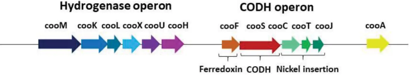
Figure 10. Comparison of the subunits encoded by the hyc operon from E. coli and the cooMKLXUH operon from
R. rubrum. Proteins without sequence similarity are shown in white. Adapted from 43.
In hydrogenogenic carboxydotrophs, such as our study model Rhodospirillum rubrum, the hydrogenase is enclosed by the CO-induced operon cooMKLXUH, present upstream of the CODH one.46 CooM and CooK (corresponding to the previously described HycC and HycD in E. coli) are two integral membrane-bound proteins, CooX (corresponding to E. coli HycF) is
described as a Fe4S4 ferredoxin-like protein and CooU has no homologue in E. coli hydrogenase
3. CooH and CooL encode for the large and small subunit, respectively, and they corresponds to HycE and HycG in E. coli hydrogenase 3 (Figure 10).
33
1.1.8 Acetyl-coenzyme A synthase/ Carbon Monoxide Dehydrogenase
Carbon monoxide dehydrogenase (CODH) can be present either as a monofunctional enzyme, as in the case for R. rubrum (discussed in the 1.1.9 session), or as a bifunctional complex with the acetyl-S-CoA synthase (ACS, ACDS in methanogens). The ACS/CODH complex catalyses the synthesis of acetyl-S-CoA from coenzyme A (CoA-SH) condensed with CO (derived from CO2
reduction catalyzed by CODH) and a methyl group (derived from a corrinoid/ Fe–S protein Co(III)-FeSP) as described in the reaction:
CO2 + 2H+ + 2e- CO + H2O CODH
CO + CoA-SH + CH3 -Co(III)-FeSP ↔ CH3C(O)-S-CoA + Co(I)-FeSP ACS
This bi-functional enzyme plays an important role in the Wood-Ljungdahl pathway, one of the six carbon fixation pathways known on Earth. It consists of two branches, defined as the methyl and the carbonyl pathways. The methyl branch differs between acetogenic bacteria and methanogenic archaea to convert the CO2 into the methyl group. The carbonyl branch is
common in acetogens and methanogens and consists of the conversion of carbon dioxide into carbon monoxide, catalyzed by the CODH. In a second step, the CO coming from the carbonyl
branch produced by the CODH is combined to the methyl coming from the methyl branch and
CoA by the Ni-ACS enzyme to form acetyl-CoA, the most common carbon source in biology (Figure 11).47
Figure 11. Wood–Ljungdahl carbon fixation pathway. The carbonyl branch is in pink, the generalized methyl branch used in acetogenic bacteria is in light blue and the generalized methyl branch used in methanogenic archaea is in green. Adapted from 48.
34
As expected, bifunctional CODH/ACS contains two distinct active sites, one present in the CODH subunit and one in the ACS subunit. The two active sites are connected by hydrophobic channels to allow the CO produced from the CODH to reach the ACS active site, regulating in this way the import-export of the substrates.49
In the model organism Moorella thermoacetica the ACS/CODH is an α2β2 tetrameric protein
formed by two CODH β-subunits in the center and an ACS α-subunit on each extremity (Figure 12). The ACS subunit contains three different domains: the N-terminal domain, responsible for the interaction of ACS with CODH, the central domain and the C-terminal domain, which contains the active site, called the A-cluster. This site consist of a [4Fe-4S] cluster bridged to a proximal Ni (Nip) via a cysteine thiolate (Cys 509). Nip is in turn bridged to a distal Ni (Nid) via
two additional cysteine thiolates (Cys 595 and 597) and its coordination is completed in a square planar geometry by an unidentified exogenous ligand, in labile position for substrate and/or intermediates binding. Historically, it was supposed that the Nip center could be
physiologically substituted by others metals, resulting in a Cu-Ni and Zn-Ni forms. However, this hypothesis has been ruled out and the scientific community now agrees that the A-cluster is formed by a [4Fe-4S]- Nid- Nip center (Figure 12).49,13 Concerning the Nid, its coordination is
held by the two thiolates and the two backbone amide groups (one from Gly and one from Cys) of a Cys-Gly-Cys motif, in a stable S2N2 square planar geometry (Figure 12). Nid does not
have redox activity and it will likely remain a diamagnetic Ni2+ through catalysis. Ni
p, on the
other hand, presents different oxidation states during the catalysis, indicating its catalytic substrate-converting role. Even though the structure of the A-cluster has been solved, the catalytic mechanism is still under debate.49,50,51
Consensus has been reach on the role of Nip as responsible for binding the CO and the methyl
group, followed by methyl migration/CO insertion to result in a Nip-acetyl bound group, to
finish with CoA substitution to produce acetyl-CoA. The role of the distant nickel Nid+2 has been
proposed either as a supporting ligand, to stabilize the proximal Nip in a low-valence redox
state, or as a hemilabile site responsible of modulating of the active conformation, facilitating its catalytic cycle.49,52 The inorganic models made for this center all point towards a neither
35
Figure 12. Structure of M. thermoacetica CODH/ACS. The tetramer α2β2 subunits are shown in different colors,
the CODH homodimer β2 is in green and cyan, the ACS α2 subunits are in pink and yellow. The metalloclusters
are shown in spheres. The active site, A-cluster, is in the zoom region where the [4Fe-4S]- Nid- Nip center in ACS
36
1.1.9 Monofunctional Carbon Monoxide Dehydrogenase (CODH)
Carbon monoxide dehydrogenase catalyzes the reversible oxidation of carbon monoxide to carbon dioxide following the reaction:
CO + H2O ↔ CO2 + 2H+ + 2e-
Two primary classes of CODH have been identified in nature, differing from each other in their active sites metal composition. One consist in a Cu-Mo-Fe-S cluster, which catalyzes a irreversible reaction and is found in aerobic microorganisms. The other class possesses a Ni-containing active site and is recurrent in anaerobic bacteria and archaea. In both case, CODH plays an essential role in the global carbon cycle.47 As already mentioned in the previous
section, Ni-CODH can occur as monofunctional or bifunctional enzyme and the following part will focus on monofunctional Ni-containing CODHs.54
To decrypt this enzyme, the majority of the information in the hand of the scientific community comes mainly from two organisms: R. rubrum (RrCODH) and Carboxydothermus
hydrogenoformans (ChCODH).
In C. hydrogenoformans five different genes encoding CODHs are present:
ChCODH-I, which is coupled to a [Ni-Fe] hydrogenase to catalyze the so-called water
gas shift (WGS) reaction (CO + H2O ↔ CO2 + 2H+ + 2e- (CODH) and 2e-+ 2H+ H2
(hydrogenase)). This process is vital for the organism considering that it is a strictly anaerobic thermophilic bacterium. In fact, it can grow using CO as a sole carbon and energy source thanks to this WGS reaction55;
ChCODH-II, involved in NADH generation56;
ChCODH-III, coupled with the ACS enzyme to perform the acetyl-CoA biosynthesis
pathway;
ChCODH-IV, probably involved in oxidative stress response as a mechanism of oxygen
detoxification57;
ChCODH-V, its role has not yet been identified. Recently, its structure has been
solved*, showing a [Fe4S3O2] cluster, which can be converted into a [Fe4S3] cluster by
37
are responsible for nitrosative stress protection during anaerobic growth in the presence of nitrate/nitrite and they harbor a 4Fe-2S-2O iron-sulfur-oxygen cluster.58
Considering than CODH-V cannot convert CO or CO2, it may represent the evolutionary
link between HCPs and CODHs. (*results published in 2015 in Fesseler J. thesis from
Dobbek group)
In R. rubrum only one CODH (RrCODH) is present and, as for the ChCODH-I, it is involved in the WGS reaction, allowing the bacteria to use CO as a sole energy source.46
Crystal structures of ChCODH-I/II/IV/V and RrCODH have been solved, displaying an overall similar structure.59,60 They are homodimers, of which each monomer is composed of three
different domains known as the N-terminal helical domain, the central domain and the C-terminal α/β (Rossmann-like) domains (Figure 13). Each homodimer contains five metalloclusters of three different natures: two unique [Ni-4Fe-4S] clusters (namely C-cluster), two [4Fe-4S] clusters (namely B-cluster) and one intermolecular [4Fe-4S] cluster (namely D-cluster). The C-cluster is a distorted Ni-3Fe-4S cubane coordinated to an additional forth iron (Feu), unique in biology.59 The global arrangement places the clusters in a C-B-D-B*-C*
position, where the bridging D-cluster sits between the two monomers (Figure 14). The reaction is catalyzed at the C-cluster, the buried active site in the protein, which oxidizes CO and releases CO2, protons and electrons. The electrons, in order to reach the CODH’s partners,
need to be transferred to the protein surface. The B- cluster is the most likely candidate to fulfil this role, due to their proximity to the active site and the protein surface.
Figure 13. Structure of R. rubrum CODH. The monomers are shown in magenta and green. The metalloclusters are shown as spheres.
38
Figure 14. Structure of R. rubrum CODH. The cluster rearrangement is highlighted on the structure of the homodimer. The active site, C-cluster, is in the zoom region where the metals are shown in spheres (green for nickel, orange for iron and yellow for sulfur). The reported distances between Fe atoms of individual clusters are in Ångstrom.
Conversely, the role of the D-cluster remains to be elucidated. It might be involved in the electron transfer with the B- cluster, but its low reducing potential (-530 mV) could be an indication of a more ‘oxygen-protective’ role.61
1.9.1 Reaction Mechanism
Different approaches have been used to elucidate the CODH reaction mechanism, allowing the identification of the main steps involved and the postulation of the following working model.
Prior to the structural characterization of the C-cluster, four different redox states were proposed via spectroscopic studies: Cox, Cred1, Cint and Cred2, all differing from their
reduction/oxidation state by one electron. Cox is catalytically inactive and EPR silent. Cred1 binds
CO and analogues such as cyanide. This state is paramagnetic (S=1/2) and displays an EPR signal with g-values of 2.03, 1.88, and 1.71. Cint is the two-electron reduced state, also EPR
silent. Cred2 is the three-electron reduced state able to bind CO2 and analogues such as cyanate
and presents an EPR signature with g-values of 1.97, 1.87 and 1.75.62 All together these data
39
and when it is oxidized to CO2 the C-cluster gains two more electrons and is in the Cred2 form.
The cluster is then re-oxidized via electron transfer to the B-cluster passing through a Cint state
(Figure 15). Where the two additional electrons are located in the Cred2 state is still not clear,
with the hypotheses of a possible Ni0 state, or hydride-bound Ni(II) or a dative Ni-Fe bond
under debate.63,64,65
The difference in redox potential among these states is a key feature in order to activate correctly the enzyme. It has been shown that the Cox/Cred1 redox potential is -200 mV and
Cred1/Cred2 has a redox potential of -530 mV, matching almost perfectly with that of CO2/CO
which is -558 mV at pH 7.66
Figure 15. Redox states of CODH active site. The electronic states are reported with their relative spin states and mid-point potentials. The inhibitors of each state are also reported in red. CN- inhibits CO oxidation by binding
to the Cred1 but it does not inhibit CO2 reduction. NCO- inhibits CO2 reduction binding to the Cred2 but it does not
inhibit COoxidation. Adapted from 67. (nBIC : n-Butyl Isocyanide a CO analogue, CN-: cyanide a CO analogue, NCO
40
Figure 16. Proposed reaction mechanism of [NiFe]-CODH. Adapted from 43.
The determination of diverse crystal structures, in complex with the CO-CO2 substrates,
inhibitors or analogues (CN- and NCO-), was also a great support to allow the proposition of
the following mechanistic model (Figure 16):
Step 1. CO binds to Ni(II) and the H2O to the Fe(II) ion of the C-cluster (Cred1) ;
Step 2. Fe-H2O deprotonates, resulting in a nucleophilic hydroxide species. OH- nucleophilic
attacks of the CO results in a Ni-Fe bridged carboxylic group;
Step 3. Second deprotonation. The Ni-Fe now is bridged by a COO- intermediate;
Step 4. Release of the CO2 and two electron cluster reduction (Cred2);
Step 5. Electrons are transferred to the B-cluster and the C-cluster goes back to its Cred1 state.
Even though the mechanism appears clear, different points still need to be elucidated. For example during the nucleophilic attack (Figure 16 step 2) a “carbon shift” should occur in order to place the carbon closer to the resulting hydroxide, which is otherwise too far away.68
Moreover, the Cint state role during the CO2 release process is still not clear. Electron transfer
via the Ni center is too rapid to visualize reduced Ni states. To obtain more insights into this last step, structures of ChCODH-II bound to CO2 and cyanate (NCO− a CO2- analogue) have
been recently determined to 1.03 and 1.06 Å resolution, using anaerobically poised crystals at -600 mV to mimic the Cred2 state.69 Both structures showed a μ2, ƞ2 bent bridging CO2/NCO
-41
coordination between the Ni and the Fe (step 2), with two unexpected identical C-O bonds lengths (1.32 and 1.3 Å). CO2 is stabilized by H-bonding with His93 and Lys563. Interestingly,
the certitude of the CO2 binding geometry, ensured by the high atomic resolution, allowed the
identification of the O-C-O angle as 1170, different from the previously reported value of 1300.
This smaller value suggests that the CO2 activation is mediated by a particularly nucleophilic
Ni species that pushes electrons into CO2, making it appear as a two-electron-reduced
carboxylate (Ni-COO−) waiting to be protonated, in favor of a 2e- transfer process. However,
the existence of a transient CO2˙- intermediate radical cannot be completely excluded.
In addition, access of CO/CO2 and proton transfer must be allowed between the buried active
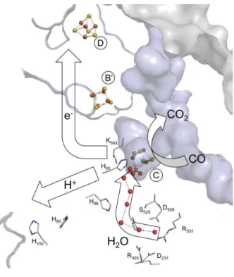
site and the solvent. Hydrophobic channels have been detected between the Ni ion and the protein surface as a possible gateway for the gas molecules.67 A polar channel would be
needed for the proton transfer and several water molecules linked by histidine residues were observed concerning the Ni to the solvent in all of the CODH structures (Figure 17). Moreover, a recent IR-X-ray-in silico combined study of the cyanide (CN-)/CODH complex showed that
the proton channel between the C-cluster and the solution phase is strictly directional and dependent on the oxidation state of the cluster.70,71
43
1.2 Nickel in the active site: where is it coming from?
Considering all the peculiar nickel active sites catalyzing the so far exposed reactions, the natural question that should arise from the reader should be: from where is this nickel coming from? How does it appear in the active site?
First, it is important to consider that this metal needs to be imported by the cell from the natural environment, passing through the cytoplasmic membrane. A tightly regulated import mechanism is essential and many organisms have developed high-affinity nickel transport systems to fulfil this role. In the case of gram-negative bacteria, nickel needs to first cross the outer membrane. Small molecules and ions can pass through this membrane by passive diffusion via non specific transmembrane porins but when their concentration is low, an energized import is required. Lately, the existence of dedicated transporters, namely TonB-dependent transporter (TBDT), has been postulated also in the outer membrane for nickel.72
Once in the periplasm, two main classes of import systems are found in the cytoplasmic membrane: the ABC-type importers and the Ni/Co permeases.12 (NikABCDE transporter in E. coli is probably the best studied ABC-type importer, whereas HoxN in Cupriavidus necator is a good representative for nickel/cobalt permeases).
Once nickel is present in the cell, its trafficking needs to be tightly controlled to avoid the presence of free toxic metal and it has to be correctly delivered and incorporated into the desired enzyme active sites. These operations often require an intricate team of accessory proteins, working in concert for the biosynthesis of these complex metalloclusters enzymes. Understanding the steps necessary for the cluster assembly and insertion into the enzyme, deciphering the maturation machinery involved, are some of the main challenges in the metallo-enzymology field. In the next session, the identified accessory proteins involved in the Ni-enzyme maturation pathways for urease, hydrogenase and CODH are discussed. A special attention will be devoted to nickel chaperones, a class of proteins able to reversibly bind the metal, transport it in the cell and deliver it to the apo-enzyme metallocenter.
44
1.2.1 Urease maturation
As already mentioned, urease is first produced in the apo-form which then needs to be activated through two main steps: lysine carbamylation and nickel insertion into the active site. Considering the buried position of this site in the apo-protein, the occurrence of a protein folding modification is predicted to allow nickel insertion.
To carry out these steps and fully complete urease biosynthesis, four accessory proteins are usually needed: UreD (UreH in H. pylori), UreF, UreG and UreE. From the studies of Klebsiella
aerogenes urease, it was evidenced that the apo-enzyme can be partially activated upon nickel
and CO2 exposure but its activity is enhanced when it is co-expressed with UreD.13 This
suggests the need for the co-expression of structural and accessory proteins to fully activate the enzyme.
Figure 18. Proposed urease maturation process. A) Classical proposed mechanism B) the new proposed one. Adapted from 13.
45
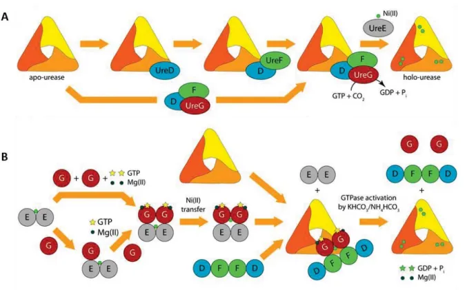
The classical model, currently proposed for the maturation mechanism even though the role of each single protein are not fully understood to date, is the following:
UreD, the first protein to interact with the apo-urease, binds to the enzyme and acts as a protein scaffold to promote the interactions of the other partners.73 UreF interacts with UreD
possible provoking a conformational change of the enzyme around the active site and enhancing Ni accessibility.74 At this point, UreG binds to UreF and promotes the lysine
carbamylation, which relies upon GTP hydrolysis by UreG, defining this protein as the energy driving force. Finally, the urease-UreDFG complex interacts with UreE, responsible for the nickel insertion and completes the enzyme maturation. (Figure 18A) This model derives from the ability to isolate the apo-urease-UreDFG complex and for this reason it is proposed to be the functional unit.73 However, no crystal structure is available for urease in complex with any
of these accessory proteins. The complex UreHFG from H. pylori has been recently crystallized, showing a dimer of heterotrimers.75 The interaction between UreG and UreE in H. pylori has
been proven both in vivo (yeast two-hybrid analysis76,77 and immunoprecipitation essay77) and
in vitro (calorimetry and NMR spectroscopy78).
In view of the most recent results, a second model was postulated for the maturation of urease active site (Figure 18B).79 In this scenario, a first interaction between Ni-UreE and UreG
is proposed, forming a complex Ni-(UreEG)2. The complex (UreDF)2 will then interact with
Ni-(UreEG)2, competing for the Ni-UreG2 binding. Nickel is delivered to the apo-urease by this last
the complex Ni-(UreDFG)2.79 A more detailed explanation about this last mechanism is
46
UreE
Historically, the role of this protein as nickel chaperone during urease maturation was suggested offer examining the UreE sequences from K. aerogenes and Proteus mirabilis. Both sequences are, in fact, histidine rich in their C-terminal domains, well known as potential nickel binding motifs. However, the His-rich terminal region is not an essential prerequisite for the UreE family, considering that some members from different organisms have been identified lacking this, such as the one from H. pylori (HpUreE) (Figure 19). Moreover, their role in the enzyme maturation differs for each organism, from being essential for several
Helicobacter species or substitutable by nickel addition in the growth medium for others.80
Figure 19. Top part: UreE from K. aerogenes. The full sequence is reported, the brackets show the truncated version in the case of the mutant H144*. The crystal structure (PDB:1GMW) comes from this truncated version. Bottom part: UreE from H. pylori. The proteins are both in a dimeric conformation and present a nickel-binding site at the interface between the two monomers. In the sequences, the histidines are colored in red, the cysteines in green and the amino acids involved in nickel binding are underlined.

![Figure 16. Proposed reaction mechanism of [NiFe]-CODH. Adapted from 43 .](https://thumb-eu.123doks.com/thumbv2/123doknet/12875975.369651/45.892.144.714.108.473/figure-proposed-reaction-mechanism-nife-codh-adapted.webp)
![Figure 22. Over simplified allegedly maturation mechanism for [Ni-Fe]-hydrogenase. Adapted from 88](https://thumb-eu.123doks.com/thumbv2/123doknet/12875975.369651/55.892.123.714.651.956/figure-simplified-allegedly-maturation-mechanism-ni-hydrogenase-adapted.webp)