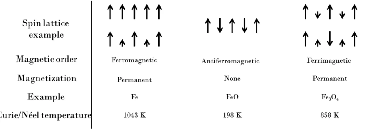
Magnetosomes used as biogenic MRI contrast agent for molecular imaging of glioblastoma model
Texte intégral
Figure

Documents relatifs
Measurement of skin blood flux on different skin sites of the forearm (numbered 1 to 5): unheated, heated to 36°C, to 39°C, to 42°C and to 44°C, respectively, using laser
A magnetic hyperthermia treatment protocol is proposed, in which nanoparticles yielding an amount of 25 of iron per mm 3 of tumor are administered and exposed to 11 to 15
ischemic stroke in transgenic CX3CR1-GFP/+ mice using three complementary imaging modalities: MRI, 30.. intravital two-photon microscopy and phase contrast imaging with
thrombosis rat model was acquired and analyzed for 10 min after injection of MBs. 50 frames after the destructive pulse were selected as a reference, and the
Right, bar plot showing the TRAM-34-sensitive KCa3.1 current density in control and IPTG-induced cells *Po0.05; (c) control and IPTG-treated cell migration toward CXCL12 or EGF
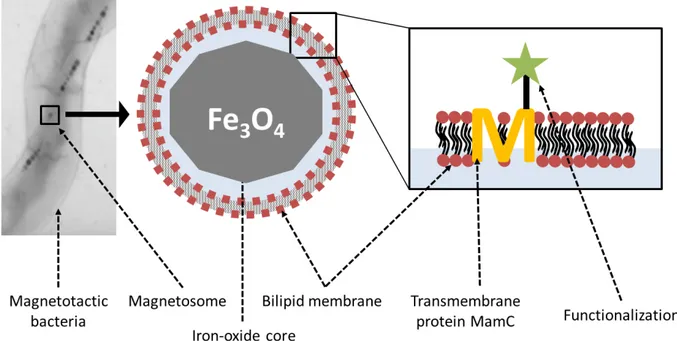
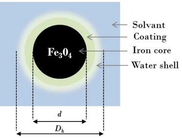
The apparent co-localization observed in the fluorescence images between cell membranes and magnetosomes (Fig. 2) could result either from nanoparticles bound at the cell surface
In order to get insight into solution equilibria of the PC2A-BP 2– ligand and its complexes formed with biologically relevant metal ions (Mg(II), Ca(II), Mn(II),
Dynamic contrast enhanced (DCE) images showed considerably higher signal enhance- ment in the kidney medulla and cortex after Gd 3 L injection than after GdDOTA injection at
