HAL Id: tel-01397186
https://tel.archives-ouvertes.fr/tel-01397186
Submitted on 15 Nov 2016
HAL is a multi-disciplinary open access
archive for the deposit and dissemination of sci-entific research documents, whether they are pub-lished or not. The documents may come from teaching and research institutions in France or abroad, or from public or private research centers.
L’archive ouverte pluridisciplinaire HAL, est destinée au dépôt et à la diffusion de documents scientifiques de niveau recherche, publiés ou non, émanant des établissements d’enseignement et de recherche français ou étrangers, des laboratoires publics ou privés.
Magnetosomes used as biogenic MRI contrast agent for
molecular imaging of glioblastoma model
Marianne Boucher
To cite this version:
Marianne Boucher. Magnetosomes used as biogenic MRI contrast agent for molecular imaging of glioblastoma model. Medical Physics [physics.med-ph]. Université Paris Saclay (COmUE), 2016. English. �NNT : 2016SACLS234�. �tel-01397186�
NTT: 2016SACLS234
Electrical Optical Bio-physics and Engineering Doctoral School, ED575 Unit´e d’Imagerie par R´esonance Magn´etique et de Spectroscopie
(NeuroSpin, I2BM, CEA Saclay)
Magnetosomes used as biogenic MRI contrast agent for
molecular imaging of glioblastoma model
Marianne Boucher
Ph.D. thesis defended at Gif-sur-Yvette the 30
thof September, 2016,
in front of the following jury:
Dr. Florence Gazeau DR, Paris Diderot Referee
Dr. Damien Faivre Group Leader, Max Plank Institute missing Referee
Dr. Nicolas Ginet CR1, Universit´e Aix Marseille Examiner
Dr. Nicolas Tsapis DR, Universit´e Paris Saclay - Paris Sud Examiner Dr. Olivier Cl´ement Pr, Hopital Europ´een Georges Pompidou PARCC President Dr. S´ebastien M´eriaux Ing´enieur Chercheur, Universit´e Paris Saclay - CEA Supervisor Dr. Michel Bottlaender Ing´enieur Chercheur, Universit´e Paris Saclay - CEA PhD Director
Magnetosomes used as biogenic MRI contrast agent for
molecular imaging of glioblastoma model
Benoˆıt Desforˆet
`
Remerciements
Je voudrais tout d’abord remercier les membres de mon jury, rapporteurs et examinateurs, pour m’avoir fait l’honneur d’accepter d’´evaluer mon travail de th`ese. Merci pour leur retours constructifs et pour m’avoir permis de d´efendre ce travail.
Je souhaiterai ensuite remercier S´ebastien M´eriaux, mon encadrant de th`ese, pour m’avoir prise en stage de Master 2 et pour m’avoir gard´e en th`ese, sans qui cette exp´erience n’aurait pas ´
et´e possible. Merci pour m’avoir mis le pied `a l’´etrier au d´ebut et pour l’ind´ependance laiss´ee `
a la fin.
Je remercie ´egalement Michel Bottlaender pour avoir accept´e d’ˆetre mon directeur de th`ese jusqu’au bout.
Ce manuscrit n’aurait pu ˆetre si fourni sans le travail de Fran¸coise Geffroy et Erwan Selingue ! Pour toute votre aide et votre bonne humeur merci ! Fran¸coise, chaque hiver, je penserai `a toi aux premi`eres chutes de temp´erature en reniflant le fameux m´elange Men-the/Ravinstara/Niaouli/Eucalyptus ! Erwan je n’oublierai pas nos interminables discussions `a l’aimant, ni tes mythiques blagues !
Le projet collaboratif MEFISTO m’a ´egalement permis de d´ecouvrir un autre labo : le LBC ! Un immense merci `a David Pignol, Nicolas Ginet, Christopher Lef`evre, Sandra Pr´everal, G´ eral-dine Adryanczyk et Michel P´ean pour m’avoir fait d´ecouvrir le monde magique des bact´eries magn´etotactiques ! D´esol´ee de vous avoir fait avoir des sueurs froides `a l’annonce des quantit´es de magn´etosomes n´ecessaires `a mes manips ! J’esp`ere en avoir fait bon usage. Merci pour m’avoir permis d’aller au congr`es MTB, ou j’ai appris ´enorm´ement de choses passionnantes sur les bact´eries magn´etotactiques et les magn´etosomes.
Merci `a Alice Gaudin pour m’avoir aid´e `a mettre en place mes manips au tout d´ebut de cette th`ese.
Merci `a Laurent Bellanger du LI2D pour la production de l’anticorps qui a permis de d´ eblo-quer pas mal de manips.
Merci `a Fabrice Richard pour les superbes images de microscopie ´electronique `a transmis-sion sur cellules et tissus biologiques.
J’ai aussi eu l’occasion de participer `a d’autres projets, notamment le projet TROPICA. Je VII
souhaiterai remercier Didier Boquet et Amaury Herbert pour m’avoir fait d´ecouvrir un autre mod`ele tumoral. Merci `a Laurence Motte, Yoann Lalatonne et Sophie Richard pour m’avoir permis de travailler sur des nanoparticules, synth´etis´ees par voie chimique, de tailles, de coatings et de fonctionnalisations diff´erentes. Merci pour les caract´erisations physico-chimique des mag-n´etosomes. J’ai beaucoup appris lors de nos discussions et j’ai pu ainsi diversifier mon travail. Sophie, belle rencontre au d´etour d’une paillasse. . . qui se poursuit maintenant r´eguli`erement autour d’une bonne table, je voudrais te remercier pour ton soutien, en particulier pendant ma derni`ere ann´ee de th`ese.
Cette th`ese s’est d´eroul´ee `a NeuroSpin, au laboratoire UNIRS. Merci `a Benoˆıt Larrat (pour m’avoir pr´esent´e `a S´ebastien, et pour mettre une bonne ambiance au labo) `a Fawzi Boumezbeur, Luisa Ciobanu et Cyril Poupon pour d’int´eressantes discussions. Merci aux post-doc que j’ai eu la chance de rencontrer : Elodie P. (pour m’avoir chaperonn´e au 11,7, entre autres), David (pour nos discussions anim´ees), Allegra (for your unfailing joy of life), Hermes (for your com-ment on the defense) et Elodie G. (pour nos interminables discussions).
J’ai eu la chance de prendre part `a l’animation des NeuroBreakfast puis du SpinDating, ´
ev`enements permettant des ´echanges scientifiques informels au sein du labo. Merci Benoˆıt (Larrat) pour m’avoir incit´e y prendre part. Thanks Valentina for your amazing energy. Merci Gabrielle pour cette aventure et pour m’avoir soutenu, surtout lors de la r´edaction. Merci Benoˆıt M. et Carole pour cette superbe organisation. Enfin, un grand merci `a Arthur, pour cette aventure et pour avoir rˆev´e avec moi d’un autre monde de la recherche ! Courage pour la suite !
Enfin, merci `a tous ceux qui ont ´egalement partag´e l’openspace 1025A ou le premier ´etage! Merci `a R´emi (pour d´etester le RER autant que moi et pour avoir fait passer le temps en par-lant gastronomie et campagne, entre autre), Alfredo (pour ton optimisme), Guillaume (pour nos discussions photos), Solenne (tu me tiendra au courant ?), Justine (pour ton enthousiasme), Matthieu (pour ta bonne humeur), Andrea, Pavel, Jacques et Khieu.
Malgr´e le temps pass´e au labo et dans les transports. . . j’ai quand mˆeme pu profiter du soutien de mes amis ! Merci `a tous mes amis de l’ESPCI (7 ans d´ej`a), en th`ese comme moi (ou pas), avec qui j’ai pu partager les bons et les mauvais moments ! Merci `a Benoˆıt G., Marine, Joachim, Antoine, Nicolas et R´emi. Merci aux PICMiens, Jean-Christophe, Romain et Marion, et Paul N. pour m’avoir accueilli parmi vous et pour m’avoir montr´e une autre vision de la th`ese. Merci `a mes amies et t´emoins : B´en´edicte, Celia, Claire, Fanny et Pascaline pour votre soutien sans faille et pour tous ces supers moments pass´es et futurs.
Pour finir, je souhaiterai remercier le plus chaleureusement possible ma famille ! Merci `a mes beaux-fr`eres Basile et Benoˆıt, ma belle-sœur Fanny et mes beau parents Laurent et Evelyne, pour tous les diners/ week-end/ vacances pass´e(e)s ensembles qui m’ont permis de m’´echapper du quotidien. Merci aussi pour votre ´ecoute et votre soutien. Merci `a mes fr`eres, Paul et Laurent, et mes belles-sœurs, Liliane et Laurence, pour avoir ´et´e ma famille « m´etropolitaine » pendant ces 10 derni`eres ann´ees loin de maman ! Merci pour m’avoir ´epaul´e dans toutes les ´etapes de mes ´etudes, pr´epa, ´ecole et th`ese ! Merci pour m’avoir offert votre pr´esence `a la soutenance ainsi que le super week-end qui a suivi. Merci pour avoir imagin´e ce petit magn´ e-tosome en 3D. Merci `a mes neveux Marius et Isaac, qui sont n´es pendant cette th`ese et qui m’ont apport´e tant de joie ! Recevoir vos adorables petites bouilles en photo ou vid´eo sur
Georges, cette th`ese m’a tenue ´eloign´ee de la R´eunion, et je compte bien rapidement reprendre le rythme de mes retours annuels « `a la maison ». Un immense merci maman, pour ˆetre une source infinie d’inspiration ! Merci pour m’avoir permis de rˆever et pour avoir tout fait pour me donner les outils pour les r´ealiser. Merci aussi pour avoir fait le d´eplacement depuis la R´eunion pour m’´epauler pendant la soutenance.
Je clˆoture ces remerciements, et par la mˆeme occasion la r´edaction de ce manuscrit, en remerciant mon meilleur ami et mon plus grand soutien. Bastien, depuis ces 7 derni`eres ann´ees, on en a v´ecu des aventures ! Et cette th`ese en fait partie ! Comme toujours tu m’as donn´e l’audace d’oser, la folie d’essayer, la force de tenir bon, les raisons de ne pas abandonner et le recul de voir ce qui est beau en chaque chose. Merci Bastien, pour ˆetre mon amour et maintenant mon mari.
Abstract
This work takes place in the context of molecular imaging, which aims at tailoring new diag-nostic and therapeutic tools to the needs of the individual patient, by revealing molecular or cellular biomarkers of interest in the less invasive manner. Among the several areas of research conducted in this field, the emphasis is here on MR-based molecular imaging using engineered iron-oxide contrast agent.
This PhD thesis focuses on the study of a new class of contrast agent for MRI, the mag-netosomes, which are natural iron-oxide vesicles produced by magnetotactic bacteria. These bacteria synthesize such magnetic vesicles and order them like a nano-compass in order to faci-litate their navigation in sediments. Magnetosomes are thus awarded with interesting magnetic properties: iron-oxide core around 50 nm, mono-crystalline, single magnetic domain and high saturation magnetization. Furthermore, several bacterial strains exist in nature, each of them producing magnetosomes with specific size, shape and chemical composition. Finally, mag-netosomes are naturally coated with a lipid bilayer which content is genetically determined. Lately, researchers have unraveled the proteins content of magnetosome membranes, opening the way to produce functionalized magnetosomes thanks to a fusion between the gene coding for a protein of interest and the gene coding for an abundant protein at magnetosome membrane. This PhD work aims at investigating a new alternative path using magnetotactic bacteria to tackle the production of high efficiency MR-based molecular imaging probes. The engineering and production of magnetosomes, carried out by our collaborators from the LBC, the Labora-toire de Bio´energ´etique Cellulaire (LBC, CEA Cadarache), are presented and discussed. We firstly characterize wild type magnetosomes as contrast agent for high field MRI, and compare them with chemically produced iron-oxide nanoparticles. Our results confirm that these mag-netosomes present very promising contrasting properties in vitro, and therefore they can be used in vivo as efficient blood pool agent, for vasculature imaging of mouse brain after intra-venous injection. Afterward, engineered magnetosomes are tested in a molecular imaging study of a U87 mouse model of glioblastoma. Knowing that ανβ3 integrins over-expressed by tumor
cells can be actively targeted by RGD peptide, AMB-1 magnetotactic bacteria are genetically modified to produce RGD functionalized magnetosomes. After verifying their good affinity properties for U87 tumor cells in vitro, we demonstrate in vivo this specific affinity with MRI acquisitions on a U87 mouse model. Finally, these in vivo results are cross-validated with post mortem acquisition of histological data.
Keywords: Molecular imaging, MRI, iron-oxide nanoparticle, contrast agent, magneto-somes, glioblastoma.
R´esum´e court en Fran¸cais
Ces travaux de th`ese s’inscrivent dans le contexte de l’imagerie mol´eculaire, qui vise `a d´ evelop-per de nouveaux outils diagnostiques et th´erapeutiques adapt´es aux besoins de chaque patient, grˆace `a l’imagerie non invasive de biomarqueurs cellulaires ou mol´eculaires. Parmi les nom-breuses disciplines contribuant `a ce domaine de recherche, l’accent est mis ici sur l’imagerie par r´esonance magn´etique (IRM) mol´eculaire `a l’aide de nanoparticules d’oxyde de fer innovantes. Cette th`ese porte sur l’´etude d’une nouvelle classe d’agent de contraste pour l’IRM, les magn´etosomes, qui sont des v´esicules d’oxyde de fer produites naturellement par des bact´eries magn´etotactiques. Ces bact´eries les synth´etisent et les alignent pour qu’elles agissent comme une boussole afin de faciliter leurs d´eplacements dans les s´ediments. Les magn´etosomes pr´ esen-tent des propri´et´es magn´etiques tr`es int´eressantes : un cœur d’oxyde de fer d’environ 50 nm de diam`etre, mono-cristallin, mono-domaine magn´etique et avec une forte magn´etisation `a satura-tion. De plus, de nombreuses souches bact´eriennes existent dans la nature, chacune d’entre elles produisant des magn´etosomes de taille, forme et composition chimique sp´ecifiques. Enfin, les magn´etosomes sont naturellement entour´es par une membrane bilipidique dont la composition est d´etermin´ee g´en´etiquement. R´ecemment, le contenu prot´eique de la membrane des magn´ e-tosomes a ´et´e mis `a jour, ouvrant la voie `a la production de magn´etosomes fonctionnalis´es par ing´enierie g´en´etique.
L’ing´enierie et la production des magn´etosomes, r´ealis´ees le Laboratoire de Bio´energ´etique Cellulaire (LBC, CEA Cadarache), sont pr´esent´ees et discut´ees. Des magn´etosomes sauvages sont caract´eris´es en tant qu’agents de contraste pour l’IRM, et nos r´esultats confirment qu’ils pr´esentent des propri´et´es contrastantes int´eressantes pour l’IRM et qu’ils permettent de r´ealiser l’imagerie de la vascularisation c´er´ebrale chez la souris apr`es une injection intraveineuse. En-suite, l’´etude de faisabilit´e de la production d’un agent de contraste IRM fonctionnalis´e en une seule ´etape, `a l’aide de bact´eries magn´etotactiques, est r´ealis´ee grˆace des exp´eriences d’imagerie mol´eculaire sur un mod`ele U87 de souris porteuse de glioblastome. Sachant que les cellules tumorales sur-expriment les int´egrines ανβ3, et que ces derni`eres peuvent ˆetre cibl´ees
active-ment par le peptide RGD, des bact´eries magn´etotactiques sont g´en´etiquement modifi´ees pour produire des magn´etosomes exprimant le peptide RGD `a leur membrane. Apr`es avoir v´erifi´e in vitro leur propri´et´es d’affinit´e pour les cellules tumorales U87, nous d´emontrons in vivo cette affinit´e sp´ecifique `a l’aide d’acquisitions IRM sur un mod`ele souris U87. Finalement, ces r´ esul-tats in vivo sont cross-valid´es par l’acquisition post mortem de donn´ees histologiques.
Mots cl´es : Imagerie mol´eculaire, IRM, nanoparticule d’oxyde de fer, agent de contraste, magn´etosomes, glioblastome.
R´esum´e en fran¸cais
Contexte de la th`
ese
Les magn´
etosomes
Cette th`ese s’inscrit dans le cadre du projet MEFISTO, portant sur l’´etude d’agents de contraste originaux et novateurs : les magn´etosomes. Il s’agit de cristaux d’oxyde de fer entour´es d’une membrane bilipidique, synth´etis´es par des bact´eries magn´etotactiques. Le travail d’optimisation de la production des magn´etosomes est men´e par le Laboratoire de Bio´energ´etique Cellulaire (CEA Cadarache), partenaire du projet MEFISTO, tandis que l’´etude de leur utilisation comme agent de contraste pour l’Imagerie par R´esonance Magn´etique (IRM) est men´ee dans le cadre de cette th`ese. Dans le domaine de l’IRM, les suspensions de nanocristaux d’oxyde de fer sont principalement utilis´ees comme agents de contraste ”n´egatifs”, car leur pr´esence se manifeste par des hypo-signaux sur les images pond´er´ees T2 et T2∗. L’effet de tels agents est souvent d’autant
plus fort que le champ magn´etique est ´elev´e, c’est pourquoi il a ´et´e choisi de les ´etudier avec des scanners pr´ecliniques `a haut champ magn´etique (7 T , 11, 7 T et 17, 2 T ).
Les magn´etosomes pr´esentent comme int´etˆet d’ˆetre int´egralement produits biologiquement, ce qui induit un contrˆole optimis´e sur la min´eralisation du fer, ainsi qu’une forme, une taille et une composition chimique du cristal propres `a la souche bact´erienne choisie. De plus, une fois extraits des bact´eries, les magn´etosomes sont directement utilisables in vivo car la membrane lipidique qui entoure leur cœur d’oxyde de fer assure leur biocompatibilit´e. Cette voie de production de nanocristaux d’oxyde de fer pour des applications biom´edicales semble donc particuli`erement int´eressante pour pallier la multiplication d’´etapes n´ecessaires `a la r´ealisation de tels objets par voie de synth`ese chimique.
Les agents de contraste pour l’imagerie mol´
eculaire par IRM
Les magn´etosomes sont pressentis pour ˆetre d’excellents agents de contraste pour l’imagerie mol´eculaire par IRM. En effet, l’imagerie mol´eculaire vise `a fournir de nouveaux outils aux techniques conventionnelles d’imagerie m´edicale, afin de permettre l’acquisition d’images de marqueurs mol´eculaires ou cellulaires, en plus des donn´ees anatomiques et fonctionnelles d´ej`a disponibles en routine clinique. De telles informations pourraient permettre de d´evelopper une m´edecine dite ”personnalis´ee” o`u le traitement s’adapte aux biomarqueurs exprim´es par chaque patient. Pour cela, il est n´ecessaire de mettre en place de nouveaux agents de contraste poss´ e-dant la particularit´e d’ˆetre affins pour le biomarqueur d’int´erˆet, tout en offrant une excellente sensibilit´e de d´etection. L’ajout d’une fonctionnalisation aux agents de contraste d´ej`a existants
requiert habituellement une ´etape de plus dans le processus de fabrication, qui est d´ej`a constitu´e de la synth`ese du cœur d’oxyde de fer contrastant et de la couche de mat´eriaux biocompatibles qui l’entoure.
Les magn´etosomes pr´esentent l’avantage de pouvoir ˆetre fonctionnalis´es en modifiant le g´enome de la bact´erie magn´etotactique qui les produit, afin d’induire l’expression d’un peptide d’interˆet directement `a leur membrane. Ces nano-objets biologiques peuvent donc ˆetre produits en une ´etape unique, la culture des bact´eries. Par ailleurs, les magn´etosomes pr´esentent une ex-cellente cristallinit´e et une taille plus grande comparativement aux autres agents de contraste `a base d’oxyde de fer. Ces caract´eristiques pourraient conf´erer aux magn´etosomes des propri´et´es contrastantes tr`es ´elev´ees, et donc une tr`es bonne sensibilit´e de d´etection, confirmant leur in-t´erˆet en imagerie mol´eculaire par IRM.
Caract´
erisation des magn´
etosomes et de nanoparticules d’oxyde
fer synth´
etis´
ees par voie chimique pour l’IRM
Caract´
erisation de la relaxivit´
e transverse
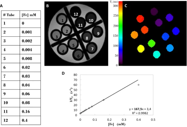
L’efficacit´e contrastante en IRM de ces nanocristaux biog´eniques a ´et´e caract´eris´ee par la mesure des relaxivit´es, param`etres qui quantifient l’influence d’un compos´e sur les temps de relaxation des protons environnants en fonction de sa concentration. Les nanoparticules d’oxyde de fer ont principalement une influence sur les temps de relaxation transversale T2,
comparative-ment `a leur influence sur les temps de relaxation longitudinale T1. Il a donc ´et´e entrepris de
mesurer principalement la relaxivit´e transverse pour caract´eriser l’efficacit´e contrastante des nanoparticules d’oxyde de fer. Les r´esultats de cette ´etude de relaxom´etrie ont montr´e que les magn´etosomes pr´esentent une excellente relaxivit´e transverse, autour de 500 mM−1s−1 `a 7 T , alors que des agents commerciaux ont une relaxivit´e transverse environ 3 `a 5 fois plus faible.
Caract´
erisation des propri´
et´
es contrastantes in vivo
Ces r´esultats prometteurs nous ont amen´e `a tester les suspensions de magn´etosomes in vivo comme agents de contraste capables de r´ev´eler la vascularisation c´er´ebrale de la souris, grˆace au r´ehaussement du signal vasculaire apr`es leur injection intra-veineuse. En effet, les mag-n´etosomes circulent uniquement dans les vaisseaux c´er´ebraux car ils ne peuvent pas acc´eder au parenchyme c´er´ebral `a cause de la barri`ere h´emato-enc´ephalique, ce qui en fait de bons agents de contraste pour l’imagerie du compartiment vasculaire c´er´ebral. Les angiogrammes c´er´ebraux ainsi acquis constituent un bon compl´ement de la mesure de relaxivit´e, qui caract´erise l’efficacit´e de l’agent in vitro, par la mesure du contraste induit par les magn´etosomes dans un environnement biologique in vivo.
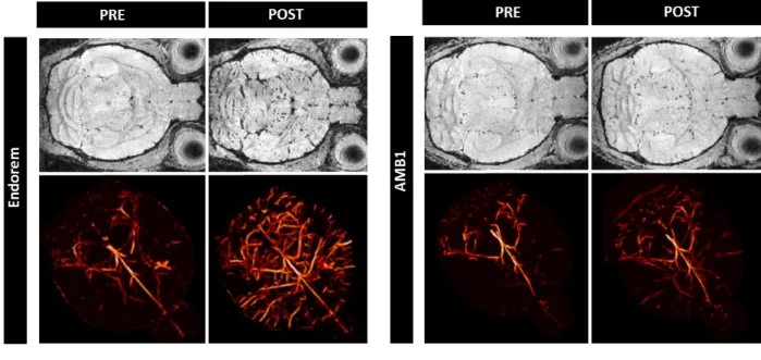
Cette ´etude a d´emontr´e que les magn´etosomes induisent un contraste significatif entre les vaisseaux et le parenchyme c´er´ebral, mˆeme si les produits commerciaux semblent plus efficaces dans les conditions exp´erimentales utilis´ees (voir Figure 1). Ce r´esultat illustre l’importance de tester in vivo les agents de contraste afin de tenir compte, dans leur ´evaluation, de leur comportement en milieu biologique et pas uniquement de leur potentiel contrastant. En effet,
cial peut ˆetre expliqu´ee par une moins bonne biocompatibilit´e des magn´etosomes. La couche biocompatible pr´esente autour du cœur d’oxyde de fer d´emontre ici toute son importance. En effet, ´etant plus ´epaisse dans les particules commerciales, elle peut conduire `a ´ecranter les perturbations locales du champ magn´etique induites par les nanoparticules sur les protons environnants, ce qui a pour effet de diminuer l’effet contrastant. Cependant, cette couche bio-compatible permet aussi de diminuer la clairance et ainsi allonger les temps de circulation dans la vascularisation, ce qui peut induire un meilleur contraste in vivo. Par ailleurs, les m´ethodes d´evelopp´ees pour ´etudier les magn´etosomes en tant qu’agent de contraste IRM ont ´et´e ´ egale-ment utilis´ees afin de caract´eriser des agents de contraste synth´etis´es par voie chimique par des collaborateurs du laboratoire CSPBAT. Cela a permis notamment d’´etudier plus finement l’impact de la taille du cœur d’oxyde de fer sur le potentiel contrastant in vitro, ainsi que l’influence de la nature la couche biocompatible sur le contraste in vivo.
Fig. 1 – Comparaison des propri´et´es contrastantes in vivo de l’Endorem (gauche,R 17 T ) et des magn´etosomes AMB1 (droite, 11.7 T ). La premi`ere et la troisi`eme colonnes pr´esentent les images avant l’injection alors que la deuxi`eme et quatri`eme, celles apr`es l’injection. Les images FLASH (premi`ere ligne) permettent d’appr´ecier le contraste entre le parenchyme c´er´ebral et les vaisseaux, `a partir duquel les angiogrammes 3D (deuxi`eme ligne) sont calcul´es.
Imagerie mol´
eculaire d’un biomarqueur de tumeur c´
er´
ebrale avec
des magn´
etosomes fonctionnalis´
es g´
en´
etiquement
Choix du biomarqueur : les int´
egrines α
νβ
3Un mod`ele de tumeur c´er´ebrale a ´et´e choisi afin d’´etudier la possibilit´e d’utiliser des magn´ eto-somes fonctionnalis´es g´en´etiquement pour des applications d’imagerie mol´eculaire par IRM. Le d´eveloppement anarchique des cellules tumorales n´ecessite un apport croissant en oxyg`ene et en
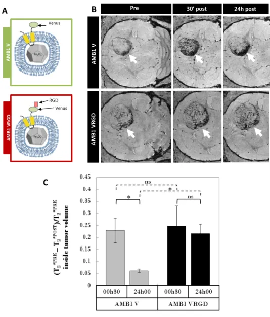
Fig. 2 – D´emonstration in vivo de l’affinit´e des magn´etosomes RGD pour le mod`ele animal de tumeur c´er´ebrale U87. A/ Sch´ema des deux magn´etosomes, fonctionnalis´e RGD (AMB1 VRGD) et contrˆole (AMB1 V), produits pour l’exp´erience. B/ Images FLASH repr´esentatives du contraste induit dans les tumeurs par l’injection des mag-n´etosomes fonctionnalis´es RGD ou contrˆoles `a diff´erents temps apr`es l’administration. C/ Quantification de l’augmentation du contraste dans la tumeur induit par l’injection des magn´etosomes.
nutriments : lorsque la tumeur atteint une certaine taille, elle va former son propre r´eseau vascu-laire pour poursuivre sa croissance. Ce ph´enom`ene, appel´e n´eo-angiog´en`ese, est souvent utilis´e comme biomarqueur pour le diagnostic tumoral, mais aussi comme cible pour des strat´egies th´erapeutiques anti-tumorales, puisque sans ces n´eo-vaisseaux, la tumeur ne peut pas continuer sa progression. Il a ´et´e d´emontr´e que les cellules n´eo-angiog´eniques tumorales sur-expriment l’int´egrine ανβ3 et qu’il ´etait possible de cibler ces int´egrines avec le peptide RGD (Arginine
-Glycine - Acide aspartique), notamment avec un peptide RGD li´e `a une nanoparticule d’oxyde de fer. Des magn´etosomes porteurs du peptide RGD `a leur membrane ont ´et´e produits afin de r´ealiser une premi`ere preuve de concept de l’utilisation de magn´etosomes fonctionnalis´es
A).
´
Etudes in vitro et in vivo d’affinit´
e des magn´
etosomes fonctionnalis´
es pour
les cellules tumorales U87
Une premi`ere ´etude a permis de montrer in vitro que les magn´etosomes fonctionnalis´es RGD se fixent sp´ecifiquement sur la lign´ee cellulaire tumorale U87 en culture, cellules de glioblastome humain connues pour sur-exprimer les int´egrines ανβ3. De plus, il a ´et´e d´emontr´e que cette
fixation sp´ecifique induit une internalisation des magn´etosomes au sein des cellules tumorales en culture. La deuxi`eme ´etude s’est int´eress´ee `a l’affinit´e in vivo de ces magn´etosomes fonction-nalis´es pour leur cible sur un mod`ele souris de tumeur c´er´ebrale. Pour cela, des souris nudes (immunod´eprim´ees) ont re¸cu une injection intra-c´er´ebrale de cellules U87. Au bout d’une quin-zaine de jours, elles ´etaient porteuses de tumeurs de quelques millim`etres de diam`etre (autour de 3 mm) `a l’emplacement de l’implantation. Une injection intra-veineuse de magn´etosomes, fonctionnalis´es ou contrˆoles, a alors ´et´e administr´ee `a ces souris, avant de les imager par une s´erie de s´equences IRM sensibles au fer, `a diff´erents temps avant et apr`es l’injection (s´equences T2∗, pond´er´ee et param´etrique). Cette ´etude a permis de d´emontrer que les magn´etosomes fonc-tionnalis´es avec le peptide RGD sont toujours pr´esents en quantit´e significative dans la tumeur 24 h apr`es l’injection intra-veineuse, contrairement aux magn´etosomes contrˆoles qui semblent ˆ
etre ´elimin´es plus vite (voir Figure 2 B et C). De plus, ces r´esultats IRM ont ´et´e valid´es par histologie, grˆace `a des marquages immunohistochimiques sur des coupes de cerveaux de souris, permettant de r´ev´eler la pr´esence des magn´etosomes dans les vaisseaux tumoraux post mortem. Ainsi, un des objectifs principaux du projet MEFISTO est atteint : il est possible de faire fabriquer `a une bact´erie un agent de contraste fonctionnalis´e pour r´ealiser de l’imagerie mol´eculaire in vivo par IRM de tumeurs c´er´ebrales.
Optimisation de l’administration d’agents de contraste
fonction-nalis´
es pour am´
eliorer leur accumulation dans une tumeur c´
er´
e-brale
´
Etude de l’accumulation des magn´
etosomes RGD dans les cellules U87 in
vitro
Partant de l’hypoth`ese que la clairance observ´ee in vivo est le facteur limitant de l’accumulation des magn´etosomes RGD dans les tumeurs c´er´ebrales, il a ´et´e entrepris d’optimiser le protocole d’administration des magn´etosomes fonctionnalis´es, afin de maximiser la dose de fer restant dans la tumeur apr`es leur administration intra-veineuse. Tout d’abord, il a ´et´e v´erifi´e in vitro, sur les cellules tumorales U87 en culture, que l’augmentation du temps d’incubation ou de la dose de magn´etosomes RGD induit une augmentation de la quantit´e de magn´etosomes accu-mul´es dans les cellules. L’accumulation de nanoparticules fonctionnalis´ees par un peptide RGD dans une cellule tumorale exprimant des int´egrines ανβ3 peut s’expliquer par le recyclage des
int´egrines : en effet, ces prot´eines transmembranaires, en se liant aux magn´etosomes RGD, permettent leur internalisation dans les cellules U87, puis elles sont `a nouveau pr´esent´ees `a la
membrane cellulaire et peuvent donc interagir avec de nouveaux magn´etosomes RGD.
Multi-injection intra-veineuse de magn´
etosomes RGD in vivo
Le parall`ele in vivo de l’exp´erience pr´ec´edente implique de ralentir la clairance effectu´ee par le syst`eme r´eticulo-endoth´elial de l’animal, pour allonger le temps de circulation des magn´etosomes dans les vaisseaux tumoraux. Il a donc ´et´e propos´e de r´ealiser plusieurs injections de doses plus faibles, afin de d´eterminer si cette augmentation du temps de contact entre l’agent de contraste et sa cible menait ´egalement in vivo `a l’augmentation de la charge en fer dans la tumeur. Les r´esultats des exp´eriences, comparant une injection de 200 µmol[F e]/kgBW `a deux
injections de 100 µmol[F e]/kgBW espac´ees de 6 h, semblent montrer une accumulation un peu
plus importante des magn´etosomes RGD dans la tumeur pour l’injection double. Toutefois, la variabilit´e de la mesure ne permet pas d’extraire une diff´erence significative entre les groupes, ce qui peut ˆetre expliqu´e par l’inclusion d’animaux porteurs de tumeurs de taille assez diff´erente (entre 2 et 3 mm). Cependant, cette exp´erience illustre l’importance du choix du protocole d’administration de l’agent de contraste, et ouvre la discussion sur des m´ethodes plus proches de l’infusion intra-veineuse par exemple, afin d’optimiser le contraste obtenu.
Conclusion
Ce travail de th`ese, aux fronti`eres de la physique, de la biologie et de l’imagerie m´edicale, a permis de mettre en ´evidence qu’un nouveau moyen de produire des agents de contraste pour l’IRM ´etait possible, tirant profit de bact´eries pr´esentes dans la nature. De plus, ces nouveaux agents de contraste peuvent ˆetre fonctionnalis´es pour des applications d’imagerie mol´eculaire, ce qui a ´et´e d´emontr´e ici sur un mod`ele de tumeur c´er´ebrale. Ces travaux pourraient ´egalement ouvrir la voie vers une nouvelle m´ethode d’hyperthermie magn´etique pour induire une r´ egres-sion tumorale, consistant `a injecter des magn´etosomes affins pour la zone tumorale par voie intra-veineuse, ce qui ferait des magn´etosomes fonctionnalis´es un potentiel agent th´eranostique contre les tumeurs c´er´ebrales.
Par ailleurs, les m´ethodes d´evelopp´ees au cours de cette th`ese, de la standardisation de la mesure des relaxivit´es, `a l’acquisition d’angiogrammes c´er´ebraux par injection d’agent de contraste, ou encore la recherche de co-localisation de marquages sur les images d’histologie, sont des outils qui seront `a nouveau utilis´es par de futurs projets d’imagerie mol´eculaire.
Contents
1 Introduction 1
1.1 Molecular imaging combining iron-oxide nanoparticles with MRI acquisitions . . 2
1.2 Targeting ανβ3 integrins for brain tumor molecular imaging. . . 3 1.3 The MEFISTO project . . . 5
1.4 Outline of this manuscript . . . 6
References . . . 9
2 Iron-oxide nanoparticles for diagnostic and therapeutic applications 15
2.1 Why iron nanoparticles? . . . 16
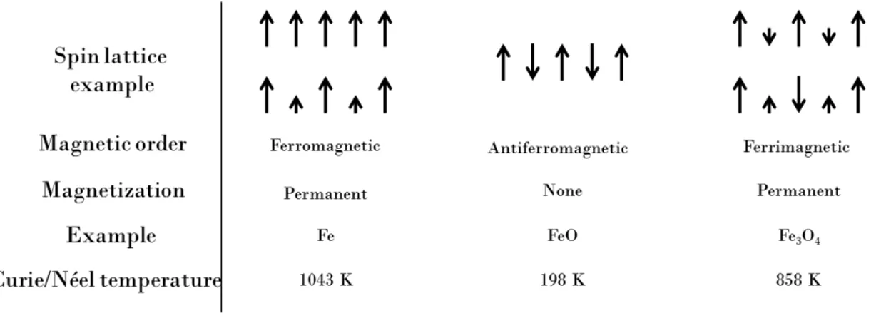
2.1.1 Physical origin of magnetism. . . 16
2.1.2 Magnetism of nanoparticles . . . 18
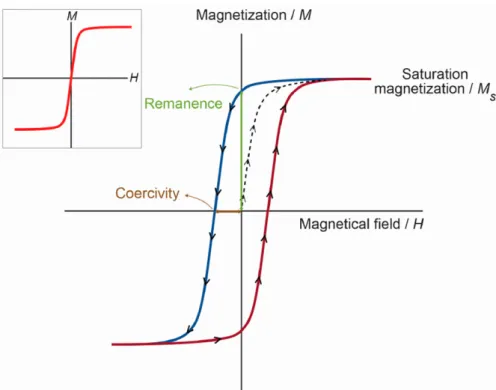
2.1.3 The interest of IONP in MRI . . . 20
2.1.4 Relaxation models for IONP . . . 22
2.2 How to optimize IONPs contrasting properties? . . . 25
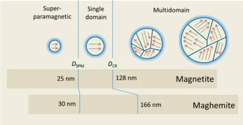
2.2.1 Superparamagnetic iron core optimization . . . 26
2.2.2 Coating optimization . . . 29
2.3 Interest of magnetsosomes as MRI contrast agent . . . 31
2.3.1 Magnetotactic bacteria and magnetosomes . . . 31
2.3.2 Magnetosomes physico-chemical properties . . . 34
2.4 Other biomedical applications of IONP . . . 36
2.4.1 IONP labeled cells . . . 37
2.4.2 Magnetic hyperthermia . . . 40
References . . . 42
3 Magnetosomes and other chemical IONPs used as MRI blood pool contrast agents 49
3.1 Physico-chemical properties of magnetosomes . . . 50
3.1.1 Iron core characterization . . . 50
3.1.2 Membrane characterization . . . 52
3.1.3 Benefits and drawbacks of magnetosomes . . . 54
3.2 MRI contrasting properties of magnetosomes and chemical IONPs . . . 55
3.2.1 Chemical IONPs transverse relaxivities . . . 56
3.2.2 Size effect in chemical IONPs transverse relaxivities . . . 58
3.2.3 Magnetosomes transverse relaxivities . . . 60
3.3 Brain angiography of healthy mouse with contrast-enhanced MRI . . . 63
3.3.1 In vivo contrasting properties of chemical IONPs . . . 64
3.3.2 In vivo contrasting properties of magnetosomes . . . 66 XXIII
XXIV CONTENTS
3.4 Brain tumor contrast-enhanced MRI in a mouse model of glioblastoma . . . 68
3.4.1 Tumor growth follow-up in diameter and volume. . . 68
3.4.2 Tumor vessels follow-up . . . 69
3.4.3 Tumor iron uptake measurements . . . 72
3.5 Summary and perspectives . . . 75
References . . . 77
4 Magnetosomes for molecular imaging of a mouse glioblastoma model 83
4.1 Genetic functionalization of AMB1 magnetosomes . . . 84
4.1.1 Genetic modification of AMB1 strain . . . 85
4.1.2 Assessment of successful genetic modification. . . 85
4.2 In vitro characterization of functionalized magnetosomes . . . 86
4.2.1 MR contrasting properties of functionalized magnetosomes . . . 86
4.2.2 Affinity study on U87 cells culture . . . 88
4.2.3 Study of RGD functionalized magnetosomes internalization into U87 cells 89
4.2.4 Affinity study on spheroid culture of U87 cells . . . 91
4.3 In vivo molecular imaging of a mouse glioblastoma model with RGD functional-ized magnetosomes . . . 94
4.3.1 Dynamic continuous follow-up: assessment of magnetosomes circulation in tumor vessels . . . 95
4.3.2 24 h follow-up: in vivo assessment of RGD magnetosomes affinity for U87 tumor . . . 96
4.3.3 Cross-validation of MRI results with post mortem histology. . . 99
4.3.4 Post-mortem MRI acquisitions: toward histology-like MRI? . . . 100
4.4 In which cells can magnetosomes be found after intravenous injection?. . . 103
4.4.1 Interaction between liver macrophages and magnetosomes . . . 104
4.4.2 Interaction between microglia and magnetosomes . . . 104
4.5 Summary and perspectives . . . 107
References . . . 108
5 Multi-injections protocol to enhance iron uptake in U87 tumor 111
5.1 In vitro enhancement of magnetosomes uptake by U87 tumor cells . . . 112
5.1.1 In vitro kinetics of RGD magnetosomes uptake . . . 113
5.1.2 Dose effect on in vitro RGD magnetosomes uptake . . . 115
5.2 In vivo comparative study of tumor iron uptake for single versus multiple injec-tions of magnetosomes . . . 118
5.2.1 Two injections of half dose . . . 118
5.2.2 Toward the injection of smaller doses? . . . 120
5.3 Summary . . . 122
References . . . 123
Conclusion and perspectives 125
References . . . 128
PhD student role within the different collaborations 131
A Protocols for in vitro experiments 137
A.1 Relaxometry . . . 137
A.2 U87 cells culture . . . 140
A.3 U87 cells spheroid culture . . . 140
A.4 Immunohistochemical stainings performed on U87 cells . . . 141
A.5 Perl’s blue staining of U87 cells . . . 142
A.6 TEM images of magnetosomes into U87 cells . . . 142
B Protocols for in vivo experiments 145
B.1 In vivo mouse brain imaging . . . 145
B.2 Mouse brain MRI scans . . . 145
B.3 Induction of U87 mouse model of human glioblastoma. . . 146
B.4 Histology . . . 146
B.5 Immunochemical staining. . . 147
C Data processing protocols 149
C.1 Brain angiography using Frangi filter . . . 149
C.2 Brain angiogram skeletonization and quantification . . . 150
C.3 Analysis of immunohistochemical stainings co-localization. . . 151
List of Figures 154
List of Tables 159
List of Acronyms 161
Chapter
1
Introduction
Contents
1.1 Molecular imaging combining iron-oxide nanoparticles with MRI acquisitions . . 2
1.2 Targeting ανβ3 integrins for brain tumor molecular imaging. . . 3
1.3 The MEFISTO project. . . 5
1.4 Outline of this manuscript . . . 6
References . . . 9
2
1.1. MOLECULAR IMAGING COMBINING IRON-OXIDE NANOPARTICLES WITH MRI ACQUISITIONS
1.1
Molecular imaging combining iron-oxide nanoparticles with
MRI acquisitions
This PhD work takes place in the general context of molecular imaging, and more particularly the development of new iron-oxide contrast agents dedicated to MRI applications. The rise of molecular medicine, which aims at tailoring personal medical diagnostics and therapies but also human well-being monitoring, is highly supported worldwide by public research policies. However, while medical imaging, such as X rays, echography, Positron Emission Tomography (PET) and MRI (Magnetic Resonance Imaging), is widely and daily used to get anatomical and functional information, subcellular and even molecular detection of prognostic and diagnostic physiological markers is striving to reach the patient’s bed. In this context, molecular imaging is a growing field that aims at overcoming the lack of specificity and sensibility of conventional imaging strategies, to reveal molecular or cellular phenomenon of medical interest in the less invasive manner.
Unraveling molecular information in vivo enables to diagnose diseases at earlier stage and to tailor therapeutic strategies to individual patient variability. Molecular imaging thus re-quires the development of specific functionalized contrast agent to reveal molecular or cellular biomarkers with dedicated imaging modality.
Currently, the most advanced modality for this purpose is PET imaging1, with a high sensi-tivity for biomarkers detection, but a low spatial resolution coupled with the use of radioactive compounds as the main drawbacks. Fluorescence imaging is also very promising since it ex-hibits a very good sensitivity. However, the poor penetration and complex diffusion of photons in living tissues currently restrain its use mainly to intraoperative applications. On the con-trary, MRI suffers from poor sensitivity but offers high spatial resolution2, does not rely on
radionuclides and can non-invasively provide in-depth information. Its sensitivity can yet be enhanced firstly by increasing the intensity of static magnetic field, secondly by improving de-tection sensitivity of radiofrequency coils, and thirdly by optimizing the contrasting part of molecular imaging probes. Advantages and drawbacks of widely used imaging modalities are listed in Table 1.1 adapted from Mahmoudi et al2: this table exhibits that the main
disadvan-tage of MRI compared to other techniques regarding molecular imaging applications is its lack of sensitivity, which therefore needs to be overcome.
One active field of research for improving MRI sensitivity is the design of new types of probes exhibiting very efficient contrasting properties, and in particular, iron-oxide nanoparticles are well studied MRI contrast agents3. Some Ultrasmall Superparamagnetic Iron-Oxide nanopar-ticles (USPIO) have been already tested as MRI contrast agents (Sinerem , CombidexR ,R ClariscanTM) for clinical applications, e.g. to differentiate metastatic from inflammatory lymph nodes4. UPSIO nanoparticles also showed their potential for providing important information about angiogenesis in tumors5, and for helping physicians to identify vulnerable atherosclerotic
plaques6.
1F. Reynolds et al., Molecular Imaging, 10: 407–419, 2011. 2M. Mahmoudi et al., Nanoscale, 3: 3007–3026, 2011.
3C. Corot et al., Advanced Drug Delivery Reviews, 58: 1471–1504, 2006. 4M.-F. Bellin et al., European Journal of Radiology, 34: 257–264, 2000. 5Jill Fredrickson et al., Magnetic Resonance in Medicine, , 2016.
Imaging techniques
Disadvantages Advantages
PET
-SPECT
Low spatial resolution, radiation risks, high cost
High sensitivity, quantitative, no penetration limit
CT Not quantitative, radiation risks, limited soft tissue resolution, li-mited molecular applications
Anatomical imaging, bone and tumor imaging
MRI Low sensitivity, high cost, time consuming scan and processing
Morphological and functional imaging, no penetration limit, high spatial resolution
Optical imaging
Photobleaching, limited penetra-tion, low spatial resolution; auto-fluorescence disturbing
Low cost, easy manipulation, high sensitivity, detection of fluo-rochrome in live and dead cells US Limited resolution and
sensiti-vity, low data reproducibility
Safety, low cost, wide availability, real time
Tab. 1.1 – Different non-invasive imaging modalities, with their main advantages and disadvantages, adapted from Mahmoudi et al2.
The last advances in engineering such particles have paved the way for designing more sen-sitive probes that could be suitable for molecular imaging. It is actually possible to tune their contrasting properties by changing their size and/or shape, or even by doping them with other atoms7–9. In addition, functionalization of the probe surface by suitable ligands can confer
selectivity for the targeted biomarker, tissue or organ. Additional probe engineering for drug delivery and other therapeutic effects can convert these vectors into theranostic agents. In this context, functionalization of iron-oxide nanoparticles represents a dynamic field of research, in-vestigating different strategies such as antibodies, aptamers, cell penetrating peptides or drugs, for a variety of applications in cancer or inflammation diagnosis for example7,8. Thanks to their metallic cores, such agents can also play a key role in cell tracking after transplantation9, and
can also deliver thermal energy locally for thermoablation therapies10.
1.2
Targeting α
νβ
3integrins for brain tumor molecular imaging
Molecular imaging of brain tumor is particularly valuable since it could help performing early diagnosis as well as monitoring treatment efficiency, and might even be coupled with therapy7,11,12.To date, brain tumor targeting has been successfully achieved through the EPR (Enhanced 7Z. Bakhtiary et al., Nanomedicine: Nanotechnology, Biology and Medicine, 12: 287–307, 2015.
8S. Sharifi et al., Contrast Media and Molecular Imaging, 10: 329–355, 2015. 9C. A. Pacak et al., PLoS ONE, 9: e108695, 2014.
10P. Soares et al., Recent patents on anti-cancer drug discovery, 7: 64–73, 2012.
7Z. Bakhtiary et al., Nanomedicine: Nanotechnology, Biology and Medicine, 12: 287–307, 2015. 11S. C. Baetke et al., The British Journal of Radiology, 88: 20150207, 2015.
4 1.2. TARGETING ανβ3 INTEGRINS FOR BRAIN TUMOR MOLECULAR IMAGING
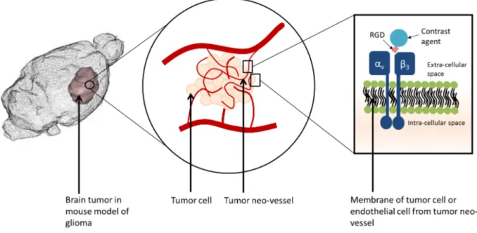
Fig. 1.1 – Principle of targeting ανβ3 integrin in vivo with RGD peptide.
Permeability and Retention) effect13 (a passive targeting due to the leakiness of tumor blood
vessels), or by active targeting of molecular biomarkers of tumor angiogenesis11, such as α
νβ3
integrins over-expressed by tumor cells and endothelial cells of tumor neo-vessels14, and involved in development of new blood vessels and cell motility15,16.
ανβ3 integrin can be targeted in vivo by RGD peptide (Arginine-Glycine-Aspartic Acid),
which binding efficiency has already been demonstrated by several studies12,14,17.
To develop new RGD functionalized probes dedicated to brain tumor molecular imaging, preclinical studies are usually performed on rodent model of glioma. Indeed, glioma has already been deeply studied, and several cellular and animal models are now available for research18.
In particular, orthotopic xenografts of cultured tumor cells originated from human biopsies lead to reproducible tumors, which explains why they are widely used in preclinical research. Advantages of such glioma models are their very good reproducibility and a good characteriza-tion of their biomarkers expression. On the other hand, these xenografts models do not induce brain tumors exhibiting all characteristics of human gliomas, mainly because of the drift during sequential passage of cell culture, but also because of the implantation method. Highly con-centrated cells suspension is usually implanted through the skull inside rodent brain, creating a wound on needle path. Therefore, the brain tumor is growing from an environment very rich in tumor cells and in a wounded brain, which is very different from the original human disease where the tumor is supposed to grow from few tumor cells in a healthy brain. Finally, one consequence of this protocol is a rapid tumor growth in rodent brain (only few weeks), which is a drawback in terms of likeness with human glioma, but an advantage for shortening induction time in preclinical research.
13J. Fang et al., Advanced Drug Delivery Reviews, 63: 136–151, 2011. 14F. Danhier et al., Molecular Pharmaceutics, 9: 2961–2973, 2012. 15P. T. Caswell et al., Traffic, 7: 14–21, 2006.
16P. C. Brooks et al., Science, 264: 569–571, 1994. 17C. Zhang et al., Cancer Research, 67: 1555–1562, 2007.
For the molecular imaging studies performed during this PhD, we chosen the U87 mouse model, consisting in the orthotopic xenograft of U87 human glioblastoma cells, this model being especially used for assessing tumor angiogenesis19. Thus, the RGD functionalized probes tested
in vivo on this model could actively target brain tumors, through the over-expression of ανβ3
integrins by tumor cells and endothelial cells from tumor neo-vessels, as presented in Figure
1.1.
1.3
The MEFISTO project
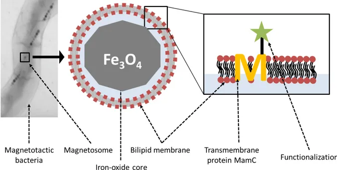
Fig. 1.2 – Principle of using magnetotactic bacteria to produce magnetosomes used as functionalized contrast agent for MRI.
This PhD work was conducted in the framework of the MEFISTO project, funded by an ANR grant in 2012 for 4 years, which aims at developing strategies, based on the use of mag-netosomes, for in vivo molecular imaging applications dedicated to brain tumor diagnosis and therapy. Magnetosomes are magnetic nanoparticles naturally encapsulated in a lipid vesicle, produced by magnetotactic bacteria. They offer in a single nano-object many of the charac-teristics that one seeks for multifunctional agent used as theranostic agent: a solid mineral iron-oxide core with contrasting properties for MRI, a therapeutic effect induced by magnetic hyperthermia, a lipid coating for solubilization and biocompatibility, and the possibility to use genetic tools for functionalizing the surface with bioactive ligands.
The production of functionalized synthetic nanoparticles usually requires several chemical procedures, including iron-oxide core synthesis, surface activation, chemical grafting of ligands and coating for solubilization. Each step is performed separately, using dedicated protocols subjected to duration, yield and cost parameters. In the MEFISTO project, we propose a
6 1.4. OUTLINE OF THIS MANUSCRIPT
methodology that gathers all these steps in one, letting the magnetotactic bacteria do the job. After a suitable genetic modification, bacteria are cultivated, and then broken to harvest func-tionalized magnetosomes by magnetic separation taking advantage of their magnetic properties, leading to a low cost and easy fabrication process.
The principle of magnetosome genetic functionalization is based on the transformation of magnetotactic bacteria with a translational fusion between the gene coding for an abundant transmembrane protein specific to the magnetosome membrane, like MamC20, and the gene en-coding the targeting ligand. The decoration is thus naturally sorted to the membrane leading to a functionalized magnetosome21,22.
The Figure 1.2 briefly illustrates the principle of using magnetotactic bacteria to produce functionalized MRI contrast agents for molecular imaging applications.
1.4
Outline of this manuscript
This manuscript is divided into five chapters, this introduction being the first one.
The second chapter draws a non-exhaustive state of the art regarding the use of iron-oxide nanoparticles in biomedical applications, starting form the physical origin of their contrasting properties, to the different strategies to optimize these properties, before focusing on their po-tential as MRI contrast agent. Besides, magnetotactic bacteria and magnetosomes specificity are also presented.
The third chapter is dedicated to the use of non-functionalized iron-oxide nanoparticles for MRI applications. Both chemically synthesized nanoparticles and magnetosomes are studied in terms of contrast enhancement efficiency in vitro and in vivo. Transverse relaxivities are measured by suspending iron-oxide nanoparticles in agar to characterize their in vitro contras-ting properties. Then, the contrast enhancement between brain parenchyma and blood vessels, induced by the intravenous injection of iron-oxide nanoparticles to mouse, is used to evaluate their in vivo contrasting properties. Besides, intravenous injections of iron-oxide nanoparti-cles are also performed on U87 mouse model of glioblastoma, in order to investigate vascular changes inside the tumor in terms of vessels geometric parameters and contrast agent retention. The results presented in this chapter, that focus on magnetosomes, have led to one publication in a peer-reviewed journal23 and several communications in scientific conferences: one poster
for WMIC24 (Savannah, USA, September 2013) and five oral presentations for GTRV25
(Or-l´eans, France, December 2013), NeWS Workshop26 (Gif-sur-Yvette, France, December 2014), MTB congress27 (Rio de Janeiro, Brazil, September 2014), SFRMRM Congress28 (Grenoble,
20K. Gr¨unberg et al., Applied and Environmental Microbiology, 70: 1040–1050, 2004. 21J. Xie et al., Nano Research, 2: 261–278, 2009.
22N. Ginet et al., PLoS ONE, 6: e21442, 2011.
23S. M´eriaux et al., Advanced Healthcare Materials, 4: 1076–1083, 2015.
24M. Boucher et al., World Molecular Imaging Congress, Poster, Savannah –USA, 2013.
25M. Boucher et al., Groupe Th´ematique de Recherche sur la Vectorisation (now Soci´et´e Fran¸caise de
Nanom´edecine), Oral, Orl´eans –France, 2013.
26M. Boucher et al., Neuroscience Workshop of Saclay, Oral, Saclay –France, 2014. 27N. Ginet et al., MagnetoTactic Bacteria, Oral, Rio de Janeiro –Brazil, 2014.
28M. Boucher et al., Soci´et´e Fran¸caise de R´esonance Magn´etique en Biologie et M´edecine, Oral, Grenoble
France, March 2015) and ISMRM Congress29 (Toronto, Canada, May 2015). And the results presented in this chapter, that focus on iron-oxide nanoparticles produced by our collabora-tors from CSPBAT laboratory, have led to one publication in a peer-reviewed journal30, two
other papers recently submitted to peer-reviewed journals31,32 and several communications in scientific conferences: three posters for Nanotech Meeting33 (Washington, USA, May 2013),
Nanobio Europe Meeting34 (Toulouse, France, June 2013) and EMRS Spring Meeting35 (Lille,
France, May 2014), and three oral presentations for Zing Biomaterials Conference36 (Nerja, Spain, April 2014) and EMRS Spring Meeting37,38 (Lille, France, May 2014). The results on
brain angiography have also been used to evaluate a method of compressed sensing and have led to one poster for ISBI39 (New-York, USA, April 2015).
The fourth chapter presents the production and the use of RGD functionalized magneto-somes in vitro and in vivo. The specific affinity and the consecutive internalization of RGD functionalized magnetosomes by U87 human glioblastoma cell cultures are assessed in vitro, as well as the MRI contrasting properties of magnetosomes. Then, RGD-labeled and unlabeled magnetosomes are injected at the tail vein of glioblastoma bearing mice to establish in vivo the proof of concept that biogenic functionalized contrast agents can be used for MR-based molecular imaging of brain tumors. The results presented in this chapter have led to several communications in scientific conferences: four poster presentations for WMIC40 (Seoul, South
Korea, September 2014), MTB congress41 (Rio de Janeiro, Brazil, September 2014),
SFNano-ENM Congress42 (Grenoble, France, December 2015) which received the British Society prize for the best poster presentation, WMIC43 (Honolulu, USA Septembre 2015), and three oral
presentations for SFRMRM Congress28 (Grenoble, France, March 2015), ISMRM Congress
(Toronto, Canada, May 2015)29 and MTB Congress44 (Marseille, France September 2016). Fi-nally, a publication is currently on preparation based on the results of this chapter45.
The fifth chapter describes a preliminary study dealing with the administration technique of RGD magnetosomes to mice, in order to maximize their accumulation inside brain tumor in vivo. The hypothesis whereby magnetosomes are better taken up by targeted tumor cells when the contact duration is lengthened is investigated in vitro, and in vivo with an intravenous multi-injections protocol. The results of the multi-administration study have led to three poster
29M. Boucher et al., International Society of Magnetic Resonance in Medicine, Oral, Toronto –Canada, 2015. 30S. Richard et al., Journal of Materials Chemistry B, 3: 2939–2942, 2015.
31S. Richard et al., Submitted to ACS Applied Materials & Interfaces, , 2016. 32S. Richard et al., Submitted to ACS Chemical Biology, , 2016.
33S. Richard et al., Nanotech, Poster, Washington –USA, 2013. 34S. Richard et al., Nano Bio Europe, Poster, Toulouse –France, 2013.
35S. Richard et al., European Materials Research Society Spring Meeting, Poster, Lille –France, 2013. 36S. Richard et al., Zing Bionanomaterials Conference, Oral, Nerja –Spain, 2014.
37S. Richard et al., European Materials Research Society Spring Meeting, Oral, Lille –France, 2014. 38J. Bolley et al., European Materials Research Society Spring Meeting, Oral, Lille –France, 2014. 39N. Chauffert et al., International Symposium on Biomedical Imaging, Poster, New–York –USA, 2015. 40M. Boucher et al., World Molecular Imaging Congress, Poster, Seoul –Korea, 2014.
41S. Pr´ev´eral et al., MagnetoTactic Bacteria, Poster, Rio de Janeiro –Brazil, 2014.
42M. Boucher et al., European Nanomedecine Meeting and Soci´et´e Fran¸caise de Nanom´edecine, Poster,
Greno-ble –France, 2015.
43F. Geffroy et al., World Molecular Imaging Congress, Poster, Honolulu –USA, 2015. 44M. Boucher et al., MagnetoTactic Bacteria, Oral, Marseille –France, 2016.
8 1.4. OUTLINE OF THIS MANUSCRIPT
presentations for WMIC46,47 (New York, USA, September 2016) and MTB Congress48 (Mar-seille, France, September 2016).
Finally this manuscript ends with a brief conclusion, followed by a summary of the PhD student role within the different collaborations that made this work possible.
46M. Boucher et al., World Molecular Imaging Congress, Poster, New–York –USA, 2016. 47F. Geffroy et al., World Molecular Imaging Congress, Poster, New–York –USA, 2016. 48F. Geffroy et al., MagnetoTactic Bacteria, Poster, Marseille –France, 2016.
References
[1] F. Reynolds and K. A. Kelly. Techniques for molecular imaging probe design. Molecular Imaging, 10: 407–419, 2011. (see p. 2)
[2] M. Mahmoudi, V. Serpooshan, and S. Laurent. Engineered nanoparticles for biomolecular imaging. Nanoscale, 3: 3007–3026, 2011. doi:10.1039/c1nr10326a (see pp. 2, 3)
[3] C. Corot, P. Robert, J. Idee, and M. Port. Recent advances in iron oxide nanocrystal technology for medical imaging. Advanced Drug Delivery Reviews, 58: 1471–1504, 2006.
doi: 10.1016/j.addr.2006.09.013(see p. 2)
[4] M.-F. Bellin, C. Beigelman, and S. Precetti-Morel. Iron oxide-enhanced MR lymphogra-phy: initial experience. European Journal of Radiology, 34: 257–264, 2000. doi:10.1016/
S0720-048X(00)00204-7 (see p.2)
[5] Jill Fredrickson, Natalie J. Serkova, Shelby K. Wyatt, Richard A. D. Carano, Andrea Pirzkall, Ina Rhee, Lee S. Rosen, Alberto Bessudo, Colin Weekes, and Alex de Crespigny. Clinical translation of ferumoxytol-based vessel size imaging (VSI): Feasibility in a phase I oncology clinical trial population. Magnetic Resonance in Medicine, 2016. doi: 10 . 1002/mrm.26167 (see p.2)
[6] T. Y. Tang, S. P. S. Howarth, S. R. Miller, M. J. Graves, J. M. U-King-Im, Z. Y. Li, S. R. Walsh, P. D. Hayes, K. Varty, and J. H. Gillard. Comparison of the Inflamma-tory Burden of Truly Asymptomatic Carotid Atheroma with Atherosclerotic Plaques in Patients with Asymptomatic Carotid Stenosis Undergoing Coronary Artery Bypass Grafting: An Ultrasmall Superparamagnetic Iron Oxide Enhanced Magnetic Resonance Study. European Journal of Vascular and Endovascular Surgery, 35: 392–398, 2008. doi:
10.1016/j.ejvs.2007.10.019(see p. 2)
[7] Z. Bakhtiary, A. A. Saei, M. J. Hajipour, M. Raoufi, O. Vermesh, and M. Mahmoudi. Targeted superparamagnetic iron oxide nanoparticles for early detection of cancer: Pos-sibilities and challenges. Nanomedicine: Nanotechnology, Biology and Medicine, 12: 287– 307, 2015. doi:10.1016/j.nano.2015.10.019 (see p.3)
[8] S. Sharifi, H. Seyednejad, S. Laurent, F. Atyabi, A. A. Saei, and M. Mahmoudi. Super-paramagnetic iron oxide nanoparticles for in vivo molecular and cellular imaging: SPIONs for Molecular and Cellular Imaging. Contrast Media and Molecular Imaging, 10: 329–355, 2015. doi:10.1002/cmmi.1638 (see p. 3)
[9] C. A. Pacak, P. E. Hammer, A. A. MacKay, Rory P. Dowd, K. R. Wang, A. Masuzawa, B. Sill, J. D. McCully, and D. B. Cowan. Superparamagnetic Iron Oxide Nanoparticles Func-tion as a Long-Term, Multi-Modal Imaging Label for Non-Invasive Tracking of Implanted Progenitor Cells. PLoS ONE , 9: e108695, 2014. doi: 10.1371/journal.pone.0108695
(see p.3)
[10] P. Soares, I. Ferreira, R. Igreja, C. Novo, and J. Borges. Application of hyperthermia for cancer treatment: recent patents review. Recent patents on anti-cancer drug discovery, 7: 64–73, 2012. (see p. 3)
[11] S. C. Baetke, T. Lammers, and F. Kiessling. Applications of nanoparticles for diagnosis and therapy of cancer. The British Journal of Radiology, 88: 20150207, 2015. doi: 10. 1259/bjr.20150207(see pp. 3,4)
10 REFERENCES
[12] F. Zhang, X. Huang, L. Zhu, N. Guo, G. Niu, M. Swierczewska, S. Lee, H. Xu, A. Y. Wang, K. A. Mohamedali, M. G. Rosenblum, G. Lu, and X. Chen. Noninvasive monitoring of orthotopic glioblastoma therapy response using RGD-conjugated iron oxide nanoparticles. Biomaterials, 33: 5414–5422, 2012. doi: 10.1016/j.biomaterials.2012.04.032 (see pp. 3, 4)
[13] J. Fang, H. Nakamura, and H. Maeda. The EPR effect: Unique features of tumor blood vessels for drug delivery, factors involved, and limitations and augmentation of the effect. Advanced Drug Delivery Reviews, 63: 136–151, 2011. doi:10.1016/j.addr.2010.04.009
(see p.4)
[14] F. Danhier, A. Le Breton, and V. Pr´eat. RGD-Based Strategies To Target Alpha(v) Beta(3) Integrin in Cancer Therapy and Diagnosis. Molecular Pharmaceutics, 9: 2961– 2973, 2012. doi: 10.1021/mp3002733 (see p.4)
[15] P. T. Caswell and J. C. Norman. Integrin Trafficking and the Control of Cell Migration: Integrin Trafficking and Cell Migration. Traffic, 7: 14–21, 2006. doi:
10.1111/j.1600-0854.2005.00362.x(see p. 4)
[16] P. C. Brooks, R. A. Clark, and D. A. Cheresh. Requirement of vascular integrin alpha v beta 3 for angiogenesis. Science, 264: 569–571, 1994. doi: 10.1126/science.7512751
(see p.4)
[17] C. Zhang, M. Jugold, E. C. Woenne, T. Lammers, B. Morgenstern, M. M. Mueller, H. Zentgraf, M. Bock, M. Eisenhut, W. Semmler, and F. Kiessling. Specific Targeting of Tu-mor Angiogenesis by RGD-Conjugated Ultrasmall Superparamagnetic Iron Oxide Parti-cles Using a Clinical 1.5-T Magnetic Resonance Scanner. Cancer Research, 67: 1555–1562, 2007. doi:10.1158/0008-5472.CAN-06-1668 (see p.4)
[18] S. S. Stylli, R. B. Luwor, T. M.B. Ware, F. Tan, and A. H. Kaye. Mouse models of glioma. Journal of Clinical Neuroscience, 22: 619–626, 2015. doi:10.1016/j.jocn.2014.10.013
(see p.4)
[19] Nienke A. de Vries, Jos H. Beijnen, and Olaf van Tellingen. High-grade glioma mouse models and their applicability for preclinical testing. Cancer Treatment Reviews, 35: 714– 723, 2009. doi:10.1016/j.ctrv.2009.08.011 (see p.5)
[20] K. Gr¨unberg, E.-C. Muller, A. Otto, R. Reszka, D. Linder, M. Kube, R. Reinhardt, and D. Schuler. Biochemical and Proteomic Analysis of the Magnetosome Membrane in Magnetospirillum gryphiswaldense. Applied and Environmental Microbiology, 70: 1040– 1050, 2004. doi: 10.1128/AEM.70.2.1040-1050.2004(see p. 6)
[21] J. Xie, K. Chen, and X. Chen. Production, modification and bio-applications of magnetic nanoparticles gestated by magnetotactic bacteria. Nano Research, 2: 261–278, 2009. doi:
10.1007/s12274-009-9025-8(see p. 6)
[22] N. Ginet, R. Pardoux, G. Adryanczyk, D. Garcia, C. Brutesco, and D. Pignol. Single-Step Production of a Recyclable Nanobiocatalyst for Organophosphate Pesticides Biodegrada-tion Using FuncBiodegrada-tionalized Bacterial Magnetosomes. PLoS ONE , 6: e21442, 2011. doi:
10.1371/journal.pone.0021442 (see p.6)
[23] S. M´eriaux, M. Boucher, B. Marty, Y. Lalatonne, S. Pr´ev´eral, L. Motte, C. T. Lef`evre, F. Geffroy, F. Lethimonnier, M. P´ean, D. Garcia, G. Adryanczyk-Perrier, D. Pignol, and N. Ginet. Magnetosomes, Biogenic Magnetic Nanomaterials for Brain Molecular Imaging with 17.2 T MRI Scanner. Advanced Healthcare Materials, 4: 1076–1083, 2015. doi: 10.
[24] M. Boucher, N. Ginet, B. Marty, C. T. Lef`evre, D. Garcia, D. Pignol, and S. M´eriaux. In Vivo characterization of MV1-magnetosomes as biogenic contrast agents dedicated to high field MRI: a dose study. World Molecular Imaging Congress, Poster, Savannah –USA, 2013. (see p. 6)
[25] M. Boucher, N. Ginet, B. Marty, C. T. Lef`evre, D. Garcia, D. Pignol, and S. M´eriaux. In Vivo charachterization of MV1-magnetosomes as biogenic contrast agents dedicated to high field MRI: a dose study. Groupe Th´ematique de Recherche sur la Vectorisation (now Soci´et´e Fran¸caise de Nanom´edecine), Oral, Orl´eans –France, 2013. (see p.6) [26] M. Boucher, N. Ginet, F. Geffroy, S. Pr´ev´eral, G. Adryanczyk-Perrier, M. P´ean, C.T.
Lef`evre, D. Garcia, D. Pignol, and S. M´eriaux. Genetically functionalized magnetosomes as MRI contrast agent for molecular imaging: in vitro proof of binding and in vivo proof of contrast. Neuroscience Workshop of Saclay, Oral, Saclay –France, 2014. (see p. 6) [27] N. Ginet, S. M´eriaux, M. Boucher, B. Marty, Y. Lalatonne, S. Pr´ev´eral, L. Motte, C.
T. Lef`evre, F. Geffroy, F. Lethimonnier, M. P´ean, D. Garcia, G. Adryanczyk, and D. Pignol. Towards molecular imaging with high-field MRI and biogenic contrast agents. MagnetoTactic Bacteria, Oral, Rio de Janeiro –Brazil, 2014. (see p. 6)
[28] M. Boucher, N. Ginet, F. Geffroy, S. Pr´ev´eral, G. Adryanczyk-Perrier, M. P´ean, C. T. Lef`evre, D. Garcia, D. Pignol, and S. M´eriaux. Fonctionnalisation par g´enie g´en´etique de magn´etosomes pour l’ imagerie mol´eculaire par IRM de biomarqueurs tumoraux. Soci´et´e Fran¸caise de R´esonance Magn´etique en Biologie et M´edecine, Oral, Grenoble –France, 2015. (see pp. 6,7)
[29] M. Boucher, N. Ginet, F. Geffroy, S. Pr´ev´eral, G. Adryanczyk-Perrier, M. P´ean, C. T. Lef`evre, D. Garcia, D. Pignol, and S. M´eriaux. Genetically functionalized magnetosomes as MRI contrast agent for molecular imaging: in vitro proof of binding and in vivo proof of contrast. International Society of Magnetic Resonance in Medicine, Oral, Toronto – Canada, 2015. (see p.7)
[30] S. Richard, M. Boucher, A. Herbet, Y. Lalatonne, S. M´eriaux, D. Boquet, and L. Motte. Endothelin B receptors targeted by iron oxide nanoparticles functionalized with a specific antibody: toward immunoimaging of brain tumors. Journal of Materials Chemistry B , 3: 2939–2942, 2015. doi: 10.1039/C5TB00103J (see p.7)
[31] S. Richard, M. Boucher, A. Saric, A. Herbet, Y. Lalatonne, P. X. Petit, S. M´eriaux, D. Boquet, and L. Motte. Optimization of pegylated iron oxide nanoplatforms for Antibody coupling and bio-targeting. Submitted to ACS Applied Materials & Interfaces, 2016. (see p.7)
[32] S. Richard, A. Saric, M. Boucher, Fran¸coise Geffroy, S. M´eriaux, Y. Lalatonne, P. X. Petit, and L. Motte. Anti-oxidative theranostic iron oxide nanoparticles towards brain tumors imaging and ROS production. Submitted to ACS Chemical Biology, 2016. (see p.7)
[33] S. Richard, M. Boucher, A. Herbet, E. Gu´enin, Y. Lalatonne, S. M´eriaux, J. P. Hugnot, D. Boquet, and L. Motte. Functionalization of nanoplatform: toward a new contrast agent specific of brain tumors. Nanotech, Poster, Washington –USA, 2013. (see p. 7)
[34] S. Richard, M. Boucher, A. Herbet, Y. Lalatonne, S. M´eriaux, J. P. Hugnot, D. Boquet, and L. Motte. Nanoplatform for immuno imaging of brain tumors by MRI. Nano Bio Europe, Poster, Toulouse –France, 2013. (see p. 7)
12 REFERENCES
[35] S. Richard, A. Herbet, M. Boucher, Y. Lalatonne, S. M´eriaux, J. P. Hugnot, D. Boquet, and L. Motte. Superparamagnetic nanoparitcles for immuno imaging of brain tumors by MRI. European Materials Research Society Spring Meeting, Poster, Lille –France, 2013. (see p.7)
[36] S. Richard, M. Boucher, A. Herbet, Y. Lalatonne, S. M´eriaux, J. P. Hugnot, D. Boquet, and L. Motte. Superparamagnetic nanoparticles for MRI brain tumors molecular imaging. Zing Bionanomaterials Conference, Oral, Nerja –Spain, 2014. (see p.7)
[37] S. Richard, A. Herbet, M. Boucher, Y. Lalatonne, S. M´eriaux, J. P. Hugnot, D. Boquet, and L. Motte. Development of nanoplateform for multimodal imaging of brain tumors. European Materials Research Society Spring Meeting, Oral, Lille –France, 2014. (see p.7) [38] J. Bolley, M. Boucher, S. M´eriaux, N. Pinna, L. Motte, and Y. Lalatonne. Iron oxide nanoparticles size effect on T1 and T2 MRI contrast. European Materials Research Society
Spring Meeting, Oral, Lille –France, 2014. (see p. 7)
[39] N. Chauffert, P. Weiss, M. Boucher, S. M´eriaux, and P. Ciuciu. Variable density sampling based on physically plausible gradient waveform: application to 3D MRI angiography. International Symposium on Biomedical Imaging, Poster, New–York –USA, 2015. (see p.7)
[40] M. Boucher, N. Ginet, F. Geffroy, S. Pr´ev´eral, G. Adryanczyk-Perrier, M. P´ean, C. T. Lef`evre, D. Garcia, D. Pignol, and S. M´eriaux. Genetically functionalized RGD-magnetosomes as MRI contrast agent: in vitro proof of concept. World Molecular Imaging Congress, Poster, Seoul –Korea, 2014. (see p. 7)
[41] S. Pr´ev´eral, G. Adryanczyk-Perrier, F. Geffroy, M. P´ean, M. Boucher, C.T. Lef`evre, D. Garcia, S. M´eriaux, D. Pignol, and N. Ginet. RGD-functionalized magnetosomes, a contrast agent with molecular affinity for diagnostic in Magnetic Resonance Imaging. MagnetoTactic Bacteria, Poster, Rio de Janeiro –Brazil, 2014. (see p.7)
[42] M. Boucher, F. Geffroy, E. Selingue, N. Ginet, S. Pr´ev´eral, L. Bellanger, G. Adryanczyk-Perrier, M. P´ean, C.T. Lef`evre, D. Garcia, D. Pignol, and S. M´eriaux. Genetically func-tionalized magnetosomes used as MRI probe for molecular imaging of U87 tumor. Euro-pean Nanomedecine Meeting and Soci´et´e Fran¸caise de Nanom´edecine, Poster, Grenoble –France, 2015. (see p. 7)
[43] F. Geffroy, M. and. Boucher, N. Ginet, S. Pr´ev´eral, L. Bellanger, G. Adryanczyk-Perrier, M. P´ean, C.T. Lef`evre, D. Garcia, D. Pignol, and S. M´eriaux. In vitro characterization of AMB1 magnetosomes as biogenic functionalized contrast agents dedicated to molecular MRI. World Molecular Imaging Congress, Poster, Honolulu –USA, 2015. (see p.7) [44] M. Boucher, F. Geffroy, E. Selingue, N. Ginet, S. Pr´ev´eral, L. Bellanger, G.
Adryanczyk-Perrier, M. P´ean, C.T. Lef`evre, D. Garcia, D. Pignol, and S. M´eriaux. Genetically func-tionalized magnetosomes used as MRI contrast agent for in vivo molecular imaging of brain tumor in U87 mice. MagnetoTactic Bacteria, Oral, Marseille –France, 2016. (see p.7)
[45] M. Boucher, F. Geffroy, N. Ginet, S. Pr´ev´eral, L. Bellanger, Selingue, G. Adryanczyk-Perrier, M. P´ean, C. T. Lef`evre, D. Garcia, D. Pignol, and S. M´eriaux. Genetically tailored iron-oxide nanoparticles used as MRI probe for in vivo molecular imaging of brain tumor. In preparation, 2016. (see p.7)
[46] M. Boucher, F. Geffroy, E. Selingue, N. Ginet, S. Pr´ev´eral, G. Adryanczyk-Perrier, M. P´ean, C.T. Lef`evre, D. Garcia, D. Pignol, and S. M´eriaux. Multi-injection of RGD func-tionalized magnetosomes to improve in vivo iron uptake by U87 brain tumor. World Molecular Imaging Congress, Poster, New–York –USA, 2016. (see p. 8)
[47] F. Geffroy, M. and. Boucher, N. Ginet, S. Pr´ev´eral, L. Bellanger, G. Adryanczyk-Perrier, M. P´ean, C.T. Lef`evre, D. Garcia, D. Pignol, and S. M´eriaux. In vitro multimodal imaging characterization of RGD functionalized magnetosomes accumulation in U87 glioblastoma cells. World Molecular Imaging Congress, Poster, New–York –USA, 2016. (see p. 8) [48] F. Geffroy, M. and. Boucher, N. Ginet, S. Pr´ev´eral, L. Bellanger, G. Adryanczyk-Perrier,
M. P´ean, C.T. Lef`evre, D. Garcia, D. Pignol, and S. M´eriaux. In vitro multimodal imaging characterization of RGD functionalized magnetosomes accumulation in U87 glioblastoma cells. MagnetoTactic Bacteria, Poster, Marseille –France, 2016. (see p. 8)
Chapter
2
Iron-oxide nanoparticles for diagnostic and
therapeutic applications
Contents
2.1 Why iron nanoparticles? . . . 16
2.1.1 Physical origin of magnetism . . . 16
2.1.2 Magnetism of nanoparticles . . . 18
2.1.3 The interest of IONP in MRI . . . 20
2.1.4 Relaxation models for IONP . . . 22
2.2 How to optimize IONPs contrasting properties? . . . 25
2.2.1 Superparamagnetic iron core optimization . . . 26
2.2.2 Coating optimization. . . 29
2.3 Interest of magnetsosomes as MRI contrast agent . . . 31
2.3.1 Magnetotactic bacteria and magnetosomes. . . 31
2.3.2 Magnetosomes physico-chemical properties . . . 34
2.4 Other biomedical applications of IONP . . . 36
2.4.1 IONP labeled cells . . . 37
2.4.2 Magnetic hyperthermia . . . 40
References . . . 42