HAL Id: hal-02037760
https://hal-normandie-univ.archives-ouvertes.fr/hal-02037760
Submitted on 20 Feb 2019
HAL is a multi-disciplinary open access
archive for the deposit and dissemination of
sci-entific research documents, whether they are
pub-lished or not. The documents may come from
teaching and research institutions in France or
abroad, or from public or private research centers.
L’archive ouverte pluridisciplinaire HAL, est
destinée au dépôt et à la diffusion de documents
scientifiques de niveau recherche, publiés ou non,
émanant des établissements d’enseignement et de
recherche français ou étrangers, des laboratoires
publics ou privés.
Distributed under a Creative Commons Attribution - NonCommercial| 4.0 International
License
Emergency surgery for epidural abcess secondary to
sacral fistula after laparoscopic proctectomy
Jeremie Zeitoun, Benjamin Menahem, Audrey Fohlen, Gil Lebreton, Jean
Lubrano, Arnaud Alves
To cite this version:
Jeremie Zeitoun, Benjamin Menahem, Audrey Fohlen, Gil Lebreton, Jean Lubrano, et al.. Emergency
surgery for epidural abcess secondary to sacral fistula after laparoscopic proctectomy. Journal of
Surgical Case Reports, Oxford University Press, 2016, 2016 (7), pp.rjw108. �10.1093/jscr/rjw108�.
�hal-02037760�
Journal of Surgical Case Reports, 2016;7, 1–3
doi: 10.1093/jscr/rjw108 Case Report
C A S E R E P O R T
Emergency surgery for epidural abcess secondary
to sacral
fistula after laparoscopic proctectomy
Jeremie Zeitoun
1,
*, Benjamin Menahem
1
, Audrey Fohlen
2
, Gil Lebreton
1
,
Jean Lubrano
1
, and Arnaud Alves
1
1
Department of Digestive Surgery, University Hospital of Caen (CHU) Caen, 14032 Caen Cedex, France and
2
Department of Radiology, University Hospital of Caen (CHU) Caen, 14032 Caen Cedex, France
*Correspondence address. Department of Digestive Surgery, University Hospital of Caen, Avenue de la Côte de Nacre, 14032 Caen Cedex, France. Tel:+33-2-31-06-32-21; Fax: +33-2-31-06-45-35. E-mail: menahem-b@chu-caen.fr
Abstract
A 61-year-old man presented via the emergency department with a few days history of abdominal and colic occlusion symptoms. He presented signs of sepsis, midline lumbar spine tenderness and reduced hipflexion. Computer tomog-raphy of the abdomen and pelvis showed a presacral collection contiguous with the posterior part of the colo-rectal anastomosis, and MRI lumbar spine revealed abscess invation into the epidural space. He underwent a laparotomy with washout of the presacral abscess and a colostomy with a prolonged course of intravenous antibiotic therapy. At 3 weeks after initial presentation he had made a full clinical recovery with progressive radiological resolution of the epidural abscess. The objective of the case report is to highlight a unique and clinically significant complication of a rare post-operative complication after rectal surgery and to briefly discuss other intra-abdominal sources of epidural abscess.
INTRODUCTION
This was a very unusual presentation of a post-operative com-plication after rectal surgery. This presentation necessitated early and aggressive surgical and medical treatment to avoid major complications of sepsis. In this case the patient had had a rectal surgery 2 years before that disrupted normal pre-sacral and retroperitoneal anatomy.
CASE REPORT
A 61-year-old man presented to the emergency department for fever since few days with abdominal pain and lack of gas and stools. The pain had become worse and was associated with electric shocks in both lower extremities. Two years before, the patient had a laparoscopic proctectomy with ileal pouch–anal anastomosis protected by an ileostomy for a
voluminous villous tumor of the lower rectum. This interven-tion was complicated at the third day of a fever that prompted an abdominal Computer tomography (CT)-scan revealing intra-abdominal effusion in the leftflank drained by the per-operative Blake drain, syringed daily and treated by intravenous antibiotics.
The patient had a high fever (T°C 39.3°C) but had no sign of severe sepsis (heart rate was 88 bpm and blood pressure was 113/78 mmHg). The abdomen was painful in the lower median and lower left quadrant. Rectal examination was normal. Neurological examination found still diffuse electric shocks in both lower extremities. The remainder of the full neurological examination was normal.
Blood test revealed elevated inflammatory markers (white cell count 21.49 g/L, C reactive protein 359 mg/L, Fibrinogen 7.4 g/L). An abdominal CT-scan showed a recurrence of digestive tumor in
Received: April 15, 2016. Accepted: May 24, 2016
Published by Oxford University Press and JSCR Publishing Ltd. All rights reserved. © The Author 2016.
This is an Open Access article distributed under the terms of the Creative Commons Attribution Non-Commercial License (http://creativecommons.org/ licenses/by-nc/4.0/), which permits non-commercial re-use, distribution, and reproduction in any medium, provided the original work is properly cited. For commercial re-use, please contact journals.permissions@oup.com
1
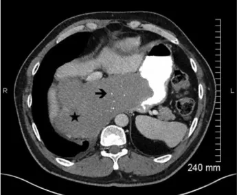
pelvis with a digestive presacral fistulization without circum-scribedfluid collection. A sagittal section abdominal CT and mag-netic resonance imaging (MRI) (Figs.1–2) showed colicfistula to the presacral collection measured to 38.8× 9.54 mm with infiltra-tion of adjacent soft tissue and which continues to the spinal canal through the sacral S1 left hole with multiple epidural abscess from L4 to S4. There was osteitis of the sacrum and arachnoiditis.
The patient underwent a new surgery by laparotomy. An intraoperative rectoscopy was performed. At the joining up end-to-end anastomozing colon, there was a hole that may cor-respond to a fistula orifice. A colostomy was performed to defunctionalize the fistula. On consultation with the micro-biology treatment, an empiric intravenous antibiotic therapy was started by Piperacillin–Tazobactam with Amikacin and Metronidazol then switched by Piperacillin–Tazobactam against the septicaemia of Escherichia coli (found in blood culture) during 2 months.
DISCUSSION
Previous studies published about epidural abcess secondary to abdominal sepsis concern a context of appendicitis, stercoral ulcer rupture, immunocompromised patients, acutization of Crohn’s disease, disadvantaged social status and bad oral con-ditions. One study described presacral pelvic abscess continu-ous with the tip of the appendix in a patient of antecedent of rectal adenocarcinoma. The other one concerns presacral pelvic abscess at the recto sigmoid junction extended into the presa-cral musculature and gas bubbles extended into the epidural space at L3. However, there are no studies dealing with a presa-cral fistula between a chronic presacral collection and spinal canal after of laparoscopic proctectomy for tubulo-villous resection of rectum with osteitis and arachnoiditis. It was sometimes necessary to realize intervertebral disc biopsy [1], bacteriologicalfluid drainage [2], parasitological examination of stools [3] and lumbar puncture [4]. The variation of symptoms depended on neurological involvement and the importance of the fistula. The origin of the abscess was different between cases reported: presacral pelvic abscess continuous with the tip of appendix after abdominoperineal resection for adenocarcin-oma and chemo radiotherapy [2], stercoral ulcer rupture[5], immunocompromised patients, acutization of Crohn’s disease [5], disadvantaged social status, bad oral conditions [1],fistula after laminectomy L4 between the wound and thefirst sacral foramen [6]. The abscess concerns more frequently the cervical spine but any spinal cord could be reached and cause afistula with other soft tissue. There is no precise data in the literature about the time between the abscess and this cause.
In our case, the patient underwent a defunctioning pelvic loop colostomy by laparotomy. An empiric intravenous anti-biotic therapy was started to Piperacillin–Tazobactam with Amikacin and Metronidazol then switched by Piperacillin– Tazobactam against Escherichia coli (found in blood culture) dur-ing 2 months.
On the one hand, the duration of antibiotherapy was 6 weeks to 6 months in the literature. There is no data in this one about the choice of duration. The choice may be chosen by the degree of clinical neurological and imagery damage as well as the germs found in bacteriological samples.
Negative Gram bacilli were most frequently found especially Bacteroid Fragilis and E. coli and positive Gram bacilli (Streptocoque intermedius). On the other hand, all teams operate their patients except for two case reports of Lechiche et al. in 2006 who decided to use only medical treatment with antibiotics [5]. For the others, decompressed laminectomy was each time neces-sary in the case of neurological deficit at the clinical examin-ation with afluid drainage [2, 4]. In our case, as in the M.H. Jamison’s case report in 1984, a defunctionning pelvic loop col-ostomy was necessary [3]. An appendectomy was necessary with a drainage of the collection for the case of Carter et al. in 2014 [2], an urgent Hartmann’s resection for focal perforation of
Figure 1: Axial view contrast-enhanced MDCT (portal phase) with oral opacifi-cation, in a x year-old men with a large esophageal GIST, showing a lesion (★), well-circumscribed, with small calcifications (è) (rare). The lesion is homoge-neous (no kystic or necrotic parts) without hypervascularization.
Figure 2: Coronal view of contrast-enhanced MDCT showing the exophytic, non-obstructive lesion (★) located at the esogastric junction.
2
|
J. Zeitoun et al.the recto sigmoid junction for the case of Uy et al. [7] and ileoce-cal resection of the affected bowel with drainage of the multiple abscesses in R. Maggiore’s Case [4].
Concerning the type of antibiotherapy, there was no consen-sus and each team used different initial empiric antibiotics. What can be noticed is that after antibiogram most teams used a G penicillin associated with metronidazole. Only one team has chosen to switch with intravenous Piperacillin–Tazobactam [7]. Forfive of these studies, the evolution has been favorable [2–4, 6,7]. One death occurred in the published literature [5]. Definitive paraplegia occurred in one case [1]. A resolution of intraspinal gas and abscess but persistent sacral osteomyelitis occurred in two cases [5,7].
To conclude, this is a very rare affection but it can lead to serious complications. Successful treatment involves prompt drainage and prolonged intravenous antibiotic therapy.
ACKNOWLEDGEMENTS
We thank Dr Audrey Fohlen, MD-MSc, Radiologist, for the help in the MRI review.
CONFLICT OF INTEREST STATEMENT
None declared.
REFERENCES
1. Chandesris MO, Schleinitz N, Gayet S, Bernit E, Crebassa C, Veit V, et al. Anaerobic deep abscesses with unusual loca-tion: report of 5 cases. Rev Med Intern 2005;26:534–40. 2. Carter M, Meshkat B, El-Masry S. Epidural abscess secondary
to acute appendicitis. BMJ Case Rep 2014;doi:10.1136/bcr-2014-207446.
3. Jamison MH, Stanworth P, Maclennan I. An unusual rectal fistula: extra-dural abscess discharging per rectum. Br J Surg 1984;71:651–2.
4. Maggiore R, Miller F, Stryker S, Buchman AL. Meningitis and epidural abscess associated withfistulizing Crohn’s disease. Dig Dis Sci 2004;49:1461–5.
5. Lechiche C, Le Moing V, Marchandin H, Chanques G, Atoui N, Reynes J. Spondylodiscitis due to Bacteroides fragilis: two cases and review. Scand J Infect Dis 2006;38:229–31.
6. Smith C, Kavar B. Extensive spinal epidural abscess as a complication of Crohn’s disease. J Clin Neurosci 2010;17:144–6. doi:10.1016/j.jocn.2009.02.038. [Epub 2009 Nov 13].
7. Uy CE, Jiang SY, Murray MC. Epidural abscess and feculent meningitis secondary to stercoral ulcer rupture. BMJ Case Rep 2015;2015:pii:bcr2014207858. doi:10.1136/bcr-2014-207858. Emergency surgery for epidural abcess secondary to sacralfistula after laparoscopic proctectomy