JON 3026
B. Meissner
K. Kallenberg
P. Sanchez-Juan
S. Ramljak
A. Krasnianski
U. Heinemann
S. Eigenbrod
E. Gelpi
B. Barsic
H. A. Kretzschmar
W. J. Schulz-Schaeffer
M. Knauth
I. Zerr
MRI and clinical syndrome in dura
mater-related Creutzfeldt-Jakob disease
Introduction
Studies in iatrogenic CJD are extremely useful to
under-stand some aspects of infections in humans, as the
infec-tious agent may reach the brain via different routes:
di-rect inoculation (EEG depth electrodes), oral infection
(growth hormone therapy, vCJD), peripheral infection
(blood transfusion) and superficial infection (dura
ma-ter and corneal transplant).
Received: 9 September 2007
Received in revised form: 29 April 2008 Accepted: 4 June 2008
Published online: 23 January 2009
B. Meissner, MD (쾷) · A. Krasnianski, MD · U. Heinemann, MD · I. Zerr, MD
National TSE Reference Center Dept. of Neurology University of Göttingen Robert-Koch-Str. 40 37075 Göttingen, Germany Tel.: +49-551/39-6636 Fax: +49-551/39-7020 E-Mail: epicjd@med.uni-goettingen.de present address: B. Meissner, MD Dept. of Gerontopsychiatry University of Zürich Minervastr. 145 8008 Zuerich, Switzerland K. Kallenberg, MD · M. Knauth, MD Dept. of Neuroradiology and MR research in Neurology and Psychiatry
Göttingen, Germany P. Sanchez-Juan, MD, PhD
Foundation Marqués de Valdecilla, IFIMAV Santander, Spain
and
Centro de Investigación Biomédica en Red sobre Enfermedades Neurodegenerativas (CIBERNED) S. Ramljak · W. J. Schulz-Schaeffer, MD Dept. of Neuropathology Georg-August University Göttingen, Germany S. Eigenbrod, MD · H. A. Kretzschmar, MD Center for Neuropathology and Prion Research
Ludwig-Maximilians-Universität München, Germany
E. Gelpi, MD
Institute of Neurology Medical University of Vienna Vienna, Austria
B. Barsic, MD
University Hospital of Infectious Diseases Zagreb, Croatia
■
Abstract
Objective Iatrogenic
Creutzfeldt-Jakob disease (iCJD) is
mainly associated with dura mater
(DM) grafts and administration of
human growth hormones (hGH).
Data on disease course in DM-CJD
are limited. We describe the clinical
and diagnostic findings in this
pa-tient group with special emphasis
on MRI signal alterations.
Methods
Ten DM-CJD patients were studied
for their clinical symptoms and
diagnostic findings. The MRIs were
evaluated for signal increase of the
cortical and subcortical structures.
Results DM-CJD patients had a
median incubation time of 18 years
and median disease duration of 7
months. The majority of patients
were MM homozygous at codon
129 of the prion protein gene
(
PRNP) and presented with gait
ataxia and psychiatric symptoms.
No correlation between the graft
site and the initial disease course
was found. The MRI showed
corti-cal and basal ganglia signal
in-crease each in eight out of ten
pa-tients and thalamic hyperintensity
in five out of ten cases. Of interest,
patients with thalamic signal
in-crease were homozygous for
me-thionine.
Conclusion The MRI
find-ings in DM-CJD largely resemble
those seen in sporadic CJD, as the
cortex and basal ganglia are mainly
affected.
■
Key words CJD · MRI ·
dementia · cerebellar disorder ·
CNS infection
The diagnosis of iCJD is based on the presence of
typical symptoms (dementia, ataxia, extrapyramidal
and pyramidal signs, myoclonus and akinetic mutism)
and a history of iatrogenic exposure. The finding of
14-3-3 protein in the CSF may support the diagnosis (77 %
sensitivity) [1, 2].
Dura mater grafts have been found as disease cause
in 200 CJD patients worldwide [1]. Due to the long
incu-bation periods ranging from 16 months to 31 years,
DM-CJD cases are still detected nowadays [1, 3–9].
Increased rates of early ataxia have been reported in
DM-CJD, hGH-CJD and vCJD [10–13]. In DM-CJD, a
possible influence of the graft location on the clinical
onset has been suggested [14, 15]. Only limited data were
reported on paraclinical tests in iCJD, such as MRI. Since
the MRI pattern of high signal abnormalities follows the
lesion pattern of the brain as detected by pathology, the
analysis of MRI in iCJD might be extremely useful.
The goals of our study were to describe MRI and
clinical findings in a larger number of DM-CJD patients
and to correlate them with the graft location and codon
129 genotype.
Methods
Patients with suspected CJD are reported to the CJD Surveillance Unit in Goettingen, (established in 1993) and examined by a study physi-cian at the hospital reporting the case. A questionnaire about the pa-tient’s history (profession, habits, meat consumption) is filled out and copies are made of the medical charts (clinical findings, laboratory tests, EEG and MRI). The patients are classified as possible, probable
or no CJD case according to established criteria [16]. One Croatian
case (case 10) was included in this study because material was sent for genetic analysis and all clinical data were available.
■ Genetic analysis
Analysis of the prion protein gene (PRNP) was performed after
isola-tion of genomic DNA from blood according to standard methods [17]. PRNP mutations were excluded in all but one patient by full PRNP sequencing (case 9).
■ Neuropathology
Histological examination was performed according to standard methods [18, 20].
■ Clinical examination
The patients were examined for the presence of typical CJD symp-toms at onset (within first month) and during the disease course. Symptoms representing a residual state after surgery were not in-cluded in the analysis. Residual symptoms which progressed at the time of the disease onset were considered to be CJD signs and in-cluded in the study.
■ MRI
MRIs were available in nine out of ten patients. All MR examinations had been obtained from a 1.5 Tesla scanner (a total of 11 T2w, 9 FLAIR (fluid attenuated inversion recovery), 6 PDw (proton density weighted imaging) and 4 DWI (diffusion-weighted imaging), including serial MRIs in four patients) and were mainly available as hardcopy. Digital MRI data were available in four patients (cases 2, 3, 5 and 8). The MRI scans were reported independently and in consensus by two neurora-diologists (KK and MK) aware of the CJD diagnosis but not aware of the disease aetiology. The interobserver agreement was moderate for all the sequences (Concordance 80,. and κ = 0.52) and high for FLAIR (Concordance 83.6 and κ = 0.57), PDw (Concordance 83.3 and κ = 0.67) and DWI (Concordance 83.9 and κ = 0.65). The results of the consen-sus review were used for the study.
Using a standardized protocol, seven cortical areas (the frontal,
parietal, temporal, occipital and insula cortex, as well as the cingulate gyrus and hippocampus), the basal ganglia (caudate nucleus,
puta-men, globus pallidus), the thalamus and the cerebellum were assessed
for signal increase in relation to suspected normointense tissue.
Results
■
Patients with dura mater grafts
In the years 1993 to 2006, ten DM-CJD cases (four
defi-nite and six probable) were identified (nine in Germany,
one in Croatia) (Table 1). All surgeries were performed
between 1980 and 1987 (Fig. 1). The median age at onset
was 55 years (range 25–70); the median disease duration
was 8 months (range 2–30). Incubation periods ranged
from 9 to 23 years (median 18 years) (Fig. 1).
The codon 129 genotype was available in all patients
(8 MM, 1 VV and 1 MV). Codon 219 was not examined
in any case. The type of pathological prion protein (PrP
Sc1 or 2) was analyzed in three out of four autopsied cases
(only formalin fixed brain tissue was available in one
patient): Three patients displayed PrP
Sctype 1. In one
further case, the PrP
Sctype could not be clearly
identi-fied as type one or two (case 7) (as previously discussed)
[21] (Table 1).
■
Clinical presentation
The main symptoms at onset (within the first month)
and during the course of the disease are displayed in
Table 2. Seven out of ten patients presented with gait
ataxia (in three cases in isolation). Psychiatric
abnor-malities (depression and aggression) occurred in four
out of ten patients (in one case as the only presenting
symptom). Visual signs (double vision) were found in
three patients, pyramidal signs (hemiparesis) in two and
extrapyramidal signs (rigidity and choreatic
move-ments) in one out of ten patients.
Only one patient displayed early signs of dementia.
As unspecific clinical signs, vertigo (five patients) and
headaches (five patients, data not displayed) were found
most frequently. Further symptoms observed in the
early disease stage were dysaesthesias in three patients
and loss of hearing, tinnitus, dysarthria, aphasia,
abnor-mal sense of taste in one patient each.
■
Time course of symptoms
The median duration from the beginning of the disease
to the onset of typical CJD symptoms is given in Fig. 2.
Ataxia and psychiatric signs were among the earliest
symptoms and followed by visual signs (median 1
month) and dementia (median 2 months). Myoclonus,
extrapayramidal and pyramidal signs were typical signs
of the advanced disease stage (median 3 to 4 months).
Akinetic mutism was found in only two patients.
■
Dura graft location and initial clinical symptoms
Details on the graft placement and the clinical signs are
shown in Table 2.
Early ataxia occurred most frequently in patients
with suboccipital graft (four cases) but also in three
pa-tients with different graft location. Psychiatric signs
were predominant in patients with frontal graft (three
cases) but also found in one patient with suboccipital
graft. Two patients with early pyramidal signs
(hemi-paresis) had a dura graft in the parietal area. Early
rigid-ity was found in one patient with frontobasal graft. In
four patients, the early disease course appeared to be
monosymptomatic: three patients with suboccipital
graft showed isolated ataxia for a time period of 4 weeks
(case 9), 6 weeks (case 7) and 8 weeks (case 10). In one
Table 1 Clinical and diagnostic data in ten dura-related iatrogenic CJD cases Case Sex/age
(years)
Codon 129 genotype (PrPSc type)
Cause for surgery CJD diagnosis (WHO criteria) Incubation (years) Disease duration (months) EEG: PSWC CSF: 14-3-3
1 m/38 MV Brain trauma Probable 10 4 + +
2 m/44 MM (1) Brain trauma Definite 19 25 + +
3 f/70 MM Meningeoma Probable 19 3 – +
4 m/42 MM AV-angioma Probable 13 4 + +
5 m/54 MM Meningioma Probable 18 11 – +
6 f/56 MM (1) Acousticus neurinoma Definite 9 6 + +
7 m/57 MM Hemangioblastoma Definite 20 10 – +
8 f/65 MM Trigeminal neuralgia Probable 15 12 – +
9 f/64 MM (1) Pilocytic astrocytoma Definite 23 2 + +
10 f/25 VV Pilocytic astrocytoma Probable 17 30 (+) (–)
Median 55 years
Median 18 8 5/9 9/9
M male; f female; PSWC periodic sharp wave complexes
(+), (–) = assessed as positive or negative at the hospital reporting the case (not included in the present analysis)
17 23 15 20 9 18 13 19 19 10 3 0 1 1 1 1 0 0 2 0 1960 1965 1970 1975 1980 1985 1990 1995 2000 2005 2010 10 9 8 7 6 5 4 3 2 1
Fig. 1 Incubation periods (years) and disease dura-tion (months) in patients with dura mater graft
further patient with frontobasal graft, only psychiatric
signs were present for a time period of 6 months (case
2).
■
MRI
A synopsis of the MRI findings and applied sequences is
given in Table 3. The cingulate gyrus was the most
fre-quently affected cortex region (eight out of ten),
fol-lowed by the frontal, temporal and parietal lobes (six out
of ten, each).The occipital cortex was least affected
(three out of ten). Basal ganglia hyperintensity was
found in eight out of ten patients (in one case only
dur-ing follow-up), and thalamic hyperintensity in five (in
three cases only on PDw). In one patient (MV type), no
signal abnormalities were found (Fig. 3). The current
criteria for a pulvinar- or hockey stick sign (pulvinar or
dorsomedial thalamus brighter than the other grey
mat-ter) were not met in any of the patients [22, 23].
Two patients showed pronounced signal increase in
the brain hemisphere containing the graft: In one
pa-tient with right temporal graft, the signal increase was
more pronounced in the right temporal and parietal
lobes (Case 4). In one patient with left suboccipital graft,
the left frontal cortex appeared to be more hyperintense
than on the right side (Case 10). In two other patients,
the findings were reversed (signal more pronounced in
the opposite brain hemisphere) (Cases 6 and 9).
Discussion
In a recent study on DM-CJD cases, MRI sensitivity was
reported to be high but no details were given on the
in-volved brain areas [24]. According to a literature review,
basal ganglia signal increase was found in eight out of 27
patients [6, 8, 9, 21, 25, 26], additional cortical signal
in-crease in three [9, 25, 26] and thalamic hyperintensity in
one [9]. In a large number of further cases, only atrophy
was reported, which is most likely due to the less
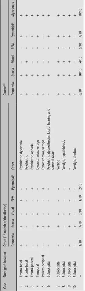
com-Table 2
Dura graft location and clinical signs at onset and during course of the disease
Case
Dura graft location
Onset (1
st month of the disease)
Course** Dementia Ataxia Visual EPM Pyramidal* Other Dementia Ataxia Visual EPM Pyramidal* Myoclonus 1 Fronto-basal + + – + – Psychiatric, dysarthria + + – + + + 2 Fronto-basal – – – – – Psychiatric + + + – – + 3 Fronto-parietal – – + – + Psychiatric, aphasia – + – – + + 4 Temporal – + + – – Dysaesthesias, vertigo + + – + + + 5 Parieto-occipital – + – – + Dysaesthesias, vertigo + + – – – + 6 Suboccipital – + – – –
Psychiatric, dysaesthesias, loss of hearing and sense of taste
–+ – – + + 7 Suboccipital – + – – – Vertigo + + + + + + 8 Suboccipital – – + – – Vertigo, hyperhidrosis + + + + + + 9 Suboccipital – + – – – – + + + + + + 10 Suboccipital – + – – – Vertigo, tinnitus + + – + – + – 1/10 7/10 3/10 1/10 2/10 8/10 10/10 4/10 6/10 7/10 10/10
* corresponding to hemiparesis (two patients at onset) or spasticity (seven patients during the course) ** During the disease course all but one patient (case 5) developed psychiatric signs. Only two patients (cases 6 and 8) became
akinetic and mute
EPM extrapyramidal signs Ataxia 0 Median (months) 0 1 2 3 3 4 4 5 4 3 2 1 0 Psychiatric Visual Dementia Myoclonus EPM Pyramidal Akinetic mutism
mon use of FLAIR and DW images in the past (Table 4)
[3, 5–9, 21, 25–41].
We studied the MRI findings in ten iatrogenic CJD
patients after dura mater transplant by highly sensitive
sequences. Cortex involvement was found in all but two
patients. The basal ganglia were affected in eight, the
thalamus in five and the cerebellum in two cases. The
majority of our patients was MM homozygous at codon
129 of the
PRNP and presented with gait ataxia.
At first sight, the MRI findings in our patients seem
to largely resemble those seen in sporadic CJD. In sCJD,
six molecular types (codon 129 genotype + PrP
Sctype 1
or 2; MM1, MM2, MV1, MV2, VV1, VV2) have been
de-scribed with various clinical and diagnostic findings
[42, 43]. Basal ganglia and cortical signal increase are
most frequently found on the MRI [44–48]. Thalamic
signal increase represents a comparatively rare finding,
which seems to largely depend on the application of DW
images [45–48].
Although the majority of our patients were not
dis-tinguishable from sporadic cases by means of MRI, the
finding of thalamic hyperintensity in five out of eight
MM homozygous patients is notable. According to
pre-vious literature reports, thalamic hyperintensity appears
to be rather typical of MV2 type sporadic CJD and
atyp-ical of MM homozygous sCJD patients [49–51]. However,
as only small patient groups have been studied so far
with limited numbers of DW images, it remains to be
confirmed that thalamic hyperintensity is rather not
typical of MM homozygous sporadic CJD.
From the clinical point of view, sporadic CJD cases
with MM homozygosity may present with rapidly
pro-gressive dementia (MM1 individuals), rather slowly
de-veloping cognitive signs (MM2-
cortical individuals) or
early ataxia (MM2-
thalamic individuals with variable
disease onset). The latter patients, who
clinically most
resemble our iatrogenic patients, are however well
dis-Table 3
MRI signal increase in eight iatrogenic CJD cases caused by dura mater transplant
Case
Dura graft location
Genotype Frontal Temporal Parietal Occipital Insula Gc Hc Bg Thalamus Cerebellum
MRI 1 (time point)* MRI 2 (time point)*
1 Fronto-basal MV – – – – – –––– – T2, FLAIR (1/3) – 2 Fronto-basal MM + –/+ –/+ – – + – –/+ – – FLAIR, DWI (2/3) T2, FLAIR (2/3) 3 Fronto-parietal MM – + – + + + – + + – T2, FLAIR (1/3) – 4 Temporal MM + + + +/– + –/+ + + – – T2, FLAIR (1/3) T2, FLAIR, DWI (3/3) 5 Parieto-occipital MM + + + – + ++++ – /+ T2, PD, DWI (2/3) T2, PD, FLAIR, DWI (2/3) 6 Suboccipital MM + –/+ + +/– + + –/+ + –/+ – T2, PD (2/3) T2, PD, FLAIR (3/3) 7 Suboccipital MM – – – – – – – + + + PD (2/3) – 8 Suboccipital MM + + + – + + – + + – T2, PD, DWI (1/3) – 9 Suboccipital MM – – – – – + – + – – T2, FLAIR (2/3) – 10 Suboccipital VV + – + – – + – – – – T2, FLAIR (1/3) – – 6/10 6/10 6/10 3/10 5/10 8/10 3/10 8/10 5/10 2/10
–/+ = signal increase during follow-up; +/– = signal increase not visible on follow-up MRI;
Bg
Basal ganglia;
Gc
Cingulate gyrus;
Hc
Hippocampus; * Time point given in thirds of the disease course
Fig. 3 Axial FLAIR (a) and diffusion-weighted (b) MRI of a 55-year old male patient (Case 5) six months after the disease onset showing signal increase in the basal ganglia, thalamus and cortex
Table 4
Review of the literature: MRI findings and codon 129 genotype in dura-related iatrogenic CJD
Author
Gender/Age at onset (years) Codon 129 genotype (PrP
Sc type)
Dura mater introduction
Incubation (years) Disease Duration (months)
MRI signal increase
Atrophy (CT/MRI)
MRI sequence
MRI performed (m.a.o.)
Kopp et al. 1996 [31]
f/52
MM (1)
Left sylvian artery aneurism 1984
11 5 – – n.m. n.m. Hannah et al. 2001 [30] f/39 MM (1)
Arnold Chiari I malformation 1992
6 4 – – n.m. n.m. Nishida et al. 2002 [34] f/69 MM (1)
Hemifacial spasm 1984 (Janetta’s operation)
14 6 – – n.m. n.m. Mochizuki et al. 2003 [7] f/79 MM (1) Parasaggital meningeoma 1982 15 18 – + T1, T2 n.m. Wakisaka et al. 2006 [9] f/19 MM (1) Brain trauma 1985 17 10
Basal ganglia, pulvinar, left fronto-temporal cortex
–
T2, PD, DWI
1 week prior to death
Preusser et al. 2006 [8] m/28 MM (2) Brain trauma 1982 23 6 Basal ganglia – T2 n.m. Delisle et al. 1993 [29] m/18 MM Craniopharyngeoma 1981, hGH 1982 8 12 – + n.m. n.m. Yamada et al. 1994 [41] f/31 MM Pituitary adenoma 1985 5 27 – + T1 12 Martinez-Lage et al. 1994 [33] m/17 MM Cerebellar astrocytoma 1983 7 3 – + T2
2nd week after admission or later
Martinez-Lage et al. 1994 [33]
m/25
MM
Arnold Chirai malformation 1983
9
15
–
+
T2
2nd week after admission or later
Yamada, et al. 1997 [40]
m/52
MM
Right frontal convexity meningeoma 1984
8 10 White matter + T2 9 Takashima at al. 1997 [37] f/38 MM
Aneurysm of right middle cerebral artery 1985
9 17 – – n.d. – Shimizu et al. 1999 [34] m/68 MM Hemifacial spasm 1985 11 9 – + n.m. 1 Shimizu et al. 1999 [36] f/68 MM
Right parasaggital meningeoma 1986
10 12 – + n.m. n.m. Kimura et al. 2001 [5] m/42 MM Pituitary adenoma 1984 14 13 – –
DWI and other
n.m. Croes et al. 2002 [28] m/51 MM Arteriovenous malformation 1983 14 6 – – n.m. n.m. Croes et al. 2002 [28] m/44 MM Hemangioblastoma 1988 10 4 – – n.m. n.m. Kobayashi et al. 2003 [25] f/37 MM
Cerebellar astrocytoma (at the age of 14)
23
n.m.
Basal ganglia, Cortex
+ DWI n.m. Kretzschmar et al. 2003 [21]* m/58 MM Angioblastoma 1980 19 18 Basal ganglia – n.m. n.m. Pocchiari et al. 1992 [35] f/32 VV Ethmoidal meningocoele 1981 10 8 – + n.m. n.m. Liscic et al. 1999 [32]* m/24 VV Brain trauma 1986 12 10 – + T2 8 Boutoleau et al. 2003 [3] m/35 VV Brain trauma 1991 10 2 – – n.m. n.m. Antoine et al. 1997 [27] m/25 MV
Dura mater embolization in the external carotid artery for a nasopharyngeal angiofibroma
8 8 – – n.m. n.m. Thadani et al. 1988 [39] f/28 n.m. Cholesteatoma 1985 1.6 3 – – n.m. n.m. Takayama et. al 1993 [38] m/38 n.m.
Cranial nerve decompression for hemifacial spasm 1985
5 n.m. – + n.m. n.m. Lang et al. 2001 [6] m/57 n.m. Cerebellar angioblastoma 1980 19 19 Basal ganglia – DWI n.m. Ukisu et al. 2005 [26] f/57 n.m. n.m. n.m. n.m.
Basal ganglia, Cortex
– T2, DWI 2 and 3 months – = no abnormalities reported; n.m. not mentioned; m.a.o.
months after onset;
n.a.
tinguished by MRI, as signal abnormalities are usually
not observed in this particularly rare disease type. In
MM2-
cortical individuals, signal increase was mainly
found in the cortex (Table 5) [7, 9, 22, 30, 31, 34, 44, 50–
54].
Variant CJD patients, who may also present with early
psychiatric signs and gait ataxia, may be differentiated
from our cases by the presence of the
pulvinar sign on
the MRI. In the early disease stages, this finding may
however be missing and follow-up exams should thus be
performed [22]. Finally, a typical history of surgery may
shift the diagnosis to iatrogenic CJD rather than
spo-radic or variant (Table 5) [7, 9, 22, 30, 31, 34, 44, 50–54].
Three of our MM homozygous patients did not show
thalamic signal increase on the MRI. This might be
ex-plained by the MR examination time point and applied
MR sequences, as thalamic signal changes were mainly
seen in the later disease stages and on PDw images.
Al-though the PDw sensitivity for thalamic hyperintensity
has been reported to be high, it should be considered
that information on the specificity of this sequence is
very limited [22, 47].
Thalamic hyperintensity has also been reported in a
Japanese dura recipient (MM type), four growth
hor-mone recipients (two MV types, one VV type and one
with unknown genotype) and one case of vCJD after
blood transfusion (MM type) [9, 55, 56]. In conclusion,
we have to consider the possibility that thalamic
hyper-intensity might be a peculiarity of acquired CJD forms.
In line with other studies, our patients were mainly
MM homozygous at codon 129 of the
PRNP [1, 11, 12,
26]. The prevalence of MM or VV homozygosity in iCJD
has been discussed as due to increased susceptibility.
Methionine and valine homozygosity are associated
with shorter incubation periods in iCJD [1, 57–59].
How-ever, in DM-CJD cases, no such influence was found [60].
Compared to other reports, the incubation periods
ob-served in our patients were significantly prolonged
(mean 18 years vs. 8 years), which may be best explained
by the fact that German CJD surveillance was only first
established in 1993 [11]. Iatrogenic CJD cases with
shorter incubation periods were thus probably missed.
No clear correlation between the original graft
loca-tion, the clinical syndrome and the MRI signal
altera-tions was found, which may be due to varying
examina-tion time points and techniques or may indicate the
independence of MRI signal alterations in iatrogenic
CJD. Another possibility to discuss is that the clinical
syndrome and MRI changes might be functions of the
infectious agent’s characteristics rather than the site of
inoculation.
DM-CJD cases have recently been divided into a
plaque type and non-plaque type on the basis of
clinico-pathological findings. The plaque type usually presents
with ataxia, whereas classic CJD features are found in the
non-plaque type [24]. MRI hyperintensities were
fre-quent in both types (not described in detail). In our
study group, neuropathological data were limited and
only one patient with florid plaques was found. This case
presented with isolated ataxia, and the MRI showed
hy-perintensities of the cortex, basal ganglia and
thala-mus.
Summarizing, the MRI findings in DM-CJD are
simi-lar to sporadic CJD, showing frequent cortex and basal
ganglia involvement. Thalamic hyperintensities in MM
homozygotes might however be suggestive of an
iatro-genic CJD form, rather than sporadic.
■ Conflict of interest The authors declare no conflict of interest.
■ Acknowledgements The authors thank Ms Bodemer, Ms Ciesiel-czyk and Mr Guettlich for technical assistance, Mr Giess for material supply and Ms Ehrlich and Ms Crozier for the manuscript processing. This study was funded by the Robert Koch Institute through funds from the Federal Ministry of Health (grant no. 1369-341) and by the (BMBF 01GI0301 and KZ: 0312720).
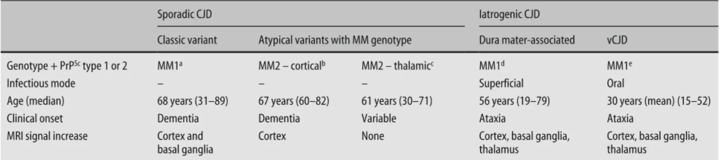
Table 5 Disease phenotypes in sporadic and iatrogenic CJD with MM genotype
Sporadic CJD Iatrogenic CJD
Classic variant Atypical variants with MM genotype Dura mater-associated vCJD Genotype + PrPSc type 1 or 2 MM1a MM2 – corticalb MM2 – thalamicc MM1d MM1e
Infectious mode – – – Superficial Oral
Age (median) 68 years (31–89) 67 years (60–82) 61 years (30–71) 56 years (19–79) 30 years (mean) (15–52)
Clinical onset Dementia Dementia Variable Ataxia Ataxia
MRI signal increase Cortex and basal ganglia
Cortex None Cortex, basal ganglia,
thalamus
Cortex, basal ganglia, thalamus
a [52] Collins et al. 2006; b [50] Krasnianski et al. 2006, [51] Hamaguchi et al. 2005; c 7 cases reported [51] Hamaguchi et al. 2005, [44] Shiga et al. 2004, [53] Yamashita et al. 2001; d 5 literature cases ([31] Kopp et al. 1996, [30] Hannah et al. 2001, [34] Nishida et al. 2002, [33] Mochizuki et al. 2003, [9] Wakisaka et al. 2006) and 3 own cases; e [54] Zeidler et al. 2000, [22] Collie et al. 2003
References
1. Brown P, Brandel JP, Preese M, Sato T (2006) Iatrogenic Creutzfeldt-Jakob disease: the waning of an era. Neurol-ogy 67:389–393
2. Sanchez-Juan P, Green A, Ladogana A, Cuadrado-Corrales N, Sanchez-Valle R, Mitrova E, Stoeck K, Sklaviadis T, Kulczycki J, Hess K, Bodemer M, Slivarichova D, Saiz A, Calero M, Ingrosso L, Knight R, Janssens C, Van Duijn C, Zerr I (2006) Cerebrospinal fluid tests in the differential diagnosis of CJD. Neurology 67:637–643 3. Boutoleau C, Guillon B, Martinez F,
Vercelletto M, Faure A, Feve JR (2003) Iatrogenic Creutzfeldt-Jakob disease subsequent to dural graft: persisting risk after 1987. Eur J Neurol 10: 521–523
4. Croes EA, Jansen GH, Lemstra AW, Frijns CJ, van Gool WA, van Duijn CM (2001) The first two patients with dura mater associated Creutzfeldt-Jakob disease in the Netherlands. J Neurol 248:877–880
5. Kimura K, Nonaka A, Tashiro H, Yaginuma M, Shimokawa R, Okeda R, Yamada M (2001) Atypical form of dural graft associated Jakob disease: report of a postmortem case with review of the literature. J Neurol Neurosurg Psychiatry 70: 696–699
6. Lang CJ, Heckmann JG, Querner V, Neundorfer B, Kornhuber J, Buchfelder M, Kretzschmar H (2001) Disease latency in Creutzfeldt-Jakob disease via dural grafting: a case report. Eur J Epidemiol 17:1013–1014
7. Mochizuki Y, Mizutani T, Tajiri N, Oinuma T, Nemoto N, Kakimi S, Kiamoto T (2003) Creutzfeldt-Jakob disease with florid plaques after cadaveric dura mater graft. Neuro-pathology 23:136–140
8. Preusser M, Strobel T, Gelpi E, Eiler M, Broessner G, Schmutzhard E, Budka H (2006) Alzheimer-type neuropathology in a 28 year old patient with iatrogenic Creutzfeldt-Jakob disease after dural grafting. J Neurol Neurosurg Psychia-try 77:413–416
9. Wakisaka Y, Santa N, Doh-ura K, Kitamoto T, Ibayashi S, Iida M, Iwaki T (2006) Increased asymmetric pulvinar magnetic resonance imaging signals in Creutzfeld-Jakob disease with florid plaques following a cadaveric dura mater graft. Neuropathology 26:82–88 10. Fradkin JE, Schonberger LB, Mills JL,
Gunn WJ, Piper JM, Wysowski DK, Thomson R, Durako S, Brown P (1991) Creutzfeldt-Jakob disease in pituitary growth hormone recipients in the United States. JAMA 265:880–884
11. Hoshi K, Yoshino H, Urata J, Nakamura Y, Yanagawa H, Sato T (2000)
Creutzfeldt-Jakob disease associated with cadaveric dura mater grafts in Japan. Neurology 55:718–721 12. Lang CJ, Heckmann JG, Neundorfer B
(1998) Creutzfeldt-Jakob disease via dural and corneal transplants. J Neurol Sci 160:128–139
13. Will RG, Zeidler M, Stewart GE, Macleod MA, Ironside JW, Cousens SN, Mackenzie J, Estibeiro K, Green AJ, Knight RS (2000) Diagnosis of new variant Creutzfeldt-Jakob disease. Ann Neurol 47:575–582
14. Iwasaki Y, Mimuro M, Yoshida M, Hashizume Y, Kitamoto T, Sobue G (2008) Clinicopathologic characteris-tics of five autopsied cases of dura mater-associated Creutzfeldt-Jakob disease. Neuropathology 28:51–61 15. Heath CA, Barker RA, Esmonde TFG,
Harvery P, Roberts R, Trend P, Head MW, Smith C, Bell JE, Ironside JW, Will RG, Knight RSG (2006) Dura associated Creutzfeldt-Jakob disease: experience from surveillance in the UK. J Neurol Neurosurg Psychiatry 77:880–882
16. WHO (1998) Human transmissible spongiform encephalopathies. Wkly Epidemiol Rec 47:361–365
17. Windl O, Giese A, Schulz-Schaeffer W, Zerr I, Skworc K, Arendt S, Oberdieck C, Bodemer M, Poser S, Kretzschmar HA (1999) Molecular genetics of human prion diseases in Germany. Hum Genet 105:244–252
18. Kretzschmar HA, Ironside JW, DeArmond SJ, Tateishi J (1996) Diagnostic criteria for sporadic Creutzfeldt-Jakob disease. Arch Neurol 53:913–920
19. Parchi P, Castellani R, Capellari S, Ghetti B, Young K, Chen SG, Farlow M, Dickson DW, Sima AAF, Trojanowski JQ, Petersen RB, Gambetti P (1996) Molecular basis of phenotypic vari-ability in sporadic Creutzfeldt-Jakob disease. Ann Neurol 39:767–778 20. Schulz-Schaeffer WJ, Tschoke S,
Kranefuss N, Drose W, Hause-Reitner D, Giese A, Groschup MH, Kretzschmar HA (2000) The paraffin-embedded tissue blot detects PrP(Sc) early in the incubation time in prion diseases. Am J Pathol 156:51–56
21. Kretzschmar H, Sethi S, Földvári Z, Windl O, Querner V, Zerr I, Poser S (2003) Iatrogenic Creutzfeldt-Jakob Disease with florid plaques. Brain Pathol 13:245–249
22. Collie DA, Summers DM, Sellar RJ, Ironside JW, Cooper S, Zeidler M, Knight R, Will R (2003) Diagnosing variant Creutzfeldt-Jakob disease with the pulvinar sign: MR imaging find-ings in 86 neuropathologically con-firmed cases. Am J Neuroradiol 24: 1560–1569
23. WHO (2001) The Revision of the Surveillance Case Definition for Variant Creutzfeldt-Jakob Disease (vCJD): Report of a WHO Consultation Edinburgh, United Kingdom, 17 May 2001. Available at: http://www.who.int/ csr/resources/publications/bse/ whocdscsreph20015.pdf
24. Noguchi-Shinohara M, Hamaguchi T, Kitamoto T, Sato T, Nakamura Y, Mizusawa H, Yamada M (2007) Clinical features and diagnosis of dura mater graft associated Creutzfeldt-Jakob disease. Neurology 69:360–367 25. Kobayashi Y, Hirata K, Tanaka H,
Yamada T (2003) Quinacrine adminis-tration to a patient with Creutzfeldt-Jakob disease who received a cadaveric dura mater graft – an EEG evaluation. Rinsho Shinkeigaku 43:403–408 26. Ukisu R, Kushihashi T, Kitanosono T,
Fujisawa H, Takenaka H, Ohgiya Y, Gokan T, Munechika H (2005) Serial diffusion-weighted MRI of Creutzfeldt-Jakob disease. Am J Roentgenol 184:560–566
27. Antoine JC, Michel D, Bertholon P, Mosnier JF, Laplanche JL, Beaudry P, Hauw JJ, Veyret C (1997) Creutzfeldt-Jakob disease after extracranial dura mater embolization for a nasopharyn-geal angiofibroma. Neurology 48: 1451–1453
28. Croes EA, Roks G, Jansen GH, Nijssen PC, van Duijn CM (2002) Creutzfeldt-Jakob disease 38 years after diagnostic use of human growth hormone. J Neu-rol Neurosurg Psychiatry 72:792–793 29. Delisle MB, Fabre N, Rochiccioli P,
Doerr-Schott J, Rumeau JL, Bes A (1993) Creutzfeldt-Jakob disease after treatment with human extracted growth hormone. A clinicopathologi-cal study. Rev Neurol Paris 149: 524–527
30. Hannah EL, Belay ED, Gambetti P, Krause G, Parchi P, Capellari S, Hoffman RE, Schonberger LB (2001) Creutzfeldt-Jakob disease after receipt of a previously unimplicated brand of dura mater graft. Neurology 56: 1080–1083
31. Kopp N, Streichenberger N, Deslys JP, Laplanche JL, Chazot G (1996) Creutzfeldt-Jakob disease in a 52-year-old woman with florid plaques (letter). Lancet 348:1239–1240
32. Liscic RM, Brinar V, Miklic P, Barsic B, Himbele J (1999) Creutzfeldt-Jakob disease in a patient with a lyophilized dura mater graft. Acta Med Croatica 53:93–96
33. Martinez-Lage JF, Poza M, Sola J, Tortosa JG, Brown P, Cervenakova L, Esteban JA, Mendoza A (1994) Acci-dental transmission of Jakob disease by dural cadaveric grafts. J Neurol Neurosurg Psychiatry 57: 1091–1094
34. Nishida Y, Yamada M, Hara K, Tsunemi T, Yamawaki M, Shimokawa R, Okeda R, Tsutsumi T, Mizusawa H (2002) Creutzfeldt-Jakob disease after Jannet-ta’s operation with cadaveric dura mater graft: initial manifestations related to the grafted site. J Neurol 249:480–483
35. Pocchiari M, Masullo C, Salvatore M, Genuardi M, Galgani S (1992) Creutzfeldt-Jakob disease after non-commercial dura mater graft (letter). Lancet 340:614–615
36. Shimizu S, Hoshi K, Muramoto T, Homma M, Ironside JW, Kuzuhara S, Sato T, Yamamoto T, Kitamoto T (1999) Creutzfeldt-Jakob disease with florid-type plaques after cadaveric dura mater grafting. Arch Neurol 56: 357–362
37. Takashima S, Tateishi J, Taguchi Y, Inoue H (1997) Creutzfeldt-Jakob dis-ease with florid plaques after cadaveric dural graft in a Japanese woman. Lancet 350:865–866
38. Takayama S, Hatsuda N, Matsumura K, Nakasu S, Handa J (1993) Creutzfeldt-Jakob disease transmitted by cadaveric dural graft: a case report. No Shinkei Geka 21:167–170
39. Thadani V, Penar PL, Partington J, Kalb R, Janssen R, Schonberger LB, Rabkin CS, Prichard JW (1988) Jakob disease probably acquired from a cadaveric dura mater graft. Case report. J Neurosurg 69:766–769 40. Yamada M, Itoh Y, Suematsu N,
Matsushita M, Otomo E (1997) Panen-cephalopathic type of Jakob disease associated with cadaveric dura mater graft. J Neurol Neurosurg Psychiatry 63:524–527 41. Yamada S, Aiba T, Endo Y, Hara M,
Kitamoto T, Tateishi J (1994) Creutzfeldt-Jakob disease transmitted by a cadaveric dura mater graft. Neurosurgery 34:740–743
42. Parchi P, Giese A, Capellari S, Brown P, Schulz-Schaeffer W, Windl O, Zerr I, Budka H, Kopp N, Piccardo P, Poser S, Rojiani A, Streichemberger N, Julien J, Vital C, Ghetti B, Gambetti P, Kretzschmar HA (1999) Classification of sporadic Creutzfeldt-Jakob disease based on molecular and phenotypic analysis of 300 subjects. Ann Neurol 46:224–233
43. Zerr I, Schulz-Schaeffer WJ, Giese A, Bodemer M, Schröter A, Henkel K, Tschampa HJ, Windl O, Pfahlberg A, Steinhoff BJ, Gefeller O, A. KH, Poser S (2000) Current clinical diagnosis in CJD: identification of uncommon variants. Ann Neurol 48:323–329 44. Shiga Y, Miyazawa K, Sato S,
Fuku-shima R, Shibuya S, Sato Y, Konno H, Dohura K, Mugikura S, Tamura H, Higano S, Takahashi S, Itoyama Y (2004) Diffusion-weighted MRI ab-normalities as an early diagnostic marker for Creutzfeldt-Jakob disease. Neurology 63:443–449
45. Young GS, Geschwind MD, Fischbein NJ, Martindale JL, Henry RG, Liu S, Lu Y, Wong S, Liu H, Miller BL, Dillon WP (2005) Diffusion-weighted and fluid-attenuated inversion recovery imaging in Creutzfeldt-Jakob disease: high sensitivity and specificity for diagno-sis. Am J Neuroradiol 26:1551–1562 46. Tschampa HJ, Kallenberg K, Urbach H,
Meissner B, Nicolay C, Kretzschmar HA, Knauth M, Zerr I (2005) MRI in the diagnosis of sporadic Creutzfeldt-Jakob disease: a study on inter-ob-server agreement. Brain 128:2026–2033 47. Kallenberg K, Schulz-Schaeffer WJ,
Jastrow U, Poser S, Meissner B, Tschampa HJ, Zerr I, Knauth M (2006) Creutzfeldt-Jakob disease: Compara-tive Analysis of MR Imaging Sequences. Am J Neuroradiol 27: 1459–1462
48. Schroeter A, Zerr I, Henkel K, Tschampa HJ, Finkenstaedt M, Poser S (2000) Magnetic Resonance Imaging in the Clinical Diagnosis of
Creutzfeldt-Jakob Disease. Arch Neurol 57:1751–1757
49. Krasnianski A, Schulz-Schaeffer WJ, Kallenberg K, Meissner B, Collie DA, Roeber S, Bartl M, Heinemann U, Varges D, Kretzschmar HA, Zerr I (2006) Clinical findings and diagnostic tests in the MV-2 subtype of sporadic CJD. Brain 129:2288–2296
50. Krasnianski A, Meissner B, Schulz-Schaeffer W, Kallenberg K, Bartl M, Heinemann U, Varges D, Kretzschmar H, Zerr I (2006) Clinical features and diagnosis of the MM2 cortical subtype of sporadic Creutzfeldt-Jakob disease. Arch Neurol 63:876–880
51. Hamaguchi T, Kitamoto T, Sato T, Mizusawa H, Nakamura Y, Noguchi M, Furukawa Y, Ishida C, Kuji I, Mitani K, Murayama S, Kohriyama T, Katayama S, Yamashita M, Yamamoto T, Udaka F, Kawakami A, Ihara Y, Nishinaka T, Kuroda S, Suzuki N, Shiga Y, Arai H, Maruyama M, Yamada M (2005) Clinical diagnosis of MM2-type sporadic Creutzfeldt-Jakob disease. Neurology 64:643–648
52. Collins SJ, Sanchez-Juan P, Masters CL, Klug GM, van Duijn C, Poleggi A, Almonti S, Cuadrado-Corrales N, de Pedro-Cuesta J, Budka H, Gelpi E, Glatzel M, Seeger HE, Zerr I, Kretzschmar H, Jansen GH, Olsen E, Mitrova E, Alpérovitsch A, Brandel JP, Murray K, Will RG (2006) Determi-nants of diagnostic investigation results across the clinical spectrum of sporadic CJD. Brain 129:2278–2287 53. Yamashita M, Yamamoto T, Nishinaka
K, Udaka F, Kameyama M, Kitamoto T (2001) Severe brain atrophy in a case of thalamic variant of sporadic CJD with plaque-like PrP deposition. Neuropathology 21:138–143
54. Zeidler M, Ironside JW (2000) The new variant of Creutzfeldt-Jakob disease. Rev Sci Tech 19:98–120
55. Gurau C, Zeghoudi AC, Atlaoui-Rabia S, Charitanski D, Mallecourt J (2005) Cerebral magnetic resonance imaging in iatrogenic Creutzfeldt-Jakob disease. Presse Med 34:1080–1081
56. Wroe SJ, Pal S, Siddique D, Hyare H, Macfarlane R, Joiner S, Linehan JM, Brandner S, Wadsworth JD, Hewitt P, Collinge J (2006) Clinical presentation and pre-mortem diagnosis of variant Creutzfeldt-Jakob disease associated with blood transfusion: a case report. Lancet 368:2061–2067
57. Brandel JP, Preece M, Brown P, Croes E, Laplanche JL, Agid Y, Will RG, Alpéro-vitsch A (2003) Distribution of codon 129 genotype in human growth hor-mone-treated CJD patients in France and the UK. Lancet 362:128–130 58. Deslys JP, Jaegly A, d’Aignaux JH,
Mouthon F, de Villemeur TB, Dormont D (1998) Genotype at codon 129 and susceptibility to Creutzfeldt-Jakob disease (letter). Lancet 351:1251 59. Huillard d’Aignaux J, Costagliola D,
Maccario J, Billette de Villemeur T, Brandel JP, Deslys JP, Hauw JJ, Chaus-sain JL, Agid Y, Dormond D, Alpéro-vitch A (1999) Incubation period of Creutzfeldt-Jakob disease in human growth hormone recipients in France. Neurology 53:1197–1201
60. Brown P, Preece M, Brandel J-P, Sato T, McShane L, Zerr I, Fletcher A, Will RG, Pocchiari M, Cashmann NR, d’Aignaux JH, Cervenáková L, Fradkin J, Schon-berger LB, Collins SJ (2000) Iatrogenic Creutzfeldt-Jakob disease at the millennium. Neurology 55:1075–1081