ORIGINAL ARTICLE DOI 10.1007/s00774-006-0732-x
A.A. Kurth · S.-Z. Kim
Department of Orthopaedic Surgery, University Hospital Frankfurt/Main, Frankfurt/Main, Germany
M. Shea
Orthopaedic Biomechanics Laboratory, Department of Orthopaedics & Rehabilitation, Oregon Health and Science University, Portland, OR, USA
F. Bauss
Roche Diagnostics GmbH, Pharma Research, Penzberg, Germany W.C. Hayes
Orthopaedic Biomechanics Laboratory, Beth Israel Deaconess Medical Center and Harvard Medical School, Boston, MA, USA R. Müller (*)
Institute for Biomedical Engineering, Swiss Federal Institute of Technology (ETH) and University of Zürich, Moussonstrasse 18, 8044 Zürich, Switzerland
Tel./Fax +41-44-632-4592; Fax +41-44-632-1214 e-mail: ralph.mueller@ethz.ch
Andreas A. Kurth · Soo-Zin Kim · Marie Shea Frieder Bauss · Wilson C. Hayes · Ralph Müller
Preventative ibandronate treatment has the most benefi cial effect on
the microstructure of bone in experimental tumor osteolysis
Abstract We investigated the effect of ibandronate on three-dimensional (3-D) microstructure and bone mass in experimentally induced tumor osteolysis. Walker carcino-sarcoma cells were implanted into the left femur of female rats that received 26-day ibandronate pretreatment fol-lowed by continued therapy or ibandronate posttreatment only. A tumor-only group received isotonic saline. At end-point, excised femurs were scanned using microcomputed tomography (µCT) to assess bone volume density, bone mineral content, trabecular number/thickness, and separa-tion for cortical plus trabecular bone or trabecular bone alone. Compared with the nonimplanted right femur, bone volume and surface density and trabecular number and thickness were reduced in the distal left femur following tumor cell implantation. µCT analysis revealed greater cor-tical and trabecular bone mineral content in the preventa-tive and interventional (pre-post tumor) ibandronate group, and the interventional (post-tumor) ibandronate group, versus the tumor-only group. Bone volume density was signifi cantly higher in pre-post and post-tumor groups compared to the tumor-only group. After preventative and interventional ibandronate, bone volume density and tra-becular thickness were 13% and 60% greater, respectively,
than in the post-tumor treatment group. 3-D µCT images confi rmed microstructural changes. We conclude that combined interventional and preventative ibandronate pre-serves bone strength and integrity more than intervention alone.
Key words bisphosphonate · preclinical · computer tomog-raphy · skeletal integrity
Introduction
Skeletal metastases are the most frequent complication of malignant tumors and are associated with severe pain and pathological fractures. Although treatment for metastatic bone lesions does not alter life expectancy, preventing frac-tures can improve the quality of life of the cancer patient. Bisphosphonates are one class of therapeutic agents cur-rently being used or under investigation for various dis-eases related to pathologically increased bone resorption, such as Paget’s disease, postmenopausal osteoporosis, and tumor-induced hypercalcemia and osteolysis [1]. Over re-cent years, bisphosphonates have also become an estab-lished treatment for bone metastases, and are currently the only available treatment option for bone complications as-sociated with malignancies. Bisphosphonates are able to reduce the number of skeletal complications by inhibiting osteoclast-mediated bone destruction. Numerous experi-mental and clinical studies have shown that interventional treatment can reduce the occurrence of pathological frac-tures, bone pain, hypercalcemia, and the need for radio-therapy and surgery [2].
Preclinical studies and initial clinical experience show that preventative administration of bisphosphonates seems very promising and more benefi cial for the patient than palliative administration alone [3–9]. Investigations into the use of bisphosphonates are now directed to understand the effects of a preventative treatment on tumor osteolysis [10]. A recent investigation on bone quality in tumor osteolysis showed that tumor growth in bone resulted in a
87
decrease in bone mass and the loss of biomechanical com-petence of bone, but they are not correlated [11]. In addi-tion, new imaging techniques for determining the alteration of the three-dimensional (3-D) structure of trabecular bone, a factor known to infl uence its mechanical behavior, have been introduced [12].
We utilized this new technology in a previous study and demonstrated that microcomputed tomography (µCT) can provide 3-D parameters of bone mass and trabecular struc-ture in an animal model for tumor-induced bone loss [13]. We found signifi cant differences in 3-D morphometric pa-rameters between the tumor-bearing bones and contralat-eral controls.
In the current experimental study, we investigated the effect of a preventative administration of ibandronate on the 3-D microstructure of bone in a rat model for tumor-induced osteolysis. Building on previously published ex-periments [11,13–15], this study aimed to investigate whether interventional treatment with ibandronate affects the tumor-induced destruction of the 3-D structure of tra-becular bone, and whether preventative treatment com-bined with interventional treatment preserves the 3-D structure of trabecular bone in tumor osteolysis better than interventional treatment alone.
Materials and methods
Animal model and treatment protocol
We used a previously described animal model to evaluate densitometric and biomechanical properties in tumor oste-olysis [11,13–15]. The widely used Walker Carcinosarcoma 256 malignant breast cancer cells (W256) were cultured in 0.6% agarose gel solution. After anesthesia by an intraperi-toneal injection of ketamine hydrochloride (75 mg/kg) and xylazine (5 mg/kg), equivalent volumes of tumor cells in agarose gel containing approximately 2 × 10⁶ cells were
surgically implanted into the medullary canal of the left femur via a drill hole in the intercondylar notch (see [15] for additional details). Forty-fi ve female Sprague–Dawley rats aged 4 months were randomly assigned to three study groups. All animals were maintained in accordance with federal regulations, and the study was conducted with the approval of the Institutional Animal Care and Use Committee (Harvard Medical School). In the fi rst group (tumor-only), Walker 256 tumor cells were implanted into the left femur without treatment. The second group (pre-post) fi rst underwent a daily pretreatment with ibandronate (Roche Diagnostics, Mannheim, Germany; 25 µg/kg sub-cutaneously) for 26 days; then, tumor cells were implanted and the daily treatment was continued until death. The third group (post) received daily ibandronate treatment only after tumor cell implantation. There were no compli-cations from the surgical procedure. Twenty-eight days after surgery, all animals were killed and both femora from each rat were harvested and cleaned of soft tissue for image analysis. Before microtomographic scanning, all femora were radiographed in an anteroposterior plane using a
Faxitron machine (Hewlett-Packard, McMinnville, OR, USA).
Structural analysis using microcomputed tomography (µCT)
As in our recent study [13], we employed a desktop µCT imaging system (µCT 20; Scanco Medical, Bassersdorf, Switzerland: 30 µm, nominal resolution) to assess 3-D den-sitometric and architectural parameters in small animal whole bones. The µCT system has a micro-X-ray source (10 µm focal spot, 25 keV) directed toward the sample. The quantitative modifi cation of the X-ray beam by apatite crystals contained in the bone sample is analyzed by a plane detector (CCD array; 1024 elements). The process is piloted by an Alpha workstation (HP, Palo Alto, CA, USA) and an OpenVMS system in cluster confi guration to per-form the 3-D analysis. For each specimen, a total of 200 microtomographic slices were taken, each 30 µm thick, starting from a reproducible anatomical landmark right behind the boundary of the condyles, thereby covering a 6-mm region of the distal femur. From the obtained two-dimensional slices, 3-D reconstruction of bone was auto-matically performed using the triangulation algorithm. Architectural indices were calculated in the spherical vol-ume of interest according to standard defi nitions used in histomorphometry [11]. Bone volume density (bone vol-ume/tissue volume), bone surface density (bone surface/ bone volume), trabecular thickness, trabecular separation, and trabecular number were determined directly from the 3-D images. Bone mineral content, calculated using the measured bone volume and assuming a constant tissue density, was measured in a metaphyseal region consisting of both cortical and trabecular bone, and a sphere consist-ing of trabecular bone only. In a previous study, calcula-tions of bone mineral content using this method were shown to correlate closely to dual-energy X-ray absorpti-ometry (DXA)-based methods [16], and in a separate study, the bone volume densities of metastatic cancer bone and normal bone were found to be similar [17]. A total of 90 specimens (left and right femora) were scanned. All 90 specimens were analyzed using both visual assessment and quantitative morphometry.
Statistical analysis
A Wilk–Shapiro test for normality was conducted on all parameters. All data were expressed as the mean ± stan-dard deviation. Analysis of variance (ANOVA) with Student–Newman–Keuls post hoc analysis, or a paired t test was performed to determine if the measurements dif-fered signifi cantly (statistical signifi cance level, P < 0.05).
Results
This model of tumor induction in bone has been character-ized previously [11,13–15]. As with previous studies,
radiographs confi rmed that all femora implanted with tu-mor cells contained osteolytic lesions [11,15]. In addition, there was macroscopic evidence of soft tissue tumors sur-rounding affected bones.
Compared with the nonimplanted right femur, bone vol-ume density, bone surface density, trabecular number, and trabecular thickness were signifi cantly reduced in the dis-tal left femur following the implantation of tumor cells (P < 0.001). Trabecular separation was signifi cantly in-creased in the left femur (Table 1).
The µCT analysis revealed greater values for cortical and trabecular bone mineral content in the preventative and interventional (pre-post tumor) ibandronate treatment group, and the interventional (post-tumor) ibandronate treatment group, than in the tumor-only group (Table 2). Morphometric analysis of trabecular volume revealed architectural changes in accordance with the quantitative changes seen in bone density (Figs. 1–4).
Changes in architectural parameters of the left femur following treatment with ibandronate are summarized in Table 3. Bone volume density was signifi cantly higher in the pre-post and post-tumor groups compared to the tumor-only group, showing an increase of 133% and 106%, respectively (P < 0.05) (Table 3). These results are most likely caused by a signifi cant increase in trabecular thick-ness (up by 211% and 95% versus the tumor-only group, respectively; P < 0.05) (Table 3). In addition, trabecular separation signifi cantly decreased in the pre-post (−74%)
Table 1. Architectural parameters of the distal left (implanted) and right (nonimplanted) femur (no ibandronate treatment)
Group (n) Bone volume Bone surface Trabecular Trabecular Trabecular
density (%) density (mm2/mm3) number (1/mm) thickness (mm) separation (mm) Left femur (15) 33.78 (11.30) 6.66 (1.72) 2.79 (1.03) 0.09 (0.01) 0.33 (0.21) Right femur (15) 48.24 (5.95) 8.41 (0.64) 3.69 (0.51) 0.10 (0.02) 0.18 (0.05)
Group differences (%) −30* −21* −24* −10* 83*
Data are means (SD) * P < 0.001 (paired t test)
Table 2. Bone mineral content for full and trabecular regions in the
left operated femur measured using micro-computed tomography (µCT)
Groupa (n) Full bone Trabecular bone
mineral content mineral content Pre-post (15) 0.17 (0.02) 0.07 (0.01) Post (15) 0.16 (0.02) 0.06 (0.01) Tumor only (15) 0.10 (0.02) 0.02 (0.01) Group differences (%)
Pre-post vs. tumor only 74* 174*
Pre-post vs. post 11* 21*
Post vs. tumor only 57* 127*
Data are mean in grams (SD)
a Group title indicates treatments received: pre-post, ibandronate administered both before and after tumor cell implantation; post, ibandronate administered after tumor implantation but not before; tumor only, tumor implantation with no ibandronate treatment (see Materials and methods for further details)
* P < 0.05 (ANOVA with Student–Newman–Keuls post hoc comparisons)
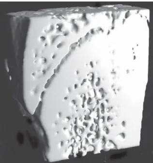
Fig. 1. Three-dimensional (3-D) reconstruction of microcomputed
tomography (µCT) slices for a control bone with no tumor and pla-cebo treatment (a 6-mm region is shown)
Fig. 2. 3-D reconstruction of µCT slices for a tumor-bearing bone with placebo treatment (a 6-mm region is shown)
89
Fig. 3. 3-D reconstruction of µCT slices for a tumor-bearing bone with interventional ibandronate treatment (a 6-mm region is shown)
Fig. 4. 3-D reconstruction of µCT slices for a tumor-bearing bone with preventative and interventional ibandronate treatment (a 6-mm region is shown)
Table 3. Architectural parameters of the distal left (operated) femur measured using microcomputed tomography (µCT)
Groupa (n) Bone volume Bone surface Trabecular Trabecular Trabecular
density (%) density (mm2/mm3) number (1/mm) thickness (mm) separation (mm) Pre-post (15) 78.58 (4.52) 5.83 (0.85) 2.80 (0.52) 0.28 (0.07) 0.09 (0.02) Post (15) 69.52 (7.32) 7.49 (0.61) 3.77 (0.37) 0.18 (0.04) 0.09 (0.02) Tumor only (15) 33.78 (11.30) 6.66 (1.72) 2.79 (1.03) 0.09 (0.01) 0.33 (0.21) Group differences (%) Pre-post vs. tumor only 133* −12 0 211* −74* Pre-post vs. post 13* −22* −26* 60* −3
Post vs. tumor only 106* 12 35* 95* −73*
Data are mean (SD)
a Group title indicates treatments received: pre-post, ibandronate administered both before and after tumor cell implantation; post, ibandro-nate administered after tumor implantation but not before; tumor only, tumor implantation with no ibandroibandro-nate treatment (see Materials and methods for further details)
* P < 0.05 (ANOVA with Student–Newman–Keuls post hoc comparisons)
and post-tumor (−73%) treatment groups (P < 0.05 versus tumor only) (Table 3).
Values recorded in the pre-post ibandronate treatment group were 13% greater for bone volume density and 60% greater for trabecular thickness than in the post-tumor treatment group (P < 0.05) (Table 3). Bone surface density and trabecular number were not affected by ibandronate treatment (see Table 3).
Discussion
The aim of the present study was to investigate the effect of a bisphosphonate, ibandronate, on bone density and 3-D bone structure in a rat model of tumor-induced osteolysis. Previously, we presented biomechanical and
mineralization data with this model and treatment regi-men, including DXA-based measurements, and also dem-onstrated no effects on serum calcium with the ibandronate doses used [11]. Other studies have also demonstrated that ibandronate does not induce either hypomineralization or hypermineralization, even at doses exceeding any thera-peutically intended dose by multiples [11,18,19].
From clinical experience, it is known that bone metas-tases frequently occur in the metaphyseal regions of long bones and the vertebral bodies of the spine. In those areas, the turnover rate of trabecular bone is much greater than that of cortical bone; consequently, dramatic changes in tumor osteolysis occur in trabecular bone much later than in cortical bone. This also is the region of bone where the effect of an interventional treatment with bisphosphonates is more pronounced [20,21]. In recent years, 3-D analysis of bone has become possible using µCT technology. This
tool rapidly evaluates the internal morphology and struc-ture of trabecular and cortical bone, and has been widely used in osteoporosis research and to examine the effects of pharmacological intervention on bone [12,22–27]. In par-ticular, µCT imaging allows the noninvasive examination of the quantity, orientation, and connection of trabecular bone elements separately from cortical bone by focusing on a selected volume of interest containing trabecular bone only.
The results of the present study confi rm that ibandro-nate administered either as a daily interventional treat-ment starting after the onset of tumor growth in bone, or as both a preventative and interventional treatment, pre-serves bone mass and 3-D structure compared with an un-treated tumor-only control group. However, we found a greater benefi cial effect on density, mineral content, and trabecular thickness in the preventative and interventional ibandronate group than the intervention-only group. In a previous study, we found that preventative and interven-tional treatment increased failure load by 9% versus inter-ventional treatment alone in this model, although this was not statistically signifi cant [11]. These data may suggest a potential for preventative ibandronate treatment to reduce the risk of fractures for patients. The similar number of trabeculae in the treatment groups indicates that ibandro-nate affected mostly existing trabeculae (i.e., increasing their width) rather than inducing formation of new trabec-ulae. These results are corroborated by two recent in vivo studies of bisphosphonate action on trabecular microarchi-tecture: a macaque study of preventative ibandronate for ovariectomy-induced bone loss in the lumbar spine [16], and a human trial of risedronate for preserving iliac bone architecture in postmenopausal women with osteoporosis [28]. In both studies, trabecular number was preserved and trabecular thickness was increased in treatment groups compared with controls or baseline values.
Several studies in different experimental models have shown that preventative bisphosphonate treatment reduces the development of bone metastases. Preventative etidro-nate treatment reduced osteolysis and prolonged survival in a rat model of bone metastasis using bladder carcinoma cells [29]. In other studies using parathyroid hormone-related protein-producing Walker 256 carcinosarcoma cells inoculated intraosseously into rats, pretreatment with clo-dronate or pamiclo-dronate was osteoprotective, with the ex-tent of the effect correlating with duration and intensity of treatment [7–9,30]. Preventative treatment with pamidro-nate was also shown to increase trabecular volume, al-though an increase in skeletal tumor burden was observed, with no effects on nonosseous metastases [6]. However, other studies of human breast carcinoma cells injected into nude mice have shown a reduced tumor burden following pretreatment with ibandronate [31] or risedronate [32]. In addition, in a similar rat model, risedronate pretreatment decreased the incidence of bone metastases with no effect on visceral metastases [4]. Previous studies performed in our laboratory showed that combined preventative and interventional treatment with ibandronate most effec-tively preserved bone mass and strength, although all
ibandronate treatment schedules resulted in signifi cant im-provements compared with untreated tumor-bearing con-trols [11,14]. Supporting these results, a recent study of experimentally induced multiple tumor osteolysis in rats found that initiation of ibandronate therapy 3 days before tumor cell inoculation substantially reduced the develop-ment of osteolytic lesions compared with postoperative ibandronate administration [33].
Our study did not investigate the mechanism of action for the observed benefi ts of preventative ibandronate treat-ment. Bisphosphonates have several potential inhibitory effects on bone metastases. In particular, these agents have a well-documented inhibitory effect on osteoclast function. Studies have shown that bisphosphonates induce apoptosis in murine osteoclasts both in vivo and in vitro, and a great-er effect is seen with more potent agents [34,35]. In addi-tion, bisphosphonates may promote the secretion of an osteoclast inhibitory factor by osteoblasts [36]. Bisphos-phonates also induce apoptosis in macrophages [37,38] and human myeloma cell lines [39] in vitro. Other biological effects include inhibiting the adhesion of breast or prostate carcinoma cells to bone matrices [40,41], inhibiting matrix metalloproteinases in vitro and in vivo, and decreasing the invasive properties of malignant melanoma and fi brosar-coma cell lines [31,42].
In a previous study from our laboratory, we assessed the effects of preventative and interventional ibandronate treatment on nonimplanted femora [11]. As is well estab-lished, ibandronate increased various bone parameters (such as bone mineral density) in femora without tumors; however, the effects were less than those observed in tumor-implanted femora. Although this confi rms that ibandronate has additional effects on tumor-containing bone, it is unclear whether this is a result of timing of treat-ment or total dose administered. Regardless of mechanism, the present study further demonstrates that combined pre-ventative and interventional ibandronate treatment is most benefi cial in our model, and this fi nding has direct rele-vance to clinical dosing.
Conclusions
In our animal model investigating the structural conse-quences of tumor destruction in trabecular bone, both ibandronate treatment schedules resulted in signifi cantly greater bone mass and improved structural parameters compared with no treatment. Combined preventative and continued treatment provided the best protection against tumor-mediated bone destruction and preserved the integ-rity of bone, even in the presence of a malignant tumor. Our experimentally obtained results for bone structure are in concordance with accumulating clinical evidence sug-gesting that preventative treatment with bisphosphonates can reduce the occurrence of skeletal complications in can-cer patients and thus may prolong survival [43,44]. These animal experiments and the current clinical experience with bisphosphonates suggest that further investigations
91
that focus on preventative treatment are likely to be very promising.
Acknowledgments We thank Dan Michaeli, M.S., and John Hipp,
Ph.D., for their input. This work was supported in part by a grant of the German National Science Foundation (Deutsche Forschungsge-meinschaft) and the G. Wallrabenstein Found (AAK). The Maurice E. Müller Professorship at the Harvard Medical School partly pro-vided the fi nancial support for the µCT system. Gardiner-Caldwell US provided editorial assistance.
References
1. Fleisch H (2000) Bisphosphonates in Bone Disease. Academic Press, San Diego
2. Diel IJ, Solomayer EF, Bastert G (2000) Bisphosphonates and the prevention of metastasis: fi rst evidences from preclinical and clinical studies. Cancer (Phila) 88:3080–3088
3. Guaitani A, Polentarutti N, Filippeschi S, Marmonti L, Corti F, Italia C, Coccioli G, Donelli MG, Mantovani A, Garattini S (1984) Effects of disodium etidronate in murine tumor models. Eur J Cancer Clin Oncol 20:685–693
4. Hall DG, Stoica G (1994) Effect of the bisphosphonate risedro-nate on bone metastases in a rat mammary adenocarcinoma model system. J Bone Miner Res 9:221–230
5. Jung A, Bornand J, Mermillod B, Edouard C, Meunier PJ (1984) Inhibition by diphosphonates of bone resorption induced by the Walker tumor of the rat. Cancer Res 44:3007–3011
6. Kostenuik PJ, Orr FW, Suyama K, Singh G (1993) Increased growth rate and tumor burden of spontaneously metastatic Walker 256 cancer cells in the skeleton of bisphosphonate-treated rats. Cancer Res 53:5452–5457
7. Krempien B, Diel IJ, Jöckle-Kretz B, Buchele R, Andre L (1984) The Walker carcinosarcoma 256 as an experimental model of bone metastasis. Infl uence of skeletal metabolism on the development of bone metastases. Verh Dtsch Ges Pathol 68:211–216
8. Krempien B, Manegold C (1993) Prophylactic treatment of skel-etal metastases, tumor-induced osteolysis, and hypercalcemia in rats with the bisphosphonate Cl2MBP. Cancer (Phila) 72:91–98 9. Krempien B, Wingen F, Eichmann T, Muller M, Schmahl D
(1988) Protective effects of a prophylactic treatment with the bisphosphonate 3-amino-1-hydroxypropane-1,1-bisphosphonic acid on the development of tumor osteopathies in the rat: experi-mental studies with the Walker carcinosarcoma 256. Oncology 45:41–46
10. Diel IJ (2000) Antitumour effects of bisphosphonates: fi rst evi-dence and possible mechanisms. Drugs 59:391–399
11. Kurth AA, Kim SZ, Sedlmeyer I, Bauss F, Shea M (2002) Iban-dronate treatment decreases the effects of tumor-associated lesions on bone density and strength in the rat. Bone (NY) 30:300–306
12. Muller R, Hildebrand T, Ruegsegger P (1994) Non-invasive bone biopsy: a new method to analyse and display the 3-dimensional structure of trabecular bone. Phys Med Biol 39:145–164 13. Kurth AA, Muller R (2001) The effect of an osteolytic tumor on
the 3-dimensional trabecular bone morphology in an animal model. Skeletal Radiol 30:94–98
14. Kurth AHA, Kim S-Z, Sedlmeyer I, Hovy L, Bauss F (2000) Treatment with ibandronate preserves bone in experimental tumour-induced bone loss. J Bone Joint Surg 82B:126–130 15. Kurth AA, Wang CO, Hayes WC, Shea M (2001) The evaluation
of a rat model for the analysis of densitometric and biomechanical properties of tumor-induced osteolysis. J Orthop Res 19: 200–205
16. Müller R, Hannan M, Smith SY, Bauss F (2004) Intermittent ibandronate preserves bone quality and bone strength in the lumbar spine after 16 months of treatment in the ovariectomized cynomolgus monkey. J Bone Miner Res 19:1787–1796
17. Nazarian A, von Stechow D, Rho JY, Grynpas M, Zurakowski D, Müller R, Snyder BD (2005) Structure dominates tissue
properties in metastatic cancer. Cancer Treat Rev 31(suppl 1): S53–S54 (abstract 103)
18. Lalla S, Hothorn LA, Haag N, Bader R, Bauss F (1998) Lifelong administration of high doses of ibandronate increases bone mass and maintains bone quality of lumbar vertebrae in rats. Osteopo-ros Int 8:97–103
19. Bauss F, Russell RG (2004) Ibandronate in osteoporosis: pre-clinical data and rationale for intermittent dosing. Osteoporos Int 15:423–433
20. Pataki A, Muller K, Green JR, Ma YF, Li QN, Jee WS (1997) Effects of short-term treatment with the bisphosphonates zole-dronate and pamizole-dronate on rat bone: a comparative histomor-phometric study on the cancellous bone formed before, during, and after treatment. Anat Rec 249:458–468
21. Peyruchaud O, Winding B, Pécheur I, Serre CM, Delmas P, Clezardin P (2001) Early detection of bone metastases in a mu-rine model using fl uorescent human breast cancer cells: applica-tion of to the use of the bisphosphonate zoledronic acid in the treatment of osteolytic lesions. J Bone Miner Res 16:2027–2034 22. Laib A, Kumer JL, Majumdar S, Lane NE (2001) The temporal
changes of trabecular architecture in ovariectomized rats as-sessed by microCT. Osteoporosis Int 12:936–941
23. Lane NE, Kumer JL, Majumdar S, Khan M, Lotz J, Stevens RE, Klein R, Phelps KV (2002) The effects of synthetic conjugated estrogens A (cenestin) on trabecular bone structure and strength in the ovariectomized rat model. Osteoporosis Int 13:816–823 24. Rüegsegger P, Koller B, Müller R (1996) A microtomograpic
system for nondestructive evaluation of bone architecture. Calcif Tissue Int 58:24–29
25. Hildebrand T, Laib A, Müller R, Dequeker J, Ruegsegger P (1999) Direct 3-D morphometric analysis of human cancellous bone: microstructural data from spine, femur, iliac crest, and calcaneus. J Bone Miner Res 14:1167–1174
26. Borah B, Dufresne TE, Chmielewski PA, Gross GJ, Prenger MC, Phipps RJ (2002) Risedronate preserves trabecular architecture and increases bone strength in vertebra of ovariectomized mini-pigs as measured by 3-dimensional microcomputed tomography. J Bone Miner Res 17:1139–1147
27. Jiang Y, Zhao JJ, Mitlak BH, Wang O, Genant HK, Eriksen EF (2003) Recombinant human parathyroid hormone (1-34) [teripa-ratide] improves both cortical and cancellous bone structure. J Bone Miner Res 18:1932–1941
28. Borah B, Dufresne TE, Chmielewski PA, Johnson TD, Chines A, Manhart MD (2004) Risedronate preserves bone architecture in postmenopausal women with osteoporosis as measured by three-dimensional microcomputed tomography. Bone (NY) 34:736–746
29. Nemoto R, Uchida K, Tsutsumi M, Koiso K, Satou S, Satou T (1987) A model of localized osteolysis induced by the MBT-2 tumor mice and its responsiveness to etidronate disodium. J Can-cer Res Clin Oncol 113:539–543
30. Wingen F, Eichmann T, Manegold C, Krempien B (1986) Effects of new bisphosphonic acids on tumor-induced bone destruction in the rat. J Cancer Res Clin Oncol 111:35–41
31. Yoneda T, Sasaki A, Dunstan C, Williams PJ, Bauss F, De Clerck YA, Mundy GR (1997) Inhibition of osteolytic bone metastasis of breast cancer by combined treatment with the bisphosphonate ibandronate and tissue inhibitor of the matrix metalloproteinase-2. J Clin Invest 99:2509–2517
32. Sasaki A, Boyce BF, Story B, Wright KR, Chapman M, Boyce R, Mundy GR (1995) Bisphosphonate risedronate reduces meta-static human breast cancer burden in bone in nude mice. Cancer Res 55:3551–3557
33. Neudert M, Fischer C, Krempien B, Bauss F, Seibel MJ (2003) Site-specifi c human breast cancer (MDA-MB-231) metastases in nude rats: model characterisation and in vivo effects of ibandro-nate on tumour growth. Int J Cancer 107:468–477
34. Hughes DE, Wright KR, Uy HL, Sasaki A, Yoneda T, Roodman GD, Mundy GR, Boyce BF (1995) Bisphosphonates promote apoptosis in murine osteoclasts in vitro and in vivo. J Bone Miner Res 10:1478–1487
35. Hiraga T, Williams PJ, Mundy GR, Yoneda T (2001) The bisphos-phonate ibandronate promotes apoptosis in MDA-MB-231 human breast cancer cells in bone metastases. Cancer Res 61:4418–4424
36. Vitte C, Fleisch H, Guenther HL (1996) Bisphosphonates induce osteoblasts to secrete an inhibitor of osteoclast-mediated resorp-tion. Eur J Cancer Clin Oncol 137:2324–2333
37. Rogers MJ, Chilton KM, Coxon FP, Lawry J, Smith MO, Suri S, Russell RG (1996) Bisphosphonates induce apoptosis in mouse macrophage-like cells in vitro by a nitric oxide independent mechanism. J Bone Miner Res 11:1482–1491
38. Selander KS, Monkkonen J, Karhukorpi EK, Harkonen P, Hannuniemi R, Vaananen HK (1996) Characteristics of clodronate-induced apoptosis in osteoclasts and macrophages. Mol Pharmacol 50:1127–1138
39. Shipman CM, Rogers MJ, Apperley JF, Russell RG, Croucher PI (1997) Bisphosphonates induce apoptosis in human myeloma cell lines: a novel anti-tumour activity. Br J Haematol 98:665–672 40. van der Pluijm G, Vloedgraven H, van Beek E, van der Wee-Pals
L, Lowik C, Papapoulos S (1996) Bisphosphonates inhibit the adhesion of breast cancer cells to bone matrices in vitro. J Clin Invest 98:698–705
41. Boissier S, Magnetto S, Frappart L, Cuzin B, Ebetino FH, Delmas PD, Clezardin P (2003) Bisphosphonates inhibit prostate and breast cancer call adhesion to unmineralized and mineral-ized bone extracellular matrices. Cancer Res 57:3890–3894 42. Teronen O, Heikkila P, Konttinen YT, Laitinen M, Salo T,
Hanemaaijer R, Teronen A, Maisi P, Sorsa T (1999) MMP inhibi-tion and down regulainhibi-tion by bisphosphonates. Ann NY Acad Sci 878:453–465
43. Diel IJ, Solomayer EF, Costa SD, Gollan C, Goerna R, Wallwiener D, Kaufmann M, Bastert G (1998) Reduction in new metastases in breast cancer with adjuvant clodronate treatment. N Engl J Med 339:357–363
44. Powles T, Paterson S, Kanis JA (2002) Randomized placebo-controlled trial of clodronate in patients with primary operable breast cancer. J Clin Oncol 20:3219–3224