ORIGINAL INVESTIGATION
Early effects of mood stabilizers on the Akt/GSK-3
β
signaling pathway and on cell survival and proliferation
Jean-Michel Aubry&Michèle Schwald&Eladia Ballmann&Félicien Karege
Received: 16 January 2009 / Accepted: 20 April 2009 / Published online: 14 May 2009
# Springer-Verlag 2009
Abstract
Rationale Lithium, some of the anticonvulsants, and several second-generation antipsychotic drugs are common medi-cations widely prescribed to treat bipolar disorder. Molecular targets and cellular events that mediate their effects have been described for these drugs but are only partially unraveled. Few comparative studies have been performed. Objectives We evaluated seven mood stabilizers (MS) in the same in vitro system and found several differences and similarities in their cellular mechanisms (proliferation and cell survival). As some MS were previously shown to activate the Akt/GSK-3β axis, this pathway was explored for other drugs.
Materials and methods The SH-SY5Y cells were cultured in RPMI-1640 medium. Effects of MS drugs on serum-induced cell proliferation and on slowing of cell death were analyzed. Phosphorylation and expression of Akt-1 and GSK-3β mRNA and protein were assessed for the seven drugs as well.
Results Lithium, Valproate, Olanzapine, and Clozapine enhance proliferation and protect cells against serum withdrawal-induced injury. These drugs also activate Akt-1 and GSK-3β phosphorylation. Interestingly, gene expres-sion of Akt-1 mRNA and protein, but not GSK-3β, was
increased. The other drugs Lamotrigine, Haloperidol, and Carbamazepine did not affect cellular events nor activate Akt/GSK-3β axis.
Conclusion Valproate and atypical antipsychotics (Olanzapine and Clozapine) regulate SH-SY5Y cell proliferation and survival, activate the Akt/GSK-3β axis, and stimulate gene expression of Akt-1 mRNA and protein, as does Lithium. The other medications have no effect. The study shows the importance of the Akt/GSK-3 axis in MS actions but also pinpoints a different dependence of these drugs on this signaling axis.
Keywords Mood stabilizers . SH-SY5Y cell proliferation . Neuroprotection . Akt–GSK-3β . Signaling axis
Introduction
Lithium and some anticonvulsant drugs such as Lamotri-gine (Ltg) or Valproate (VPA) are common medications prescribed to treat patients with bipolar disorder symptom-atology and to prevent relapses (Aubry et al.2007; Yatham et al.2006). Recently, some atypical antipsychotics such as olanzapine (Ola) or clozapine (CZ) have also been considered as mood stabilizers (MS) to prevent manic and depressive episodes (Brambilla et al. 2003; Yatham et al.
2005). All these medications are recommanded by official guidelines. Regarding conventional antipsychotics such as Haloperidol (Hal), although they have a therapeutic effect in manic episode, a prophylactic effect on relapse preven-tion has not been shown for this class of medicapreven-tion (Gelenberg and Hopkins 1996). Because bipolar disorder and other psychiatric illness are associated with changes in volume of discrete brain regions, there is much interest in potential neurotrophic effects of treatments (Sheline 2003;
M. Schwald
:
E. Ballmann:
F. Karege (*) Department of Medical Genetics,Geneva University Hospitals and Geneva University, Chemin Petit Bel-Air, 2,
CH-1225 Chêne-Bourg, Switzerland e-mail: [email protected] J.-M. Aubry
Department of Psychiatry, Bipolar Program, Geneva University Hospitals and University of Geneva,
6-8 rue du 31 Décembre, CH-1207 Geneva, Switzerland
Manji and Duman2001). Furthermore, recent research has shown interest in how mood-stabilizing agents change the activities of signal transduction systems (Chen et al.1999; Manji and Duman2001). Our study has focused on cellular events and on protein kinases (Akt-1 and GSK-3β) involved in signaling axes in order to compare effects of different classes of mood stabilizers.
Lithium, the standard mood stabilizer drug, has emerged as a neuroprotective agent against insult and apoptotic stimuli (for review, Jope and Bijur2002; Manji et al.1999; Jope and Williams1994; Li and El-Mallahk 2000). It has also been reported that long-term lithium treatment increases total gray matter volume (Moore et al. 2000). This cation is involved in a number of biochemical systems and tens of enzymatic activities and signal transduction systems, including the inositol monophosphatase, the phospholipase C, adenylate cyclase, and protein kinase C (for review, Manji et al.1999). Lithium has been shown to be a direct inhibitor of glycogen synthase kinase (GSK-3β), a serine/threonine-specific protein kinase that plays key roles in the regulation of a variety of cellular processes (Klein and Melton1996; Stambolic et al.1996). GSK-3β is
regulated upstream by another protein kinase, Akt-1, and together these two kinases are part of a signaling pathway regulated by both phosphatidylinositide 3-kinase (PI3K; Cross et al.1995; Grimes and Jope2001) and by the Wnt signal (Zeng et al. 2005). Akt and GSK-3β proteins are involved in cell cycle progression, cell survival, neuronal structure, and apoptotic cell death (Lawlor and Alessi2001; Jope and Johnson,2004). There is both direct and indirect evidence supporting the involvement of these proteins in the pathophysiology of mood disorders and schizophrenia (Lesort et al.1999; Hsiung et al.2003; Karege et al.2007; Pandey et al.2009; Beaulieu et al.2009).
Some of the other drugs of the above-mentioned list, but not all, have been shown to affect these cellular and biochemical changes. Olanzapine produces trophic effects in vitro and stimulates a number of signal transduction such as Akt, ERK, and the mitogen-activated protein kinase p38 (Lu et al. 2004). Moreover, Olanzapine was shown to enhance glucose uptake consistent with that neurotrophic role (Dwyer et al.2003). Valproate is also known to have diverse cellular effects, such as protecting against apoptotic insults, although this has been suggested to be cell-type-specific (Chen et al. 1999; Chuang 2005). Valproate was initially reported to inhibit GSK-3β activity in SH-SY5Y cells, but these effects in neuronal cells have not been confirmed by all authors (Gurvich and Klein 2002). Clozapine is an atypical or second-generation antipsychotic drug sometimes used in treatment-resistant bipolar disorder and proposed as a mood stabilizer (Ciapparelli et al.2003). This drug was reported to regulate the phopshorylation of GSK-3β through the Wnt signal mechanism (Kang et al.
2004). For the other drugs, like Lamotrigine, Carbamaze-pine, or Haloperidol, few studies have been carried out either on cell survival or on potentiation of cell proliferation (Xie and Hagan, 1998). With respect to the Akt/GSK-3β
pathway, Haloperidol effect has been examined in rat frontal cortex (Roh et al.2007). The authors indicated that haloperidol induces transiently both Ser-21/9 phosphoryla-tion of GSK-3βα/β and Ser-473 phosphorylaphosphoryla-tion of Akt, but no study on long-term expression of these proteins was performed in their protocol. Moreover, few studies have compared the effects of these drugs in the same conditions and on the same cellular model. This approach could help to identify shared mechanisms between drugs.
Therefore, the aim of this study was to compare different classes of mood stabilizers and explore possible differences or shared mechanisms in their biochemical effects on the Akt/GSK-3β signaling pathways. As bipolar disorder is associated with changes in structures of some discrete brain regions, there is much interest in the neurotrophic effects of MS. To determine possible effects of these drugs, cells were grown at low serum content (LSC, 5%). Moreover, as bipolar disorder may be due in part to alterations in signal transduction mechanisms, activation and expression of the Akt-1 and GSK-3β protein were assessed. These proteins exist in different forms (Akt-1, Akt-2, Akt-3, and GSK-3α, GSK-3β, respectively). For this study, we have chosen to study Akt-1 and GSK-3β because they are more ubiqui-tously expressed at high levels than the other isoforms. Furthermore, phosphorylation at serine473-Akt-1 and serine9-GSK-3β, which are the major physiologic mecha-nisms to activate these proteins, was used to assess rapid drug actions. The Akt/GSK-3β pathway is an important axis in neural plasticity. A better understanding of these processes may result in identification of therapeutically important sites of actions for the development of novel mood-stabilizing drugs.
Materials and methods Drugs and reagents
All drugs (Lithium Chloride, Valproate, Haloperidol, Carbamazepine, Lamotrigine, Clozapine) and other reagents used (i.e., LY294002) were purchased from Sigma (St Louis, MO, USA). Olanzapine was a gift from Eli Lilly and Co. (Indianapolis, IN, USA).
Cell culture and drug treatment: assessment of proliferation and cell survival
The human-derived neuroblastoma cells, the SH-SY5Y, purchased from ECACC (Salisbury, UK), were cultured in
a humidified atmosphere of 95% air/5% CO2 at 37°C in
RPMI-1640 medium supplemeted with 10% (v/v) fetal calf serum supplemented with 100 U/ml penicillin and 100μg/ ml streptomycin (Invitrogen, Basel, CH). Depending on the experiment, cells were plated at various densities on 100-mm dishes or six-well plates, and where indicated, serum was withdrawn 24 h or few hours before adding the drugs. For the proliferation test, cells grown in the low fetal calf serum (FCS; 5%) medium were plated at the same density and incubated up to 72 h with different drugs. In preliminary tests, different drug levels were used in dose– response assays, but only optimal drug doses (1 mM lithium, 0.6 mM VPA, 10μM clozapine) were used in different experiments. Cells were counted in each 24-h period to determine their proliferation. For the survival study, cells were seeded at 105cells/well in a six-well plate in serum-free RPMI-1640, and the drugs were added. Two controls without drugs (serum-free and serum-feed medi-um) were performed. The medium and drugs were changed every 2 to 3 days, and fresh drugs were added. Phase-contrast images were acquired from three random fields from three coverslips at different times up to 10 days, and cells were counted. Cell viability was assessed by the trypan blue staining method and further confirmed by the 3- (4,5-dimethylthiazol-2-yl)-5-(3-carboxymethoxyphenyl)-2-(4-sulfophenyl)-2H-tetrazolium, inner salt (MTS) method (Kahle and Maas1997).
Biochemical assays
Concentration–response and time course effects of different drugs, namely Lithium, Valproate, Carbamazepine, and Lamotrigine, as well as of typical and atypical antipsychot-ic, Haloperidol, Olanzapine, and Clozapine, on the Akt-1 and GSK-3β were analyzed. The range of drug concen-trations was chosen from either the previous in vitro studies, therapeutic doses, or human plasma levels when known (Bowden et al. 1994 and 1996; Perry et al. 1998; Ciapparelli et al.2000; Aubry et al.2007; Kim et al.2008; Heiser et al.2007). Concentration range of Lamotrigine and Carbamazepine correspond roughly to the therapeutic windows used in bipolar disorder treatment (Calabrese et al. 2008; Simhandl et al. 1993). Both immediate effects (473Ser-Akt and 9Ser-GSK-3β phosphorylation) and late biochemical actions (Akt-1 and GSK-3β proteins and mRNA expression) were determined. In some experiments, blockade of PI3K pathway was performed by prior addition of 20μM LY2920049, a specific inhibitor of the pathway. Protein assay by Western immunoblot
All proteins were determined by the Western immunoblot method as previously described in detail (Karege et al.
2007). Cells were homogenized in a lysis buffer (50 mM Tris pH7.4), 150 mM NaCl, 1% Triton X-100, 1% sodium decolate, 0.1% sodium dodecyl sulfate (SDS), 5 mM EDTA, 1 mM phenylmethylsulfonyl fluoride, aprotinin, leupeptin, and pepstatin (10μg/ml each). Homogenates were incubated for 20 min at 4°C with shaking, centrifuged (10,000×g for 10 min, 4°C), and the supernatant was used for the assay samples. The loading buffer contained 0.125 mM Tris (pH6.8), 20% glycerol, 10% mercaptoetha-nol, 4% SDS, and 0.02% bromophenol blue. Samples were heated at 95°C for 10 min before gel loading. Protein concentrations had previously been determined using the BCA kit (Pearse Chemical, Rockford, USA). Equal amounts of a soluble fraction of cell proteins (25μg for GSK-3β or Akt and β-actin or 50μg for ser9-pGSK-3β and 473
ser-pAkt) were electrophoresed on 10% (w/v ratio) SDS-polyacrylamide gel with a Mini Protean system (Bio Rad) with molecular weight standards. After electrophoresis, the samples were electrotransferred overnight onto nitrocellu-lose membranes (GE Amersham) then blocked for 1 h at room temperature in Tris-buffered saline Tween-20 solution with 5% (w/v) non-fat milk powder and bovine serum albumin, 2%. The blots were incubated overnight at 4°C with primary antibody for Akt-1 (1:1,000) or phospho-Akt (1:500) purchased from either Cell Signaling Technology (Beverly, MA, USA), or Santa-Cruz Technology (Santa Cruz, CA, USA). After washing, membranes were incubat-ed with an anti-rabbit IgG labelincubat-ed with HRP (Amersham Pharmacia) for 1 h at room temperature. The membranes were washed and developed with the ECL Western Blotting system (Amersham Pharmacia) followed by exposure of the membranes with radiographic films (Hyperfilm ECL, Amersham Pharmacia). In most cases, the membranes were stripped with Reprob Plus (Chemicon, Temecula, CA, USA) and reincubated with β-actin antibody (1:1,000 dilution; Abcam Ltd. Cambridge, UK) as the reference protein. Quantification of the immunoreactivities was performed by densitometric scanning using an image analysis system (Molecular Analyst, BioRad). The optical density (OD) obtained from each band was normalized against the correspondingβ-actin band.
Total RNA extraction and quantitative reverse transcription polymerase chain reaction
Approximately 5×10⁶ cells were collected after different periods of drug treatment, washed in phosphate-buffered saline, and then submitted to lysis with the RNAqueous kit from Ambion (Austin, TX, USA). Total RNA concentration was determined from a spectrophotometer optical density measurement ( 260 and 280 nm), and the purity of RNA was tested on a Bioanalyzer Agilent Technologies instru-ment (Basel, CH) which yield two sharp bands of 18 S and
28 S RNA. For all samples tested, the ratio between the spectrophotometer readings at 260 and 280 nm (OD 260/ OD 280) was between 1.8 and 2.0. Reverse transcription reactions were carried out on 500 ng of total RNA using the High Capacity cDNA Archive enzyme (Applied Biosys-tems) at 37°C for 2 h.
For real-time reverse transcription polymerase chain reaction (RT-PCR), the ABI Prism 7900 HT Sequence Detection System instrument and the Assay-on-demands Gene Expression Products (TaqMan MGB probes, FAM dye-labeled) from Applied Biosystems (Applera Europe, Rotkreuz) were used to quantify mRNA for target gene and endogenous controls (β-actin or glyceraldehyde-3-phosphate dehydrogenase (GAPDH)). The thermal condi-tions were given by the supplier : an initial step (2 min at 50°C and 10 min at 95°C) was followed by 40 cycles of amplification (15 s at 95°C and 1 min at 60°C). All samples were run in triplicate. Data were analyzed by the SDS 2.2 software with the comparative Ct method, where mRNA levels of target genes were normalized to the corresponding β-actin or GAPDH gene expression (Livak and Schmittgen
2001). The drug-naive treated samples at time 0 h were used as calibrator samples, and results were expressed in percentage of change with respect to control.
Statistical analyses
Statistical analyses were performed with a statistical software (StatView V, Brain Power, Calabasas, CA, USA). Data from immunoblotting and from quantitative RT-PCR (qRT-RT-PCR) were expressed in relative values (percentages or ratios ± SD) with respect to housekeep-ing genes (GAPDH or β-actin). Due to transformed values, nonparametric Friedman tests for analysis of variance (ANOVA) was used for variance between groups, and multiple comparisons between was per-formed with post hoc Mann–Whitney U test. The significance was set at p < 0.05.
Results
Cell proliferation and cell survival
Cellular effects of different mood stabilizers were assessed in serum-fed (proliferating cells) and serum-deprived (dying cells) neuroblastoma cells, by applying different doses of mood stabilizers. To reveal possible effects of these drugs, cells were grown at low serum content (LSC, 5%). Figure1a, b displays a concentration–response study
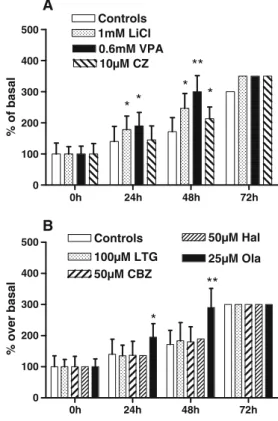
with different mood stabilizer drugs. Only Lithium, Valproate (Fig.1a), and Olanzapine (Fig. 1b) significantly potentiated serum-induced cell proliferation in a dose–
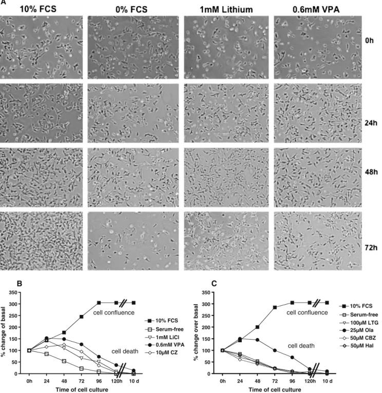
response manner. For the other drugs, there was either no change or decrease in cell proliferation (high concentrations induced toxicity). The doses with optimal responses were used to assess the time-course effects. Li+ (1 mM), VPA (0.6 mM), and Ola (25μM) gradually increased cell proliferation both at 24 and 48 h. At 72 h, cells were in a confluence state, both in controls and MS-treated samples (Fig. 2a, b), and the effect of drugs was nulled. The withdrawal of serum induced a progressive death (Fig.3), and application of drugs on cell cultures prevented or delayed cell death. Lithium, Valproate, Clozapine (Fig. 3a, b), and Ola (Fig. 3c) significantly delayed the cell death. The other drugs (Lamotrigine, Carbamazepine, and Hala-peridol) were unable to prevent cell death (Fig.3c) Phosphorylation of Akt-1 and GSK-3β proteins
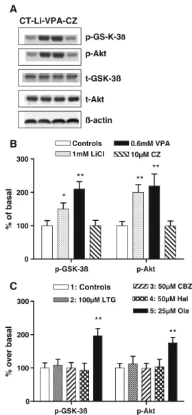
A study of immediate effects on the phosphorylation of Akt at serine-473 and GSK-3β serine-9 was performed (Fig.4),
0.1 1 10 100µM 0.01 0.1 1 10 m M 0.006 0.06 0.6 6 m M 0 25 50 75 100 0 50 100 150 200 Ltg Ola Cbz Hal * ** ** µM B 0 50 100 150 200 Li VPA CZ ** ** * * * A % change of controls % change of controls
Fig. 1 Concentration–response effect of mood stabilizer on cell proliferation on SH-SY5Y cells cultured in RPMI-1640 with low serum content (LSC, 5% FCS) and complemented with mood stabilizers for a period of 24 h. a Lithium, Valproate, and Clozapine and b Olanzapine, Lamotrigine, Haloperidol, and Carbamazepine. Results are expressed in percentage changes of controls (no drug added to LSC), and data are mean values (±SD) of four to six independent assays. Nonparametric Mann–Whitney tests: *p<0.05; **p<0.01
and result showed that addition of Lithium (1 mM), Valproate (0.6 mM), and Olanzapine (25μM) to LSC-grown samples activated GSK-3β and Akt-1 phosphoryla-tion after 30 min. There was also a late activaphosphoryla-tion by Clozapine (not shown), and no other drug did activate the Akt/GSK-3β. Respective t tests on both GSK-3β and Akt were for Lithium (p<0.05 and 0.01, respectively), for Valproate (p<0.01 and 0.01, respectively), and for Olanza-pine (p < 0.01 and 0.01, respectively). Addition of LY294002 (20μM), a specific PI3K inhibitor, 10 min before drug application, resulted in decrease or suppression of the Akt-1 phosphorylation in both Lithium (1 mM) and Olanzapine (50μM)-treated cells with respective to LSC samples (data not shown).
Akt-1 gene expression (mRNA and protein levels)
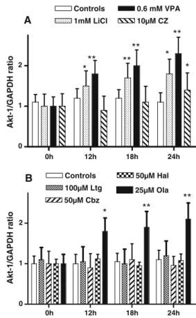
Figure 5a, b displays data of the real-time qRT-PCR for Akt-1 gene expression. Lithium and Valproate (Fig.5a) and Olanzapine (Fig.5b) induced a time-dependent increase in Akt mRNA levels (Friedman test of ANOVA:χ2=19.8 p< 0.01,χ2=20.2; p<0.01, andχ2=21.6, p<0.01, respective-ly). These changes were significant after 12 h for Lithium (p<0.05), Valproate (p<0.01), and Olanzapine (p<0.01)
and progressively increased at 18 and 24 h. For the Clozapine-treated cells, the global changes were not significant, but at 24 h, Akt-1 mRNA was significantly increased compared with control levels (p < 0.05). No change was observed in GSK-3β mRNA during the same period, whatever the drug used (data not shown).
Analysis of total Akt-1 and total GSK-3β proteins over a period of 72 h of drug treatment revealed that Akt protein levels, but not GSK-3β protein, significantly increased compared to control levels (time—0 h; Fig. 6b, c). Friedman test of ANOVA yielded for Akt changes: χ2=13.5; p<0.05. Multiple comparison analysis indicated that changes were significant after 24 h for Lithium, Valproate, and Olanzapine (p<0.05). A weak change was also observed for Clozapine but at 48 h (p< 0.05; Fig. 6b). No change in GSK-3β protein levels was
observed during this period.
Discussion
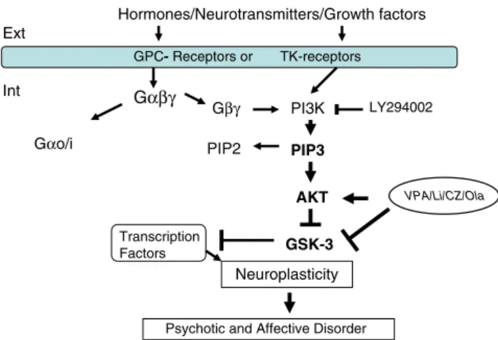
The major goal of the present study was to determine whether anticonvulsant molecules or typical and atypical antipsychotic drugs could induce cellular effects and regulate molecular pathways, namely the Akt/GSK-3β axis, with comparable efficacy and timing with those of Lithium (Fig. 7). Seven different drugs, most of them used as mood stabilizers, were analyzed in vitro by investigating both cellular and biochem-ical actions on cultures of SH-SY5Y cells. The potentiation of cell proliferation and the blockade of cell death were also assessed. Phosphorylation of both Akt-1 and GSK-3β protein kinases and expression of mRNA and proteins were analyzed as well. The study yielded the following results: 1. On the cellular level, the low serum content stimulates
the cell division and proliferation at a steady pace. Addition of the mood stabilizers to the culture medium results in significant potentiation. The effect was statistically significant after 24 h (Lithium, Olanzapine, and Valproate) and 48 h (Clozapine). In these conditions, the other drugs (Lamotrigine, Carbamazepine, and Haloperidol) have no effect on the proliferation. The withdrawal of serum initiates the apoptotic cell death. Addition of some mood stabilizers (Li+, VPA, CZ, and Ola) in the serum-free cell culture protects cells by a transient proliferation and a delayed death allowing them to survive a few more days. Lamotrigine, Carbamazepine, and Haloper-idol have no effect on the survival of SH-SY5Y cells. 2. On the molecular level, after 30 min, Lithium,
Olanzapine, and Valproate induced a significant phos-phorylation of 9Ser-GSK-3β and 473Ser-Akt-1,
com-0h 24h 48h 72h 0 100 200 300 400 500 Controls 100µM LTG 50µM CBZ 50µM Hal * ** 25µM Ola B 0h 24h 48h 72h 0 100 200 300 400 500 Controls 1mM LiCl 0.6mM VPA 10µM CZ * * ** * * A % over basal % of basal
Fig. 2 Time-course study of mood stabilizer effects on cell proliferation. a Li+, VPA, and CZ. b Ltg, Cbz, Hal, and Ola. Results
which show relative growth are expressed in percent of control values (LSC cells). Data are mean values (s ± SD) of four to six independent assays. Nonparametric Mann–Whitney tests: *p<0.05; **p<0.01
pared with LSC-feed controls. The other drugs did not induce any phosphorylation activity. The effect of these drugs was prevented by a previous application of a PI3K inhibitor (20μM LY294002), confirming that
Akt-1 was activated through PI3K signaling. At 12 h, Lithium, Olanzapine, and Valproate induced an in-crease in Akt-1 mRNA levels, compared with LSC samples. No such change was observed for GSK-3β
0h 24 48 72 96 120h 10 d 0 50 100 150 200 250 300 350 10% FCS Serum-free 100µM LTG 25µM Ola cell confluence cell death 50µM CBZ 50µM Hal C
Time of cell culture
0h 24 48 72 96 120h 10 d 0 50 100 150 200 250 300 350 10% FCS Serum-free 1mM LiCl 0.6mM VPA cell confluence cell death 10µM CZ B
Time of cell culture
% change of basal
% change over basal
A
Fig. 3 The neuroprotective effect of mood stabilizers against serum withdrawal-induced cell death. Viable cells were counted in serum-deprived medium where mood stabilizer drugs were supplemented. a Contrast images of SH-SH5Y cells in different conditons of culture. b Serum-free cells with addition of 1 mM Li+, 0.6 mM VPA, or 25μM CZ. c Serum-free cells with addition of 100μM Ltg, 50μM Cbz,
50μM Hal, or 25μM Ola. Results are mean values (±SD) of four to six independent assays. In both figures, control cases with 10% FCS-feed and serum-free cells are shown. With respect to serum-free samples, significant differences were found at 48 (Li+, VA, and CZ, p <0.05) and at 24, 48, and 72 h (Ola, p<0.05)
mRNA. Clozapine effect on Akt mRNA was significant after 24 h, and the other drugs did not induce any change in mRNA levels. After 24 h, increase in Akt-1 protein levels, but not in GSK-3β levels, was observed in Lithium-, Olanzapine-, and Valproate-treated cells. Clozapine effect was significant after 48 h, and no other drug was observed to be effective. After these observations, Lamotrigine, Haloperidol, and Carbama-zepine's effects on these cell paradigms were null. Globally, these data are in accordance with a number of previous individual reports especially the phosphorylation of Akt and GSK-3β and the cellular events here studied (Di Daniel et al.2005; Kang et al.2004; Li et al.2002; Manji and Duman 2001; Kozlovsky et al. 2006). However, we were unable to find a study investigating all these medications in a single protocol. Di Daniel compared the action of four MS drugs (VPA, Li+, Cbz, and Ltg) but on a different signaling axis, i.e. ERK/MAPK. Moreover, these authors did not include a typical antipsychotic drug, such as Haloperidol (Di Daniel et al. 2005). Using animal brain, Kozlovsky et al. (2006) have analyzed the effect of four drugs (VPA, CZ, Hal, and Li+) on the in vivo phosphor-ylation of GSK-3β and reported similar data. However, they did not test the MS effect on Akt-1 activation. Many other important studies were carried out using either one drug or compared two or three drugs, mostly Ola, CZ, VPA, and Li+ (Kim et al.2008; Heiser et al. 2007).
The novelty of this study consists in three points: first, for the first time, seven different MS drugs were used to test two cell events (proliferation and survival) and three molecular assays (protein phosphorylation, mRNA, and protein expression) on two important biochemical actors, Akt-1 and GSK-3β. To our knowledge, no other previous assay has tested a so complex mechanism. Second, to our knowledge, no other study has been conducted on the Carbamazepine or Lamotrigine's effects on Akt/GSK-3β pathway. And thirdly, for the first time, we report an increase of the Akt-1 mRNA and protein levels by mood stabilizers. The latter point is the most important result of this study, given the importance of Akt-1 protein in the proliferation processes.
Previous works conducted on animal and neuronal and non-neuronal cells have suggested that psychotropic drugs, including Lithium, Valproate, or other antipsychotics, can enhance cell viability and activate the Akt/GSK-3β signaling pathway (Li and El-Mallahk2000; Di Daniel et al. 2005; Kang et al. 2004; Manji and Duman 2001; Kozlovsky et al. 2006). However, Shin et al. reported a different effect of CZ with an inhibition of Akt and a dephosphorylation of GSK-3β (Shin et al. 2006). But as they admitted themselves, they used a special cell line, the U-87MG glioblastoma, a cell which lacks a powerful
regulator of the Akt/GSK-3β axis, the PTEN, and probably mobilizes alternative pathways.
With respect to Carbamazepine, this drug was reported to be involved in signal transduction of cyclic adenosine monophosphate (cAMP) second messenger systems, but no effect on Akt/GSK-3β has been reported up to date (Gould et al.2004). Lamotrigine has a potent activity dependent on ion channels (i.e., Na+ and Ca+) and could have indirect action on signal transduction (Xie and Hagan 1998). Whether Ltg has direct actions on this intracellular signaling molecules has not been extensively studied to
p-Akt t-GSK-3ß t-Akt ß-actin CT-Li-VPA-CZ p-GS-K-3ß p-GSK-3ß p-Akt 0 100 200 300 2: 100µM LTG 3: 50µM CBZ 1: Controls ** ** 4: 50µM Hal 5: 25µM Ola C A p-GSK-3ß p-Akt 0 100 200 300 1mM LiCl 0.6mM VPA 10µM CZ Controls * ** ** ** B % of basal % over basal
Fig. 4 Effect of mood stabilizer drugs on phosphorylation of 473 S-Akt-1 and9S-GSK-3β. a Representative Western blot images. b Cells
were treated with Li+, VPA, or CZ. c Cells were treated with Ltg, Cbz,
Hal, and Ola. Relative values of optical densities (ROD) of9
S-pGSK-3β and 473S-pAkt immunoblots expressed as percent of nontreated samples. Data are mean values (±SD) of four to six independent assays, and statistical analysis with t test yielded significant change in different samples. Nonparametric Mann–Whitney tests were: *p<0.05 and **p<0.01
date. Also, both Haloperidol and Clozapine were shown to induce GSK-3β phosphorylation, but as this study was conducted in rat brain cortex, it could be thought that the effect depends on the cell type (Roh et al.2007).
Interestingly, Akt protein levels, but not GSK-3β, were gradually increased in parallel to the drug-induced cell proliferation. It seems that the two molecular and cellular processes have a causal relationship. A number of previous reports have demonstrated that Akt is not only involved in cell growth but is also involved in glucose metabolism/ uptake (Hajduch et al.2001; Lawlor and Alessi2001). Akt was shown to be a key mediator of signal transduction process and mediates many of the survival signals (Brunet et al.2001). Therefore, it is possible to speculate that the addition of the mood-stabilizing drugs resulted in inducing more Akt protein levels available for signals that mediate the subsequent cell proliferation. Valproate was the most effective on enhancing Akt-1 protein levels. This effect
could be alternatively explained by its capacity to upregu-late gene expression through inhibiting histone-deacetylase, as has been reported (Harwood and Agam2003; De Sarno et al.2002).
Our study showed that cellular events and protein synthesis were preceded by rapid phosphorylation and mRNA synthesis. This may suggest that long-term effects must probably be previously sensitized by acute effects, such as protein phosphorylation and mRNA synthesis. To
0-24-48-72 h 0-24-48-72 h Li VPA CZ t-GSK-3ß t-Akt CT Ola 0h 24h 48h 0 50 100 150 200 250 Controls 100µM Ltg 50µM Cbz * ** 50µM Hal 25µM Ola C 0h 24h 48h 72h 0 50 100 150 200 250 Controls 1mM LiCl 0.6mM VPA 10µM CZ * * ** * * B A % change of controls % change of controls
Fig. 6 Time-course of mood stabilizer drugs on Akt-1 protein levels in SH-SY5Y cells grown in LSC medium supplemented with mood stabilizers at their respective optimal doses. Proteins were assayed by the Western immunoblot method, and results are relative optical densities (ROD). a Represntative Western blot images. b Li+, VPA, and CZ. c Ltg, Cbz, Hal, and Ola. Data, expressed as percent of change with respect to controls, are means (±SD) of four to six independent assays. Nonparametric Mann–Whitney tests were signif-icant: *p<0.05; **p<0.01 0h 12h 18h 24h 0 1 2 Controls 100µM Ltg 50µM Cbz ** ** * 50µM Hal 25µM Ola B 0h 12h 18h 24h 0 1 2 Controls 1mM LiCl 0.6 mM VPA 10µM CZ ** ** ** * ** * * A Akt-1/GAPDH ratio Akt-1/GAPDH ratio
Fig. 5 Time course of Akt-1 mRNA expression in SH-SY5Y cells grown in LSC medium supplemented with mood stabilizers at their respective optimal doses. Akt-1 mRNA was assayed by the real-time quantitative RT-PCR method, and results were expressed in ratio values with respect to a house-keeping gene (i.e., GAPDH). a Li+, VPA, and CZ at optimal levels. b Ltg, Cbz, Hal, and Ola. Data, expressed as percent of change with respect to controls, are mean values (±SD) of four to six independent assays. Friedman test of ANOVA:χ2=19.8, p<0.01;χ2=20.2, p<0.01; andχ2=21.6, p<0.01, respectively. Post hoc nonparametric Mann–Whitney tests were significant: *p<0.05; **p<0.01
reveal this sequential process, the use of cells with high rates of division such as SH-SY5Y could be an advantage. Globally, our results indicate that cellular mechanisms observed with Lithium are shared by Valproate, Clozapine, and Olanzapine but not by Carbamazepine, Lamotrigine, and Haloperidol. This could suggest that either the SH-SY5Y cell is not a universal model to investigate these cell events or that the MS drug effects are cell-type-specific. Moreover, all drugs do not activate the Akt/GSK-3β. This indicates that this signaling pathway is not shared by all mood stabilizers.
This study raised some points which require more clarification. First, comparison to clinical situations was allowed by performing at optimal concentrations which correspond nearly to their therapeutically relevant concen-trations as reported by different authors (Bowden et al.
1994and 1996; Perry et al. 1998; Ciapperelli et al. 2003; Aubry et al.2007). However, the doses used for Olanzapine were higher than the corresponding clinical doses (Olesen and Linnet 1999). The serum concentrations are usually much lower, but according to some authors, the brain levels can contain much higher doses of Olanzapine (Olesen and Linnet 1999; Robertson and McMullin 2000; Lu et al.
2004). This is probably due to the fact that brain cells, especially fatty cells, can accumulate the drug and reach levels sufficient to induce the effects observed in this study (Lu et al. 2004). In addition, active metabolites can also contribute to raise the effective concentration of the drug. This is the case for haloperidol, where postmortem concentrations were found to be ten to 30 times higher than the serum concentrations (Kornhuber et al.1999).
Second, it remains also to clarify the clinical relevance of such rapid cellular and molecular effect. In clinical context, these drugs are effective in chronic administration where after several days they may enhance the resilience of
human neural cells against cell death elicited by diverse insults (Manji and Duman 2001). The in vitro studies remain a remote mirror of what really happen in vivo and in pathological conditions. The increase of Akt-1 gene expression could be an indication of the MS drugs' effect to sustain the lag period for onset action.
Another interesting issue concerns the specificity of this pathway to MS drugs. In other words, do antidepressant drugs have the same effect? There are few reports on the effects of antidepressants on either Akt or GSK-3β. According to Tsai et al. 2008, GSK-3β gene is associated
with antidepressant response in a Chinese population. Moreover, administration of imipramine and fluoxetine was found to increase 9S-GSK-3β in mouse brain (Li et al. 2004). These studies show the importance of this pathway which can be activated by both mood stabilizers and antidepressants.
Despite the above-mentioned limitations, this study has shown that in human-derived neuroblastoma SH-SY5Y cells, four drugs used as mood stabilizers or antipsychotics, namely Li+, VPA, Ola, and CZ can mimic the postulated cellular events occurring in mood disorder. These drugs were able to delay cell death and therefore to act as neuroprotectors and to increase serum-induced cell prolif-eration. The possibility that these drugs may protect cells from injury-induced death or may enhance growth is particularly relevant given the reported morphological deficits associated with bipolar disorder. On a molecular level, these three drugs activate Akt-1 protein by a rapid phosphorylation, resulting in deactivating phosphorylation of GSK-3β. These drugs also induce a time-dependent increase in Akt-1 gene and protein expression but not in GSK-3β. The enhanced expression of Akt-1 was parallel to enhanced cell proliferation, thus confirming its role in anti-apoptotic processes. This study may suggest that long-term effects must probably be previously sensitized by acute effects that only in vitro assays with SH-SY5Ycells can reveal because of their high rate of division. The study provides a framework for understanding how protein kinases involved in signal transduction may contribute to study MS mechanisms of action.
Aknowledgments The authors thank Mrs Pascale Marin for her technical support. All the experiments complied with the current laws of Switzerland.
References
Aubry JM, Ferrero F, Schaad N (2007) Pharmachotherapy of bipolar disorders. J Wiley (ed.), Chester
Beaulieu JM, Gainetdinov RR, Caron MG (2009) Akt/GSK3 signaling in the action of psychotropic drugs. Annu Rev Pharmacol Toxicol 49:327–347 Hormones/Neurotransmitters/Growth factors Gβγ PI3K PIP3 PIP2 AKT Gαβγ Ext Int GSK-3 Transcription Factors Neuroplasticity
Psychotic and Affective Disorder
VPA/Li/CZ/Ola
Gαo/i
GPC- Receptors or TK-receptors
LY294002
Fig. 7 Simplified overview of the PI3K/Akt/GSK-3β signaling pathway. The potential sites of MS actions are indicated. GPC-receptors G-protein coupled-receptors, TK-receptors tyrosine kinase-receptors, PIP3 phosphatidylnositol-triphosphate, PIP2 phosphatidylinositol-biphospahte
Bowden CL, Brugger AM, Swann AC, Calabresse JR, Janicak PG (1994) Efficacy of divalproex vs lithium and placebo in the treatment of mania. The Depakote Mania Study group. J Am Med Association 271:918–924
Bowden CL (1996) Dosing strategies and time course of response to antimanic drugs. J Clin Psychiatry 57(Suppl 13):4–9
Brambilla P, Barale F, Soares JC (2003) Atypical antipsychotics and mood stabilization in bipolar disorder. Psychopharmacology 166:315–332
Brunet A, Datta SR, Greenberg M (2001) Transcription-dependent and– independent control of neuronal survival by the PI3K-Akt signaling pathway. Curr Opin Neurobiol 11:297–305
Calabrese JR, Huffman RF, White RL, Edwards S et al (2008) Lamotrigine in the acute treatment of bipolar depression: results of five double-blind, placebo-controlled clinical trials. Bipolar Disord 2008(10):323–333
Chen G, Hasanat KA, Bebchuk JM, Moore GJ, Glitz D, Manji HK (1999) Regulation of signal transduction pathways and gene expression by mood stabilizers and antidepressants. Psychoso-matic Med 61:599–617
Chuang DM (2005) The Antiapoptotic Actions of Mood Stabilizers: Molecular Mechanisms and Therapeutic Potentials. Annals of New York Academy of Science 1053:195–204
Ciapparelli A, Dell'Osso L, Pini S, Chiavacci MC, Fenzi M, Cassano GB (2000) Clozapine for treatment-refractory schizophrenia, schizoaffective disorder, and psychotic bipolar disorder: a 24-month naturalistic study. J Clin Psychiatry 61:329–334 Ciapparelli A, Dell'Osso L, Bandettini di Poggio A, Carmassi C,
Cecconi D, Cassano GB (2003) Clozapine in treatment-resistant patients with schizophrenia, schizoaffective disorder or psychotic bipolar disorder: a naturalistic 48-month follow-up study. J Clin Psychiatry 64:451–458
Cross DA, Alessi DR, Cohen P, Andjelkovich M, Hemmings BA (1995). Inhibition of glycogen synthase kinase-3 by insulin-mediated Protein kinase B. Nature 378:785–789
De Sarno P, Li X, Jope RS (2002) Regulation of Akt and glycogen synthase kinase-3β phosphorylation by sodium valproate and lithium. Neurpharmacology 43:1158–1164
Di Daniel E, Mudge AW, Maycox PR (2005) Comparative analysis of the effects of four mood stabilizers in SH-SY5Y cells and in primary neurons. Bipolar Disorder 7:33–41
Dwyer DS, Lu X-H, Freeman AM (2003) Neuronal glucose metabolism and schizophrenia: therapeutic prospects? Expert Rev Neurotherapeutics 3:29–40
Gelenberg AJ, Hopkins HS (1996) Antipsychotics in bipolar disorder. J Clin Psychiatry 57(Suppl 9):49–52
Gould TD, Quiroz JA, Sing J, Zarate CA, Manji HK (2004) Emerging experimental therapeutics for bipolar disorder; insights from the molecular and cellular actions of current mood stabilzers. Mol Psychiatry 9:734–755
Grimes RS, Jope CA (2001) The multifaceted roles of glycogen synthase kinase 3β in cellular signalling. Progr Neurobiology 65:391–426
Gurvich N, Klein PS (2002) Lithium and Valproic acid: parallels and contrasts in diverse signaling contexts. Pharmacol Ther 96:45–66 Hajduch E, Litherland GJ, Hundal HS (2001) Protein kinase B (PKB/Akt)
—a key regulator of glucose transport? FEBS Lett 492:199–203 Harwood AJ, Agam G (2003) Search for common mechanism for mood
stabilizers as plasticity enhancers in the treatment of neuropsychi-atric disorders. J Clin Psychiatry 64(Suppl 5):179–189
Heiser P, Enning F, Krieg J-C, Vedder H (2007) Effects of haloperidol, clozapine and olanzapine on the survival of human neuronal and immune cells in vitro. J Psychopharmacol 21:851–856
Hsiung SC, Adlersberg M, Arango V, Mann JJ, Tamir H, Liu KP (2003) Attenuated 5-HT1A receptor signaling in brains of suicide victims: involvement of adenylyl cyclase, phosphatidylinositol
3-kinase, Akt and mitogen-activated protein kinase. J Neurochem 87:162–194
Jope RS, Williams MB (1994) Lithium and brain signal transduction systems. Bioch Pharmacology 47:429–441
Jope RS, Johnson GVW (2004) The glamour and gloom of glycogen synthase kinase-3. Trends Biochem Sci 29:95–102
Jope RS, Bijur GN (2002) Mood stabilizers, glycogen synthase kinase-3beta and cell survival. Mol Psychiatry 7(Suppl 1):S35–S45 Kahle PJ, Maas JW (1997) Use of CellTiter 96 reagents in semi
automatic assay of neuronal survival. Neural Notes 3:12–14 Kang UG, Seo MS, Roh MS, Kim Y, Yoon SC, Kim YS (2004) The
effects of clozapine on the GSK-3-mediated signaling pathway. FEBS Lett 560:11–119
Karege F, Perroud N, Burkhardt S, Schwald M, Ballmann E, La Harpe R, Malafosse A (2007) Alteration in Kinase activity but not in protein levels of kinase B and glycogen synthase kinase-3β in ventral prefrontal cortex of depressed suicide victims. Biol Psychiatry 61:240–245
Kim NR, Park SW, Lee JG, Kim YH (2008) Protective effects of olanzapine and haloperidol on serum withdrawal-induced apo-ptosis in SH-SY5Y cells. Prog Neuro-Psychopharmacol Biol Psychiatry 32:633–642
Klein PS, Melton DA (1996) A molecular mechanism for the effect of lithium on development. PNAS USA 93:8455–8459
Kornhuber J, Schulz A, Wiltfang J et al (1999) Persistance of haloperidol in human brain tissue. Am J Psychiatry 156:885–890 Kozlovsky N, Amar S, Belmaker TH, Agam G (2006) Psychoptropic drugs affect ser9-phosphorylated GSK-3β protein levels in rident frontal cortex. Int J Neuropsychopharmacol 9:337–342 Lawlor M, Alessi D (2001) PKB/Akt: a key mediator of cell
proliferation, survival and insulin responses? J Cell Science 114:2903–2910
Lesort M, Greendorfer A, Stockmeier C, Johnson GV, Jope RS (1999) Glycogen synthase kinase-3beta, beta-catenin, and tau in post-mortem bipolar brain. J Neural Transm 106:1217–1222 Li R, El-Mallahk RS (2000) A novel evidence of different
mechanisms of lithium and valproate neuroprotective action on human SY5Y neuroblatoma cells: caspase-3 dependency. Neuro-sci Lett 294:147–150
Li X, Bijur GN, Jope RS (2002) Glycogen synthase kinase-3beta, mood stabilizers, and neuroprotection. Bipolar Disorder 4:137– 144
Li X, Zhu W, Roh MS, Friedman AB, Rosborough K, Jope RS (2004) In vivo regulation of glycogen synthase kinase-3beta (GSK3beta) by serotonergic activity in mouse brain. Neuropsychopharmacol-ogy 29:1426-31
Livak KJ, Schmittgen TD (2001) Analysis of relative gene expression data using real-time quantitative PCR and the 2(delta-delta) Ct method. Methods 25(4):412–408
Lu X-H, Bradley RJ, Dwyer DS (2004) Olanzapine produces trophic effects in vitro and stimulates phopshotylation of Akt/PKB, ERK1/2 and the mitogen-activated protein kinase p38. Brain Research 1011:58–68
Manji HK, Moora GJ, Chen G (1999) Lithium at 50: have the neuroprotective effects of this unique cation been overlooked? Biol Psychiatry 46:949–940
Manji HK, Duman RS (2001) Impairments of neuroplasticity and cellular resilience in severe mood disorders: implications for the development of novel therapeutics. Psychopharmacol Bull 35:5– 49
Moore GJ, Bebchuk JM, Wids IB, Chen G, Manji HK (2000) Lithium-induced increase in human brain grey matter. Lancet 356:1241–1242
Olesen OV, Linnet K (1999) Olanzapine serum concentration in psychiatric patients given standard doses: the influence of comediacation. Ther Drug Monit 21:87–90
Pandey GN, Dwivedi Y, Rizavi HS et al (2009) GSK-3β gene expression in human postmortem brain: regional distribution, effects of age and suicide. Neurochem Res 34:274–285 Perry PJ, Bever KA, Arndt S, Combs MD (1998) Relationship
between patient variables and plasma clozapine concentrations: a dosing nomogram. Biol Psychiatry 44:733–738
Robertson MD, McMullin MM (2000) Olanzapine concentrations in clinical serum and postmortem blood specimens–when does therapeutic become toxic? J Forensic Sci 45:418–421
Roh M-S, Seo MS, Kim Y et al (2007) Haloperidol and clozapine differently regulate signals upstream of glycogen synthase kinase-3 in the rat frontal cortex. Exp Mol Med 39:353–360 Sheline YI (2003) Neuroimaging studies of mood disorder effects on
the brain. Biol Psychiatry 54:338–352
Shin SY, Choi BH, Ko J, Kim SH, Kim YS, Lee YH (2006) Clozapine, a neuroleptic agent, inhibits Akt by counteracting Ca±2/calmodulin in PTEN-negative U-87MG human glioblastoma cells. Cell Signalling 18:1876–1886
Simhandl C, Denk E, Thau K (1993) The comparative efficacy of carbamazepine low and high serum level and lithium carbonate in the prophylaxis of affective disorders. J Affect Disord 28(4):221–231
Stambolic V, Ruel L, Woodgett JR (1996) Lithium inhibits glycogen synthase kinase-3 activity and mimics wingless signaling in intact cells. Curr Biol. 6:1664–1668
Tsai SJ, Liou YJ, Hong CJ, Yu YW, Chen TJ (2008) Glycogen synthase kinase-3β gene is associated with antidepressant treatment response in Chinese major depressive disorder. Phar-macogenomics J 8:384–390
Xie X, Hagan RM (1998) Cellular and molecular actions of lamotrigine: possible mechanisms of efficacy in bipolar disorder. Neuropsychobiology 38:119–130
Yatham LN, Goldstein JM, Vieta E et al (2005) Atypical antipsy-chotics in Bipolar depression: potential mechanisms of action. J Clin Psychiatry 66:40–48
Yatham LN, Kennedy SH, O'Donovan C, Parikh SV, MacQueen G, McIntyre RS, Sharma V, Beaulieu S (2006) Canadian Network for Mood and Anxiety Treatments (CANMAT) guidelines for the management of patients with bipolar disorder: update 2007. Bipolar Disorder 8:721–739
Zeng X, Tamai K, Doble B, Li S, Huang H, Habas R, Okamura H, Woodgett J, He X (2005) A dual-kinase mechanism for Wnt co-receptor phosphorylation and activation. Nature 438:873–877