www.elsevier.com / locate / cardiores
Felodipine inhibits nuclear translocation of p42 / 44 mitogen-activated
protein kinase and human smooth muscle cell growth
a b c c b
Zhihong Yang , Anna Madinova , Toshiyuki Kozai , Hana Joch , Ueli Aebi ,
c,d ,*
¨
Thomas F. Luscher
aVascular Biology, Institute of Physiology, University of Fribourg, Fribourg, Switzerland b
¨
Maurice-Muller Institute of Structural Biology, Biocenter, University of Basel, Basel, Switzerland c
¨ ¨
Cardiovascular Research, Institute of Physiology, University of Zurich, Zurich, Switzerland d
¨
Cardiovascular Center, Division of Cardiology, University Hospital, Ramistrasse 100, CH-8091 Zurich, Switzerland Received 25 May 2001; accepted 25 July 2001
Abstract
21
Objective: Smooth muscle cell (SMC) proliferation contributes to vascular structural changes in cardiovascular disease. Ca
antagonists exert antiproliferative effects and may also be clinically beneficial in the patients. However, the underlying mechanisms of action remain elusive. Activation of mitogen-activated protein kinases (MAPK), in particular p42 / 44mapk plays a central role in cell
21
proliferation. We hypothesise that Ca antagonists inhibit cell proliferation by interfering with the p42 / 44mapk pathway in human SMC.
3
Methods: SMC were cultured from human aorta. Cell proliferation was analysed by [ H]thymidine incorporation. Activation of
p42 / 44mapk and the nuclear target protein Elk-1 was analysed by phosphorylation and p42 / 44mapk nuclear translocation by confocal
3
microscope. Results: PDGF-BB (10 ng / ml) stimulated [ H]thymidine incorporation, phosphorylated p42 / 44mapk, caused nuclear
27 25
translocation of the enzymes and phosphorylated the nuclear target protein Elk-1. Felodipine (10 to 10 mol / l) inhibited
3
[ H]thymidine incorporation to PDGF-BB, had no effect on p42 / 44mapk phosphorylation. However, p42 / 44mapk nuclear translocation
21
and Elk-1 activation stimulated by PDGF-BB were prevented by the Ca antagonist. Conclusion: Activation of p42 / 44mapk,
21
subsequent nuclear translocation and activation of Elk-1 are essentially associated with human SMC proliferation. The Ca antagonist felodipine prevents p42 / 44mapk nuclear translocation (but not its activation) associated with inhibition of human SMC growth. 2002 Elsevier Science B.V. All rights reserved.
Keywords: Growth factors; Protein kinases; Protein phosphorylation; Smooth muscle
1. Introduction coronary artery disease which are thought at least in part to be due to inhibition of SMC proliferation. Previous studies Vascular smooth muscle cell (SMC) growth plays an demonstrated that expression of proto-oncogenes induced
21
important role in intimal thickening in atherosclerosis and by growth factors are inhibited by Ca antagonists [6]. restenosis as well as in venous bypass graft disease [1,2]. However, the exact molecular mechanisms have not been Furthermore, the process contributes to certain forms of elucidated, yet.
vascular remodeling in hypertension [3]. Platelet-derived Mitogen-activated protein kinases (MAPKs) are impor-growth factor (PDGF) released mainly from activated tant mediators of a variety of cellular responses to growth platelets or produced by vascular cells is an important factors, hormones and cytokines [7]. The best studied growth factor of SMC and involved in restenosis after p42 / p44mapk cascade plays a pivotal role in proliferation angioplasty in animal models and in patients [4,5]. of various cells including human SMC [8,9]. p42 /
21
Ca antagonists exert beneficial effects in patients with p44mapk is activated by dual phosphorylation of threonine and tyrosine residues, achieved by the dual-specificity
*Corresponding author. Tel.: 141-1-255-2121; fax: 141-1-255-4251.
¨
E-mail address: [email protected] (T.F. Luscher). Time for primary review 24 days.
0008-6363 / 02 / $ – see front matter 2002 Elsevier Science B.V. All rights reserved. P I I : S 0 0 0 8 - 6 3 6 3 ( 0 1 ) 0 0 4 1 9 - 9
kinase MEK1 / 2. Whereas MEK1 / 2 remains in the cyto- 2.4. Activation of p42 /44mapk and Elk-1 activation plasm, p42 / p44mapk are translocated from the cytoplasm
to the nucleus upon stimulation with growth factors [10– Activation of p42 / 44mapk and Elk-1 activation was 13]. This process allows activation of nuclear transcription analysed by phosphorylation of the enzymes and nuclear factors such as c-Fos and Elk-1 and thereby induces factor on Western blots using anti-phospho-p42 / 44 and downstream gene expression and cell cycle progression anti-phospho-Elk-1 antibodies (1:1000). p42mapk
activa-[14–16]. tion was also analysed by the slower mobility of the
We hypothesised that the integrity of the p42 / 44mapk phosphorylated form on Western blot using anti-p42mapk pathway (i.e. its activation and subcellular relocalisation) is antibody. Staining of the membranes with Ponseau S was
21
essential for human SMC growth. Ca antagonists such as performed to make sure that the protein loading was the felodipine may interfere with the p42 / 44mapk pathway same. The cells were stimulated with PDGF-BB (10 ng /
and thereby arrest cell growth. ml) for 15 min in the presence or absence of felodipine
25
(10 mol / l) and then harvested in extraction buffer as described [17]. Twenty mg of the cell lysates were subjected to 10% SDS–PAGE for electrophoresis. Western
2. Methods blotting was performed as described and phosphorylation
of p42 / 44mapk was visualised by ECL system (Amersham
2.1. Chemicals and materials Pharmacia Biotech Europe GmbH).
Bovine serum albumin (BSA, 7.5%), a monoclonal
2.5. p42 /44mapk nuclear translocation antibody against a-smooth muscle actin and all chemicals
for immunoblotting were from Sigma (Buchs,
Switzer-2.5.1. Immunofluorescence labeling land); recombinant PDGF-BB was from R&D System
Quiescent SMC were cultured on coverslips and stimu-GmbH (Wiesbaden-Nordenstadt, Germany); all tissue
cul-lated with PDGF-BB (10 ng / ml) for the time periods ture materials and media were from Gibco Life Technol
3 indicated. They were then washed twice with modified
(Basel, Switzerland); [ H]methyl-thymidine was from
21
Ca -free Hank’s buffer (MHB) containing 2 mM EGTA ¨
Amersham Pharmacia Biotech Europe GmbH (Dubendorf,
and 5 mM MES (2-morpholino-ethanesulfonic acid, pH Switzerland); trichloroacetic acid was from Fluka (Buchs,
6.2 to 6.4) and quickly substituted with ‘permeabilization Switzerland); rabbit p42 / 44mapk ([9102) and
anti-buffer’, i.e. MHB containing 2% octyl-POE (n-oc-phospho-p42 / 44mapk antibodies ([9101), and rabbit
anti-tylpolyoxyethylene; Alexis, Switzerland) and 0.125% phospho-Elk-1 antibody ([9181) were from Cell Signaling
glutaraldehyde (Electron Microscopy Sciences, USA). Technol (Allschwil, Switzerland). Felodipine was kindly
After 5 min of permeabilization and prefixation, cells were provided by AstraZeneca AG (Zug, Switzerland).
fixed for 20 min with MHB containing 1% glutaraldehyde. The cells were then washed 3 to 4 times with MHB.
2.2. Cell culture Aldehyde groups were reduced by treating the cells twice
for 10 min each with NaHB (0.5 mg / ml) in MHB on ice.4
Human aortic SMCs were purchased from Clonetics Immunofluorescence labeling was carried out as described Corp. The cells were cultured in DMEM containing 10% [18] by incubating the cells with appropriate concen-FCS supplemented with 20 mmol / l L-glutamine and trations of primary (anti-p42 / 44mapk, 1:250) and fluoro-HEPES buffer solution, 100 U / ml penicillin, and 100 chrome-conjugated secondary antibodies (1:500) for 1 h mg / ml streptomycin in a humidified atmosphere (378C; each. Finally, the cells were washed with MHB, mounted 95% air / 5% CO ) [17]. Cells were passaged by 0.01%2 bottom-up (i.e. inverted prior to mounting) in Mowiol 488
EDTA-trypsin. The medium was changed every 2 to 3 (Hoechst, Germany) containing 0.75% n-propyl-gallate as days. All experiments were performed between passages 3 an anti-bleaching agent. Mounted slides were left to dry for
and 5. 24 h at room temperature in the dark, and then stored at
48C in the dark until viewed. 2.3. DNA synthesis
2.5.2. Confocal laser scanning microscopy
SMC were seeded on 12-well plates at a density of Micrographs were taken with a confocal microscope
4
2310 / well and rendered quiescent in culture medium consisting of a Zeiss Axiovert fluorescence microscope containing 0.2% BSA for 48 h. Cells were then stimulated with a Zeiss Plan Apo 63 / 1.4 oil objective lens and an with PDGF-BB (10 ng / ml) for 24 h in the presence or Odyssey XL confocal laser-scanning unit (NORAN, USA),
27 25
absence of felodipine (10 to 10 mol / l). DNA syn- driven by the Intervision software package run on an
3
thesis was measured by [ H]thymidine incorporation (1 INDY workstation (Silicon Graphics Inc., USA). The light mCi / ml; 70–85 Ci / mol), as previously described [17]. source was an argon laser, tuned so that the excitation
2.6. Statistics
All measurements are presented as means6S.E.M.
3
Stimulation of [ H]thymidine incorporation was expressed as mean6S.E.M. In all experiments, n equals the number of independent experiments. ANOVA followed by Schef-fe’s test for repeated measurements were used. A two-tailed P value #0.05 was considered statistically signifi-cant.
3. Results
Stimulation of human smooth muscle cells with
PDGF-3
BB (10 ng / ml) for 24 h enhanced [ H]thymidine incorpo-ration from 1866110 to 1493626 cpm / well (n53, P,
3 0.05; Fig. 1). This process was prevented in the presence
Fig. 1. Felodipine inhibited [ H]thymidine incorporation in human SMC:
21 27 25
of the Ca antagonist felodipine (10 to 10 mol / l,
stimulation of the cells with PDGF-BB (10 ng / ml) for 24 h markedly 3
enhanced [ H]thymidine incorporation in human VSMC, which was 174620 cpm / well; n53, P,0.005 vs. PDGF alone, Fig.
27 25
inhibited by felodipine (10 to 10 mol / l; n53). *P,0.05 vs. control, 1).
[
P,0.005 vs. PDGF. 3
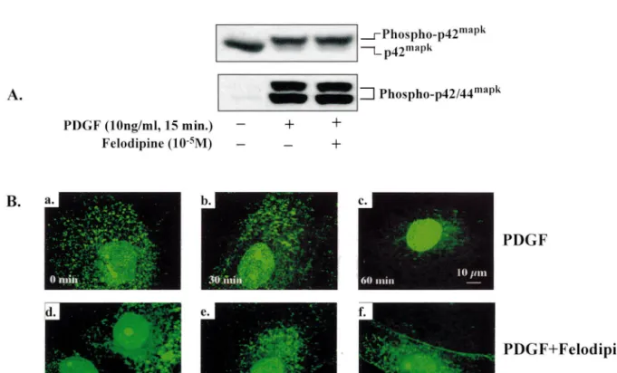
The stimulation of [ H]thymidine incorporation by the growth factor PDGF-BB (10 ng / ml, 15 min) was associ-ated with phosphorylation of p42 / 44mapk (Fig. 2A). wave length for Cy3 was 529 nm, and that for FITC and Activation of p42 / 44mapk (also measured by slower Cy2 488 nm. The confocal images were processed with the mobility of activated p42mapk on the gel), however, was
25
¨
Imaris software from Bitplane AG (Zurich, Switzerland). not influenced by felodipine (10 mol / l; Fig. 2A).
Fig. 2. Effects of felodipine on activation and nuclear translocation of p42 / 44mapk in human VSMC: (A) PDGF-BB (10 ng / ml, 15 min) phosphorylated p42 / 44mapk as measured directly by anti-phospho-p42 / 44mapk (lower panel) or slower mobility (upper panel) of p42mapk on 10% SDS–PAGE, which
25
was not influenced by felodipine (10 mol / l). (B) PDGF-BB (10 ng / ml) induced nuclear translocation at 30 (inset b) and 60 min (inset c) which was
25
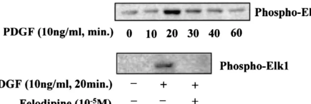
Fig. 3. Time course of PDGF-BB-induced Elk-1 phosphorylation (upper panel). The phosphorylation of Elk-1 induced by the growth factor was prevented
25
by felodipine (10 mol / l, lower panel). The experiments were repeated three times.
PDGF-BB (10 ng / ml) induced translocation of p42 / transcription factors such as TCF / Elk-1 resulting in c-Fos 44mapk from the cytoplasm to the cell nucleus (Fig. 2B). production, which then modulates transcription of other This process could be documented by confocal microscopy target genes via AP-1 complex formation [14,19]. 30 (inset b) and 60 min (inset c) after stimulation with the Indeed, in the present study we demonstrated that in growth factor compared to baseline (inset a). Nuclear human SMC enhanced DNA synthesis induced by PDGF
25
was associated with phosphorylation of p42 / 44mapk and translocation was inhibited by felodipine (10 mol / l) at
subsequent translocation of the kinases from the cytoplasm 30 and 60 min (insets e and f) as illustrated by a
into the nucleus. Moreover, this process also involved representative experiment (n53).
21
activation of the nuclear protein target Elk-1. The Ca Furthermore, one of the nuclear protein substrates of
antagonist felodipine prevented DNA synthesis induced by p42 / 44mapk, Elk-1, was phosphorylated by PDGF-BB (10
PDGF as previously described for most compounds of this ng / ml; Fig. 3). Phosphorylation of Elk-1 reached its peak
class [20], yet had no effect on p42 / 44mapk phosphoryla-at 20 min and this process was inhibited by felodipine
25
tion or activation. The fact that felodipine inhibited p42 / (10 mol / l; Fig. 3). This further confirmed that the entry
44mapk entry into the cell nucleus and phosphorylation of of p42 / 44mapk into the nucleus was prevented by the
21
the nuclear protein Elk-1 without inhibition of p42 / Ca antagonist felodipine.
44mapk activation as shown by confocal microscopy and Western blot using specific antibodies indicates that trans-location of p42 / 44mapk from cytoplasm to the nucleus
4. Discussion instead of activation of the enzymes was blocked by the
21
Ca antagonist. Thus, in human SMC, entry of activated In this study we were able to further delineate the p42 / 44mapk into the cell nucleus is essentially associated molecular mechanisms underlying human SMC prolifer- with cell proliferation. The necessity of p42 / 44mapk
21
ation and its inhibition by a Ca antagonist. In particular, nucleus translocation in cell proliferation has also been we could confirm the antiproliferative properties of this observed in fibroblasts [8] and PC12 cells [21].
class of drugs and, for the first time, demonstrate that The mechanism of p42 / 44mapk translocation into the nuclear translocation of phosphorylated p42 / 44mapk and nucleus is not completely understood. It is proposed that subsequent activation of Elk-1 are essentially associated phosphorylation of p42 / 44mapk is associated with and has
21
with human SMC proliferation. The Ca antagonist been proposed as an essential step for the nuclear accumu-felodipine prevents p42 / 44mapk from entering the nu- lation [22,23]. However, our present study showed that this cleus, while the drug does not interfere with the activation association could be pharmacologically interrupted. The of the kinases. As a consequence, the nuclear protein Elk-1 results also indicate that analysis of integrity of p42 / cannot get phosphorylated. These effects of felodipine are 44mapk pathway should include activation of the enzymes associated with inhibition of human SMC proliferation. and nuclear translocation as well. It remains to be
investi-21
Mitogen-activated protein kinases (MAPKs), in particu- gated which mechanisms are influenced by the Ca lar p42 / 44mapk, play a critical role in transmitting ex- antagonist and whether inhibition of p42 / 44mapk nuclear
21
tracellular signals into the cell nucleus to initiate the translocation is a general mechanism of Ca antagonists expression of a number of genes responsible for cell requires testing of other members of this class of drugs.
21
proliferation [7–9]. Upon stimulation with growth factors Ca antagonists are widely used in hypertension and p42 / 44mapk are phosphorylated at tyrosine and threonine angina and exhibit an array of biological effects in the residues by the dual specific upstream kinases MEK1 / 2. cardiovascular system. In animal models and in patients, The phosphorylated or activated p42 / 44mapk then enter the drugs are able to prevent or reduce endothelial into the cell nucleus and phosphorylate a number of dysfunction in atherosclerosis and hypertensive
remodel-[11] Gonzalez FA, Seth A, Raden DL et al. Serum-induced translocation
ling of resistance arteries [24–26]. The cellular
mecha-of mitogen-activated protein kinase to the cell surface ruffling
nisms delineated in this study might be important for the
membrane and the nucleus. J Cell Biol 1993;122:1089–1101.
ability of these drugs to prevent or reverse structural [12] Lenormand P, Sardet C, Pages G et al. Growth factors induce vascular changes in atherosclerosis, hypertension and nuclear translocation of MAP kinases (p42mapk and p44mapk) but
restenosis. It may play an essential role in restenosis. The not of their activator MAP kinase kinase (p45 mapkk) in fibroblasts. J Cell Biol 1993;122:1079–1088.
fact that clinical trials revealed inconsistent results in this
[13] Zheng C-F, Guan K-L. Cytoplasmic localization of the
mitogen-condition [27] may relate to low local concentrations of the
activated protein kinase activator MEK. J Biol Chem
drug with systemic application. Indeed, the concentrations 1994;269:19947–19952.
required to block p42 / 44mapk nuclear translocation might [14] Gille H, Sharrocks AD, Shaw PE. Phosphorylation of transcription
be higher than those commonly achieved in vivo to lower factor p62TCF by MAP kinase stimulates ternary complex forma-tion at c-fos promoter. Nature 1992;358:414–424.
blood pressure or relieve angina. Thus, local drug delivery
[15] Janknecht R, Ernst WH, Pingoud V, Nordheim A. Activation of
via specially designed balloons or drug-eluting stents
ternary complex factor Elk-1 by MAP kinases. EMBO J
might be a promising approach. 1993;12:5097–5104.
[16] Marais R, Wynne J, Treisman R. The SRF accessory protein Elk-1 contains a growth factor-regulated transcriptional activation domain. Cell 1993;73:381–393.
Acknowledgements
[17] Yang Z, Krasnici N, Luscher TF. Endothelin-1 potentiates human smooth muscle cell growth to PDGF: effects of ET-A and ET-B
This work was supported by the Swiss National Science receptor blockade. Circulation 1999;100:5–8.
Foundation (no. 32-51069.97 / 1 and 31-63811.00), the [18] Mandinova A, Atar D, Schafer BW et al. Distinct subcellular
Swiss Heart Foundation, the Roche Research Foundation localization of calcium binding S100 proteins in human smooth muscle cells and their relocation in response to rises in intracellular
and Prof. Max Cloetta Foundation (Zhihong Yang).
calcium. J Cell Sci 1998;111(Pt 14):2043–2054.
[19] Karin M, Hunter T. Transcriptional control by protein phosphoryla-tion: signal transmission from the cell surface to the nucleus. Curr
References Biol 1995;5:747–757.
[20] Yang Z, Noll G, Luscher TF. Calcium antagonists differently inhibit proliferation of human coronary smooth muscle cells in response to [1] Schwartz SM. The intima: a new soil. Circ Res 1999;85:877–879.
pulsatile stretch and platelet-derived growth factor. Circulation [2] Yang Z, Luscher TF. Basic cellular mechanisms of coronary bypass
1993;88:832–836. graft disease. Eur Heart J 1993;14(suppl I):193–197.
[21] Traverse S, Gomez N, Paterson H, Marshall C, Cohen P. Sustained [3] Moreau P, Takase H, Kung CF et al. Structure and function of the
activation of the mitogen-activated protein kinase cascade may be rat basilar artery during chronic nitric oxide synthase inhibition.
required for differentiation of PC12 cells. Comparison of the effects Stroke 1995;26:1922–1928.
of nerve growth factor and epidermal growth factor. Biochem J [4] Ferns GA, Raines EW, Sprugel KH et al. Inhibition of neointimal
1992;288:351–355. smooth muscle accumulation after angioplasty by an antibody to
[22] Khokhlatchev AV, Canagarajah B, Wilsbacher J et al. Phosphoryla-PDGF. Science 1991;253:1129–1132.
tion of the MAP kinase ERK2 promotes its homodimerization and [5] Maresta A, Balducelli M, Cantini L et al. Trapidil
(tri-nuclear translocation. Cell 1998;93:605–615. azolopyrimidine), a platelet-derived growth factor antagonist,
re-[23] Lenormand P, Brondello JM, Brunet A, Pouyssegur J. Growth duces restenosis after percutaneous transluminal coronary
angioplas-factor-induced p42 / p44 MAPK nuclear translocation and retention ty. Results of the randomized, double-blind STARC study.
Circula-requires both MAPK activation and neosynthesis of nuclear anchor-tion 1994;90:2710–2715.
ing proteins. J Cell Biol 1998;142:625–633. [6] Ko Y, Totzke G, Graack GH et al. Action of dihydropyridine
[24] Henry PD. Atherogenesis, calcium and calcium antagonists. Am J calcium antagonists on early growth response gene expression and
Cardiol 1990;66:3I–6I. cell growth in vascular smooth muscle cells. J Hypertens
[25] Jost S, Deckers JW, Nikutta P et al. Evolution of coronary stenoses 1993;11:1171–1178.
is related to baseline severity — a prospective quantitative angiog-[7] Schaeffer HJ, Weber MJ. Mitogen-activated protein kinases: specific
raphic analysis in patients with moderate coronary disease. INTACT messages from ubiquitous messengers. Mol Cell Biol
Investigators. International Nifedipine Trial on Antiatherosclerotic 1999;19:2435–2444.
Therapy. Eur Heart J 1994;15:648–653. [8] Pages G, Lenormand P, L’Allemain G et al. Mitogen-activated
[26] Park JB, Schiffrin EL. Effects of antihypertensive therapy on protein kinases p42mapk and p44mapk are required for fibroblast
hypertensive vascular disease. Curr Hypertens Rep 2000;2:280–288. proliferation. Proc Natl Acad Sci USA 1993;90:8319–8323.
[27] Opie LH, Yusuf S, Kubler W. Current status of safety and efficacy of [9] Mii S, Khalil RA, Morgan KG, Ware JA, Kent KC.
Mitogen-calcium channel blockers in cardiovascular diseases: a critical activated protein kinase and proliferation of human vascular smooth
analysis based on 100 studies. Prog Cardiovasc Dis 2000;43:171– muscle cells. Am J Physiol 1996;270:H142–H150.
196. [10] Chen RH, Sarnecki C, Blenis J. Nuclear localization and regulation
of ERK- and RSK-encoded protein kinases. Mol Cell Biol 1992;12:915–927.