Tricuspidisation of the aortic valve with creation of a crown-like
annulus is able to restore a normal valve function in
bicuspid aortic valves
§Rene
´ Pre
ˆtre
a,*
, Alexander Kadner
a, Hitendu Dave
a,
Dominique Bettex
b, Michele Genoni
aaDepartment for Congenital Cardiac Surgery, University Children’s Hospital Zurich, Steinwiesstrasse 75, 8032 Zurich, Switzerland bDepartment of Anesthesia, University Children’s Hospital Zurich, Zurich, Switzerland
Received 27 September 2005; received in revised form 18 January 2006; accepted 26 January 2006; Available online 3 May 2006
Abstract
Objective: To evaluate the early results of a new method to repair malfunctioning bicuspid aortic valves by creating a tricuspid valve with a crown-like (i.e. anatomic) annulus. Material and methods: Twelve patients (ages from 10 to 27 years) with chronic regurgitation (and flow-dependent stenosis) of a bicuspid aortic valve underwent repair with the principle of creating a tricuspid valve and a crown-like annulus. The fused leaflets were trimmed and reinserted underneath the existing aortic annulus to create one new native cusp. The third leaflet was fashioned out of a xenopericard patch and was inserted underneath the existing annulus as well to restore the crown-like anatomy of a normal aortic annulus. A tricuspid aortic valve with a morphologically normal annulus was thus created, which resulted in improved coaptation of the leaflets. The repair was immediately assessed by transesophageal echocardiography (TEE) with the heart loaded at 50%. In two patients, a second run helped fine-tune the repair. Median cross-clamping time was 82 min. Follow-up ranged from 3 to 46 months (median 13 months). Results: No significant complication occurred. The function of the aortic valve was excellent with trivial or mild regurgitation in 11 patients and moderate regurgitation in 1 patient. There was no stenosis across the valve. The repair remained stable over time. Remodelling of the left ventricle occurred as expected. Conclusions: Aortic valve repair is feasible in some dysfunctioning bicuspid aortic valves. Tricuspidisation of the valve can result in excellent systolic and diastolic functions. The creation of a crown-like annulus results in improved coaptation of the cusps and could lead to more reliable outcome. Although long-term results are needed, this anatomic correction seems to be a good alternative to valvular replacement in certain sub-groups of patients.
#2006 Elsevier B.V. All rights reserved.
Keywords: Reconstruction; Bicuspid; Aortic valve; Valve repair
1. Introduction
Bicuspid aortic valve (BAV) is the second most common congenital cardiac anomaly, with an estimated prevalence of 0.9—1.36%, in the general population [1,2]. Clinically significant isolated valvular regurgitation has been reported to occur in up to 15—20% of all BAVs. Roberts and Ko[1]have established an age-related pattern of clinical presentation. In infancy, the stenotic forms of BAV are prominent, while later in childhood or adolescence the regurgitation forms are more prevalent, often slowly evolving in the patient who previously received intervention in the cardiac catheterisation suite or
operating theatre. The stenotic forms re-emerge in adulthood, usually due to calcification of the fused leaflets.
Pure or predominant aortic regurgitation is therefore the typical presentation of BAV in the teenager or the young adult. Although chronic aortic valve insufficiency is well tolerated for a long time, progressive dilatation of the left ventricle is a sign with ominous perspective if correction is not timely undertaken. Because of the prospect of an aortic valve replacement, the threshold of cardiologists to refer those patients to surgeons (even to those surgeons performing the Ross procedure) is high. This at times excessively conservative attitude could change if surgeons were able to develop reliable and reproducible techniques of aortic valve reconstruction. This need has been well perceived by surgeons who have strived to develop efficient repair techniques[3—8]. At the moment, no single concept or technique has imposed itself. We report our approach aimed at restoring the morphological characteristics of the www.elsevier.com/locate/ejcts
§
Presented at the joint 19th Annual Meeting of the European Association for Cardio-thoracic Surgery and the 13th Annual Meeting of the European Society of Thoracic Surgeons, Barcelona, Spain, September 25—28, 2005.
* Corresponding author. Tel.: +41 1 266 8020; fax: +41 1 266 8021. E-mail address:[email protected](R. Preˆtre).
1010-7940/$ — see front matter # 2006 Elsevier B.V. All rights reserved. doi:10.1016/j.ejcts.2006.01.068
aortic valve: the presence of three leaflets and a crone-like annulus.
2. Material and methods
All the patients who underwent valve repair with tricuspidisation of a defective bicuspid aortic valve were reviewed. In the young population that we considered for the procedure, symptomatic bicuspid aortic valve presented with asymmetric leaflets comprising a relatively normal left coronary leaflet and a fusion between the non-coronary and the right coronary leaflets. Our reconstruction technique has been used only in this setting of BAV. Demographic and operative data are summarised inTable 1.
The decision to attempt such a repair was done according to echocardiographic and operative findings. In transeso-phageal echocardiography (TEE), the location, quality and mobility of the leaflets were established. A relatively normal (in morphology and mobility) posterior leaflet was our requirement to consider the described repair. The origin and direction of regurgitation jets were identified. A specific look was also given at the commissure between the left and the non-coronary leaflets. If present, a plasty of the commissure and/or of the sub-commissural trigone was performed in addition.
As expected, the jet due to the fused leaflets had a central origin, was eccentric and directed to the anterior leaflet of the mitral valve. In the long-axis midesophageal view, the height of the annulus at the level of the raphe and the morphology of the fused coronary leaflets (degree of relative prolapsus or doming) was determined. A relatively wide and concave leaflet (mild relative prolapsus) was a good sign while a narrow and straight leaflet (more pronounced prolapsus) was an indicator of more difficult reconstruction. Finally, the anatomy of the aortic root, the function of the left ventricle and of the other cardiac valves was assessed. The operative findings forecasting possible repair were a relatively normal (even if thickened) left aortic valve leaflet, enough tissue and mobility of the fused non-coronary and right coronary leaflets. The degree of prolapsus of the fused leaflet at the raphe level had also a prognosis. When the prolapsus was relative (with a still concave curvature of the leaflet) the chances for repair were good, while when it was
absolute (a straight or convex curvature) the chances were compromised.
2.1. Surgical technique
After establishment of CPB, venting of the left ventricle, cross-clamping the aorta and establishment of cardioplegic arrest, the ascending aorta was opened transversally (over three quarters of its circumference) a few millimetres above the sino-tubular junction. This opening exposes the aortic valve correctly and allows a possible conversion to a Ross procedure. The aortic valve was pulled up symmetrically with stay sutures set above the commissures and the valve was inspected. The left coronary leaflet was normal, although thickened, in all the cases considered for repair. The deep insertion of its annulus was ascertained and used as a guide for the reconstruction of the defective facing annulus. The fused leaflets were assessed to see whether one functional leaflet could be created out of them. The amount of tissue, its mobility and the stress necessary to strain a normal concave curve were evaluated. To gain a better idea regarding the possibility of this remodelling, the right coronary leaflet was disinserted from the annulus, starting at the commissure between left and right leaflets (Figs. 1 and 2). The disinsertion was continued across the rudimentary or fictive commissure down to the deepest point of the non-coronary leaflet. This long tongue of tissue was trimmed and modelled to create a normal leaflet with an annulus set at a deeper level. If this could not be obtained with the available tissue, a Ross procedure was performed. Otherwise, the valve tissue was
Table 1
Patient characteristics
Demographic and clinical data
Male/female (n) 9/3
Median (range) age (years) 18 (10—28) Previous operation/balloon dilatation (n) 3/2 LV function <50% (n) 4
Operative data
Median (range) CPB time (min) 108 (76—205) Median (range) cross-clamp time (min) 82 (55—188)
Second cross-clamp 2
Associated procedure (n) 6 Reduction plasty of the AA 5 Resection of LV aneurysm 1
n: number; CPB: cardiopulmonary bypass; LV: left ventricle; AA: ascending aorta.
Fig. 1. Superior view of a BAV. The fused leaflet is disinserted from annulus to the deepest point of the non-coronary leaflet. The fused leaflet is trimmed to create a ‘normal’ non-coronary leaflet. The right coronary leaflet is created with a patch of xenopericardium (L: left; R: right; NC: non-coronary).
inserted along a fictive annulus (set deeper than the original one) to the rudimentary commissure with a running 5/0 or 6/0 prolene suture. Once this was accomplished, the length of the missing right coronary leaflet (which corresponds to the diameter of the aorta) was measured. A U-shaped leaflet was fashioned out of a patch of xenopericard (Bovine pericardial patch, Edwards Lifesciences, Irvine, CA, USA). The height of the patch was let longer than that of a normal leaflet. The patch was inserted (starting in its middle) with a 5/0 or 6/0 prolene underneath the native annulus on the interventricular septum. The patch was often pushed within the left ventricle and inserted laterally to the intercommissural triangle area in both directions. It was then extracted from the left ventricle. Its broadness was again controlled to avoid any wrinkle at the coaptation line. After possible adjustment, the patch was further inserted along the adjoining leaflet to the correspond-ing commissure. The excessive height of the patch was trimmed after the traction on the commissures had been relieved. Additional procedures on the valve (as described by El Khoury et al.[4,9]) were performed in eight patients and consisted in narrowing of the intercommissural triangle between the left and right leaflets in six patients, partially adapting this commissure in two patients and stabilization of the free edge of a native leaflet with a 7/0 Goretex suture in four patients (Fig. 3). The aortotomy was closed, warm retrograde cardioplegia given and deairing manoeuvres performed. The vent in the left ventricle was set on the venous line of the CPB machine. After resumption of a cardiac rhythmus (eventually conducted by epicardial pacing), the aortic cross-clamp was removed. Return of blood through the vent was evaluated and the left ventricle carefully inspected visually and with TEE for absence of distension. The heart was loaded to obtain ejection of blood, and the repair was assessed by TEE. The cause and amount of residual regurgitation was established and if, improvement could be expected, the cross-clamp was re-established, cardioplegia given, the aortotomy
re-opened and the repair fine-tuned. This happened two times because of central regurgitation due to a restricted leaflet. The intercommissural triangle was reduced with a pledgeted stitch and the corresponding sinus was plicated. The aorta was closed again and the same loading manoeuvres performed once more.
The repair was evaluated during the operation with a TEE and postoperatively, during hospital stay and thereafter after Fig. 2. Deployment of the aortic root showing the detachment of the fused
leaflets and the re-attachment of a native and an artificial (dark gray) leaflet over a wider height, thus re-creating the crown-like annulus. Inserts right demonstrate the reason of the eccentric regurgitation (top) and the improved coaptation obtained with the annular remodelling.
Fig. 3. Operative results after tricuspidisation of the aortic valve: note the two native leaflets (NC and L) and the leaflet out of a xenopericard patch (R), and the coaptation over a few millimetres. In this case, both native leaflets had been stabilized with a Goretex running suture.
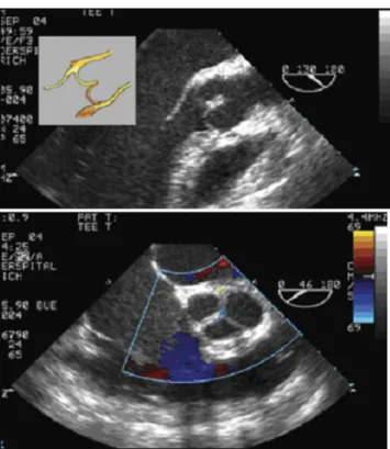
Fig. 4. TEE diastolic frames in long (top) and short (bottom) axes. The restored curve of the leaflets with a broad coaptation segment is well illustrated in the long-axis view (insert). Colour Doppler flow (short axis) shows a trivial residual regurgitation in the centre of the valve.
3—6 months with a transthoracic echocardiography (Figs. 4 and 5). During operation, the excursion of the leaflets and the opening surface area of the valve were evaluated in the short axis. The mean and maximal pressure gradients across the valve were evaluated in the long-axis view by determining the flow velocity spectrum integrated in the modified Bernoulli equation. The amount of regurgitation was assessed with colour Doppler, according to classical criteria. Under normal afterload conditions, the regurgitation was graded as trivial if the central jet length was smaller then 10% of the LVOT diameter, mild if it was between 10 and 25% and moderate if it was between 25 and 65% and severe if it was longer than 65%.
Clinical and echocardiographic data were obtained in all the patients. Follow-up ranged from 3 to 46 months (median: 13 months).
3. Results
Tricuspidisation of the aortic valve could be performed in 12 patients. During the same time period, we performed 46 Ross procedures, 8 other valve repairs and 3 conventional replacements in a similar population (younger than 35 years) with a congenital aortic valve defect, usually a BAV. The majority of the patients were deemed unsuitable for a reconstruction because of their previous history, TEE findings and/or initial valve visualization. It is impossible in retro-spect to establish the number of patients who were initially considered for a repair and eventually converted to a Ross procedure because of the common initial sequence we use for both procedures. In many cases, the Ross procedure was
decided after disinsertion and modelling of the fused leaflets. The repair was interrupted in a more advanced phase and converted to a Ross procedure in four patients. In two of them, the conversion occurred after declamping of the aorta.
There was no significant postoperative morbidity. No patient had a complete heart block. They all left the hospital within 14 days after operation.
Aortic valve regurgitation was trivial to mild in 11 patients and moderate in 1 patient. The regurgitation was always central and eccentric. Peroperative mean transaortic gradient was inferior to 5 mmHg in 10 patients and inferior to 10 mmHg in all 12 patients. The transaortic gradient improved to normal in these two patients. The repair remained stable over the follow-up time. The size of the left ventricle regressed und function remained good or improved in all the patients (including the patients with moderate regurgitation) and were normal in eight patients at last ambulatory control.
4. Discussion
The established procedure for severely defective aortic valve in young patients is a replacement, usually using the Ross procedure[10]. In a minority of patients with a bicuspid aortic valve, however, the morphological properties of the valve permit an anatomic reconstruction, which, at least initially, can show excellent clinical and echocardiographic results[4,5,7]. In our opinion, the most important require-ment to contemplate reconstruction is the presence of leaflets that have a good mobility and a dystrophy limited to their fused portion.
Techniques to repair a defective aortic valve, especially a bicuspid valve, are emerging [4—7,11—13]. Morphologists described more precisely the surgical anatomy of the aortic valve and stressed the fact, that the valve is a three-dimensional structure and that the height of the valve is determined by the interleaflet triangles [14—16]. The morphology of the triangles plays a crucial role for the proper function of the valve. Normally the triangles create a crown-like morphology of the leaflet attachment. If one triangle is vestigial or very small (as in a fused leaflet), the valve attachment is more like a ring, and the surface of coaptation of the leaflets is reduced [16]. El Khoury and co-workers
[4,9,11,12]are certainly pioneers in the repair approach of the aortic valve and have established sound and practical guidelines both in delineating the defect and in repair techniques. Their approach centres on simple principles: correcting any prolapsus of the leaflets by correcting the length of the free border, reducing the aortic annulus by plicating the inter-commissural trigone and, often, reducing the aortic sinuses with a graft reimplanted on the annulus. With this systematic approach, they have achieved an incredible high rate of reconstruction, which proved to be stable over mid-term time[9]. Although the surgical technique is suitable for a classical population, it appears less attractive for the young and active patient. It leaves a bicuspid aortic valve, which limits the absolute valvular opening and can result in a significant trans-annular gradient during increase of cardiac output[17,18]. Finally, the aortic root is replaced by a graft, which lacks any elastic properties.
Fig. 5. TEE systolic frames in long (top) and short (bottom right) axis. The laminar flow through the aortic annulus (top) acknowledges the absence of transaortic gradient. The opening surface of the aortic valve (bottom right) with wide excursion of all three leaflets is exhibited in the bottom panel.
We transferred most of these repair principles to our young population with a BAV but tried instead to obtain a functional tricuspid aortic valve (which provides the largest opening area) with preservation of the native aortic root. In this population (unlike in an older one), the fusion between leaflets is usually located between the non-coronary and the right coronary cusps. The regurgitation is due to a relative or absolute prolapsus and to an improper coaptation of the fused leaflets (Fig. 2). The distorted anatomy of the fused leaflets is associated with a corresponding defective position of the aortic annulus. The normal aortic valve annulus has a crown-like morphology with leaflets deeply implanted in the LVOT and commissures highly suspended in the aortic root
[11,19]. Furthermore, the aortic leaflets can be viewed as a prolongation of the aortic sinuses which inner layers would be prolonged in the valve leaflets. The mechanical stress during motion is absorbed within a broad hinging segment. From the annulus, the folding (concavity) of the cusp is steep so that each cusp meets its counterpart in an almost parallel fashion and already before the leaflets free edge (Fig. 2). In the closed position, the normal leaflets have a coaptation area of a few millimetres and the tangential vector of the leaflet curve is close to vertical (i.e. parallel to the axis of the LVOT). The aortic annulus in a BAV has lost the crown-like morphology and is confined within a narrower height at the level of the fusion: the height between the bottom and the commissures of the fused leaflets is greatly reduced. As a consequence, the fused leaflets extend more directly to the facing leaflet (its tangential vector is more perpendicular to the axis of the LVOT), which results in relative prolapsus of the free edge and in an extremely narrowed coaptation area. In extreme cases, the curve of the leaflets is inversed (no longer concave) and account for the so-called doming appearance. The coaptation no longer takes place and an eccentric regurgitation occurs. The position of the fused leaflets within the LVOT, associated with the absence of opening of the fused commissure, results in a restricted opening area of the valve and therefore in a stenosis, especially if a significant regurgitation exists.
Our technique tends to restore the crown-like morphology of the annulus (Fig. 1). The deep insertion of the leaflets — within the LVOT — restores a progressive, smooth coaptation over a wider area of the leaflets. We used the tissue of the fused leaflets to reconstruct a native leaflet. The creation of this leaflet has been the limiting factor in our experience. Frequently, the height of the leaflet at the raphe is too short or the curve of the leaflet is opposite the naturally concave one. Inverting the curvature of the leaflet can create excessive tension and has been a frequent reason for us to abandon the repair. The right component of the fused leaflet was used as an additional source of tissue to create one normal non-coronary leaflet. The leaflet was disinserted from the defective annulus down to the nadir of the non-coronary sinus. The excessive tissue of the right coronary leaflet was trimmed. The remodelled leaflet was reinserted on a fictive annulus (underneath the present one) to the rudimentary commissure. Care was taken to generate during the reinsertion a normal curvature of the reconstructed leaflet. The right coronary leaflet was constructed out of a patch of xenopericard. It, too, was inserted on a fictive deep annulus. The crown-like annulus was therefore reconstituted which
resulted in a more effective coaptation of the valve leaflets (Figs. 2 and 3).
Our immediate results were good as assessed by clinical and echocardiographic examination (Figs. 4 and 5). Still, a close scrutiny of the repair is necessary. The fate of the xenopericard and of the reconstructed leaflets is unknown. Calcification of the pericardial leaflet and to a lesser extent of the reconstructed one is prone to occur with time. The progression, however, should be slow due to the minimal mechanical stress set on the leaflets and should be easily controllable with serial echocardiographies.
Valve repair is enjoying a new revival[4—7,11—13]. The ideal technique for the various forms of defective anatomy is not clearly established at the moment even though a few groups have achieved a large experience with standardized techniques. Our approach has the advantage to restore the largest possible efficient opening area, but at the expense of a non-native, non-living leaflet. The choice of material to create the missing leaflet can be endlessly debated. Native or tanned autologous pericard or even a leaflet of the tricuspid valve has been used successfully for this purpose [3,5— 7,13,20]. Our choice to use a xenopericard patch was dictated by the easiness of its handling characteristics and the fact that this material, employed to create biological prosthesis, achieves excellent long-term results.
References
[1] Roberts WC, Ko JM. Frequency by decades of unicuspid, bicuspid, and tricuspid aortic valves in adults having isolated aortic valve replacement for aortic stenosis, with or without associated aortic regurgitation. Cir-culation 2005;111(7):920—5.
[2] Lewin MB, Otto CM. The bicuspid aortic valve: adverse outcomes from infancy to old age. Circulation 2005;111(7):832—4.
[3] Al-Halees Z, Gometza B, Duran CM. Aortic valve repair with bovine pericardium. Ann Thorac Surg 1998;65(2):601—2.
[4] El Khoury G, Vanoverschelde JL, Glineur D, Poncelet A, Verhelst R, Astarci P, Underwood MJ, Noirhomme R. Repair of aortic valve prolapse: experi-ence with 44 patients. Eur J Cardiothorac Surg 2004;26(3):628—33. [5] Tolan MJ, Daubeney PE, Slavik Z, Keeton BR, Salmon AP, Monro JL. Aortic
valve repair of congenital stenosis with bovine pericardium. Ann Thorac Surg 1997;63(2):465—9.
[6] Kadri MA, Hovaguimian H, Starr A. Commissurotomy and bileaflet peri-cardial augmentation-resuspension for bicuspid aortic valve stenosis. Ann Thorac Surg 1997;63(2):548—50.
[7] Fraser Jr CD, Wang N, Mee RB, Lytle RB, McCarthy PM, Sapp SK, Rosenkranz ER, Cosgrove DM. Repair of insufficient bicuspid aortic valves. Ann Thorac Surg 1994;58(2):386—90.
[8] Cosgrove DM, Rosenkranz ER, Hendren WG, Bartlett JC, Sterwart WJ. Valvuloplasty for aortic insufficiency. J Thorac Cardiovasc Surg 1991;102(4):571—6. discussion 576—7.
[9] El Khoury G, Glineur D, Rubay J, Verhelst R, D’Acoz Y, Poncelet A, Astarci P, Noirhomme P, van Dyck M. Functional classification of aortic root/valve abnormalities and their correlation with etiologies and surgical proce-dures. Curr Opin Cardiol 2005;20(2):115—21.
[10] Oury JH, Mackey SK, Duran CM. Critical analysis of the Ross procedure: do its problems justify wider application? Semin Thorac Cardiovasc Surg 1999;11(4 Suppl. 1):55—61.
[11] Underwood MJ, El Khoury G, Deronck D, Glineur D, Dion R. The aortic root: structure, function, and surgical reconstruction. Heart 2000;83(4): 376—80.
[12] Haydar HS, He GW, Hovaguimian H, McIrvin DM, King DH, Starr A. Valve repair for aortic insufficiency: surgical classification and techniques. Eur J Cardiothorac Surg 1997;11(2):258—65.
[13] Doss M, Moid R, Wood JP, Miskovic A, Martens S, Moritz A. Pericardial patch augmentation for reconstruction of incompetent bicuspid aortic valves. Ann Thorac Surg 2005;80(1):304—7.
[14] Angelini A, Ho SY, Anderson RH, Devine WA, Zuberbuhler JR, Becker AE, Davies MJ. The morphology of the normal aortic valve as compared with the aortic valve having two leaflets. J Thorac Cardiovasc Surg 1989;98 (3):362—7.
[15] Anderson RH. Clinical anatomy of the aortic root. Heart 2000;84(6): 670—3.
[16] Sutton III JP, Ho SY, Anderson RH. The forgotten interleaflet triangles: a review of the surgical anatomy of the aortic valve. Ann Thorac Surg 1995;59(2):419—27.
[17] Robicsek F, Thubrikar MJ, Cook JW, Fowler B. The congenitally bicuspid aortic valve: how does it function? Why does it fail? Ann Thorac Surg 2004;77(1):177—85.
[18] Robicsek F. Bicuspid versus tricuspid aortic valves. J Heart Valve Dis 2003;12(1):52—3.
[19] Kunzelman KS, Grande KJ, David TE, Cochran RP, Verrier ED. Aortic root and valve relationships. Impact on surgical repair. J Thorac Cardiovasc Surg 1994;107(1):162—70.
[20] Duran CM, Gometza B, Kumar N, Gallo R, Martin-Duran R. Aortic valve replacement with freehand autologous pericardium. J Thorac Cardiovasc Surg 1995;110(2):511—6.
Appendix A. Conference discussion
Dr B. Maruszewski (Warsaw, Poland): First, what’s your age and size, meaning the valve annulus limit, for this technique?
And secondly, did you observe, during your follow-up, any signs of calcification or deterioration of the xeno material?
Dr Preˆtre: We have a population of patients with primarily aortic valve regurgitation. Except for a 10-year-old boy, who was operated upon on an emergency basis — and I was very happy to be able to get out with this repair — the next youngest patient was 16 years. So, we have dealt mainly with patients having an adult size of their aortic annulus.
Our follow-up is not that long (our median follow-up is 13 months) and, at this time, we haven’t seen any calcification.
Dr C. Brizard (Melbourne, Australia): We have used a similar technique of tricuspidisation since 1996, but we use cusp extension. However, initially, when we started, we were quite aggressive and have at times replaced one
cusp almost completely. Based on our follow-up, we do not offer repair to patients where we have to replace one cusp and therefore have a suture line at the hinge point. I think you may find in the long-term follow-up of your technique that this will be a weak point.
Dr Preˆtre: This is also one of my concerns. Normally, an aortic valve leaflet has its hinging point spread over a segment. When you do a patch augmentation of a leaflet, the hinging segment stays the same and the mechanical stress is distributed over the segment. In our technique, the hinging point and stress energy are concentrated exactly on the suture line. I agree that this could be the weak point of this technique.
We haven’t seen any growth problem, as stated before. If you do this repair at an earlier age, however, you will have to look at this as well.
Dr Brizard: And using a more conservative approach, we have a greater proportion of patients that can be amenable to repair.
Dr R. Cesnjevar (Erlangen, Germany): A technical question: How would you do the sizing to get an appropriate leaflet for this replacement?
Dr Preˆtre: Well, we pick up the centre of the two native leaflets, pull them and then measure the length of the additional leaflet. We cut it longer, implant the curved part under the annulus and then toward the commissures. Eventually you have to trim its length. One has to be careful that there is no fold of the free margin; otherwise you will have a regurgitation. It takes a lot of concentration.
Dr Cesnjevar: You have not developed any sizers for this? Dr Preˆtre: No. As you understood, there is a lot of eyeballing there. Dr Z. Al-Halees (Riyadh, Saudi Arabia): Just a comment on the behaviour of the material used in the aortic valve reconstruction. We just published our experience in the European Journal of around 16-year follow-up of aortic reconstruction with both bovine and autologous pericardium. At the mean of about 8 years, the survival of that material is about 40—50% and is basically the same for both.
The mode of failure between the two materials is a little bit different. The bovine pericardium, calcifies; while the autologous pericardium, becomes fibrosed. [The autologous pericardium is treated with glutaraldehyde.]
Dr Preˆtre: Your point is well taken. But calcification is also a consequence of the hemodynamic stress. And if the stress is extremely low, like the result I showed you, we can expect that this problem comes later. But I’m well aware, this is still a palliative approach.
Editorial comment
Demystifying the anatomic arrangement of the aortic valve
It was, perhaps, paradoxical that as I prepared to writethis editorial comment to accompany the article describing ‘tricuspidisation’ of the bifoliate aortic valve[1], I also had at my elbow the December issue of the Journal for the year 2005. In this issue, I discovered another editorial comment, entitled ‘The aortic valve: an everlasting mystery to the surgeon’ [2]. In the light of my own comments that will follow, it is worth quoting in its entirety the opening sentence of this editorial. Manuel Antunes started his own comment by stating ‘Despite its apparent simplicity, the anatomy, physiology, and pathophysiology of the aortic valve have persistently defied the comprehension of the surgeons.’ I do not consider myself qualified to express an opinion on the reasons why understanding of the physiology and pathophy-siology have defeated the combined attentions of the surgical world, but I do consider myself able to point to one of the major reasons why the anatomic arrangement continues to raise problems.
If the interested reader studies the article[3]that formed the focus of the editorial comment provided by Antunes[2], he or she will discover that Thubrikar and colleagues describe the aortic root as extending between the sinutubular
junction and the ‘annulus diameter’. Although they do not specifically define the level of this ‘annulus’, it is clear that they refer to the diameter of the virtual ring constructed by joining together the most proximal attachments of the leaflets of the aortic valve within the left ventricle. Pretre and colleagues[1], however, when discussing their technique for surgical reconstruction of the aortic valve with only two leaflets, describe how they create a crown-like arrangement for the new valve, with the leaflets suspended at semilunar lines of attachment which extend proximally to the level of the sinutubular junction. Such an arrangement is intuitively optimal, since it replicates the arrangement seen in the normally trifoliate aortic valve [4]. But does it help our understanding when the group from Zurich then describe these semilunar attachments as a ‘crown-like annulus’? I cannot speak for the entirety of the surgical world in this regard, and most surgeons might well agree with Pretre and colleagues[1]in viewing the semilunar attachments of the leaflets as the ‘annulus’, but it is evident from analysis of the publication of Thubrikar and colleagues [3] that the group from Charlotte do not share this opinion. And the level of the aortic root chosen by Thubrikar and colleagues