Toward Mechanical Systems Biology in Bone
A
NDREAST
RU¨SSEL, R
ALPHM
U¨LLER, and D
UNCANW
EBSTERInstitute for Biomechanics, ETH Zu¨rich, Wolfgang-Pauli-Strasse 10, 8093 Zurich, Switzerland
(Received 20 March 2012; accepted 10 May 2012; published online 22 May 2012)
Associate Editor Michael R. King oversaw the review of this article.
Abstract—Cyclic mechanical loading is perhaps the most important physiological factor regulating bone mass and shape in a way which balances optimal strength with minimal weight. This bone adaptation process spans multiple length and time scales. Forces resulting from physiological exercise at the organ scale are sensed at the cellular scale by osteocytes, which reside inside the bone matrix. Via bio-chemical pathways, osteocytes orchestrate the local remod-eling action of osteoblasts (bone formation) and osteoclasts (bone resorption). Together these local adaptive remodeling activities sum up to strengthen bone globally at the organ scale. To resolve the underlying mechanisms it is required to identify and quantify both cause and effect across the different scales. Progress has been made at the different scales experimentally. Computational models of bone adap-tation have been developed to piece together various exper-imental observations at the different scales into coherent and plausible mechanisms. However additional quantitative experimental validation is still required to build upon the insights which have already been achieved. In this review we discuss emerging as well as state of the art experimental and computational techniques and how they might be used in a mechanical systems biology approach to further our understanding of the mechanisms governing load induced bone adaptation, i.e., ways are outlined in which experimen-tal and computational approaches could be coupled, in a quantitative manner to create more reliable multiscale models of bone.
Keywords—Bone adaptation, Multiscale modeling, Osteo-cytes, Mechanobiology, Mechanical systems biology.
INTRODUCTION
Mechanical conditioning of the tissue has been identified as one of the major contributors to both cortical and trabecular bone adaptation.81Trabecular bone, in particular, has been shown to have a more enduring sensitivity to mechanical stimulation in
human adults,4,26with this in mind this review focuses primarily on trabecular bone adaptation. Mechanically driven trabecular-bone-adaptation is a process which spans multiple spatial and temporal scales. At cell -level, molecular mechanisms respond within seconds to forces defined at the organ-level, while orchestrat-ing, at tissue-level, the addition or removal of bone over time scales which vary between months and years.56 The exact mechanisms governing trabecular bone mechanobiology remain unknown. Current experimental approaches aimed at providing further insight are typically limited to one particular level of hierarchy only, neglecting the effect of other scales. In contrast, computational approaches are not physically limited to one scale only, but have, up until recently, existed in isolation. Furthermore, their outputs lack quantitatively precise definition and validation. To move toward, a more precise description of trabecular bone mechanobiology will be necessary to combine quantitatively precise in vivo and in vitro data retrieved from the different scales with multiscale in silico models of trabecular bone adaptation. This approach, which we would like to term mechanical systems biology, demands the development of (a) experimental methods using existing in vivo and in vitro model sys-tems for load-induced bone adaptation; and (b) a novel computational framework capable of merging or nesting existing computational models for bone adap-tation which attempt to simulate bone mechanobiology at the different scales. With this in mind, we review state-of-the-art in vivo and in vitro systems used for studying load induced bone adaptation along with the technologies used to quantify gene expression, bone formation and the mechanical environment at the cell-level and the tissue-cell-levels, respectively. We also discuss state-of-the-art in silico models of load-induced bone adaptation and how they are beginning to be merged into multiscale frameworks. Finally, we outline future strategies for extracting data from multiple scales using
Address correspondence to Duncan Webster, Institute for Bio-mechanics, ETH Zu¨rich, Wolfgang-Pauli-Strasse 10, 8093 Zurich, Switzerland. Electronic mail: dwebster@ethz.ch
DOI: 10.1007/s10439-012-0594-4
0090-6964/12/1100-2475/0Ó2012 Biomedical Engineering Society
experimental model systems with the latest imaging and molecular technologies in a high-throughput manner. Furthermore, we discuss how this can be used to shape, feed and validate the next generation of multiscale computational models for bone mechano-biology.
STATE-OF-THE-ART CELL- AND TISSUE-LEVEL EXPERIMENTAL SYSTEMS Bone is a dynamic tissue which responds to mechanical stimulation. Cyclic overloading causes a net increase in bone mass, while disuse has been shown to result in significant reductions. Cortical as well as trabecular bone tissue is formed and resorbed by cells known as osteoblasts and osteoclasts, respectively. These cells are derived from mesenchymal and hema-topoietic stem cells respectively, which reside in the bone marrow. It is widely hypothesized that osteo-cytes, residing in the bone matrix, control osteoblasts, and osteoclasts in response to cyclic mechanical load-ing.9,10,46These cells are ideally located and distributed to act as mechanosensors: They represent 90–95% of all cells residing inside the mineral matrix in the adult skeleton. They are regularly dispersed throughout the bone matrix and they are connected to each other and cells on the bone surface via dendritic processes that occupy tiny canals called canaliculi.38,63 The exact mechanisms by which osteocytes sense mechanical load and choreograph the actions of osteoblasts and osteoclasts remain unknown. A variety of in vitro and in vivo experimental approaches at both cell- and tissue-level have been employed in an attempt to pro-vide more insight, however owing to inherent limita-tions, state-of-the-art experimental methods are only able to provide part of the picture.
At cell-level, in vitro approaches using co-culture systems have been used to investigate the dependency of osteoblast and osteoclast function on osteocytes. Heino et al.33tested the hypothesis that soluble factors secreted by osteocytes stimulate bone formation by increasing osteoblast activity. Culture medium from osteocyte-like MLO-Y4 cell line was used to condition mice osteoblasts. Results showed that calcium deposi-tion by osteoblasts was increased almost four fold. The bone matrix protein osteocalcin (OCN) as well as the osteoblast activity marker alkaline phosphatase (ALP) increased according to the amount of MLO-Y4 culture medium added. The proliferation rate of osteoblasts was also increased. The exact molecular mechanisms responsible for these observations are yet to be resolved. In another study, Zhao et al.86hypothesized
that osteocytes regulate osteoclast function.
Co-culturing MLO-Y4 with cell populations containing
osteoclast precursors (murine spleen or marrow cells) on a dentine substrate resulted in significant larger area of resorption pits and pit numbers as well as in a higher number of tartrate-resistant acid phos-phatase (TRAP) stained multinucleated osteoclasts, when compared to monocultures. The receptor acti-vator of nuclear factor-jB ligand (RANKL) has been identified both in vitro34 as well as in vivo52,83 as a major factor responsible for osteocytic activation of osteoclast activity. After secretion by osteocytes, RANKL activates the osteoclastic resorption activity by binding to the receptor activator of nuclear factor-jB (RANK) on the osteoclast membrane. Further-more increased osteocytic RANKL expression has been shown to be a major contributor to unloading-induced bone resorption.83 Other studies have looked at the effect of specific factors which are known to be produced by osteocytes following mechanical stimu-lation. Sclerostin has been identified as a molecule that inhibits bone formation59 and is preferentially expressed by osteocytes.73,80By treating human primary osteoblasts with differing concentrations of sclerostin, Atkins et al.7 demonstrated that their capacity to mineralize was significantly reduced. More specifically, treatment of human primary osteoblasts with sclero-stin concentrations down to 1 ng/ml inhibited calcium incorporation into the matrix of bone cultured in vitro. At the same time the expression of the bone matrix protein collagen 1, phosphate-regulating gene PHEX and OCN were down regulated.
To better characterize the factors released by osteocytes in response to mechanical stimulation cell culture systems have been developed to expose osteo-cytes (or their precursors) to shear stresses via fluid flow17,36,42,43 or direct mechanical deformation using deformable substrates.29,57,85 For example, using osteocytes derived from embryonic chicken calvariae, Klein-Nulend et al.43showed that intermittent hydro-static pressure or pulsating fluid flow results in an increase in the level of the osteoclast stimulatory factor Prostaglandin E2 (PGE2) throughout the culture medium. Using the same system they also demon-strated that nitric oxide (NO) was released in minutes, reaching a maximum after 5 min.42 These mechanical in vitrosystems and others, along with the biochemical pathways which they proposed are reviewed in more detail elsewhere.11,41,51 The results which are gained using in vitro systems must be interpreted with caution. It is possible that potential mechanosensitive pathways are misrepresented owing to the limitations associated with the in vitro environment. The physiological envi-ronment of osteocytes is not accurately reproduced, as such neither are the forces which they are exposed to.12 Furthermore, the absence of other cell types means that cell culture systems lack vital communication
networks which are known to facilitate bone adaptation.84
To investigate load-induced cortical and trabecular bone adaptationin vivo, a number of animal loading models have been established. These include the mur-ine, ulna,62,75,85 tibia,21,25,32 radii,31 tibia bending,72 femur,74and vertebra77loading models. Some of these models have been used to characterize the biochemical pathways which are activated in response to their specific cyclic loading formats. Kesavan et al.39used a cyclic mouse tibia loading model combined with quantitative PCR (qPCR) to investigate the behavior of bone formation and bone resorption markers in response to mechanical stimulation. They extracted large populations of osteocytes from the full bone (cortical and trabecular) by pulverizing the snap frozen tibia and homogenizing the powder in Trizol. The contents of the lysed cells were then processed to iso-late high quality total RNA. Purified total RNA was then reverse transcribed and subjected to real-time PCR amplification for selected genes. With this approach they were able to demonstrate the up-regu-lation of several genes implicated in bone remodeling including type I collagen, ALP, and OCN. In another study, Xing et al.82 used similar cell harvesting tech-niques to provide input material for microarray anal-ysis in an effort to identify other genes involved in load induced bone formation. Again using a cyclic mouse tibia loading model, they isolated high quality RNA from pulverized full bone and then used microarrays to investigate the behavior of 20,280 different genes. Results showed that 346 genes were differentially expressed in loaded bones compared to controls. Subsequent pathway analysis revealed that 28 out of the 346 genes exhibited a direct biological association. Recently, similar global gene expression analyses were performed using the mouse tail vertebra model and a protocol which isolated large populations of trabecular bone osteocytes.76 The fifth caudal verte-brae of C57BL/6 female mice were dynamically loaded three times a week for 3000 cycles at a frequency of 10 Hz and a peak load of 8 N using a previously developed caudal vertebral axial compression device (CVAD).77Large populations of trabecular osteocytes were successfully harvested from loaded vertebrae and enough high quality mRNA76 was isolated for sub-sequent DNA microarray analysis. When monitoring the expression of 34,000 genes for short and extended periods of cyclic loading it was discovered that hun-dreds of different genes were differentially expressed [data not published]. Among these were genes which have known or suspected roles in osteocyte metabo-lism, however, there were also many load-regulated genes which have not been previously connected to bone metabolism. Comparison of short and long term
cyclic loading expression profiles revealed some com-mon genes, however many genes were not similarly expressed. Besides revealing a number of novel genes these data allude to the existence of complex transient pathways and interactions.
Global gene expression essays derived fromin vivo models for bone adaptation have identified a number of candidate genes and revealed potential load regu-lated pathways. However, interpretation of the data is limited. One reason for this is the fact that these techniques report the average effect of tens of thou-sands of cells thereby concealing potentially important signals.87 For example, in the many in vivo loading models, which have been discussed, trabecular bone formation is also accompanied by structural reorga-nization which involves bone resorption. Expression signals are thus representative of both bone forming and bone resorbing genes. Furthermore, because a diverse micro-mechanical environment exists within the micro-architecture of trabecular bone i.e., osteo-cytes are subjected to a wide range of both compres-sive, tensile, and sheer strains, it is impossible to precisely define the relationship between gene expres-sion and the mechanical environment.
At tissue-level, with recent technical advances, it is now possible to precisely characterize the diverse mechanical environment in bone and relate, local
micro-mechanical environments with local
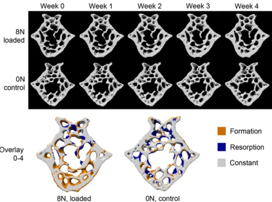
load-induced bone remodeling events. However, challenges remain on how to relate this to biological pathway information such as gene expression data. Using the previously mentioned cyclic mouse tail loading model in combination with time-lapsed in vivo micro-com-puted tomography (lCT) and the latest image regis-tration techniques,66 Lambers et al.45 were able to track changes in trabecular bone micro-architecture due to cyclic mechanical loading (Fig.1). By creating micro-finite element (lFE) models from in vivo lCT images prior to loading, Schulte et al.67 resolved the mechanical strains which occur throughout the tra-becular micro architecture at a resolution of 10 lm. Projection of formed and resorbed surfaces (identified in the registered lCT images) onto the surface of the finite element model revealed that bone was formed in regions of high mechanical strain and resorbed in region of low mechanical strain. The availability of this combined computational and experimental approach makes it possible for researches to investigate, locally, the relationship between bone’s mechanical environ-ment and the action of osteoblasts and osteoclasts. Retrieval of molecular information associated with these local events would therefore go some way to further elucidating the mechanisms responsible for load-induced bone adaptation. At a resolution of 10 lm, the lFE model is unable to capture the specific
geometry of osteocyte lacunae and canaliculi where mechanical strains are likely to be amplified.12,54lFE models are thus only able to provide a continuum level measure of the osteocyte’s mechanical environment, nevertheless this technique will still be able to distin-guish between those osteocytes in regions of high and low mechanical strains, allowing associated gene expression patterns to be grouped accordingly.
Retrieval of molecular information could be
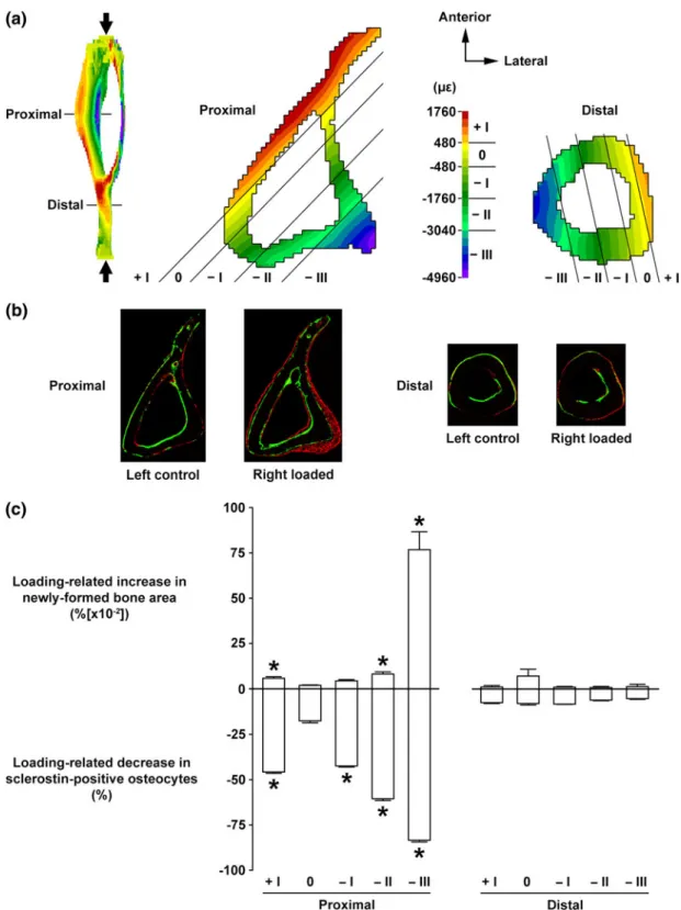
achieved in manner similar to that performed by Moustafa et al.50 In this study, the cyclic ulna mice loading model21 was used to investigate the relation-ship between the expression of sclerostin in osteocytes and the local mechanical environment. Immunohisto-chemistry was used to identify the fractions of osteo-cytes expressing sclerostin in standard 2D histological sections. Voxel based lFE models with a resolution of 40 lm were then constructed to resolve the mechanical strains throughout the tibia’s micro-architecture. By comparing the 2D histology sections of cortical bone with the equivalent 2D plane in the lFE model rela-tionships between sclerostin expression and mechanical strains were searched for (Fig. 2). Results show that sclerostin seems to be more closely related to the local areas of bone formation (as identified by standard 2D
dynamic bone histomorphometry) than to local strain levels. This disagrees to some extent with a similar study performed by Robling et al.61 who found that the relative numbers of osteocytes expressing sclerostin were directly associated with the magnitude of the mechanical environments. While these approaches allow biochemical information to be spatially related to both mechanics and osteoblastic/osteoclastic activ-ity at a local level, only one molecular target can be investigated at a time. If the molecular networks involved in load induced bone adaptation are to be fully characterized experimental approaches are required which can enable similar correlations to be performed with more molecular targets.
STATE-OF-THE-ART MULTISCALE MODELING OF LOAD-REGULATED BONE ADAPTATION
Owing to limitations in technology, experimental methods alone are unable to fully characterize the governing mechanisms of a biological system. Com-putational modeling can help to fill the missing gaps in knowledge. The simple act of modeling a biological system forces the synthesis of mechanisms, which by
FIGURE 1. Bone microstructure (cross-section) of representative mice from the 8 and 0 N groups at time points when in vivo micro-CT scans were performed. In the top row (8 N group) thickening of the trabecular structure can be observed, while on the bottom row (0 N group) little changes in the bone microstructure can be seen between time points. This becomes even clearer when an overlay of the first with the last scan is observed (bottom). For the 8 N group many yellow sites, indicating bone formation, can be observed around the trabeculae, while for the 0 N mouse formation and resorption are rather balanced (equal amount of blue and yellow structures). Reprinted with permission from Lambers et al.45with permission from Elsevier.
FIGURE 2. Relationship between mechanical loading-related changes in osteocyte sclerostin expression and magnitudes of local strain engendered vs. subsequent osteogenesis in cortical bone. (a) Transverse loading induced strain distribution by FE analysis at the proximal and distal sites (37 and 75% of the bone’s length from its proximal end, respectively) of the tibia. Bone area was divided into five regions parallel to the neutral axis (region 0) corresponding to different magnitudes of strain in tension (region +I) or compression (regions 2I to 2III). (b) Representative transverse fluorochrome-labeled images at the proximal and distal sites of the left control and right loaded tibiae. Green: calcein label injected on the first day of loading. Red: alizarin label injected on the last day of loading. (c) Loading-related increase in newly formed bone area and decrease in sclerostin positive osteocytes in each of the five regions (corresponding to different strain magnitudes) at the proximal and distal sites. Bars represent the means 6 SE (n 5 6). *p < 0.05 vs. region 0. Reprinted with permission from Moustafa et al.50and Springer Science and Business Media B.V.
being able to replicate real world events, help to deduce and further elucidate how the biological system in question functions. As such computational models can be used as a platform to develop and test hypotheses and to build theoretical models of real-world events.
A plethora of computational models exist which propose a variety of mechanisms by which bone is remodeled at both and tissue-levels. At the cell-level computational models reconstruct the probable paracrine networks using partial differential equations to describe the dynamics of osteoblast-osteoclast interaction. For example, based on the RANK– RANKL–osteoprotegerin pathway, Lemaire et al.47 derived a series of partial differential equations which describe osteoblastic and osteoclastic activity as a closed loop control network, mediated by paracrine factors such as parathyroid hormone (PTH) and transforming growth factor beta (TGF-b). In doing so they were able to depict the differentiation, prolifera-tion, and death of populations of each cell type as a function of time. Recently, the model used by Lemaire et al.58 has been extended to include a more explicit description of ligand-receptor kinetics. Furthermore, this and other models have been developed to model the cell–cell dynamics in simple spatial domains,14,65 providing insights into how osteoblasts, osteoclasts, and their progenitors are coordinated spatially. The primary focus of most cell-level models is osteoclast– osteoblast interaction, most are yet to incorporate the proposed role of the osteocyte and its molecular mechanosensitive pathways. Currently, there is only one model which addresses this issue. Maldonado et al.48 have described a preliminary model which integrates the osteocyte with osteoblast-osteoclast interactions as described by Lemaire et al.47 Here it is assumed that osteocytes respond to mechanical stimuli and initiate osteoblast activity through pathways involving NO and PGE2.16,18 Besides resolving the proliferation and number of active osteoblasts/osteo-clasts, Maldonado et al. have also tentatively linked cellular activities with geometric changes in the x–y plane of a cylindrical idealization of cortical bone. For a more detailed description of the different cell-level models which have been developed for bone, the reader is referred to a more focused review.78
At the tissue-level computational models attempt to deduce the mechanisms by which trabecular micro-architecture adapts to external mechanical loads. In an early approach Fyhrie and Carter27 developed the theory of self optimization whereby the distribution of structural elements was determined according to structural optimization theory that determines, for a given load, the most efficient structural arrangement. This theory was applied to the proximal femur in order to predict the of trabecular bone density distribution.28
When compared with the actual trabecular density distributions in proximal femurs predictions were shown to be relatively good. However, this approach does not explicitly describe the micro-architecture of trabecular bone.
Later approaches described the geometry of tra-becular bone along with its local micro-mechanical environment and simple empirical relationships have been implemented to describe the otherwise complex biochemical relationships between osteocyte, -blast and -clast populations. For example, the model developed by Adachi et al.3 subscribes to the theory that osteoblast/osteoclast activity is proportional to the magnitude of the mechanical signal sensed by neighboring osteocytes.2 This theoretical premise was
implemented using representative 3D trabecular
structures, composed of small cubic elements (or vox-els) together with lFE analysis.40 Assuming that osteocytes were uniformly distributed throughout voxels space, their local micro-mechanical environ-ments were computed for a specific set of external boundary (loading) conditions. The magnitude of the mechanical signal sensed locally by a group of osteo-cytes (i.e., the voxel strain) then dictated if osteoblasts formed new bone (additional voxels were added) or osteoclasts resorbed old bone (voxels were removed). Ruimerman et al.64used a similar approach, however instead of linking both osteoclast and osteoblast activity to the strength of the mechanical signal sensed by osteocytes, osteoclast activity was assumed to be random.35 This particular aspect is based on the assumption that osteoclasts are active at sights of micro-fractures, which occur in a spatially random manner owing to daily loading activities.
A limitation of these micro-architectural approaches is that they lack the definition of realistic boundary conditions, which in reality are derived from loading patterns at the organ-scale. This issue has recently
been addressed by both Coelho et al.20 and
Be’eyr-Lipperman et al.8 Both used a global–local hierarchical approach in which a global model of an entire bone supplies strain and density information to a series of local models characterizing the trabecular microstructure at a specific location in the global model. A common limitation of the aforementioned mod-eling approaches is that they assume trabecular bone to be a homogeneous isotropic material, where as in reality owing to the orientation of collagen fibers it is not.6This heterogeneity is further compounded by the fact the levels of mineralization differ throughout the trabecular architecture.55 Furthermore, assumptions are made about the nature of the mechanical signal responsible for initiating adaptation. It is common to use strain energy density as the stimulus which is a measure of the amount of strain sensed by osteocytes,
implying that structural deformation is responsible for load-induced bone adaptation. However, fluid flow (strain gradients)44 or fatigue damage60 could be con-tributing factors all of which are dependent on the dynamic aspect of the mechanical signal, i.e., fre-quency which is rarely included.1For a more detailed description of tissue-level models the reader is referred to more focused reviews.30,78
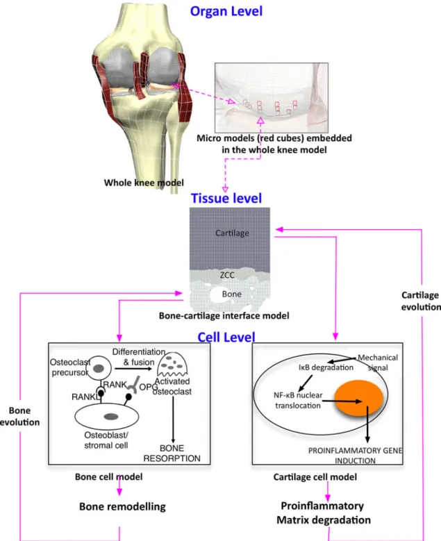
The insight which can be provided by cell- or tissue-level models is limited. Cell-tissue-level models have the potential to explain the biochemical mechanisms con-trolling osteocyte, osteoblast, and osteoclast activities, however, they are uninformative about how these processes are coordinated in space and time to selec-tively reorient and thicken certain individual trabecu-lae. Tissue-level models on the other hand are constrained to the latter case. To provide a more complete description of load-induced bone adaptation, Shim et al.69 recently linked cell- and tissue-level models using a multiscale framework. The main aim of this study was to develop a computational frame work, which could be used to investigate the biochemical and biophysical processes, spanning multiple scales at the bone-cartilage interface which lead to osteoarthritis. The authors hypothesized that the progression of osteoarthritis can be attributed to the following chain of events: Altered patterns of mechanical loading, due to knee injury, induce abnormal patterns of remodel-ing in subchondral bone. This augments and shifts peak cartilage strains, the supra-physiological nature of which causes the release of pro-inflammatory cyto-kines, which in turn initiates cartilage degradation. To test this hypothesis it was necessary to link tissue-level models defining the physical micro-mechanical envi-ronments in both bone and cartilage with cell-level models defining the biochemical processes involved in bone remodeling and the pathological release of cyto-kines in cartilage (Fig.3). This was achieved by using the open-source markup languages FieldML19,24 and CellML.15,53 By implementing a generic web-based language, FieldML is able to capture the spatially varying information for both bone and cartilage, whereas CellML is used to define the intracellular networks and paracrine kinetics which determine cell activity. The spatial organization of bone and cartilage was defined with a 2D electron microscope image showing cartilage and subchondral bone (Fig.3). Mechanical signals defined at the tissue-level were then passed to and processed by cell-level models unique to both bone and cartilage. The outcome of the cell-level models was then used to instruct tissue-level models to add or remove structural elements.
The simulated outcome of these simulations were consistent with observations reported in separate studies which showed that altered mechanical loads led
to increased loading in more fibrillated and thinner regions of cartilage,5,23 thereby supporting the hypothesis that osteoarthritis is indeed mediated by altered loading patterns in the knee. This study dem-onstrates the ability of multiscale approaches to pro-vide a more complete description of load-regulated bone biology. Furthermore, its modular structure permits the extension of code at the cell-level to incorporate the most up-to-date pathways as well the interchange of different tissue-level models. However, without quantitative validation its ability to assess the theories which it implements is limited. Other than being able to reproduce outcomes which are qualita-tively consistent with experimental observations, the models which form such multiscale approaches have received little in the way of quantitative validation both as independent and co-dependent entities, a fact which is also true of all the aforementioned cell- and tissue-level models. For example, at the cell-level out-comes of Lemaire’s simulations were only validated in the sense that the directional change of osteoblasts/ osteoclast populations and activities (increase or decrease) agreed with those reported experimentally. At tissue-level the approaches conceived by Adachi, Ruimerman and co-workers have only been validated in the sense that they have been shown to replicate certain aspects of adaptation observed in vivo, for example trabecular realignment and thickening. They have also been shown to yield structures with a similar bone volume density to those measured in humans. Although qualitative validation in this manner does provide a certain level of credibility it is not enough to properly authenticate the theoretical mechanisms they propose. With this in mind, if multiscale computa-tional models are to provide further insight into load adaptive mechanisms, experimental approaches are required which are (i) suitably aligned with the in silico setup and (ii) capable of retrieving quantitative data from cell- and tissue-levels which can be directly compared to multiscale simulations.
A MECHANICAL SYSTEMS BIOLOGY DESCRIPTION OF LOAD-REGULATED BONE
ADAPTATION
As previously established, the direct relationship between the local mechanical environment, the molec-ular pathways activated in osteocytes and the local
bone remodeling activities has not yet been
defined. Furthermore, computational models at the different scales, which attempt to model aspects of these unknown relationships require quantitative validation. Experimental methods are therefore needed which can
quantify their local micro-mechanical environments and relate these to both osteoblastic and osteoclastic activities. The availability of registered,in vivolCT for the cyclic vertebra loading model, along with the lFE models goes some way to satisfying these needs. Com-bining this data with the multiscale framework pre-sented by studies such as Shim et al.69 would provide immediate opportunities to assess the mechanisms
which are proposed in both cell and tissue level com-putational models, so long as the different cell- and tissue-level models are inserted appropriately into the multiscale framework. However, to enhance the overall picture, information is also required which describes localized molecular regulatory networks. This is not possible with current experimental protocols, i.e., pul-verization of bone and the extraction of large
FIGURE 3. Multiscale framework depicting the coupled links from the knee model at organ scale to the macro- and cell-level descriptions. Copyright 2011 IEEE, reprinted with permission, Shim et al.69
heterogeneous cell populations or the use of immuno-histochemistry (IHC). If this is to be realized, methods are required which, in essence combine the quantitative nature of microarray or PCR with the spatial attributes of IHC. In other words, methods which can isolate single or small populations of osteocytes quantify the expression of multiple molecular targets and relate these locally to regions of bone formation and the micro-mechanical environment.
Recent developments in cell isolation technology and gene expression essays have opened the doors for local gene expression analysis of small cell populations isolated from both soft and hard tissue. Emmert-Buck et al.22 introduced a technique called laser capture microdissection (LCM), which was further developed68 to allow the isolation of single cells in a non-contact way from sectioned tissue, followed by reverse
transcription PCR analysis. In short, a laser is used to cut around the area of interest on a previously sec-tioned tissue mounted on a glass slide. The cut area is then transferred to the cap of an Eppendorf tube by a short laser pulse, which catapults the tissue from the glass slide. Even though bone is difficult to section, Jacquet et al.37 demonstrated that they were able to detect the 18s RNA in as less as ten chondrocytes harvested from cryosections of murine tibia. However, owing to the amount of starting RNA required by the bench-top gene expression assays, only a limited number of genes can be quantified by RT-PCR.
With the advent of microfluidic devices it is possible to analyze and quantify gene expression for a large number of genes in single cells.13,70,87It is likely that in the near future microfluidic devices and new technol-ogy platforms will be available which are capable of
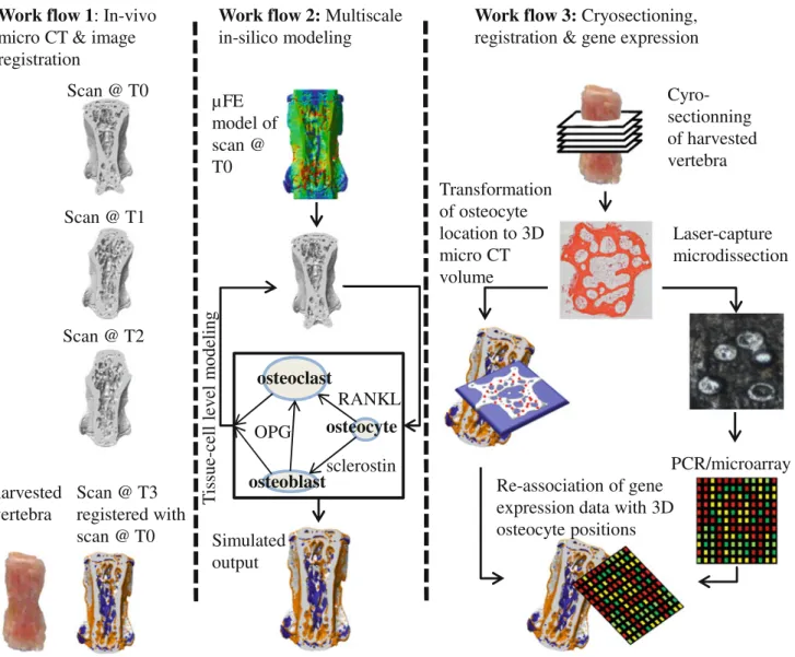
FIGURE 4. The mechanical systems biology framework for investigating load-induced bone adaptation is a combined experi-mental and computational approach which can be separated into three different workflows. Images provided with courtesy by Dr. Friederike Schulte and Reto Fortunati.
performing micro-array analysis on single cells.49,71,79 With this in mind, if cryosectioning and laser-capture microdissection techniques are coupled with micro-fluidics and applied to cyclic loaded bones, it will be possible to retrieve the local molecular information which is lacking. This has added advantages when combined with loaded bones which are monitored using in vivo lCT, for example in mice vertebrae. By developing software to translate osteocyte positions in 2D cryosections to their corresponding positions in registered 3D in vivo lCT volumes and their lFE models it should be possible to relate gene expression in individual osteocytes with their local micro-mechanical environment. Moreover, the same princi-ples can be applied to osteoblasts and osteoclasts. Pilot studies (data not shown) have shown that both oste-oblasts and osteoclasts are present on bone surfaces in the cryosections. Spatial mapping of these cells with the developed registration software as well as extrac-tion using laser capture micro-dissecextrac-tion will enable questions related to RANKL, OPG, and Sost path-ways to be addressed. Information retrieved from sequential in vivo lCT measurements can also be used to address such questions since the biochemical response of osteocytes can be correlated with markers of osteoblastic and osteoclastic activity i.e., the amount of newly formed or resorbed bone volumes and the rates at which they occur.
This experimental approach would not only provide more insight into the mechanisms governing load induced bone adaptation but would also provide data at both cell- and tissue-levels, which could be used to validate multiscale computational approaches. This strategy, which we would like to term Mechanical Systems Biology in Bone, is illustrated in Fig.4 and consists of the following workflows: (1) in vivo lCT images are generated at the start (T0) during (T1, T2) and at the end (T3) of a cyclic loading study and reg-istered to quantify local areas of osteoblast/osteoclast activity. (2) Micro finite element models are created from in vivo lCT scans describing the state of bone before cyclic loading. This is used as the input for tissue- and cell-level computational models. Using a multiscale framework similar to that proposed by Shim et al.69the effect of loading on bone is simulated. The outcome of which can be directly compared to in vivo lCT data. (3) At the endpoint of the experiment, the loaded bone is harvested and cryosectioned. These sections are registered in the three dimensional lCT images to identify the location of osteocytes. Osteo-cytes are harvested from the cryosections using LCM and transferred to microfluidic arrays for gene expression quantification. These data are used to create spatial gene expression maps which can then be cor-related with both registered lCT images and the lFE
model to better characterize the relationship between molecular activity, the local osteoblast/osteoclast activity and the associated micro-mechanical environ-ment. The experimental components of this workflow will provide more insight into the mechanisms gov-erning bone adaptation and allow quantitative vali-dation of both cell- and tissue level model.
CONCLUSIONS
This review gives an overview over the state-of-the-art in experimental and computational techniques in the field of load-induced bone adaptation. Fur-thermore, the limitations of these approaches were discussed. Only up-until recently have approaches started to span spatial scales, relating mechanical load at the tissue scale to molecular responses at the cellular scale. By coupling old and emerging technologies we have outlined a mechanical systems biology approach to further these advancements and overcome some of the limitations. When realized this should lead to a better understanding of the adaptive remodeling pro-cesses in the bone.
ACKNOWLEDGMENTS
The authors thank Dr. Friederike Schulte and Reto Fortunati for the provided images. Furthermore the authors gratefully acknowledge the funding from the SystemsX.ch/Swiss National Science Foundation.
REFERENCES
1
Adachi, T., Y. Kameo, and M. Hojo. Trabecular bone remodelling simulation considering osteocytic response to fluid-induced shear stress. Philos. Trans. R. Soc. A 368: 2669–2682, 2010.
2
Adachi, T., Y. Tomita, H. Sakaue, and M. Tanaka. Sim-ulation of trabecular surface remodeling based on local stress nonuniformity. Jpn. Soc. Mech. Eng. 40:782–792, 1997.
3
Adachi, T., K. Tsubota, Y. Tomita, and S. J. Hollister. Trabecular surface remodeling simulation for cancellous bone using microstructural voxel finite element models. J. Biomech. Eng.123:403–409, 2001.
4
Albright, J. The Scientific Basis of Orthopaedics. New York: Appleton-Century Crofts, 1987.
5Andriacchi, T. P., S. Koo, and S. F. Scanlan. Gait
mechanics influence healthy cartilage morphology and osteoarthritis of the knee. J. Bone Joint Surg. Am.
91(Suppl 1):95–101, 2009.
6Ascenzi, M. G., J. Gill, and A. Lomovtsev. Orientation of
collagen at the osteocyte lacunae in human secondary osteons. J. Biomech. 41:3426–3435, 2008.
7
Atkins, G. J., P. S. Rowe, H. P. Lim, K. J. Welldon, R. Ormsby, A. R. Wijenayaka, L. Zelenchuk, A. Evdokiou, and D. M. Findlay. Sclerostin is a locally acting regulator of late-osteoblast/preosteocyte differentiation and regu-lates mineralization through a MEPE-ASARM-dependent mechanism. J. Bone Miner. Res. 26:1425–1436, 2011.
8
Be’ery-Lipperman, M., and A. Gefen. A method of quantification of stress shielding in the proximal femur using hierarchical computational modeling. Comput. Methods Biomech.9:35–44, 2006.
9Bonewald, L. F. Osteocytes: a proposed multifunctional
bone cell. J. Musculoskelet. Neuronal Interact. 2:239–241, 2002.
10Bonewald, L. Osteocytes as multifunctional cells. J.
Mus-culoskelet. Neuronal Interact.6:331–333, 2006.
11
Bonewald, L. F., and M. L. Johnson. Osteocytes, mechanosensing and Wnt signaling. Bone 42:606–615, 2008.
12
Bonivtch, A. R., L. F. Bonewald, and D. P. Nicolella. Tissue strain amplification at the osteocyte lacuna: a microstructural finite element analysis. J. Biomech. 40: 2199–2206, 2007.
13
Bontoux, N., L. Dauphinot, T. Vitalis, V. Studer, Y. Chen, J. Rossier, and M. C. Potier. Integrating whole transcrip-tome assays on a lab-on-a-chip for single cell gene profiling. Lab Chip8:443–450, 2008.
14
Buenzli, P. R., J. Jeon, P. Pivonka, D. W. Smith, and P. T. Cummings. Investigation of bone resorption within a cor-tical basic multicellular unit using a lattice-based compu-tational model. Bone 50:378–389, 2012.
15CellML.http://www.cellml.org/. Accessed 15 March 2012. 16Chambers, T. J., S. Fox, C. J. Jagger, J. M. Lean, and J. W.
Chow. The role of prostaglandins and nitric oxide in the response of bone to mechanical forces. Osteoarthritis Car-tilage7:422–423, 1999.
17
Chen, N. X., K. D. Ryder, F. M. Pavalko, C. H. Turner, D. B. Burr, J. Y. Qiu, and R. L. Duncan. Ca2+regulates fluid shear-induced cytoskeletal reorganization and gene expression in osteoblasts. Am. J. Physiol. Cell Physiol. 278:C989–C997, 2000.
18
Chow, J. W. Role of nitric oxide and prostaglandins in the bone formation response to mechanical loading. Exerc. Sport Sci. Rev.28:185–188, 2000.
19
Christie, G. R., P. M. Nielsen, S. A. Blackett, C. P. Bradley, and P. J. Hunter. FieldML: concepts and imple-mentation. Philos. Trans. A Math. Phys. Eng. Sci. 367:1869–1884, 2009.
20Coelho, P. G., P. R. Fernandes, H. C. Rodrigues, J. B.
Cardoso, and J. M. Guedes. Numerical modeling of bone tissue adaptation—a hierarchical approach for bone apparent density and trabecular structure. J. Biomech. 42:830–837, 2009.
21
De Souza, R. L., M. Matsuura, F. Eckstein, S. C. F. Rawlinson, L. E. Lanyon, and A. A. Pitsillides. Non-invasive axial loading of mouse tibiae increases cortical bone fori-nation and modifies trabecular organization: a new model to study cortical and cancellous compartments in a single loaded element. Bone 37:810–818, 2005.
22
Emmert-Buck, M. R., R. F. Bonner, P. D. Smith, R. F. Chuaqui, Z. P. Zhuang, S. R. Goldstein, R. A. Weiss, and L. A. Liotta. Laser capture microdissection. Science 274:998–1001, 1996.
23
Fernandez, J. W., M. Akbarshahi, K. M. Crossley, K. B. Shelburne, and M. G. Pandy. Model predictions of increased knee joint loading in regions of thinner articular cartilage
after patellar tendon adhesion. J. Orthop. Res. 29:1168–1177, 2011.
24
FieldML.http://www.fieldml.org/. Accessed 15 March 2012.
25
Fritton, J. C., E. R. Myers, T. M. Wright, and M. C. H. van der Meulen. Loading induces site-specific increases in mineral content assessed by microcomputed tomography of the mouse tibia. Bone 36:1030–1038, 2005.
26
Frost, H. M. The mechanostat: a proposed pathogenic mechanism of osteoporoses and the bone mass effects of mechanical and nonmechanical agents. Bone Miner. 2:73–85, 1987.
27Fyhrie, D. P., and D. R. Carter. A unifying principle
relating stress to trabecular bone morphology. J. Orthop. Res.4:304–317, 1986.
28
Fyhrie, D. P., and D. R. Carter. Prediction of cancellous bone apparent density with 3-D stress analysis. In: Trans-actions 32nd Annual Orthopedic Research Society, p. 133, 1986.
29
Galea, G. L., A. Sunters, L. B. Meakin, G. Zaman, T. Sugiyama, L. E. Lanyon, and J. S. Price. Sost down-regu-lation by mechanical strain in human osteoblastic cells involves PGE2 signaling via EP4. FEBS Lett. 585:2450– 2454, 2011.
30
Gerhard, F. A., D. J. Webster, G. H. van Lenthe, and R. Muller. In silico biology of bone modelling and remodel-ling: adaptation. Philos. Trans. A Math. Phys Eng. Sci. 367:2011–2030, 2009.
31
Gross, T. S., J. L. Edwards, K. J. McLeod, and C. T. Rubin. Strain gradients correlate with sites of periosteal bone formation. J. Bone Miner. Res. 12:982–988, 1997.
32Gross, T. S., S. Srinivasan, C. C. Liu, T. L. Clemens, and
S. D. Bain. Noninvasive loading of the murine tibia: an in vivo model for the study of mechanotransduction. J. Bone Miner. Res.17:493–501, 2002.
33
Heino, T. J., T. A. Hentunen, and H. K. Vaananen. Con-ditioned medium from osteocytes stimulates the prolifera-tion of bone marrow mesenchymal stem cells and their differentiation into osteoblasts. Exp. Cell Res. 294:458–468, 2004.
34
Henriksen, K., M. Karsdal, J. M. Delaisse, and M. T. Engsig. RANKL and vascular endothelial growth factor (VEGF) induce osteoclast chemotaxis through an ERK1/2-depen-dent mechanism. J. Biol. Chem. 278:48745–48753, 2003.
35
Huiskes, R., R. Ruimerman, G. H. van Lenthe, and J. D. Janssen. Effects of mechanical forces on maintenance and adaptation of form in trabecular bone. Nature 405:704– 706, 2000.
36Jacobs, C. R., C. E. Yellowley, B. R. Davis, Z. Zhou, J. M.
Cimbala, and H. J. Donahue. Differential effect of steady versus oscillating flow on bone cells. J. Biomech. 31:969– 976, 1998.
37
Jacquet, R., J. Hillyer, and W. J. Landis. Analysis of connective tissues by laser capture inicrodissection and reverse transcriptase-polymerase chain reaction. Anal. Biochem.337:22–34, 2005.
38
Kamioka, H., T. Honjo, and T. A. Takano-Yamamoto. Three-dimensional distribution of osteocyte processes revealed by the combination of confocal laser scanning microscopy and differential interference contrast micros-copy. Bone 28:145–149, 2001.
39
Kesavan, C., S. Mohan, S. Oberholtzer, J. E. Wergedal, and D. J. Baylink. Mechanical loading-induced gene expression and BMD changes are different in two inbred mouse strains. J. Appl. Physiol. 99:1951–1957, 2005.
40
Keyak, J. H., J. M. Meagher, H. B. Skinner, and C. D. Mote. Automated three-dimensional finite element modelling of bone: a new method. J. Biomed. Eng. 12:389–397, 1990.
41
Klein-Nulend, J., R. G. Bacabac, and M. G. Mullender. Mechanobiology of bone tissue. Pathol. Biol. 53:576–580, 2005.
42
Klein-Nulend, J., C. M. Semeins, N. E. Ajubi, P. J. Nijweide, and E. H. Burger. Pulsating fluid flow increases nitric oxide (NO) synthesis by osteocytes but not periosteal fibroblasts—correlation with prostaglandin upregulation. Biochem. Biophys. Res. Commun.217:640–648, 1995.
43Klein-Nulend, J., A. Vanderplas, C. M. Semeins, N. E.
Ajubi, J. A. Frangos, P. J. Nijweide, and E. H. Burger. Sensitivity of osteocytes to biomechanical stress in vitro. FASEB J.9:441–445, 1995.
44
Knothe Tate, M. L. Top down and bottom up engineering of bone. J. Biomech. 44:304–312, 2011.
45
Lambers, F. M., F. A. Schulte, G. Kuhn, D. J. Webster, and R. Mueller. Mouse tail vertebrae adapt to cyclic mechanical loading by increasing bone formation rate and decreasing bone resorption rate as shown by time-lapsed in vivo imaging of dynamic bone morphometry. Bone 49:1340–1350, 2011.
46
Lanyon, L. E. Osteocytes, strain detection, bone modeling and remodeling. Calcif. Tissue Int. 53(Suppl 1):S102–S106; discussion S106–S107, 1993.
47
Lemaire, V., F. L. Tobin, L. D. Greller, C. R. Cho, and L. J. Suva. Modeling the interactions between osteoblast and osteoclast activities in bone remodeling. J. Theor. Biol. 229:293–309, 2004.
48Maldonado, S., S. Borchers, R. Findeisen, and F.
Allgower. Mathematical modeling and analysis of force induced bone growth. Conf. Proc. IEEE Eng. Med. Biol. Soc.1:3154–3157, 2006.
49
Marcus, J. S., W. F. Anderson, and S. R. Quake. Micro-fluidic single-cell mRNA isolation and analysis. Anal. Chem.78:3084–3089, 2006.
50
Moustafa, A., T. Sugiyama, J. Prasad, G. Zaman, T. S. Gross, L. E. Lanyon, and J. S. Price. Mechanical loading-related changes in osteocyte sclerostin expression in mice are more closely associated with the subsequent osteogenic response than the peak strains engendered. Osteoporos. Int. 23:1225–1234, 2012.
51
Mullender, M., A. J. El Haj, Y. Yang, M. A. van Duin, E. H. Burger, and J. Klein-Nulend. Mechanotransduction of bone cells in vitro: mechanobiology of bone tissue. Med. Biol. Eng. Comput.42:14–21, 2004.
52Nakashima, T., M. Hayashi, T. Fukunaga, K. Kurata,
M. Oh-hora, J. Q. Feng, L. F. Bonewald, T. Kodama, A. Wutz, E. F. Wagner, et al. Evidence for osteocyte regula-tion of bone homeostasis through RANKL expression. Nat. Med.17:1231–1234, 2011.
53
Nickerson, D., and P. Hunter. Using CellML in compu-tational models of multiscale physiology. Proc. Ann. Int. IEEE EMBS6:6096–6099, 2005.
54
Nicolella, D. P., D. E. Moravits, A. M. Gale, L. F. Bonewald, and J. Lankford. Osteocyte lacunae tissue strain in cortical bone. J. Biomech. 39(1735–43):57, 2006.
55
Norman, J., J. G. Shapter, K. Short, L. J. Smith, and N. L. Fazzalari. Micromechanical properties of human trabecu-lar bone: a hierarchical investigation using nanoindenta-tion. J. Biomed. Mater. Res. A 87:196–202, 2008.
56
Parfitt, A. M. Osteonal and hemi-osteonal remodeling—the spatial and temporal framework for signal traffic in adult human bone. J. Cell. Biochem. 55:273–286, 1994.
57
Pitsillides, A. A., S. C. F. Rawlinson, R. F. L. Suswillo, S. Bourrin, G. Zaman, and L. E. Lanyon. Mechanical strain-induced NO production by bone cells: a possible role in adaptive bone (re)modeling? FASEB J. 9:1614–1622, 1995.
58
Pivonka, P., J. Zimak, D. W. Smith, B. S. Gardiner, C. R. Dunstan, N. A. Sims, T. J. Martin, and G. R. Mundy. Model structure and control of bone remodeling: a theo-retical study. Bone 43:249–263, 2008.
59
Poole, K. E. S., R. L. van Bezooijen, N. Loveridge, H. Hamersma, S. E. Papapoulos, C. W. Lowik, and J. Reeve. Sclerostin is a delayed secreted product of osteocytes that inhibits bone formation. FASEB J. 19:1842, 2005.
60Prendergast, P. J., and D. Taylor. Prediction of bone
adaptation using damage accumulation. J. Biomech. 27:1067–1076, 1994.
61
Robling, A. G., P. J. Niziolek, L. A. Baldridge, K. W. Condon, M. R. Allen, I. Alam, S. M. Mantila, J. Gluhak-Heinrich, T. M. Bellido, S. E. Harris, et al. Mechanical stimulation of bone in vivo reduces osteocyte expression of Sost/sclerostin. J. Biol. Chem. 283:5866–5875, 2008.
62
Robling, A. G., and C. H. Turner. Mechanotransduction in bone: genetic effects on mechanosensitivity in mice. Bone 31:562–569, 2002.
63
Rubin, M. A., and I. Jasiuk. The TEM characterization of the lamellar structure of osteoporotic human trabecular bone. Micron 36:653–664, 2005.
64
Ruimerman, R., P. Hilbers, B. van Rietbergen, and R. Huiskes. A theoretical framework for strain-related tra-becular bone maintenance and adaptation. J. Biomech. 38:931–941, 2005.
65Ryser, M. D., N. Nigam, and S. V. Komarova.
Mathemat-ical modeling of spatio-temporal dynamics of a single bone multicellular unit. J. Bone Miner. Res. 24:860–870, 2009.
66
Schulte, F. A., F. M. Lambers, G. Kuhn, and R. Mueller. In vivo micro-computed tomography allows direct three-dimensional quantification of both bone formation and bone resorption parameters using time-lapsed imaging. Bone48:433–442, 2011.
67
Schulte, F. A., F. M. Lambers, D. J. Webster, G. Kuhn, and R. Mueller. Strain energy density predicts sites of local trabecular bone formation and resorption. In: Abstracts 17th Congress of the European Society of Biomechanics (ESB), Edinburgh, UK, 5–7 July 2010, p. 697.
68
Schutze, K., and G. Lahr. Identification of expressed genes by laser-mediated manipulation of single cells. Nat. Bio-technol.16:737–742, 1998.
69Shim, V. B., P. J. Hunter, P. Pivonka, and J. W. Fernandez.
A multiscale framework based on the physiome markup languages for exploring the initiation of osteoarthritis at the bone-cartilage interface. IEEE Trans. Biomed. Eng. 58:3532–3536, 2011.
70
Tang, F., C. Barbacioru, Y. Wang, E. Nordman, C. Lee, N. Xu, X. Wang, J. Bodeau, B. B. Tuch, A. Siddiqui, et al. mRNA-Seq whole-transcriptome analysis of a single cell. Nat. Methods6:377–382, 2009.
71
Tang, F. C., P. Hajkova, S. C. Barton, K. Q. Lao, and M. A. Surani. MicroRNA expression profiling of single whole embryonic stem cells. Nucleic Acids Res. 34:e9, 2006.
72
Turner, C. H., M. R. Forwood, and M. W. Otter. Mech-anotransduction in bone—do bone-cells act as sensors of fluid flow. FASEB J. 8:875–878, 1994.
73
van Bezooijen, R. L., B. A. J. Roelen, A. Visser, L. van der Wee-Pals, E. de Wilt, M. Karperien, H. Hamersma, S. E. Papapoulos, P. ten Dijke, and C. Lowik. Sclerostin is an osteocyte-expressed negative regulator of bone formation,
but not a classical BMP antagonist. J. Exp. Med. 199:805– 814, 2004.
74
van der Meulen, M. C. H., T. G. Morgan, X. Yang, T. H. Baldini, E. R. Myers, T. M. Wright, and M. P. G. Bostrom. Cancellous bone adaptation to in vivo loading in a rabbit model. Bone 38:871–877, 2006.
75
Warden, S. J., and C. H. Turner. Mechanotransduction in cortical bone is most efficient at loading frequencies of 5–10 Hz. Bone 34:261–270, 2004.
76
Wasserman, E., D. Webster, M. Attar-Namdar, R. Mueller, and I. Bab. Method for differential isolation of RNA from mouse caudal trabecular osteoblasts and osteocytes. J. Biomech. 41:S131–S131, 2008.
77Webster, D. J., P. L. Morley, G. H. van Lenthe, and R.
Mueller. A novel in vivo mouse model for mechanically stimulated bone adaptation—a combined experimental and computational validation study. Comput. Methods Bio-mech. Biomed. Eng.11:435–441, 2008.
78
Webster, D., and R. Muller. In silico models of bone remodeling from macro to nano-from organ to cell. Wires Syst. Biol. Med.3:241–251, 2011.
79
White, A. K., M. VanInsberghe, O. I. Petriv, M. Hamidi, D. Sikorski, M. A. Marra, J. Piret, S. Aparicio, and C. L. Hansen. High-throughput microfluidic single-cell RT-qPCR. Proc. Natl Acad. Sci. USA 108:13999–14004, 2011.
80
Winkler, D. G., M. K. Sutherland, J. C. Geoghegan, C. P. Yu, T. Hayes, J. E. Skonier, D. Shpektor, M. Jonas, B. R. Kovacevich, K. Staehling-Hampton, et al. Osteocyte con-trol of bone formation via sclerostin, a novel BMP antag-onist. EMBO J. 22:6267–6276, 2003.
81
Wolff, J. Das Gesetz der Transformation der Knochen. Berlin: Hirschwald, 1892.
82
Xing, W. R., D. Baylink, C. Kesavan, Y. Hu, S. Kapoor, R. B. Chadwick, and S. Mohan. Global gene expression analysis in the bones reveals involvement of several novel genes and pathways in mediating an anabolic response of mechanical loading in mice. J. Cell. Biochem. 96:1049–1060, 2005.
83
Xiong, J., M. Onal, R. L. Jilka, R. S. Weinstein, S. C. Manolagas, and C. A. O’Brien. Matrix-embedded cells con-trol osteoclast formation. Nat. Med. 17:U1235–U1262, 2011.
84Yang, X. B., R. S. Tare, K. A. Partridge, H. I. Roach,
N. M. Clarke, S. M. Howdle, K. M. Shakesheff, and R. O. Oreffo. Induction of human osteoprogenitor chemotaxis, proliferation, differentiation, and bone formation by osteoblast stimulating factor-1/pleiotrophin: osteoconduc-tive biomimetic scaffolds for tissue engineering. J. Bone Miner. Res.18:47–57, 2003.
85
Zaman, G., H. L. Jessop, M. Muzylak, R. L. De Souza, A. A. Pitsillides, J. S. Price, and L. L. Lanyon. Osteocytes use estrogen receptor alpha to respond to strain but their ER alpha content is regulated by estrogen. J. Bone Miner. Res.21:1297–1306, 2006.
86
Zhao, S., Y. Kato, Y. Zhang, S. Harris, S. S. Ahuja, and L. F. Bonewald. MLO-Y4 osteocyte-like cells support osteoclast formation and activation. J. Bone Miner. Res. 17:2068–2079, 2002.
87
Zhong, J. F., Y. Chen, J. S. Marcus, A. Scherer, S. R. Quake, C. R. Taylor, and L. P. Weiner. A microfluidic processor for gene expression profiling of single human embryonic stem cells. Lab Chip 8:68–74, 2008.