© The Author 2013. Published by Oxford University Press on behalf of the Society for Experimental Biology. All rights reserved. For permissions, please email: [email protected]
Abbreviations: AFM, atomic force microscopy; MFA, microfibril angle.
review paper
Plant micro- and nanomechanics: experimental techniques
for plant cell-wall analysis
Ingo Burgert* and Tobias Keplinger
ETH Zurich, Institute for Building Materials, Schafmattstrasse 6, 8093 Zurich & Empa, Applied Wood Materials Laboratory, Ueberlandstrasse 129, 8600 Duebendorf, Switzerland
* To whom correspondence should be addressed. Email: [email protected]
Received 18 March 2013; Revised 9 July 2013; Accepted 12 July 2013
Abstract
In the last few decades, micro- and nanomechanical methods have become increasingly important analytical tech-niques to gain deeper insight into the nanostructure and mechanical design of plant cell walls. The objective of this article is to review the most common micro- and nanomechanical approaches that are utilized to study primary and secondary cell walls from a biomechanics perspective. In light of their quite disparate functions, the common and opposing structural features of primary and secondary cell walls are reviewed briefly. A significant part of the article is devoted to an overview of the methodological aspects of the mechanical characterization techniques with a particular focus on new developments and advancements in the field of nanomechanics. This is followed and complemented by a review of numerous studies on the mechanical role of cellulose fibrils and the various matrix components as well as the polymer interactions in the context of primary and secondary cell-wall function.
Key words: Atomic force microscopy, fibre–matrix interactions, micromechanics, nanoindentation, plant biomechanics, primary cell walls, secondary cell walls.
Introduction
Plant micro- and nanomechanics are subdisciplines of plant biomechanics, which primarily address the cell and cell-wall level of the plant body (Niklas & Spatz, 2012). Due to the hier-archical structure of plants (Speck & Burgert, 2011; Gibson, 2012), research progress in this field has a great impact on plant biomechanics in general, as properties and features at this scale inevitably influence the macroscopic appearance and performance of the plant body (Fratzl and Weinkamer, 2007). In recent years, there has been a tremendous increase in mechanical characterization studies at the cell and cell-wall level dealing with various issues of structure–property and structure–function relationships, which have been reviewed by various authors (Geitmann, 2006; Burgert & Dunlop, 2011;
Cosgrove & Jarvis, 2012; Eder et al., 2013; Kasas et al., 2013). On the one hand, this has been due to rapid methodological developments in this field that allow more precise determina-tion of mechanical properties, in particular at the nanoscale
of the biological systems. On the other hand, cell and cell-wall mechanics are increasingly considered to be highly relevant as we gain a deeper understanding of how mechanics affects growth processes and how mechanical properties are sensed, controlled, and tuned by the plant (Cosgrove, 1993, 2005;
Burgert, 2006; Telewski, 2006; Niklas, 2009). This knowledge gain substantially affects matters of plant growth and mor-phology as well as closing a feedback loop in terms of the biosynthesis and structure of cell-wall components. In this context, cell and cell-wall mechanics have become an impor-tant characterization tool for evaluating the impact of genetic modifications regarding effects on plant structure and growth processes, material performance, and mechanical stability of crops (Ryden et al., 2003; Pena et al., 2004; Bjurhager et al., 2008; Bjurhager et al., 2010; Hoenicka et al., 2012).
Besides a tight interplay of plant biomechanics and plant morphology, cell and cell-wall mechanics are highly relevant
in various fields of plant material utilization. For instance, mechanical characterization techniques provide highly valu-able information about material properties, such as fibre per-formance in pulp and paper (Jayne, 1959; Page et al., 1971;
Kersevage, 1973; Page & El-Hosseiny, 1983; Groom et al., 2002a). Furthermore, the increased knowledge offers the potential to transfer principles and mechanisms evolved by nature to the design of bio-inspired materials (Sidorenko et al., 2007; Dunlop & Fratzl, 2010; Martone et al., 2010;
Paris et al., 2010).
Surprisingly, research on primary and secondary cell walls in recent decades has been conducted largely by separate communities that have rarely interacted. While primary cell-wall research has been closely related to physiological aspects due to the interplay with the living cell, research on secondary cell walls has focused on material aspects of predominately dead cells. In recent years, however, the research communi-ties have come closer together, due in part to our increase in understanding of how genes regulate biosynthesis processes and determine the structure, chemistry, and properties of cell walls. Additionally, we see a bridging function by micro- and nanomechanical approaches, which help to identify common principles of cell-wall assembly and the resulting structure– function relationships (Burgert, 2006; Cosgrove & Jarvis, 2012). In this review, we aim to provide an overview of the basic principles of micro- and nanomechanical techniques that are applied to both primary and secondary cell-wall analysis and give examples of how these methods are used to unravel the underlying structure–property relationships of cells and cell walls in view of their specific functions.
While there has been great progress in the field of cell-wall modelling (Salmén & de Ruvo, 1985; Perez et al., 1996;
Yamamoto & Kojima, 2002; Besombes & Mazeau, 2005;
Hofstetter et al., 2005; Hanus & Mazeau, 2006; Bader et al., 2011; de Borst et al., 2012) and in combined approaches in which experimental data and modelling complement each other, here we focus on the experimental side and review the tremendous knowledge gain exclusively in this field. This comprises mechanical analysis of primary and secondary cell walls of various cell types, which are briefly introduced in terms of general mechanical constraints and specific functions.
Mechanical functions of primary and
secondary cell walls
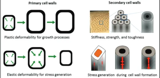
Micro- and nanomechanical characterization needs to thor-oughly consider the functional context of the investigated plant material as this is crucial in order to gain insight into the underlying structure–function relationships. The main mechanical functions that primary cell walls need to fulfil are to provide sufficient stiffness and strength to the cell but at the same time to allow for cell growth, as well as enabling revers-ible changes of cell size and shape with regard to pre-stressing and organ movements (Cosgrove, 2005; Martone et al., 2010). Primary cell walls have to be considered together with turgor pressure regulated by the living cell to attain stiffness in an
‘inflated’ state. With regard to the specific mechanical proper-ties of the primary cell wall, it is fascinating to realize that the same wall has to fulfil two mutually exclusive mechanical requirements. In order to allow growth processes, primary cell walls need to be plastically deformable, whereas to provide mechanical stability or allow reversible movements such as stomata opening and closure, the primary cell wall needs to be entirely elastic (Williams & Bennett, 1982; Proseus et al., 1999; Boyer, 2001; Cosgrove, 2001, 2005; Forterre et al., 2005;
Roelfsema & Hedrich, 2005; Moran, 2007). The mechanism by which the cell wall can ‘switch’ between elastic and plastic deformability by alterations of biomacromolecule interac-tions is still debated (Fry et al., 1992; Cosgrove, 2000, 2005;
Schopfer, 2001; Proseus & Boyer, 2006).
At a first glance, secondary cell walls appear to be by far the more pre-determined in their functionality. The rigid network of parallel aligned cellulose fibrils and matrix substances pro-vides mechanical stability, even to dead cells (Cave, 1968; Mark & Gillis, 1970, 1973). Beyond this fundamental requirement, one can see a wide variability in structure and composition of secondary cell walls that mirrors the different functions of cell and tissue types accompanied by different mechanical property profiles (Donaldson, 2001; Wegst & Ashby, 2004; Donaldson, 2008; Eder et al., 2009; Eder & Burgert, 2010). These profiles include a vast variability in terms of material stiffness, tough-ness and strength as well as the capability to generate both tensile and compressive mechanical stresses (Yamamoto, 1998;
Lichtenegger et al., 1999; Reiterer et al., 1999; Farber et al., 2001; Burgert et al., 2007; Goswami et al., 2008; Burgert & Fratzl, 2009; Clair et al., 2011; Eder et al., 2013). The impor-tant mechanical requirements that need to be fulfilled by pri-mary and secondary cell walls are illustrated in Fig. 1.
Brief overview of structure and chemistry
in relation to biomechanics
In view of the wide range of various cell types and cell-wall compositions, it is impossible to treat the topic comprehen-sively. Therefore, we will focus on a simplified illustration of the underlying principles and concepts of primary and ondary cell walls. Generally speaking, both primary and sec-ondary cell walls can be described by means of natural fibre composites consisting of a stiff fibrous phase made of cellu-lose fibrils composed of crystalline and amorphous regions as well as a pliant amorphous matrix comprising various biopolymers with hydrogen bonding between the two phases (Kerstens et al., 2001; Fratzl et al., 2004a). According to their specific functions, both cell-wall types can be distinguished by means of structural and chemical parameters. This applies to cell-wall thickness, cellulose orientation, degree of crystallization, volume fractions of cell-wall components, composition of the matrix, chemical bonding patterns, and water content (McCann & Roberts, 1991; Pauly et al., 1999;
Brändström, 2001; Donaldson, 2001; Somerville et al., 2004;
Burgert, 2006; Donaldson, 2008; Jarvis, 2009; Cosgrove & Jarvis, 2012). It is important to note that all these param-eters are also highly varied within primary and secondary
cell walls, meaning that plants manifest a vast potential to regulate properties at the nano-and microscale level of the cell walls. In Table 1, crucial parameters of primary and secondary cell walls, predominantly from a biomechanical perspective, are listed. Such a tabular comparison inevitably results in a simplification, which we assume to be tolerable for the benefit of clarity.
Plant materials and characterization
techniques
In this review, we intend to discuss plant materials that are examined intensively in micro- and nanomechanical analyses, as well as to introduce the related characterization techniques for biomechanical studies. In principle, any plant organ could be studied with at least one of the below-mentioned methods. However, screening of the relevant literature shows that par-ticular plant materials have been more prominently examined
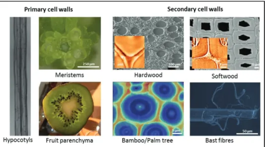
either because of being used as model systems or because of having important mechanical properties for the plant body or in application (Fig. 2).
In terms of primary cell walls, basic investigations on cell-wall structure and properties as well as growth processes have been conducted mainly on hypocotyls of various plant spe-cies (Nakahori et al., 1991; Cosgrove, 1993, 2011; Kutschera & Kohler, 1994; Ryden et al., 2003; Refregier et al., 2004). In recent years, with the technological developments in nano-mechanical studies, even meristems have become a matter of intensive studies as very local cell-wall stiffness alterations can be monitored (Peaucelle et al., 2011). More applied research activities are related to the mechanical characterization of fruit parenchyma or fruit peels (Bargel et al., 2004; Landahl et al., 2004; Matas et al., 2004; Bargel & Neinhuis, 2005). Micro- and nanomechanical studies on secondary cell walls at the level of basic research are aimed at unravelling general principles and mechanisms of strength and deformation behaviour, includ-ing how plants achieve and control certain property profiles.
Fig. 1. Main functions and mechanical requirements of primary and secondary cell walls.
Table 1. Tabular comparison of general features of primary and secondary cell walls accompanied by cell-wall illustrations
The schematic drawing of the primary cell wall is adapted from a new cell-wall model by Park & Cosgrove (2012b)
Feature Primary cell walls Secondary cell walls
Illustration
Cell wall Thin, ongoing property alterations by the cell Thick, multi-lamellar, fully differentiated structure Polymer network Flexible network, ongoing modification processes Rigid network, interlocked status
Cellulose Low content, reorientation possible, orientation more variable
High content, densely packed, strictly parallel orientation
Matrix Predominately hemicelluloses, pectin, and structural proteins Predominately hemicelluloses and lignin Water interactions Highly hydrophilic, hydrogel character More hydrophobic when lignified Fibre–matrix interactions—uncertainties Widely accepted tethered cellulose–xyloglucan model
recently challenged. Illustration above shows a new cell-wall model suggested by Park and Cosgrove (2012b)
Mechanical function and spatial orientation in particular of hemicellulose composition and lignin not fully understood
In addition to establishing a general relationship between cel-lulose orientation and mechanical performance, understand-ing cellulose–matrix interactions has developed into a major focus in recent years (Reiterer et al., 1999; Spatz et al., 1999;
Köhler & Spatz, 2002; Keckes et al., 2003; Fratzl et al., 2004b;
Altaner & Jarvis, 2008). More applied research approaches aim at determining local mechanical properties of industrially relevant crops. A major interest is on wood fibre properties of wood species with regard to pulp and paper as well as on annu-ally harvested bast fibres (hemp, flax) for utilization in natural fibre composites (Page & El-Hosseiny, 1983; Eichhorn et al., 2001a; Bos et al., 2002; Groom et al., 2002a,b; Peetla et al., 2006; Thygesen et al., 2007). Due to the excellent mechani-cal performance and fast-growing capacities of bamboo and other grasses, several micro- and nanomechanical studies have been conducted in order to unravel the underlying structure– function relationships and derive mechanical characteristics for utilization purposes (Ruggeberg et al., 2008; Shao et al., 2010; Yu et al., 2011; Wang et al., 2012).
Microtensile tests
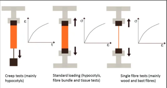
Micromechanical testing techniques address the plant level of tissues and individual cells. This includes tests on entire organs such as Arabidopsis hypocotyls, small tissue samples, and fibre bundles (e.g. wood segments, and fibre bundles of bamboo, palms, flax, and hemp), as well as individual cells mainly in the form of fibres and tracheids. The size and dimensions of the samples as set by plant tissue structure and variability limit the kind of loading conditions that can be applied. Accordingly, at the micromechanical level, predominantly uniaxial tensile tests are conducted, comprising common standard tests to gain information on stiffness, strength, and toughness of the plant material and time-dependent investigations to study relaxation and creep phenomena. When deriving information on cell-wall properties from mechanical tests at the tissue level, one needs to consider that a multitude of additional parameters affect the
obtained data, such as tissue density, variability of the tissue, cell length, cell–cell interactions, and turgor pressure.
For primary cell walls, the most widely utilized micro-mechanical test is a creep test in which a defined weight is attached to the tissue and the elongation is recorded over time. Based on creep tests, important information on pri-mary cell-wall architecture and cell-wall loosening mecha-nisms during cell growth have been gained (Cleland, 1971;
Richmond et al., 1980; Suslov & Verbelen, 2006; Cosgrove, 2011). However, it needs to be emphasized that there are vari-ous constraints on what can be inferred from creep experi-ments and uniaxial tensile tests concerning growth processes and turgor-driven expansion, respectively (Cosgrove, 1993). Rather recently, standard tensile tests and cyclic loading tests have been applied to study the impact of genetic modification treatments on mechanical properties, in particular of dark-grown hypocotyls of Arabidopsis thaliana (Ryden et al., 2003;
Pena et al., 2004; Cavalier et al., 2008; Abasolo et al., 2009). The hypocotyls are fixed in a microtensile tester and a uniax-ial tensile stress is applied until final rupture of the specimen occurs. Methodologically, besides maintaining a sufficient level of humidity of the specimen, fixation of the hypocotyls is crucial, as the fragile and turgorized samples do not allow classical clamping, which makes a gluing process more favour-able. The applied forces are recorded with load cells of small capacity, and the elongation can be derived from the machine path. However, it is always recommendable and sometimes mandatory to use optical systems such as video extensometry to record the strain more precisely and avoid measuring errors, for instance due to sample slippage in the clamps. In order to calculate the stress based on the force measurement, the cross-sectional area of the hypocotyl needs to be considered. A rather fast method is to calculate it from the diameter of the almost circular hypocotyl. However, one has to be aware that this procedure results in a stress–strain diagram for the entire hypocotyl and rules out structural differences at the tissue level such as cell size and shape, as well as cell-wall thickness.
Fig. 2. Common plant materials in mechanical analysis visualized by light microscopy, scanning electron microscopy, Raman imaging
Further parameters that influence the testing protocol and can lead to a large scatter of data and prevent direct comparisons between different studies are the influence of hypocotyl age, the germination conditions, the culture medium and the seg-ment of the hypocotyl that has been tested.
In terms of secondary cell walls, one can state that the micro-tensile testing is a bit more determined than for primary cell walls, because the specimens are less fragile and usually dead, which rules out the influence of turgor pressure. However, in particular, specimen size can have an underestimated effect on the mechanical response of the tested plant material (Navi et al., 1995). The standard tensile testing setups are similar to the one described for primary cell walls, although certain fibre bundles can be sufficiently rigid to allow fast clamping in the machine. Single fibres are usually fixed by gluing, which can be achieved either by gluing them on supporting frames that are fixed in the tensile tester or by glue droplets that are directly mounted at the fibre ends for a ball and socket setup (Kersevage, 1973; Groom et al., 2002b; Burgert et al., 2003;
Sedighi-Gilani et al., 2005). In any case, the fibres or tissue samples need to be aligned precisely, because even small devia-tions from the uniaxial loading condition have a large influence on the recorded mechanical behaviour due to the occurrence of shear stresses. The cross-sectional area of the sections in terms of tissues/fibres or cell walls can be determined by light or electron microscopy studies on samples that are cut trans-versally after testing, or cutting can be avoided when confo-cal light microscopy is applied (Groom et al., 2002a; Burgert et al., 2005). Usually, the strain required to fracture fibres and small tissue sheets containing cells with secondary cell walls is rather small, which makes a coupling of the testing setup to an optical system for high-resolution strain measurements such as video extensometry essential. Figure 3 shows in simple illustrations the most utilized micromechanical testing setups for biomechanical studies on primary and secondary cell walls.
Unique micromechanical testing approaches are so-called combined (in situ) methods in which external loading
is combined with simultaneous observation of nano- and microstructural deformation. The material response at the nano- and microstructural level reveals insights into specific deformation mechanisms and crack propagation events in the specimen. In plant biomechanics in particular, uniaxial tensile tests have been combined with various load-monitor-ing techniques to examine deformation patterns of plants at different levels of hierarchy. Detailed structural information can be obtained at the micro- and mesoscale by combining mechanical loading with light microscopy and for higher resolution with scanning electron microscopy (Mott et al., 1995; Bodner et al., 1996; Badel & Perré, 1999; Fruhmann et al., 2003; Thygesen et al., 2007; Eder et al., 2008). An in
situ approach based on computed tomography allows the
three-dimensional monitoring of deformation and fracture events at the level of cells and cell-wall layers (Nazarian et al., 2005; Zauner et al., 2012). For an even closer look into nanostructural deformation mechanisms at the level of polymer interactions of cell-wall components, spectros-copy and X-ray scattering techniques are utilized. In situ infra-red and Raman spectroscopy approaches can provide information on specific load-bearing capacities of cell-wall components, as well as collective mechanical responses of polymer assemblies (Salmén & Olsson, 1998; Eichhorn et al., 2000; Akerholm & Salmén, 2001; Eichhorn et al., 2001b; Akerholm & Salmén, 2003; Gierlinger et al., 2006;
Sturcova et al., 2006; Salmén & Bergstrom, 2009). In situ X-ray measurements at synchrotron facilities allow detailed observations of cellulose fibril–matrix interactions, includ-ing monitorinclud-ing of cellulose fibril reorientation durinclud-ing load-ing and the impact of water (Keckes et al., 2003; Kamiyama et al., 2005; Kölln et al., 2005; Zabler et al., 2010).
Nanoindentation
For many decades, traditional hardness tests (pushing a tip with a defined geometry into the surface of the sample)
Fig. 3. Most applied micromechanical testing techniques for the characterization of primary and secondary cell walls illustrated by
schematic drawings of the setup as well as of strain–time and stress–strain diagrams, respectively. (This figure is available in colour at JXB online.)
have been used for mechanical characterization in structural biology, including testing of cartilage, bones, soft tissues, plants, and wood in which hardness is defined as the ratio of a maximum load and the indent area (Kempson et al., 1971a,b; Doyle & Walker, 1985). Nanoindentation is based on the same principle but is performed at a smaller length sale and is advanced by exactly monitoring the displacement and the loading of the indenter during the measurement. The displacement is controlled via inductance or capacitance, and for the force actuation, normally piezo elements or magnetic coils are used (Ebenstein & Pruitt, 2006; Fischer-Cripps, 2011; Oyen, 2011).
In nanoindentation, it is highly important to obtain per-fectly flat surfaces in order not to distort the measurement due to surface roughness. Thus, to fulfil this requirement, an embedding material is needed (e.g. epoxy resin), which gives mechanical support during microtoming and nanoin-dentation. The question of penetration of the embedding material into the samples and its effect on the measure-ment remains unresolved. However, there is currently no conclusive evidence suggesting infiltration of the embed-ding material into the cell wall. For more details about sample preparation, see Konnerth et al. (2008) and Meng et al. (2013). A related limitation in nanoindentation stud-ies towards plant biomechanics is that it is very difficult to retain the natural wet condition of the samples due to the embedding procedure. Furthermore, one has to keep in mind that only a small area/volume is tested, which is a problem in view of the vast heterogeneity of plant cell walls (de Borst et al., 2012).
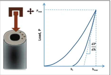
The main principle in nanoindentation is to calculate hard-ness and elastic modulus from a load-displacement (compli-ance) curve recorded during a local indentation. Unlike in conventional hardness tests (e.g. Vickers hardness), the size of the indent is too small for measurements with optical methods. Therefore, the area is indirectly determined from
the penetration depth together with the known geometry of the indenter (e.g. Berkovich, spherical indenter, power law indenters) (Fischer-Cripps, 2011; Oyen, 2011). Fig. 4 illus-trates the testing principle and shows a typical load-displace-ment curve for an elastic–plastic solid.
Based on the approach by Oliver and Pharr (1992), called compliance method, the reduced modulus and the hardness are determined by analysing the load-displacement curve. For further details on the analysis of the data and the calculation of the various parameters, see Eder et al. (2013).
In recent last years, a tremendous effort has gone into improving the technique in order to extract further param-eters such as creep compliance functions, storage modu-lus, and loss modulus (VanLandingham, 2003; Oyen, 2005;
Fischer-Cripps, 2011; Oyen, 2011).
For data interpretation, it has to be considered that the compliance method is based on the assumption of a homoge-neous and isotropic half space (Oliver & Pharr, 1992). A gen-eral restriction for biological materials is their anisotropic non-homogeneous nature. Hence, there have been huge efforts towards developing methods to reflect these constraints, such as the introduction of a holding period at peak load, con-tinuous stiffness measurements, and multi-load indentation experiments (Ebenstein & Pruitt, 2006; Tze et al., 2007; Oyen, 2011). An anisotropic indentation theory was introduced by the work of Vlassak et al. (2003) and applied for wood cell-wall characterization by Jager et al. (2011a,b). They showed that it is necessary to perform nanoindentation experiments with at least five different indention angles (compared with the orientation of the cellulose fibrils) to determine the elastic constants with the help of an error minimization procedure. As a result of the complex behaviour of the sample under the indenter, the absolute stiffness values are smaller than expected for the longitudinal modulus (Eder et al., 2013). For a comprehensive review of the mechanical modelling of cell-wall indentation, see Milani et al. (2013) in this issue.
Fig. 4. Schematic of a nanoindentation test on secondary cell walls with a typical load–displacement curve for an elastic–plastic solid.
Atomic force microscopy (AFM)
For measurements of mechanical properties at a smaller scale than in nanoindentation (for instance for primary cell walls), AFM has to be used. Figure 5 schematically illus-trates the common loading setups for primary and second-ary cell walls.
In AFM studies, a tip mounted on a cantilever spring is scanned over a sample, and the deflection of the cantilever, based on the force between the tip and the sample, is meas-ured with the help of a photodiode. With this configuration, topography images are obtained. However, to receive mechan-ical properties, so-called ‘force measurements’ are necessary, where the tip is moved towards the sample in the normal direction and force–distance curves are recorded (Green et al., 2002; Butt et al., 2005). In contrast to nanoindentation, in AFM the results of a force measurement—cantilever deflec-tion and piezo posideflec-tion—have to be converted into force and distance. First, the deflection of the photodiode has to be con-verted to vertical displacement and the spring constant of the cantilever is needed to calculate a force based on Hooke’s law. Additionally, a conversion for the height (change in piezo) is necessary, as it must be corrected for the deflection of the can-tilever. For a detailed description of the conversion methods, see Hutter & Bechhoefer (1993), Hinterdorfer et al. (1996), and Butt et al. (2005). The main objective in using AFM for mechanical characterization is not to take force–distance curves on selected points of the sample but rather to gener-ate images that are based on mechanical properties (force–dis-tance curve for every pixel). Various modes for the imaging of mechanical properties are available. However, there is still much potential for improvement and new techniques. Two possible modes are resonant contact AFM and pulsed force mode. In resonant contact AFM, the tip is used as a resona-tor whose frequency depends on the interactions between the sample and the tip, which allows measurement of the elastic properties of the sample (Clair et al., 2003). In pulsed force mode, the Z-piezo of the AFM is modulated with a sinusoidal voltage (oscillation amplitudes between 20 and 50 nm). At the lowest point of the oscillation, the tip is out of contact, and at the highest point, it reaches a deflection maximum. The out-come of pulsed force mode is force curves at high frequencies
that are recorded in real time, and high-resolution images of the mechanical properties are obtained (Krotil et al., 1999).
AFM has been used intensively to characterize the molecu-lar architecture of cell walls, such as the orientation and size of microfibrils or pore size distributions (Kirby et al., 1996;
Fahlen & Salmén, 2003, 2005; Yarbrough et al., 2009). When interpreting the obtained data, one has to consider the fact that the stiffness values extracted from force–distance curves do not exclusively reflect the elastic properties of the cell wall (Routier-Kierzkowska et al., 2012), but that indenter geometry (Bolduc et al., 2006), turgor pressure (Wang et al., 2004), and internal stresses also have an impact (Zamir & Taber, 2004). Another limiting factor in AFM examinations of primary cell walls is that the force that can be applied is too small to stretch the cell walls of turgid tissues. For this purpose, a so-called cel-lular force microscopy with the potential of applying of up to 1 mN has been developed, which allows one to indent cells until their rupture and to measure the release of the turgor pressure by changes in local stresses (Routier-Kierzkowska et al., 2012).
The AFM methods for mechanical characterization intro-duced above are based on examining force–distance curves, but it should be also mentioned that alternative methods exist for determining mechanical properties at the nanoscale. Clair et al. (2003) developed resonance contact AFM for determin-ing elastic properties of wood cells. Tetard et al. (2010a,b) used the so-called mode synthesizing AFM, which is based on exerting a multi-harmonic force on the substrate on the probe, which creates multiple orders coupling in frequency and allows the deduction of mechanical properties within the plant cell-wall structure. Furthermore, single-molecule force spectroscopy has been used to characterize interac-tions between xyloglucan molecules and a cellulose substrate (Morris et al., 2004). There is ongoing research into develop-ing new techniques for measurdevelop-ing mechanical properties, and many methods have simply not been utilized in plant cell-wall characterization yet (Krotil et al., 1999).
Biomechanics of plant cell walls
In the following, we intend to discuss micro and nanostruc-tural examinations on primary and secondary cell walls with
Fig. 5. Schematics of AFM in a transverse direction on primary cell walls and in the longitudinal direction on secondary cell walls. (This
regard to the specific mechanical role of the cell-wall compo-nents, and their interaction in cell-wall assembly.
Secondary cell walls
In terms of biomechanical studies on secondary cell walls, we intend to restrict ourselves to the basic structure–property rela-tionships, which exclude the principles of the stress-generation mechanisms mentioned and referenced above. For approaches aiming at unravelling the specific mechanical role of a cell-wall component, cellulose is probably the most easily accessible cell-wall biomacromolecule. Structure–property relationships can be derived directly due to the parallel arrangement and the partly crystalline nature of the cellulose fibrils, which allows the acquisition of detailed information on cellulose structure and formation via various methodical techniques. Hence, its crucial role in cell-wall mechanics has been shown in various studies at the tissue and fibre levels in which the tensile stiff-ness of the material has been related to the cellulose orienta-tion [microfibril angle (MFA)]. The larger the MFA, the lower the stiffness of the cell wall, which enables the cell to control and adjust mechanical performance in the cellulose spinning process. Using micromechanical tests on tissue and fibres, this relationship has been shown in various studies (Page & El-Hosseiny, 1983; Lichtenegger et al., 1999; Reiterer et al., 1999; Saren et al., 2001; Salmén & Burgert, 2009; Eder & Burgert, 2010; Eder et al., 2013). In situ tests combining ten-sile straining of single wood fibres with Raman spectroscopy have directly revealed the load-bearing capacity of the cellulose fibrils, by showing a strong correlation between applied stress/ strain and the nanodeformation of covalent bonds in the cellu-lose fibrils (Gierlinger et al., 2006). In recent years, nanoinden-tation has also been increasingly utilized to work on secondary cell walls (Wimmer et al., 1997; Gindl & Schoberl, 2004; Gindl et al., 2004; Konnerth et al., 2009; Wu et al., 2009; Adusumalli
et al., 2010) and in particular on the mechanical impact of cellulose orientation. Figure 6 shows the correlation between MFA and cell-wall stiffness measured by nanoindentation as well as microtensile tests based on values reported in litera-ture. Data obtained by both methods show the same trend fol-lowing an increase in MFA, but in particular the decrease in stiffness measured by nanoindentation above 15° MFA is less pronounced compared with the microtensile studies. This can be explained by the different mechanical loading and testing conditions, which result in discrepancies between the methods, predominately for samples with larger MFAs, when the influ-ence of the matrix properties becomes more relevant.
The mechanical role of cell-wall matrix components is more difficult to examine than that of cellulose, because of a less-ordered assembly and their amorphous nature. In conse-quence, this excludes almost entirely an unravelling of direct structure–property relationships. Hence, a common proce-dure is specifically to alter matrix components by chemical, enzymatic, or genetic treatments and to conduct mechanical tests to discriminate between the modified and the reference sample. The influence of individual cell-wall components in secondary cell walls has been shown by tensile tests on deligni-fied, and cellulose or hemicellulose extracted samples (Köhler & Spatz, 2002; Konnerth et al., 2010; Takeichi et al., 2013). For interpretation of the obtained data, it has to be consid-ered that the mechanical influence of the matrix components is largely affected by the cellulose orientation and the load-ing condition, as the influence of the matrix becomes more prominent with increasing MFA (see also Fig. 6). How this further relates to the loading condition can be seen in stud-ies on the mechanical role of lignin. While genetic alteration in aspen trees only marginally influenced the tensile stiffness (Bjurhager et al., 2010), nanoindentation tests on hardness revealed a positive correlation with the lignin content (Gindl et al., 2004; de Borst et al., 2012).
Fig. 6. Reduced moduli measured by nanoindentation plotted against microfibril angle (MFA) based on data in the literature (blue dots)
of softwood cell walls (Gindl et al., 2004; Tze et al., 2007; Jager et al., 2011a,b) and cell-wall stiffness calculated from microtensile tests plotted against MFA based on data in the literature (red dots) of spruce (Reiterer et al., 1999). Dashed lines have been inserted to accent the different decreases in stiffness measured with both methods following an increase in cellulose MFA.
These interdependencies in characterizing the mechani-cal impact of cell-wall components underpin the fact that it is very important to gain further insight into the fibril– matrix interactions. This applies in particular to the hemi-celluloses, as they are the dominating matrix components at the interface with cellulose fibrils and have a mediating function between cellulose and the other matrix components such as lignin. In this matter, in situ methods have contrib-uted significantly to an advanced understanding of cell-wall deformation processes. X-ray measurements on plant tissues under external stresses showed that the orientation of cellulose fibrils can change upon external loading, in particular in wet secondary cell walls with a high MFA in the range of 30–50°. Analysis of cyclic loading behaviour indicated a passive movement or reorientation of cellulose fibrils (Köhler & Spatz, 2002; Keckes et al., 2003). It is sup-posed that a multitude of hydrogen bonds that attach the hemicellulose chains to the cellulose surface can be opened and closed and thereby facilitate this ‘Velcro’ mechanism, which leads to a tight but highly flexible interface (Keckes et al., 2003; Altaner & Jarvis, 2008). However, it needs to be mentioned that the required strains are beyond tensile deformations that can appear in woody tissues under natu-ral conditions in living plants. Dynamic mechanical tests in combination with Fourier transform infra-red spectroscopy have allowed the division of hemicelluloses into two catego-ries: those in close affinity to cellulose and those coupled to lignin. Additionally, this technique has provided informa-tion on the structural orientainforma-tion of lignin and has thereby contributed to a more detailed model of secondary cell-wall
architecture (Salmén & Olsson, 1998; Akerholm & Salmén, 2001, 2003).
Primary cell walls
In biomechanical studies on primary cell walls, the cellulose is less easily accessible due to the low thickness of the expand-ing primary cell wall and the rather low cellulose content. In primary cell walls, a tilting of cellulose is supposed to be a consequence of the axial expansion of the cell walls during cell growth (Preston, 1974, 1982; Baskin, 2005). However, we currently have no in situ technology at hand that allows the monitoring of reorientation of cellulose fibrils during growth processes. Studies that compare the cellulose orientation before and after expansion indicate that the tilting could be less pronounced than expected (Marga et al., 2005).
Mechanical tests mainly aim at unravelling the specific function of pectin and hemicelluloses in the cell-wall net-work, as well as of assembly-modifying substances in the growth process (e.g. expansins, enzymes). Likewise, in sec-ondary cell walls, the mechanical function of matrix compo-nents is addressed mainly by an alteration/modification of the targeted polymer. In primary cell-wall research, genetic modifications are further advanced and enzyme treatments are more favourable due to the better accessibility of the cell-wall structure. In addition to investigations on the natural cell wall, cell-wall analogues can also be utilized in microme-chanical studies. Here, the cell-wall assembly is mimicked by merging bacterial cellulose with matrix polymers obtained from plant sources. Volume fractions and the composition
Fig. 7. Data plot of mean values of relative stiffness against relative ultimate stress of Arabidopsis hypocotyls to compare wild-type
properties with xyloglucan and pectin mutants (4 and 6 d old); the arithmetic means are given as a percentage of the wild-type (Col-0=1.0). Detailed information on the mechanical properties (arithmetic means, standard deviation) of Col-0, mur1, mur2, and qua2 is given in Abasolo et al. (2009), of Col-0 and mur3 in Burgert (2006), and of Col-0 and xxt1/xxt2 in Cavalier et al. (2008). Image from Mechanical integration of plant cells and plants, 2011, 27–52, Micromechanics of cell walls, Burgert I, Dunlop JWC. © Springer-Verlag Berlin Heidelberg 2011. With kind permission of Springer Science+Business Media.
of these artificial cell walls can be varied and the mechanical performance analysed (Chanliaud et al., 2002). Generally, the addition of matrix substances leads to a reduction in cell-wall stiffness in comparison with a pure cellulose network where-upon the type of hemicellulose has a crucial impact on the mechanical performance (Whitney et al., 1995, 1999).
AFM studies on local growth zones in meristems for organ initiation have revealed the prominent influence of pectin on cell-wall stiffness, as it was shown that tissue stiff-ness decreased with pectin demethylesterification (Peaucelle et al., 2008, 2011). AFM stiffness tomography has also been used to map the mechanical properties of Arabidopsis during growth. The stiffness was higher in the exponential growth phase compared with the beginning and end of the growth process (Radotic et al., 2012). Creep tests in par-ticular on hypocotyls were utilized to investigate cell-wall extension in the presence of auxin, acid conditions, and cellulose- and hemicellulose-specific enzymes as well as expansins (Kutschera & Schopfer, 1986a,b; Cleland et al., 1987; Cosgrove, 1988, 1989, 1993, 1999, 2011). These inves-tigations have largely contributed to the understanding of primary cell-wall structure and composition, as well as the mechanisms of cell-wall loosening that allow cell expan-sion. More recently, standard tensile and cycling loading tests on genetically modified Arabidopsis hypocotyls further revealed the mechanical relevance of xyloglucan composi-tion and pectin components, as well as the binding char-acteristics within and between the macromolecules. It was shown that hypocotyl stiffness and strength is highly influ-enced by the cellulose–xyloglucan network and by pectin in terms of rhamnogalacturonan II–borate complexes (Ryden et al., 2003; Pena et al., 2004). A severe reduction in the pec-tin homogalacturonan (qua2) largely affected the stiffness but only marginally affected the strength of the cell walls (Abasolo et al., 2009). Hypocotyls with an altered xyloglu-can structure (mur2, mur3, xxt1/xxt2) showed a loss in stiff-ness and strength to different extents. Interestingly, mur1, which possessed a more severe alteration in pectin than in xyloglucan, closely matched the qua2 mutant (Fig. 7).
In particular, the remaining mechanical performance of the
xxt1/xxt2 mutant could not fully be explained on the basis of
the widely accepted tethered cellulose–xyloglucan model, in spite of a severe xyloglucan alteration (Cavalier et al., 2008).
Park & Cosgrove (2012b) suggested recently a new cell-wall model with very local connections of adjacent cellulose fibrils based on observations of creep and relaxation behaviour of the same mutant (Park & Cosgrove, 2012a) and following treatments with cellulose- and hemicellulose-specific enzymes (see also the primary cell-wall illustration in Table 1).
Conclusion and outlook
Besides structural and (bio)chemical studies, micro- and nano-mechanical techniques have become increasingly important tools to gain a deeper insight into structure–function relation-ships in plant materials. The obtained mechanical data con-tributes in a highly valuable manner to the development of
new cell-wall models, as well as a better understanding of con-trol and adjustment of mechanical properties and cell-expan-sion processes during cell growth. However, one always needs to be aware of the intrinsic constraints for a comprehensive and precise experimental characterization resulting from the inhomogeneity and anisotropy of the investigated plant mate-rial as well as polymer–water interactions and pre-stresses. In consequence, the present specific limitations in the mechani-cal characterization techniques have to be considered when interpreting the obtained data, and complementary modelling approaches are required to gain insight into the underlying principles of plant cell-wall structure and function.
Although primary and secondary cell walls have to fulfil different mechanical functions, they are both fibre compos-ite structures governed by the general principles and mecha-nisms of biomacromolecular interactions. Hence, micro- and nanomechanical characterization techniques can not only reveal basic structure–function relationships of both cell-wall types but can also build a bridge between the rather independently acting research communities. New possibili-ties to gain a more advanced understanding of plant cell-wall structure and biomechanics arise both from the material and the methodology side. In terms of the material, the ongoing development in particular in the field of genetic modifica-tions will lead to a vast pool of plants with highly specific alterations of cell-wall components and bonding patterns, for both primary and secondary cell walls. In terms of meth-odology, we expect further progress with in situ techniques making them also applicable at the primary cell-wall level. An even greater impact may arise from AFM and its com-bination with other methods, such as tip-enhanced Raman spectroscopy and scanning near-field optical microscopy, as they have the potential to provide new insights into the basic structure–function relationships with nanoscale resolu-tion by simultaneously collecting the topography with AFM technology and chemical information with nano-optical methods. However, these methods are still highly challenging due to a lack of fully integrated instruments, problems with introducing artefacts, and data interpretation.
Acknowledgements
The authors thank Dr Matthew Harrington, MPI-KG, Potsdam, Germany, for proofreading this article. The financial support of the research group Wood Materials Science by the Bundesamt für Umwelt (BAFU) and Lignum, Switzerland, is gratefully acknowledged.
References
Abasolo W, Eder M, Yamauchi K, et al. 2009. Pectin may hinder
the unfolding of xyloglucan chains during cell deformation: implications of the mechanical performance of Arabidopsis hypocotyls with pectin alterations. Molecular Plant 2, 990–999.
Adusumalli RB, Mook WM, Passas R, Schwaller P, Michler J. 2010. Nanoindentation of single pulp fibre cell walls. Journal of Materials Science 45, 2558–2563.
Akerholm M, Salmén L. 2001. Interactions between wood polymers
studied by dynamic FT-IR spectroscopy. Polymer 42, 963–969. Akerholm M, Salmén L. 2003. The oriented structure of lignin
and its viscoelastic properties studied by static and dynamic FT-IR spectroscopy. Holzforschung 57, 459–465.
Altaner CM, Jarvis MC. 2008. Modelling polymer interactions of the
‘molecular Velcro’ type in wood under mechanical stress. Journal of Theoretical Biology 253, 434–445.
Badel É, Perré P. 1999. Détermination des propriétés élastiques
d’éléments individuels du plan ligneux du chêne par des essais de traction sur micro-éprouvettes. Annals of Forest Science 56, 467–478. Bader TK, Hofstetter K, Hellmich C, Eberhardsteiner J. 2011.
The poroelastic role of water in cell walls of the hierarchical composite “softwood”. Acta Mechanica 217, 75–100.
Bargel H, Neinhuis C. 2005. Tomato (Lycopersion esculentum Mill.)
fruit growth and ripening as related to the biomechanical properties of fruit skin and isolated cuticle. Journal of Experimental Botany 56,
1049–1060.
Bargel H, Spatz HC, Speck T, Neinhuis C. 2004. Two-dimensional
tension tests in plant biomechanics sweet cherry fruit skin as a model system. Plant Biology 6, 432–439.
Baskin TI. 2005. Anisotropic expansion of the plant cell wall. Annual Review of Cell and Developmental Biology 21, 203–222.
Besombes S, Mazeau K. 2005. The cellulose/lignin assembly
assessed by molecular modeling. Part 1: adsorption of a threo guaiacyl β-O-4 dimer onto a Iβ cellulose whisker. Plant Physiology and Biochemistry 43, 299–308.
Bjurhager I, Berglund LA, Bardage SL, Sundberg B. 2008.
Mechanical characterization of juvenile European aspen (Populus tremula) and hybrid aspen (Populus tremula×Populus tremuloides) using full-field strain measurements. Journal of Wood Science 54, 349–355. Bjurhager I, Olsson AM, Zhang B, Gerber L, Kumar M, Berglund LA, Burgert I, Sundberg B, Salmén L. 2010. Ultrastructure and
mechanical properties of Populus wood with reduced lignin content caused by transgenic down-regulation of cinnamate 4-hydroxylase. Biomacromolecules 11, 2359–2365.
Bodner J, Grüll G, Schlag MG. 1996. In-situ fracturing of wood in
the scanning electron microscope. Holzforschung 50, 487–490. Bolduc JF, Lewis LJ, Aubin CE, Geitmann A. 2006. Finite-element
analysis of geometrical factors in micro-indentation of pollen tubes. Biomechanics and Modeling in Mechanobiology 5, 227–236. Bos HL, Van den Oever MJA, Peters O. 2002. Tensile and
compressive properties of flax fibres for natural fibre reinforced composites. Journal of Materials Science 37, 1683–1692. Boyer JS. 2001. Growth-induced water potentials originate from
wall yieling during growth. Journal of Experimental Botany 52,
1483–1488.
Brändström J. 2001. Micro- and ultrastructural aspects of norway
spruce tracheids: a review. IAWA Journal 22, 333–353.
Burgert I. 2006. Exploring the micromechanical design of plant cell
walls. American Journal of Botany 93, 1391–1401.
Burgert I, Dunlop JWC. 2011. Micromechanics of cell walls. In:
Wojtaszek P, ed. Mechanical integration of plant cells and plants. Heidelberg: Spinger Verlag, 27–52.
Burgert I, Eder M, Frühmann K, Keckes J, Fratzl P, Stanzl-Tschegg SE. 2005. Properties of chemically and mechanically
isolated fibres of spruce (Picea abies [L.] Karst.). Part 1: mechanical characterisation. Holzforschung 59, 354–357.
Burgert I, Eder M, Gierlinger N, Fratzl P. 2007. Tensile and
compressive stresses in tracheids are induced by swelling based on geometrical constraints of the wood cell. Planta 226, 981–987. Burgert I, Fratzl P. 2009. Actuation systems in plants as prototypes
for bioinspired devices. Philosophical Transactions of the Royal Society A—Mathematical Physical and Engineering Sciences 367,
1541–1557.
Burgert I, Fruhmann K, Keckes J, Fratzl P, Stanzl-Tschegg SE. 2003. Microtensile testing of wood fibers combined with video
extensometry for efficient strain detection. Holzforschung 57,
661–664.
Butt HJ, Cappella B, Kappl M. 2005. Force measurements with the
atomic force microscope: technique, interpretation and applications. Surface Science Reports 59, 1–152.
Cavalier DM, Lerouxel O, Neumetzler L, et al. 2008. Disrupting
two Arabidopsis thaliana xylosyltransferase genes results in plants deficient in xyloglucan, a major primary cell wall component. Plant Cell
20, 1519–1537.
Cave ID. 1968. The anisotropic elasticity of the plant cell wall. Wood Science and Technology 2, 268–278.
Chanliaud E, Burrows KM, Jeronimidis G, Gidley MJ. 2002.
Mechanical properties of primary plant cell wall analogues. Planta 215,
989–996.
Clair B, Almeras T, Pilate G, Jullien D, Sugiyama J, Riekel C.
2011. Maturation stress generation in poplar tension wood studied by synchrotron radiation microdiffraction. Plant Physiology 155, 562–570. Clair B, Arinero R, Leveque G, Ramonda M, Thibaut B. 2003.
Imaging the mechanical properties of wood cell wall layers by atomic force modulation microscopy. IAWA Journal 24, 223–230.
Cleland R, Cosgrove DJ, Tepfer M. 1987. Long-term acid-induced
wall extension in an in-vitro system. Planta 170, 379–385. Cleland RE. 1971. The mechanical behavior of isolated Avena
coleoptile walls subjected to constant stress. Plant Physiology 47,
805–811.
Cosgrove DJ. 1988. Mechanism of rapid suppression of cell
expansion in cucumber hypocotyls after blue-light radiation. Planta
176, 109–116.
Cosgrove DJ. 1989. Characterization of long-term extension of isolated
cell walls from growing cucumber hypocotyls. Planta 177, 121–130. Cosgrove DJ. 1993. Wall extensibility—its nature, measurement and
relationship to plant-cell growth. New Phytologist 124, 1–23. Cosgrove DJ. 1999. Enzymes and other agents that enhance
cell wall extensibility. Annual Review of Plant Physiology and Plant Molecular Biology 50, 391–417.
Cosgrove DJ. 2000. Loosening of plant cell walls by expansins. Nature 407, 321–326.
Cosgrove DJ. 2001. Wall structure and wall loosening: a look
backwards and forwards. Plant Physiology 125, 131–134.
Cosgrove DJ. 2005. Growth of the plant cell wall. Nature Reviews Molecular Cell Biology 6, 850–861.
Cosgrove DJ. 2011. Measuring in vitro extensibility of growing plant
cell walls. In: Popper ZA, ed. Plant cell wall: methods and protocols. USA: Humana Press, 291–303.
Cosgrove DJ, Jarvis MC. 2012. Comparative structure and
biomechanics of plant primary and secondary cell walls. Frontiers in Plant Science 3, 204.
de Borst K, Bader TK, Wikete C. 2012. Microstructure–stiffness
relationships of ten European and tropical hardwood species. Journal of Structural Biology 177, 532–542.
Donaldson L. 2008. Microfibril angle: measurement, variation and
relationships—a review. Iawa Journal 29, 345–386.
Donaldson LA. 2001. Lignification and lignin topochemistry—an
ultrastructural view. Phytochemistry 57, 859–873.
Doyle J, Walker JCF. 1985. Indentation hardenss of wood. Wood and Fiber Science 17, 369–376.
Dunlop JWC, Fratzl P. 2010. Biological composites. Annual Review of Materials Research 40, 1–24.
Ebenstein DM, Pruitt LA. 2006. Nanoindentation of biological
materials. Nano Today 1, 26–33.
Eder M, Arnould O, Dunlop JWC, Hornatowska J, Salmén L.
2013. Experimental micromechanical characterisation of wood cell walls. Wood Science and Technology 47, 163–182.
Eder M, Burgert I 2010. Natural fibres—function in nature. In: Müssig
J, ed. Industrial applications of natural fibres—structure, properties and technical applications. Weinheim: Wiley-VCH.
Eder M, Rueggeberg M, Burgert I. 2009. A close-up view of
the mechanical design of arborescent plants at different levels of hierarchy—requirements and structural solutions. New Zealand Journal of Forestry Science 39, 115–124.
Eder M, Stanzl-Tschegg S, Burgert I. 2008. The fracture behaviour of
single wood fibres is governed by geometrical constraints: in situ ESEM studies on three fibre types. Wood Science and Technology 42, 679–689. Eichhorn SJ, Baillie CA, Zafeiropoulos N, et al. 2001a. Current
international research into cellulosic fibres and composites. Journal of Materials Science 36, 2107–2131.
Eichhorn SJ, Hughes M, Snell R, Mott L. 2000. Strain induced
shifts in the raman spectra of natural cellulose fibers. Journal of Materials Science Letters 19, 721–723.
Eichhorn SJ, Sirichaisit J, Young RJ. 2001b. Deformation
mechanisms in cellulose fibres, paper and wood. Journal of Materials Science 36, 3129–3135.
Fahlen J, Salmén L. 2003. Cross-sectional structure of the
secondary wall of wood fibers as affected by processing. Journal of Materials Science 38, 119–126.
Fahlen J, Salmén L. 2005. Pore and matrix distribution in the
fiber wall revealed by atomic force microscopy and image analysis. Biomacromolecules 6, 433–438.
Farber J, Lichtenegger HC, Reiterer A, Stanzl-Tschegg S, Fratzl P. 2001. Cellulose microfibril angles in a spruce branch and
mechanical implications. Journal of Materials Science 36, 5087–5092. Fischer-Cripps AC. 2011. Nanoindentation. New York: Springer. Forterre Y, Skotheim JM, Dumais J, Mahadevan L. 2005. How
the venus flytrap snaps. Nature 433, 421–425.
Fratzl P, Burgert I, Gupta HS. 2004a. On the role of interface
polymers for the mechanics of natural polymeric composites. Physical Chemistry Chemical Physics 6, 5575–5579.
Fratzl P, Burgert I, Keckes J. 2004b. Mechanical model for the
deformation of the wood cell wall. Zeitschrift für Metallkunde 95, 579–584. Fratzl P, Weinkamer R. 2007. Nature’s hierarchical materials. Progress in Materials Science 52, 1263–1334.
Fruhmann K, Burgert I, Stanzl-Tschegg SE. 2003. Detection of
the fracture path under tensile loads through in situ tests in an ESEM chamber. Holzforschung 57, 326–332.
Fry SC, Smith RC, Renwick KF, Martin DJ, Hodge SK, Matthews KJ. 1992. Xyloglucan endotransglycosylase, a new wall-loosening
enzyme activity from plants. Biochemical Journal 282, 821–828. Geitmann A. 2006. Experimental approaches used to quantify
physical parameters at cellular and subcellular levels. American Journal of Botany 93, 1380–90.
Gibson LJ. 2012. The hierarchical structure and mechanics of plant
materials. Journal of the Royal Society Interface 9, 2749–2766. Gierlinger N, Schwanninger M, Reinecke A, Burgert I. 2006.
Molecular changes during tensile deformation of single wood fibers followed by Raman microscopy. Biomacromolecules 7, 2077–2081. Gindl W, Gupta HS, Schoberl T, Lichtenegger HC, Fratzl P. 2004.
Mechanical properties of spruce wood cell walls by nanoindentation. Applied Physics A—Materials Science & Processing 79, 2069–2073. Gindl W, Schoberl T. 2004. The significance of the elastic modulus
of wood cell walls obtained from nanoindentation measurements. Composites Part A—Applied Science and Manufacturing 35,
1345–1349.
Goswami L, Dunlop JWC, Jungnikl K, Eder M, Gierlinger N, Coutand C, Jeronimidis G, Fratzl P, Burgert I. 2008. Stress
generation in the tension wood of poplar is based on the lateral swelling power of the G-layer. The Plant Journal 56, 531–538. Green NH, Allen S, Davies MC, Roberts CJ, Tendler SJB, Williams PM. 2002. Force sensing and mapping by atomic force
microscopy. Trends in Analytical Chemistry 21, 64–73. Groom L, Mott L, Shaler S. 2002a. Mechanical properties of
individual southern pine fibers. Part I. determination and variability of stress-strain curves with respect to tree height and juvenility. Wood and Fiber Science 34, 14–27.
Groom L, Shaler S, Mott L. 2002b. Mechanical properties of
individual southern pine fibers. Part III. Global relationships between fiber properties and fiber location within an individual tree. Wood and Fiber Science 34, 238–250.
Hanus J, Mazeau K. 2006. The xyloglucan–cellulose assembly at the
atomic scale. Biopolymers 82, 59–73.
Hinterdorfer P, Baumgartner W, Gruber HJ, Schilcher K, Schindler H. 1996. Detection and localization of individual
antibody–antigen recognition events by atomic force microscopy. Proceedings of the National Academy of Sciences, USA 93,
3477–3481.
Hoenicka H, Lautner S, Klingberg A, et al. 2012. Influence of
over-expression of the FLOWERING PROMOTING FACTOR 1 gene (FPF1) from Arabidopsis on wood formation in hybrid poplar (Populus tremula L.×P. tremuloides Michx.). Planta 235, 359–373.
Hofstetter K, Hellmich C, Eberhardsteiner J. 2005. Development
and experimental validation of a continuum micromechanics model for the elasticity of wood. European Journal of Mechanics A—Solids 24,
1030–1053.
Hutter JL, Bechhoefer J. 1993. Calibration of atomic force
microscoy tips. Review of Scientific Instruments 64, 1868–1873. Jager A, Bader T, Hofstetter K, Eberhardsteiner J. 2011a. The
relation between indentation modulus, microfibril angle, and elastic properties of wood cell walls. Composites Part A—Applied Science and Manufacturing 42, 677–685.
Jager A, Hofstetter K, Buksnowitz C, Gindl-Altmutter W, Konnerth J. 2011b. Identification of stiffness tensor components of
wood cell walls by means of nanoindentation. Composites Part A— Applied Science and Manufacturing 42, 2101–2109.
Jarvis MC. 2009. Plant cell walls: supramolecular assembly, signalling
and stress. Structural Chemistry 20, 245–253.
Jayne BA. 1959. Mechanical properties of wood fibers. Tappi Journal 42, 461–467.
Kamiyama T, Suzuki H, Sugiyama J. 2005. Studies of the structural
change during deformation in Cryptomeria japonica by time-resolved synchrotron small-angle X-ray scattering. Journal of Structural Biology
151, 1–11.
Kasas S, Longo G, Dietler G. 2013. Mechanical properties of
biological specimens explored by atomic force microscopy. Journal of Physics D—Applied Physics 46, 133001.
Keckes J, Burgert I, Fruhmann K, Muller M, Kolln K, Hamilton M, Burghammer M, Roth SV, Stanzl-Tschegg S, Fratzl P. 2003.
Cell-wall recovery after irreversible deformation of wood. Nature Materials 2, 810–814.
Kempson GE, Freeman MAR, Swanson SAV. 1971a. Determination
of a creep modulus for articular cartilage from indentation tests on human femoral head. Journal of Biomechanics 4, 239–250.
Kempson GE, Swanson SAV, Spivey CJ, Freeman MAR. 1971b.
Patterns of cartilage stiffness on normal and degenerate human femoral heads. Journal of Biomechanics 4, 597–609.
Kersevage PC. 1973. Moisture content effect on tensile properties
of individual Douglas-fir latewood tracheids. Wood Fiber Science 5,
105–117.
Kerstens S, Decraemer WF, Verbelen JP. 2001. Cell walls at the
plant surface behave mechanically like fiber-reinforced composite materials. Plant Physiology 127, 381–385.
Kirby AR, Gunning AP, Waldron KW, Morris VJ, Ng A. 1996.
Visualization of plant cell walls by atomic force microscopy. Biophysical Journal 70, 1138–1143.
Köhler L, Spatz HC. 2002. Micromechanics of plant tissues beyond
the linear-elastic range. Planta 215, 33–40.
Kölln K, Grotkopp I, Burghammer M, Roth SV, Funari SS, Dommach M, Müller M. 2005. Mechanical properties of cellulose
fibres and wood. Orientational aspects in situ investigated with synchrotron radiation. Journal of Synchrotron Radiation 12, 739–744. Konnerth J, Eiser M, Jager A, Bader TK, Hofstetter K, Follrich J, Ters T, Hansmann C, Wimmer R. 2010. Macro- and
micro-mechanical properties of red oak wood (Quercus rubra L.) treated with hemicellulases. Holzforschung 64, 447–453.
Konnerth J, Gierlinger N, Keckes J, Gindl W. 2009. Actual
versus apparent within cell wall variability of nanoindentation results from wood cell walls related to cellulose microfibril angle. Journal of Materials Science 44, 4399–4406.
Konnerth J, Harper D, Lee SH, Rials TG, Gindl W. 2008. Adhesive
penetration of wood cell walls investigated by scanning thermal microscopy (SThM). Holzforschung 62, 91–98.
Krotil HU, Stifter T, Waschipky H, Weishaupt K, Hild S, Marti O. 1999. Pulsed force mode: a new method for the investigation
of surface properties. Surface and Interface Analysis 27,
336–340.
Kutschera U, Kohler K. 1994. Cell elongation, turgor and
osmotic-pressure in developing sunflower hypocotyls. Journal of Experimental Botany 45, 591–595.
Kutschera U, Schopfer P. 1986a. Effect of auxin and abscisic acid
on cell wall extensibility in maize coleoptiles. Planta 167, 527–535. Kutschera U, Schopfer P. 1986b. In-vivo measurement of cell-wall
extensibility in maize coloeptiles: effects of auxin and abscisic acid. Planta 169, 437–442.
Landahl S, Herppich WB, Herold B, Geyer M, De Baerdemaeker J. 2004. A comprehensive evaluation of the interactions between
produce elasticity and water status. European Journal of Horticultural Science 69, 250–257.
Lichtenegger H, Reiterer A, Stanzl-Tschegg SE, Fratzl P.
1999. Variation of cellulose microfibril angles in softwoods and hardwoods—a possible strategy of mechanical optimization. Journal Of Structural Biology 128, 257–269.
Marga F, Grandbois M, Cosgrove DJ, Baskin TI. 2005. Cell wall
extension results in the coordinate seperation of parallel microfibrils: evidence from scanning electron microscopy and atomic force microscopy. The Plant Journal 43, 181–190.
Mark RE, Gillis PP. 1970. New models in cell-wall mechanics. Wood Fiber 2, 79–95.
Mark RE, Gillis PP. 1973. The relationship between fiber modulus
and S2 angle. Tappi Journal 56, 164–167.
Martone PT, Boller M, Burgert I, Dumais J, Edwards J, Mach K, Rowe N, Rueggeberg M, Seidel R, Speck T. 2010. Mechanics
without muscle: biomechanical inspiration from the plant world. Integrative and Comparative Biology 50, 888–907.
Matas AJ, Cobb ED, Bartsch JA, Paolillo DJ, Niklas KJ. 2004.
Biomechanics and anatomy of Lycopersicon esculentum fruit peels and enzyme-treated samples. American Journal of Botany 91, 352–360. McCann MC, Roberts K. 1991. Architecture of the primary cell wall.
In: Lloyd CW, ed. The cytosceletal basis of plant growth and form. New York: Academic Press, 109–129.
Meng Y, Wang S, Cai Z, Young TM, Du G, Li Y. 2013. A novel
sample preparation method to avoid influence of embedding medium during nano-indentation. Applied Physics A—Materials Science & Processing 110, 361–369.
Milani P, Braybrook SA, Boudaoud A. 2013. Shrinking the
hammer: micromechanical approaches to morphogenesis. Journal of Experimental Botany 64, 4651–4662.
Moran N. 2007. Osmoregulation of leaf motor cells. FEBS Letters 581, 2337–2347.