HAL Id: hal-02381756
https://hal.archives-ouvertes.fr/hal-02381756
Submitted on 28 Nov 2019HAL is a multi-disciplinary open access archive for the deposit and dissemination of sci-entific research documents, whether they are pub-lished or not. The documents may come from teaching and research institutions in France or abroad, or from public or private research centers.
L’archive ouverte pluridisciplinaire HAL, est destinée au dépôt et à la diffusion de documents scientifiques de niveau recherche, publiés ou non, émanant des établissements d’enseignement et de recherche français ou étrangers, des laboratoires publics ou privés.
A Positive Feedback between Growth and Polarity
Provides Directional Persistency and Flexibility to the
Process of Tip Growth
Armin Haupt, Dmitry Ershov, Nicolas Minc
To cite this version:
Armin Haupt, Dmitry Ershov, Nicolas Minc. A Positive Feedback between Growth and Polarity Provides Directional Persistency and Flexibility to the Process of Tip Growth. Current Biology - CB, Elsevier, 2018, 28 (20), pp.3342-3351.e3. �10.1016/j.cub.2018.09.022�. �hal-02381756�
1
A POSITIVE FEEDBACK BETWEEN GROWTH AND POLARITY PROVIDES 1
DIRECTIONAL PERSISTENCY AND FLEXIBILITY TO THE PROCESS OF TIP 2
GROWTH 3
4 5
Armin Haupt1, Dmitry Ershov1,2 and Nicolas Minc1,*,$
6 7 8
Affiliations: 9
1Institut Jacques Monod, CNRS UMR7592 and Université Paris Diderot, 15 rue Hélène
10
Brion, 75205 Paris Cedex 13, France 11
2Image Analysis Hub at Institut Pasteur, 25-28 Rue du Dr Roux, 75015 Paris.
12 13 14 15
*Correspondence to: nicolas.minc@ijm.fr (N.M) 16 $ Lead contact 17 18 KEYWORDS 19
Cell polarity, Cell growth, Fission Yeast, Fungal Hyphae, Mechanobiology 20
2 22
SUMMARY 23
24
Polar cell growth is a conserved morphogenetic process needed for survival, mating, and 25
infection [1, 2]. It typically implicates the assembly and spatial stabilization of a cortical 26
polar domain of the active form of a small GTPase of the Rho family, such as Cdc42, which 27
promotes cytoskeleton assembly and secretion needed for local surface expansion [3-6]. In 28
multiple physiological instances, polarity domains may switch from being spatially unstable 29
exhibiting a wandering behavior around the cell surface, to being stable at a fixed cellular 30
location [7-11]. In here, we show that the rate of surface growth may be a key determinant in 31
controlling the spatial stability of active-Cdc42 domains. Reducing the growth rate of single 32
rod-shaped fission yeast cells using chemical, genetic and mechanical means, systematically 33
causes polar domains to detach from cell tips and oscillate around the cell surface within 34
minutes. Conversely, an abrupt increase in growth rate improves domain stabilization. A 35
candidate screen identifies vesicular transport along actin cables as an important module 36
mediating this process. Similar behavior observed in distant filamentous fungi suggests that 37
this positive feedback between growth and polarity could represent a basal property of 38
eukaryotic polarization promoting persistent polar growth as well as growth redirection with 39
respect to the mechanical environment of cells. 40
3 RESULTS AND DISCUSSION
42
Single cell manipulation of growth rate impacts the stability of GTP-Cdc42 polarity. 43
Polar domains of the active-form of the Rho-like GTPase Cdc42 promote local growth.
44
Studies in yeast and fungal cells have shown that polar domains can oftentimes become
45
unstable, assembling and disassembling at successive positions around the cell surface [5, 7,
46
9, 11-13]. Motivated by the observation that faint and unstable domains are associated with
47
slow surface growth [7, 9, 12], we sought to dissect the causal-effect relationships between
48
growth and polarity, and assay in a systematic manner the impact of modulating surface
49
growth rate on polarity stability.
50
Using the rod-shape fission yeast Schizosaccharomyces pombe, as an established model for 51
polar growth [14], we first manipulated growth rate by reducing turgor pressure that serves as 52
a physical driver for growth in walled cells [15]. We used a gpd1Δ mutant, defective in turgor 53
adaptation, expressing the CRIB-3GFP probe to label active GTP-Cdc42, and used a defined 54
concentration of sorbitol to reduce turgor and consequent growth rates in a reproducible 55
manner [6, 16, 17]. In normal medium, gpd1Δ cells exhibited the same amount of tip-56
associated active-Cdc42 as WT, and no gross defect in cell shape, size and growth (Figure 57
S1A). Using microfluidic flow chambers [18], we rapidly rinsed cells with medium 58
containing 0.5 M sorbitol. This caused a transient cell shrinkage, followed by a stable 59
reduction in growth rate of ~ 80% within 5-10 min (Figure 1A) [6]. Strikingly, as growth rate 60
dropped, GTP-Cdc42 domains became dimmer and more spread with the appearance of 61
transient ectopic patches of activity on cell sides (Figure 1A-1B; Movie S1). Both growth and 62
polarity changes remained stable for at least one hour after treatment. CRIB-3GFP 63
polarization index (PI), computed as the ratio of membrane-associated signal at cell tips to 64
that of the whole cell contour, decreased from a mean value of 2.02 before treatment down to 65
1.50 after treatment. Importantly, rinsing cells back into normal medium led to a rapid raise 66
in growth rate and concomitant increase in CRIB PI, suggesting this effect is reversible 67
(Figure S1B). Similar treatment in WT cells, only yielded a partial and transient reduction of 68
both growth rate and CRIB PI (Figure S1C). 69
As another means to affect growth, we abruptly reduced glucose content of yeast medium to 70
0.03%. This caused WT cells to swell transiently and was followed by a marked growth rate 71
decrease (Figure 1C; Movie S1). CRIB PI also dropped following growth arrest, with cells 72
4
featuring cap spreading and occasional domains forming on cell sides (Figure 1C). Finally, 73
we exploited a global slow growth phenotype of a trk1Δtrk2Δ mutant deficient in potassium 74
import/export, which can be rescued by addition of 50 mM KCl to the medium [19]. We 75
found that addition of KCl to these mutant cells increased their growth rate by ~30% within 76
few minutes and was accompanied by a significant improvement in CRIB polarization 77
(Figure S1D; Movie S1). Together, those results suggest that growth may positively influence 78
the stability of active Cdc42 polarity domains. 79
80
Influence of growth on other polarity factors. 81
We next addressed the response of other polarisome components to turgor-reduction 82
mediated growth arrest. Landmark factors such as microtubules and tea1-3GFP did not 83
exhibit significant changes in their spatial distribution (Figure S1E and S1F). In contrast, 84
polarity factors downstream of Cdc42 including the actin-associated protein bud6-3GFP, the 85
type V myosin, myo52-3GFP, and exocyst components such as sec8-GFP, and sec6-GFP all 86
showed some degree of reduced polarization with occasional domain assembly on cell sides, 87
co-localized with CRIB (Figure 1D and S1F-G). 88
F-actin visualized with a GFP-LifeAct probe [20] remained mostly intact, suggesting that 89
polarity defects were not caused by some indirect stress effects on actin polymerization. 90
However, we noted some defects in the spatial organization of F-actin structures. At a short-91
time scale after sorbitol addition, endocytic patches exhibited a transient increase in number 92
(Figure S1H) [21] . On longer time-scales of more than 5-10 min, the actin network appeared 93
disorganized as compared to controls, with the occurrence of cable elongation and endocytic 94
patches at cell sides, consistent with the presence of GTP-Cdc42 there (Figure 1D and S1H). 95
Finally, polarized integral membrane components transported in vesicles to cell tips, such as 96
the cell wall synthase GFP-bgs4 and the SNARE GFP-syb1 exhibited near-complete 97
detachment from the membrane with consequent enrichment in internal compartments 98
(Figure 1D, S1F and S1I-J). Importantly, this complete detachment was not observed for non-99
polar integral membrane proteins such as GFP-psy1 [22], suggesting this response was not 100
the result of potential pleiotropic insults on membrane shape and structure (Figure S1E-F). 101
Those findings suggest that growth may positively influence the stability of Cdc42-based 102
polarity as a whole. 103
5
Although growth reduction did not grossly affect actin polymerization, its impact on polarity 104
was somewhat reminiscent of cells treated with the actin-depolymerizing drug latrunculin A 105
(LatA) [23]. As LatA halts growth in yeast, we asked whether its effect on growth could 106
account for some of its impact on polarity stability. Time-controlled LatA addition yielded a 107
gradual decrease of the growth rate, eventually dropping to similar levels as with sorbitol 108
treatment after 45 min (Figure 1E; Movie S1). Interestingly, CRIB polarization dropped with 109
similar kinetics, but LatA caused CRIB domains to completely detach from cell tips and to 110
reform on cell sides, as previously reported (Figure 1E) [23, 24]. 111
We reasoned that F-actin could promote some of the remnant CRIB localization at cell tips in 112
turgor-reduced cells, by mediating directly or indirectly GTP-Cdc42 domain association with 113
tip landmarks such as tea1p. To test this, we arrested growth with sorbitol in a gpd1Δ tea1Δ 114
double mutant. This led to a near complete depolarization, which recapitulated the effect of 115
LatA treatment on CRIB domains in turgid cells (Figure 1F; Movie S1). Those results 116
suggest that landmarks retain a fraction of active-Cdc42 at cell tips upon growth arrest, in an 117
actin-dependent manner. 118
Finally, by computing the dependence of CRIB PI changes on growth rate variations using all 119
aforementioned assays, we found a significant correlation (r = 0.7604) (Figure 1G). We 120
conclude that growth may promote polarity stability in a dose-dependent manner. 121
122
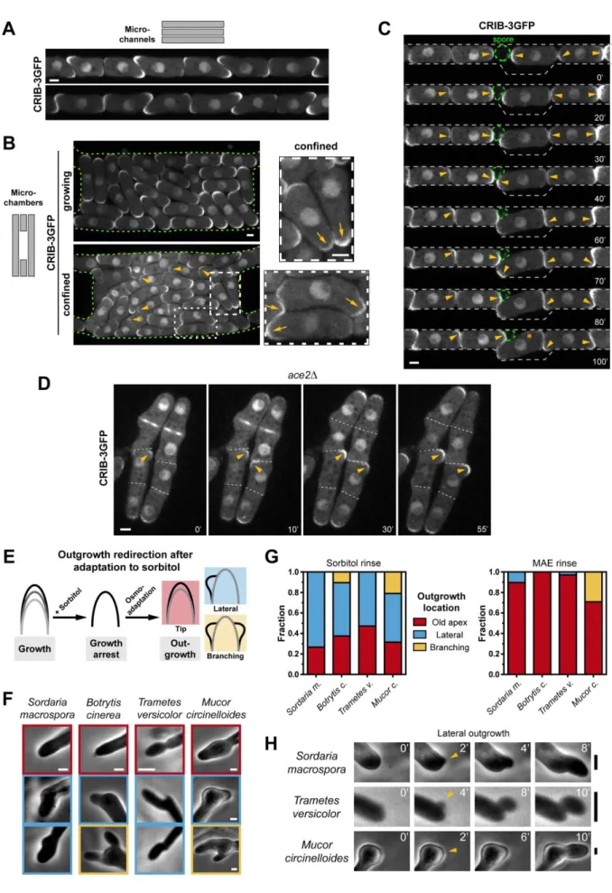
Growth inhibition by physical confinement destabilizes polarity 123
Growth in turgid cells may be influenced by external mechanical barriers such as neighboring 124
cells, physical obstacles or cell-encasing layers [6, 9, 13, 25]. To assay this, we designed 125
dedicated narrow channels to confine cells laterally but allow them to grow from their ends 126
[26, 27] (Figure 2A). WT cells growing in those channels initially proliferated with similar 127
rates as controls outside (Figure S2A); but as density increased, they started to hamper one 128
another’s growth. Cells reaching high levels of confinement ceased growth or grew very 129
slowly, and adopted small triangular or rectangular shapes (Figure 2B). Remarkably, growth-130
arrested cells exhibited weak CRIB domains oftentimes dynamically exploring the cell 131
contour away from cell tips, reminiscent of turgor reduction assays (Figure 2B; Movie S2). 132
Polar components, including bud6-3GFP, LifeAct-mCherry, sec6-GFP, sec8-GFP and GFP-133
bgs4, were also spatially destabilized as in turgor-reduction mediated growth arrest and 134
exhibited dynamic changes of localization (Figure 2C-2D and S2D). Importantly, dynamic 135
6
assembly and disassembly of ectopic domains, was still highly pronounced in mal3Δ and 136
tea1Δ mutants in channels, ruling out a role for microtubule-based polarity reorganization
137
following shape changes, in CRIB domain instability (Figure S2B-C) [28, 29]. Thus, 138
hampering growth by external mechanical means, can yield significant destabilization of 139
polarity domains. 140
Because confinement in channels is reached as a result of multiple phases of growth and 141
division; we devised an assay to rapidly suppress confinement and assay polarity response. 142
We started with fully confined and depolarized cells and used UV-laser ablation to kill a 143
subset of cells in the microchannel (Figure 2E; Movie S3) [9]. Ablated cells deflated within 144
seconds, freeing space for adjacent cells to grow (Figure 2E). Strikingly, ~10 - 20 min after 145
ablation, neighboring cells resumed rapid growth with the concomitant formation of bright 146
and stable CRIB-3GFP domains at growth sites. Importantly, this response was also observed 147
in cells at a further distance from the gaps, ruling out putative effects of chemical release 148
from ablated cells or contact-inhibition cues on polarity stability (Figure 2E). Those findings 149
demonstrate that cells can dynamically adapt their polarity behavior to different confinement 150
states altering growth. 151
152
A candidate screen identifies suppressors and enhancers for the impact of growth on 153
polarity stability 154
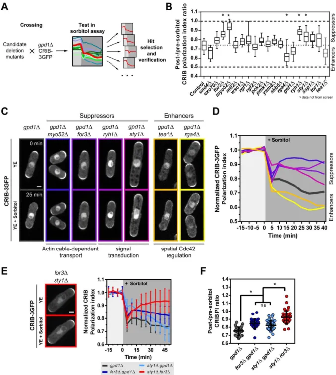
To identify potential factors mediating those effects, we then designed a candidate screen 155
using the gpd1Δ-sorbitol growth arrest assay (Figure 3A). We selected candidates from 156
different classes of sensing and regulating systems, including components of the cell wall 157
integrity pathway, factors feeding into polarity and a set of globally acting protein kinases 158
and modulators that have been implicated in growth or polarity before (Figure 3B and S3A-159
D). We crossed candidate mutants into a gpd1Δ background expressing CRIB-3GFP or 160
CRIB-3Citrine [24], and computed the ratio of CRIB PI after and before sorbitol addition 161
(Figure S3A-S3D). This screen identified suppressors, in which CRIB PI after growth 162
reduction was maintained at a higher level than gpd1Δ controls. Mutants in actin cable-163
dependent transport, like a mutant in the formin for3, and in the myosin typeV, myo52 had 164
the most pronounced suppressing effect [14]. Mutants in signaling components including the 165
stress-related MAP Kinase sty1 and in the TOR-activating Rab-family GTPase 166
ryh1exhibited a lower, yet significant suppression (Figure 3B-D and S3D) [30, 31] . This 167
7
screen also identified enhancers including mutants for the Cdc42 GAP rga4and in the 168
landmark tea1, as discussed above (Figure 1F and Figure 3B). As both factors have 169
established roles in confining GTP-Cdc42 to cell tips; this result could reflect that spatial 170
landmarks outcompete the basal destabilizing impact of growth arrest on polarity. Finally, a 171
mutant in the endocytosis-promoting factor end4 and in the Cdc42 GEF gef1 also exhibited 172
milder enhancing effects (Figure 3B-D). 173
174
To test the epistatic relationships between suppressing pathways, we compared the responses 175
of gpd1Δfor3Δ, gpd1Δsty1Δ and for3Δsty1Δ with gpd1Δ alone (Figure 3E-3F and S3E). We 176
resorted to using the for3Δsty1Δ mutant, as we were unable to obtain a viable 177
gpd1Δfor3Δsty1Δ triple mutant, but noted that this double mutant, as in gpd1Δ background
178
strains, did not adapt growth upon sorbitol treatment, due to the absence of sty1. 179
Interestingly, in for3Δsty1Δ cells we found that polarization upon growth arrest remained 180
nearly indistinguishable from the pre-treatment state (Figure 3E and F). Although this 181
analysis, cannot fully rule out unknown direct effects of gpd1 on polarity stability, it indicates 182
that actin cable-mediated transport acts independently of sty1 in this response. 183
As sty1p is an established downstream target of hyperosmotic treatments, and can drive 184
active-Cdc42 dispersal from cell tips when ectopically activated [24] the suppressing effects 185
of sty1Δ could represent an assay-specific bias. However, the effect on polarity removal here 186
is rather mild as compared to that reported for ectopic activation. Thus, an open possibility is 187
that sty1 becomes partly activated as a general response to growth arrest and contributes to 188
the detachment of GTP-Cdc42. The implication of actin-based vesicle delivery, which feeds 189
directly into Cdc42 polarity, may be more generic, and suggests that an imbalance or a mis-190
regulation of vesicles fluxes could serve as a core mechanism allowing cells to adapt polarity 191
to their growth rate [5, 8]. Vesicle flows into the tip membrane may have enhanced polarity 192
diluting effects when surface growth is slowed down [8, 32, 33]. An alternative effect could 193
come from defects in vesicle docking and/or fusion associated with alterations in tip 194
membrane biochemistry or mechanics, resulting from growth defects. 195
One interesting finding is that endocytosis appears to promote the maintenance of active-196
Cdc42 at cell tips but is required for the detachment of the cell wall synthase bgs4 from cell 197
tips (Figure S1J). Accordingly, the detachment of bgs4 happens much slower, and was 198
recently reported to depend on the cell wall mechanical sensor wsc1 [34], which did not 199
8
appear to affect active-Cdc42 detachment (Figure 3B). This suggests that different 200
mechanisms acting at various time-scales may control the response of polarity and growth-201
promoting components to growth arrest. 202
203
Feedbacks between growth and polarity as means to reorient growth around physical 204
obstacles. 205
We then asked what could be the functional relevance of a feedback between growth and 206
polarity stability. By observing cells growing in 1D or 2D microchannels, we noted that cell 207
tips that met end-on and were unable to displace one another, systematically started to grow 208
into available spaces by actively reorienting polar growth, yielding a high proportion of bent 209
cells (Figure 4A and 4B). Such behavior implicated a first phase of destabilized polarity due 210
to growth blockage by other cells, which served to locate empty spaces. This was evident 211
from monitoring cells that grew onto each other or onto an obstacle, like a non-germinated 212
spore (Figure 4C and S4A; Movie S4). In those instances, polarity could dynamically reorient 213
multiple times, sometimes stabilizing at the other cell end, before causing marked shape 214
changes, in a window as short as 20-30 min responding to newly available narrow gaps 215
(Figure 4C, 40’). 216
Furthermore, we observed a similar dynamic behavior outside a microchannel using an ace2Δ 217
mutant, in which cell separation is strongly impaired. This mutant bears defects in cell 218
separation, yielding chains of 4 or more cells, with flanked middle cells that cannot grow 219
from their tips [35]. In middle cells, CRIB-3GFP domains were weak and dynamically 220
changed their positions and became bright and stable upon outgrowth from the side of the 221
chain, much like WT cells in channels (Figure 4D). This behavior was never observed in 222
front cells free to grow from their tips. We conclude that a positive feedback between growth 223
and polarity provides cells with a functional module to dynamically reorient their growth axis 224
towards empty spaces. 225
226
Universality of feedbacks between growth and polarity 227
To test if this effect was a conserved feature of tip growing cells, we assayed responses in 228
several filamentous fungus species belonging to distant branches of the fungal tree including 229
zygomycetes (Mucor), basidiomycetes (Trametes and Coprinus) and ascomycetes (Sordaria 230
9
and Botrytis) (Figure S4B). We grew fungi in flow channels, rinsed them in hyperosmotic 231
medium (0.2 - 0.5 M sorbitol, depending on the species) and monitored their directional 232
response after osmo-adaptation. This treatment led to a complete cessation of hyphal growth, 233
followed by a resumption of rapid growth after an adaptation delay (Figure 4E and S4C). 234
Strikingly, when cell growth recommenced, hyphae frequently grew from the sides of the 235
apex in 50-70% of cases and occasionally branched (Figure 4F-4G). This response was 236
independent of hyphal diameter and also prominent in newly formed branches. Importantly, 237
this altered polarized behavior was dependent on growth arrest, as flowing in medium did not 238
affect hyphal polarization (Figure 4G and Figure S4D). In some species, like Botrytis or 239
Mucor, lateral branch formation following growth arrest, was preceded by a brief period of
240
isotropic growth, indicative of initial polarity dispersal before local stabilization (Figure 4F 241
and H). One species, Coprinus cinereus, formed lateral branches following growth arrest and 242
recovery, mainly at large distances from the apex (30-60 µm), plausibly reflecting inherent 243
mechanisms that suppress tip-proximal branching (Figure S4E). Although this growth 244
redirection behavior created some sharp shape changes at the hyphal tip, we noted that, on a 245
global scale, hyphae continued their mean growth direction away from the bulk of the colony, 246
likely guided by some tropisms [1]. Those results support a plausible universality for a 247
feedback between growth and polarity relevant to most fungal species. 248
In sum, we propose a dynamic feedback linking growth and polarity stability: when polarity 249
domains form at the surface, they become stable if growth occurs at a normal/high rate; and 250
disassemble if growth is hindered mechanically or chemically (Figure S4F). This feedback 251
may account for persistent polar growth typical of yeast and fungal cells independently of 252
guiding cues or spatial landmarks [9, 36, 37]. Accordingly, this mechanism appears more 253
prominent in the absence of internal landmarks provided by the tea1 system in fission yeast 254
and may compete with global guiding cues in fungal hyphae (Figure 1F and 4F). This 255
feedback could also synchronize, or phase-lock, Rho GTPases oscillations and pulsatile 256
growth typical of many tip-growing cells [38, 39]. One important realization however is that 257
growth rates in yeast and other walled cells remain bounded. Thus, in addition to positive 258
feedbacks, limitation systems either based upon the availability of polarity/growth factors or 259
encoded in the mechanics of the surface must exist to buffer growth [34]. 260
In addition to conferring spatial persistency to polar growth, this mechanism also provides 261
flexibility, by allowing cells to reorient growth in directions, which are mechanically 262
favorable. Many tip growing cells, including fungal hyphae and plant roots, exhibit 263
10
thigmotropisms and can redirect their growth axis in response to mechanical stimuli, when 264
navigating in tissues or in soil, for instance [40]. The feedback we document here, could in 265
principle serve as an inherited trait promoting such behavior even in a simple yeast cell. 266
While growth is the prominent driver of cell surface deformation in fungal cells, other 267
eukaryotic cells which migrate or are submitted to tissue forces may also employ similar 268
conceptual feedbacks to support the stability of polarity machineries with respect to their 269
rates of surface deformation. Accordingly, recent work has suggested near-universal 270
correlations between directional persistence and locomotion speed in migrating cells [41]. 271
Cell surface deformations, which originate form patterns of tissue stress may also promote 272
cell polarization along specific tissue axis in animal [42] and plant tissues [43]. Further work 273
addressing the interconnections between dynamic shape changes and directional behaviors 274
shall enlighten mechanisms of eukaryotic morphogenesis. 275
276
ACKNOWLEDGMENTS 277
The authors acknowledge M. Balasubramanian, Q. Chen, M. Edamatsu, S. Martin, P. Perez, 278
Y. Sanchez, K. Sawin and F. Leclerc for sharing strains and material. This work was 279
supported by the CNRS and grants from the FP7 CIG program (no. 303821), the Mairie de 280
Paris “Emergence” program, and the European Research Council (CoG Forcaster n° 647073). 281
282
AUTHOR CONTRIBUTIONS 283
A.H. performed experiments and analyzed data. D.E. developed image analysis scripts. A.H. 284
and N.M. designed the research and wrote the manuscript. 285
286
DECLARATON OF INTERESTS 287
The authors declare that they have no competing interests. 288
11 REFERENCES:
290 291
1. Brand, A., and Gow, N.A. (2009). Mechanisms of hypha orientation of fungi. Curr Opin 292
Microbiol 12, 350-357. 293
2. Merlini, L., Dudin, O., and Martin, S.G. (2013). Mate and fuse: how yeast cells do it. Open 294
Biol 3, 130008. 295
3. Li, R., and Gundersen, G.G. (2008). Beyond polymer polarity: how the cytoskeleton builds a 296
polarized cell. Nat Rev Mol Cell Biol 9, 860-873. 297
4. Johnson, J.M., Jin, M., and Lew, D.J. (2011). Symmetry breaking and the establishment of 298
cell polarity in budding yeast. Curr Opin Genet Dev 21, 740-746. 299
5. Martin, S.G. (2015). Spontaneous cell polarization: Feedback control of Cdc42 GTPase 300
breaks cellular symmetry. Bioessays 37, 1193-1201. 301
6. Minc, N., Boudaoud, A., and Chang, F. (2009). Mechanical forces of fission yeast growth. 302
Curr Biol 19, 1096-1101. 303
7. Bendezu, F.O., and Martin, S.G. (2013). Cdc42 explores the cell periphery for mate selection 304
in fission yeast. Curr Biol 23, 42-47. 305
8. Bonazzi, D., Haupt, A., Tanimoto, H., Delacour, D., Salort, D., and Minc, N. (2015). Actin-306
Based Transport Adapts Polarity Domain Size to Local Cellular Curvature. Curr Biol 25, 307
2677-2683. 308
9. Bonazzi, D., Julien, J.D., Romao, M., Seddiki, R., Piel, M., Boudaoud, A., and Minc, N. 309
(2014). Symmetry Breaking in Spore Germination Relies on an Interplay between Polar Cap 310
Stability and Spore Wall Mechanics. Dev Cell 28, 534-546. 311
10. Merlini, L., Khalili, B., Bendezu, F.O., Hurwitz, D., Vincenzetti, V., Vavylonis, D., and 312
Martin, S.G. (2016). Local Pheromone Release from Dynamic Polarity Sites Underlies Cell-313
Cell Pairing during Yeast Mating. Curr Biol 26, 1117-1125. 314
11. Dyer, J.M., Savage, N.S., Jin, M., Zyla, T.R., Elston, T.C., and Lew, D.J. (2013). Tracking 315
shallow chemical gradients by actin-driven wandering of the polarization site. Curr Biol 23, 316
32-41. 317
12. Howell, A.S., Jin, M., Wu, C.F., Zyla, T.R., Elston, T.C., and Lew, D.J. (2012). Negative 318
feedback enhances robustness in the yeast polarity establishment circuit. Cell 149, 322-333. 319
13. Thomson, D.D., Wehmeier, S., Byfield, F.J., Janmey, P.A., Caballero-Lima, D., Crossley, A., 320
and Brand, A.C. (2015). Contact-induced apical asymmetry drives the thigmotropic responses 321
of Candida albicans hyphae. Cell Microbiol 17, 342-354. 322
14. Chang, F., and Martin, S.G. (2009). Shaping fission yeast with microtubules. Cold Spring 323
Harb Perspect Biol 1, a001347. 324
15. Davi, V., and Minc, N. (2015). Mechanics and morphogenesis of fission yeast cells. Curr 325
Opin Microbiol 28, 36-45. 326
16. Ohmiya, R., Yamada, H., Nakashima, K., Aiba, H., and Mizuno, T. (1995). Osmoregulation 327
of fission yeast: cloning of two distinct genes encoding glycerol-3-phosphate dehydrogenase, 328
one of which is responsible for osmotolerance for growth. Mol Microbiol 18, 963-973. 329
17. Tatebe, H., Nakano, K., Maximo, R., and Shiozaki, K. (2008). Pom1 DYRK regulates 330
localization of the Rga4 GAP to ensure bipolar activation of Cdc42 in fission yeast. Curr Biol 331
18, 322-330. 332
18. Charvin, G., Cross, F.R., and Siggia, E.D. (2008). A microfluidic device for temporally 333
controlled gene expression and long-term fluorescent imaging in unperturbed dividing yeast 334
cells. PLoS One 3, e1468. 335
19. Calero, F., Gomez, N., Arino, J., and Ramos, J. (2000). Trk1 and Trk2 define the major K(+) 336
transport system in fission yeast. J Bacteriol 182, 394-399. 337
20. Courtemanche, N., Pollard, T.D., and Chen, Q. (2016). Avoiding artefacts when counting 338
polymerized actin in live cells with LifeAct fused to fluorescent proteins. Nat Cell Biol 18, 339
676-683. 340
21. Basu, R., Munteanu, E.L., and Chang, F. (2014). Role of turgor pressure in endocytosis in 341
fission yeast. Mol Biol Cell 25, 679-687. 342
12
22. Nakamura, T., Nakamura-Kubo, M., Hirata, A., and Shimoda, C. (2001). The 343
Schizosaccharomyces pombe spo3+ gene is required for assembly of the forespore membrane 344
and genetically interacts with psy1(+)-encoding syntaxin-like protein. Mol Biol Cell 12, 345
3955-3972. 346
23. Bendezu, F.O., and Martin, S.G. (2011). Actin cables and the exocyst form two independent 347
morphogenesis pathways in the fission yeast. Mol Biol Cell 22, 44-53. 348
24. Mutavchiev, D.R., Leda, M., and Sawin, K.E. (2016). Remodeling of the Fission Yeast Cdc42 349
Cell-Polarity Module via the Sty1 p38 Stress-Activated Protein Kinase Pathway. Curr Biol 350
26, 2921-2928. 351
25. Sampathkumar, A., Yan, A., Krupinski, P., and Meyerowitz, E.M. (2014). Physical forces 352
regulate plant development and morphogenesis. Curr Biol 24, R475-483. 353
26. Goulev, Y., Morlot, S., Matifas, A., Huang, B., Molin, M., Toledano, M.B., and Charvin, G. 354
(2017). Nonlinear feedback drives homeostatic plasticity in H2O2 stress response. Elife 6. 355
27. Zegman, Y., Bonazzi, D., and Minc, N. (2015). Measurement and manipulation of cell size 356
parameters in fission yeast. Methods Cell Biol 125, 423-436. 357
28. Minc, N., Bratman, S.V., Basu, R., and Chang, F. (2009). Establishing new sites of 358
polarization by microtubules. Curr Biol 19, 83-94. 359
29. Terenna, C.R., Makushok, T., Velve-Casquillas, G., Baigl, D., Chen, Y., Bornens, M., 360
Paoletti, A., Piel, M., and Tran, P.T. (2008). Physical mechanisms redirecting cell polarity 361
and cell shape in fission yeast. Curr Biol 18, 1748-1753. 362
30. Millar, J.B., Buck, V., and Wilkinson, M.G. (1995). Pyp1 and Pyp2 PTPases dephosphorylate 363
an osmosensing MAP kinase controlling cell size at division in fission yeast. Genes Dev 9, 364
2117-2130. 365
31. Tatebe, H., Morigasaki, S., Murayama, S., Zeng, C.T., and Shiozaki, K. (2010). Rab-family 366
GTPase regulates TOR complex 2 signaling in fission yeast. Curr Biol 20, 1975-1982. 367
32. Savage, N.S., Layton, A.T., and Lew, D.J. (2012). Mechanistic mathematical model of 368
polarity in yeast. Mol Biol Cell 23, 1998-2013. 369
33. Tay, Y.D., Leda, M., Spanos, C., Rappsilber, J., Goryachev, A., and Sawin, K. (2018). Fission 370
yeast NDR/LATS kinase Orb6 regulates exocytosis via phosphorylation of exocyst complex. 371
bioRxiv. 372
34. Davì, V., Tanimoto, H., Ershov, D., Haupt, A., De Belly, H., Le Borgne, R., Couturier, E., 373
Boudaoud, A., and Minc, N. (2018). Mechanosensation Dynamically Coordinates Polar 374
Growth and Cell Wall Assembly to Promote Cell Survival. Developmental Cell 45, 170-375
182.e177. 376
35. Dekker, N., Speijer, D., Grun, C.H., van den Berg, M., de Haan, A., and Hochstenbach, F. 377
(2004). Role of the alpha-glucanase Agn1p in fission-yeast cell separation. Mol Biol Cell 15, 378
3903-3914. 379
36. Kelly, F.D., and Nurse, P. (2011). De novo growth zone formation from fission yeast 380
spheroplasts. PLoS One 6, e27977. 381
37. Sawin, K.E., and Snaith, H.A. (2004). Role of microtubules and tea1p in establishment and 382
maintenance of fission yeast cell polarity. J Cell Sci 117, 689-700. 383
38. Das, M., Drake, T., Wiley, D.J., Buchwald, P., Vavylonis, D., and Verde, F. (2012). 384
Oscillatory dynamics of Cdc42 GTPase in the control of polarized growth. Science 337, 239-385
243. 386
39. Hwang, J.U., Gu, Y., Lee, Y.J., and Yang, Z. (2005). Oscillatory ROP GTPase activation 387
leads the oscillatory polarized growth of pollen tubes. Mol Biol Cell 16, 5385-5399. 388
40. Jaffe, M.J., Leopold, A.C., and Staples, R.C. (2002). Thigmo responses in plants and fungi. 389
Am J Bot 89, 375-382. 390
41. Maiuri, P., Rupprecht, J.F., Wieser, S., Ruprecht, V., Benichou, O., Carpi, N., Coppey, M., 391
De Beco, S., Gov, N., Heisenberg, C.P., et al. (2015). Actin flows mediate a universal 392
coupling between cell speed and cell persistence. Cell 161, 374-386. 393
42. Aw, W.Y., Heck, B.W., Joyce, B., and Devenport, D. (2016). Transient Tissue-Scale 394
Deformation Coordinates Alignment of Planar Cell Polarity Junctions in the Mammalian 395
Skin. Curr Biol 26, 2090-2100. 396
13
43. Nakayama, N., Smith, R.S., Mandel, T., Robinson, S., Kimura, S., Boudaoud, A., and 397
Kuhlemeier, C. (2012). Mechanical regulation of auxin-mediated growth. Curr Biol 22, 1468-398
1476. 399
44. Sato, M., Dhut, S., and Toda, T. (2005). New drug-resistant cassettes for gene disruption and 400
epitope tagging in Schizosaccharomyces pombe. Yeast 22, 583-591. 401
45. Fehrmann, S., Paoletti, C., Goulev, Y., Ungureanu, A., Aguilaniu, H., and Charvin, G. (2013). 402
Aging yeast cells undergo a sharp entry into senescence unrelated to the loss of mitochondrial 403
membrane potential. Cell Rep 5, 1589-1599. 404
46. Bendezu, F.O., Vincenzetti, V., Vavylonis, D., Wyss, R., Vogel, H., and Martin, S.G. (2015). 405
Spontaneous Cdc42 polarization independent of GDI-mediated extraction and actin-based 406
trafficking. PLoS Biol 13, e1002097. 407
408 409
14 FIGURE LEGENDS
410 411
15
Figure 1 (Related to Figure S1 and Movie S1): Abrupt growth rate changes alter the 413
localization of cell polarity factors. 414
A) gpd1Δ cells expressing CRIB-3GFP rinsed in YE medium with 0.5 M sorbitol to reduce 415
turgor pressure and halt growth (n=36). Top: Representative cells before and after treatment. 416
Bottom: Evolution of growth rate (black) and CRIB-3GFP polarization index (PI, blue). B) 417
Representative gpd1Δ CRIB-3GFP cells before (left) and after (right) sorbitol treatment. Right: 418
Kymographs of the same cells as in left panels showing signal intensities of a central 1.6 µm 419
and a lateral 0.5 µm segments over 80 min with 5 min intervals. Colors correspond to growth 420
conditions as labeled in the graph in A. C) Wild type cells expressing CRIB-3GFP rinsed in 421
YE medium with 0.03% Glucose (n=46). Top: Representative cells before and after treatment. 422
Bottom: Evolution of growth rate (black) and CRIB-3GFP polarization index (PI, blue). D) 423
Subcellular localization patterns of different polarity factors in gpd1Δ cells in YE and after 30 424
min of treatment in YE + 0.5 M sorbitol. E) Wild type cells expressing CRIB-3GFP treated 425
with 50 μM Latrunculin A (LatA) (n=46). Left: Representative cells before and after treatment. 426
Right: Evolution of growth rate (black) and CRIB-3GFP polarization index (PI, blue). F) gpd1Δ 427
(n=36) and gpd1Δ tea1Δ (n=45) cells treated with 0.5 M sorbitol. Left: Changes in CRIB-3GFP 428
localization in gpd1Δtea1Δ cells. Right: Evolution of PI over time for both strains. G) 429
Correlation of CRIB-3GFP PI changes as a function of growth rate changes of all cells from 430
experiments in A, C, E and F. Pearson correlation coefficient, r=0.7604. In all images 431
arrowheads label spread caps and asterisks mark lateral accumulations of polarity factors. Scale 432
bars, 2 μm. Error bars represent standard deviations. Data are from two or more independent 433
experiments. 434
16 436
17
Figure 2 (Related to Figure S2 and Movie S2 and S3): Growth arrest through 438
mechanical confinement triggers polarity domain destabilization and wandering. 439
A) Large-scale picture and schematic representation of microchannel design. Three columns, 440
each containing microchannel arrays of different length, are embedded within a large medium 441
reservoir (‘chamber’) for optimal nutrient supply. B) WT cells expressing CRIB-3GFP grown 442
to confinement in microchannels (top). Arrowheads label transiently forming CRIB domains 443
on the surface. Time-lapse of confined cells (bottom left) and close-up view of two cells as 444
labeled on the left (bottom right). C) WT cells expressing the indicated polarity markers 445
grown in free-growing and fully confined regions in the same set of microchannels. D) WT 446
cells expressing bud6-3GFP grown to full confinement in microchannels. Arrowheads label 447
transiently forming bud6-3GFP ectopic small domains wandering on the surface. E) Wild 448
type cells expressing CRIB-3GFP were grown in microchannels to full confinement and two 449
cells, labelled with red asterisks, were ablated with a UV-laser to free space for neighbors to 450
grow. Arrows label initially transient or weak CRIB domains and arrowheads depict 451
reforming stable CRIB domains. Scale bars, 2 μm. 452
453 454 455
18 456
Figure 3 (Related to Figure S3): Candidate genetic screen for modulators of active-457
Cdc42 domain destabilization upon growth reduction. 458
A) Schematic representation of the screening approach. B) Overview of screen results plotted 459
as post/pre-sorbitol ratio of CRIB-3GFP polarization index. Box plots depict the mean with 460
25th and 75th percentiles and whiskers the full data range. Dotted lines indicate upper and 461
lower limits of the range of values for no change or complete loss of polarization, 462
respectively. The middle dotted line depicts values for the control. For control vs. for3Δ, 463
myo52Δ, ryh1Δ, rga4Δ, and sty1Δ, there was a statistically significant difference between
464
groups as determined by one-way ANOVA (F(19, 330) = 40.49, p < 0.0001). tea1Δ values 465
19
plotted for comparison are derived from experiment in Figure 1F but were not included in the 466
statistical analysis because of non-matching experimental conditions. C) Representative 467
images of CRIB-3GFP response of candidates derived from the screen directly before (0 min) 468
and 25 min after exposure to 0.5 M sorbitol. D) Mean response curves of CRIB-3GFP PI for 469
hits from the screen. Colors and genotypes are matched with panel C. E) Effect of for3Δ and 470
sty1Δ deletions on CRIB-3GFP polarization upon treatment with 0.5 M sorbitol. Left: Image
471
shows CRIB-3GFP distribution in for3Δ sty1Δ cells before and after sorbitol treatment. Right: 472
CRIB-3GFP PI during sorbitol treatment (gpd1Δ (n=36), for3Δ gpd1Δ (n=28), sty1Δ gpd1Δ 473
(n=34) and for3Δ sty1Δ (n=39)). F) Comparison of post/pre-sorbitol PI ratio of CRIB-3GFP.. 474
For gpd1Δ, for3Δ gpd1Δ, sty1Δ gpd1Δ, and for3Δ sty1Δ, there was a statistically significant 475
difference between groups as determined by one-way ANOVA (F(3, 144) = 45.112, p < 476
0.0001). Scale bars, 2 μm. Error bars represent standard deviations. Data, except in B, come 477
from two or more independent experiments. 478
20 480
481
Figure 4 (Related to Figure S4 and Movie S4): Universal feedback between growth and 482
polarity as a means to reorient polar growth axis. 483
21
A) Wild type cells expressing CRIB-3GFP grown in microchannels and observed at a stage of 484
early confinement. B) Wild type cells expressing CRIB-3GFP grown in 2D microchambers 485
and observed at a stage of low (left top) and high (left bottom) confinement. Insets show 486
magnifications of cells in high confinement (right). Arrowheads point at examples of weak 487
CRIB-3GFP domains and arrows label irregularly shaped cell tips. C) WT cells expressing 488
CRIB-3GFP growing from the two opposite ends of a channel and reorienting polarity and 489
growth upon contact. Arrowheads indicate CRIB domains that change position. The green 490
dashed circle depicts the outline of a dormant spore acting as a rigid obstacle. D) ace2Δ cells 491
expressing CRIB-3GFP grown on YE agar. As a consequence of delayed cell separation some 492
cells are flanked by two sister cells and finally outgrow from the sides. Arrowheads label 493
initially transient or weak CRIB domains that become stabilized and intense as cells outgrow. 494
E) Assaying outgrowth direction of filamentous fungi after recovery from sorbitol-induced 495
growth arrest. F) Representative hyphal shapes following growth recovery after sorbitol 496
treatment in various fungal species. Colors are related to response classification in Figure 4G. 497
G) Quantification of the different hyphal phenotypes reflecting a transient alteration of 498
polarization behavior following growth arrest and recovery. For sorbitol rinse: S.m. n=30, B.c. 499
n=39, T.v. n=36, M.c. n=19. For MAE rinse: S.m. n=29, B.c. n=26, T.v. n=34, M.c. n=24. H) 500
Time-lapse of lateral outgrowth at the hyphal tips in different species. Scale bars, 2 μm (A-E) 501
and 4 μm (F-H). 502
503 504
22 STAR METHODS:
505 506
CONTACT FOR REAGENT AND RESOURCE SHARING 507
Further information and requests for resources and reagents should be directed to and will be 508
fulfilled by the lead contact, Nicolas Minc (nicolas.minc@ijm.fr). 509
510
EXPERIMENTAL MODEL AND SUBJECT DETAILS 511
Yeast strains, genetic methods, and media 512
Standard methods for S. pombe media and genetic manipulations were used (www-513
bcf.usc.edu/~forsburg; www.biotwiki.org/foswiki/bin/view/Pombe/NurseLabManual). 514
Strains used in this study are listed in the key resource table. Marker switching from Kan to 515
Nat was achieved by amplifying the NatMX cassette from a pCR2.1-nat vector with primers 516
MD1 and MD2 and transforming it into KanMX cassette-bearing strains. Positive clones 517
were selected on YE5S + 50 µg/ml Nat plates and tested for functional and visual phenotypes 518
[44]. For all experiments, liquid cultures were grown in YE5S (containing 3% glucose) 519
overnight at 25°C. For experiments in flow channels, cells were pre-stained with 10 µg/ml 520
Alexa647-labeled isolectin GS-IB4 from Griffonia simplicifolia (Thermo Fisher Scientific, 521
Waltham, Massachusetts, USA) and growth medium contained a constant Lectin supply of 5 522
µg/ml. The lectin signal labelled the cell contour and was used for cell segmentation during 523
image analysis. 524
Filamentous fungi species, media and growth: 525
Strains: Botrytis cinerea, Sordaria macrospora, Coprinus cinereus, Trametes versicolor and
526
Mucor circinelloides. All strains were grown on MAE medium (2% malt, 0.1% peptone, 2%
527
glucose) either as liquid or plates with 2% agar. To grow hyphal mycelia, spores were 528
prepared by lysing hyphae from a fully-grown MAE plate with glass beads and a FastPrep 529
device (MP Biomedicals, Santa Ana, California, USA). Crude spore preparations were used 530
to inoculate 20 ml MAE cultures and these were grown over night at 25°C. Emerging 531
mycelia from these cultures were placed in PDMS flow channels under a dialysis membrane 532
for microscopy. 533
Pharmacological inhibition 534
23
Latrunculin A (Sigma-Aldrich, St. Louis, Missouri, USA) was used at a final concentration of 535
50 μM from a 200x stock in DMSO (Euromedex, Souffelweyersheim, France). CK666 536
(Sigma-Aldrich) was used at a final concentration of 100 μM from a 500x stock in DMSO. 537
DMSO amounts in control experiments were adjusted correspondingly for each drug. 538 539 METHOD DETAILS 540 Flow channels 541
We used two kinds of flow channels depending on the type of experiment. For drug 542
treatments we used a simple glass channel built from one 24x50 mm (VWR, Radnor, 543
Pennsylvania, USA) and a 22x22 mm (Thermo Fisher Scientific) coverslip spaced by ~ 250 544
μm, using double-sided adhesive tape. To make cells adhere to the glass surface, the flow 545
channel was pre-coated with 1 mg/ml poly-L-lysine (Sigma-Aldrich) and 0.1 mg/mg isolectin 546
GS-IB4 from Griffonia simplicifolia, (Thermo Fisher Scientific). 547
In the second type of flow channels, cells were placed beneath a single layer of dialysis 548
membrane and covered with a polydimethylsiloxane (PDMS) channel, allowing for exchange 549
of the medium [18]. 550
551
Microchannels for confined growth assays 552
Design and fabrication
553
The general design of microchannels to confine fission yeast cells is shown in Figure 2A. 554
Those channels were optimized to ensure a near unlimited nutrient supply, and implicated a 555
2-step soft-lithography microfabrication technique [45]. A first thin layer of ~2.5 µm in 556
height was first fabricated using SU8 photoresists, and subsequent UV illumination through a 557
Quartz mask. Subsequently, a second layer of ~40µm in height of SU8 photoresists, was 558
spread on top of the first layer, and exposed with UV through a Quartz mask, aligned with the 559
first design using a Mask Aligner . PolyDimethylSiloxane (PDMS, Sylgard 184 from Dow 560
Corning, Midland, Michigan, USA) replica were prepared using a 10:1 ratio of PDMS:curing 561
agent. 562
Microchannel assembly
24
PDMS channels were covalently mounted onto 24x50 mm cover slips (VWR) [27]. To this 564
aim, coverslips were cleaned with Acetone, Isopropanol and ddH2O. For covalent binding,
565
both channel and coverslip were then plasma treated for 1 min, mounted and baked at 65°C 566
for ≥ 1 h. 567
Cell loading and growing
568
Cell were loaded into channels as spores, which then germinated and proliferated as 569
vegetative cells, crowding the channels [27] . Spores were obtained from h90 strains 570
sporulated on ME/3% agarose (Formedium, Hunstanton, UK)/(Sigma-Aldrich) plates at 25°C 571
for 2-3 days. Mating mixtures were digested in 1/200 Glusulase (PerkinElmer, Waltham, 572
Massachusetts, USA) at room temperature overnight, to eliminate vegetative cells. Digests 573
were cleared of debris by adding four volumes of Percoll (Sigma-Aldrich), followed by a 574
centrifugation at 600 rcf for 3min and supernatant removal. Spores were washed once with 575
five volumes of YE5S and subsequently re-suspended in YE5S medium [46]. Channels were 576
first filled with YE5S and subsequently a high density spore solution was pushed into 577
channels with a syringe, usually yielding 1-6 spores per channel. Excess spores were rinsed 578
out. Filled channels were incubated for 18-24 h in a humidified petri dish at 25 or 30°C, 579
depending on global spore density after seeding. The next morning, channels were rinsed 580
with fresh media at least 1h before live-imaging. 581
582
Microscopy 583
For immediate imaging, 1.8 µl of fresh, concentrated cells were placed between a glass slide 584
and coverslip and imaged within 20 min. For experiments with dynamic exchange of growth 585
media / drug addition, cells were placed in different types of flow channels. Microchannels 586
were directly imaged. 587
All fission yeast imaging was carried out at room temperature (22–25°C) with an inverted 588
spinning-disk confocal microscope equipped with a motorized stage and automatic focus (Ti-589
Eclipse, Nikon, Minato, Tokyo, Japan), a Yokogawa CSUX1FW spinning unit, and an EM-590
CCD camera (ImagEM-1K, Hamamatsu Photonics, Japan). Images were acquired with a 591
100× oil-immersion objective (CFI Plan Apo DM 100x/1.4 NA, Nikon). For laser ablation in 592
microchannels we used an iLas2 module (Roper Scientific) with a 355 nm laser and acquired 593
images with a 60x oil-immersion objective (CFI Apochromat 60x Oil λS, 1.4 NA, Nikon) in 594
25
combination with a 2.5x magnifying lens. The microscope was operated with Metamorph 595
software (Molecular Devices). 596
Presented images represent single confocal slices or specific z-stacks: Figure 1A, 1E, 1F, 3C 597
and 3E: Single-plane confocal images. Figure 1D: Maximum intensity projections of 19 z-598
slices spaced by 0.25 μm, except for sec8-GFP which is a projection of 3 slices spaced by 0.5 599
μm. Figure 2B: Maximum intensity projections from 5 z-slices spaced by 0.6 μm. Figure 2C: 600
Maximum intensity projections from 5 (bud6-3GFP, GFP-bgs4) or 3 (sec6-GFP, sec8-GFP) 601
z-slices spaced by 0.6 μm, and from 14 slices with 0.3 μm spacing for LifeAct-mcherry. 602
Figure 2D: Maximum intensity projections from 3 z-slices spaced by 0.6 μm. 2E: Maximum 603
intensity projections from 11 z-slices spaced by 0.4 μm. Figure 4A: Maximum intensity 604
projections from 7 z-slices spaced by 0.5 μm. Figure 4B: Maximum intensity projections 605
from 5 z-slices spaced by 0.5 μm. Figure 4C: Maximum intensity projections from 5 z-slices 606
spaced by 0.6 μm. Figure 4D: Maximum intensity projections from 3 z-slices spaced by 0.2 607
μm. 608
Filamentous fungi imaging was carried out with an inverted epifluorescence microscope (Ti-609
Eclipse, Nikon) combined with a CMOS camera (Hamamatsu). Hyphae were filmed with a 610
40x dry objective (CFI Plan Fluor DLL 40X/0.75 NA, Nikon) and a 1.5x magnifier. The 611
microscope was operated with Micro-Manager (Open Imaging). 612
613
QUANTIFICATION AND STATISTICAL ANALYSIS 614
Image analysis 615
To analyze changes in growth rates and localization of polar factors following growth 616
perturbations, we developed dedicated Matlab scripts [34]. We first segmented cells using the 617
signal from the lectin-labelled cell wall. To this aim, we first smoothed the image with 618
median and Gaussian filters and detected cell edges using the Laplacian of the Gaussian 619
filter. The resultant binary image was then filtered to remove small segments. Given that the 620
signal of the labelled cell wall has a finite thickness, we detected the inner and the outer 621
border of this signal. All spaces in this image were then filled in white except for the spaces 622
between the inner and outer border of the wall, yielding a black band representing the cell 623
wall. Using the watershed algorithm, we finally extracted the whole-cell contour defined as 624
the middle of this band. To compute cell length, we fitted the long axis of the segmented cell 625
26
with a polynomial of degree 3. This fit was then used to define a “cell spine” and its length 626
was calculated and used as a measurement for cell length. 627
The whole-cell contour could then be manipulated using morphological and logical 628
operations to obtain a set of arbitrary regions (tips, membrane, cytoplasm, etc.). The tip 629
regions are for instance shaped as a cut off from the whole-cell mask perpendicular to the cell 630
spine at specific distances along the spine. A segment outside of the cell can be shaped to 631
compute the background. 632
Fluorescent signals of interest were then extracted from fluorescent images by using a mask 633
based on corresponding sub-regions and are background corrected. Polarization index (PI) 634
was computed by normalizing the corrected tip signal with the background-635
corrected plasma membrane signal: 636
𝑃𝐼 = 𝐼𝑡𝑖𝑝 𝑟𝑎𝑤− 𝐼𝑏𝑔
𝐼𝑝𝑙𝑎𝑠𝑚𝑎 𝑚𝑒𝑚𝑏𝑟𝑎𝑛𝑒 𝑟𝑎𝑤− 𝐼𝑏𝑔
637
Additional image analysis and processing for Figures was done in ImageJ (National Institutes 638
of Health). 639
640 641
Extraction of single cell data 642
Because of marked rapid changes in cell shape (shrinkage or swelling) upon sorbitol or other 643
treatments, PI and growth rate quantification for single cells was done as an average of data 644
from -15 to 0 min (‘pre’) and from +10 min to 40 min (‘post’) relative to treatment, omitting 645
data corresponding to these rapid responses. This calculation was used for the screen results 646
(Figure 3B, 3F, S3C, S3D). Data to compute Figure 1G were extracted in the same manner 647
from experiments in panels 1A, 1C, 1E, 1F and S1D. Data post-treatment from 1E was split 648
into two phases of slowing growth (+10 to +30 min in LatA) and no growth (+35 to +50 min 649
in LatA). To compute the changes in PI or growth rate the difference between pre- and post-650
treatment values (e.g. diffPI = PIpost – PIpre) was calculated.
651 652
Statistical analysis 653
27
Statistical and correlation analyses were carried out using Prism 6 software (GraphPad 654
Software, La Jolla, CA). We used one-way analysis of variance (ANOVA) followed by 655
Dunnett multiple comparisons test or two-tailed, unpaired t-test. Statistically significant 656
difference between groups are *p < 0.0001. Linear correlation was analysed by computing 657
the Pearson correlation coefficient. 658
28 KEY RESOURCES TABLE
660 661
REAGENT or RESOURCE SOURCE IDENTIFIER
Experimental Models: S. pombe Strains
h- gpd1::NatMX CRIB-3GFP:ura leu1-32 ura4-D18 This study AH313
h- CRIB-3GFP:ura leu1-32 ura4-D18 ade- Lab stocks NM123
h+ gpd1::NatMX Pcof1-mEGFP-LifeAct:KanMX leu1-32 ura4-D18 ade-
This study
AH371
h+ gpd1::NatMX GFP-syb1:KanMX leu1-32 ura4-D18 ade- This study AH409
h+ gpd1::NatMX sec6-GFP:ura4 leu1-32 ura4 This study AH415
h+ gpd1::NatMX sec8-GFP:ura4 leu1-32 ura4 This study AH421
h+ gpd1::NatMX bud6-3GFP:KanMX leu1-32 ura4-D18 ade- This study AH413
gpd1::KanMX GFP-bgs4-Leu bgs4::ura leu1-32 ura4-D18 ade- Lab stocks NM266
h- gpd1::KanMX CRIB-GFP:ura leu1-32 ura4-D18 ade- Lab stocks NM434
gpd1::KanMX CRIB-tdTomato:ura sec6-GFP:ura4 leu1-32 ura4-D18 This study AH477
h- gpd1::KanMX tea1::NatMX CRIB CRIB-3GFP:ura leu1-32 ura4-D18
This study
AH432
h- trk1::KanMX trk2::KanMX CRIB-3GFP:ura This study AH229
h+ KanMX6-Pcof1-mEGFP-LifeAct leu- ura- ade- Chen Lab QC596
h90 CRIB-3GFP:ura ade6-M216 leu1-32 Lab stocks DB301
h90 tea1::NatMX CRIB-3GFP:ura leu1-32 ura4-D18 This study AH242
h90 bud6-3GFP:KanMX leu1-32 ura4-D18 Lab stocks DB169
h90 bgs4::ura GFP-bgs4:leu leu1-32 ura4-D18 Lab stocks DB124
h90 CRIB-3GFP:ura Pact1-Lamcherry:leu ade6-M216 Lab stocks DB337
h90 sec6-GFP:ura leu1-32 ura4-D18 ade- Lab stocks DB361
h90 sec8-GFP:ura leu1-32 ura4-D18 Lab stocks DB362
h90 CRIB-3GFP:ura mCherry-atb2:hph leu1-32 ura4-D18 This study AH148
ace2::KanMX4 CRIB-3GFP:ura leu1-32 ura4-D18 ade- This study AH213
h90 CRIB-GFP:ura bgs4::ura RFP-bgs4:leu leu1-32 ura4-D18 Lab stocks DB287
h- gpd1::NatMX end4::KanMX CRIB-3GFP:ura leu1-32 ura4-D18 This study AH332
h+ gpd1::KanMX exo70::NatMX CRIB-3GFP:ura ura4-D18 This study AH419
h+ gpd1::NatMX for3::KanMX CRIB-3GFP:ura leu1-32 ura4-D18 This study AH423
h- gpd1::NatMX myo52::ura ars1(BlpI):Padh13:CRIB-3xmCitrine:leu2 leu1-32 ura4-D18 ade-
This study
AH454 gpd1::NatMX ryh1::ura ars1(BlpI):Padh13:CRIB-3xmCitrine:leu2
leu1-32 ura4-D18
This study
AH374 h- gpd1::NatMX mtl2::KanMX CRIB-3GFP:ura leu1-32 ura4-D18
ade-
This study
AH321 h- gpd1::NatMX wsc1::KanMX CRIB-3GFP:ura leu1-32 ura4-D18
ade-
This study
AH320 h+ gpd1::NatMX rgf1::KanMX CRIB-3GFP:ura leu1-32 ura4-D18
ade-
This study
AH346
h- gpd1::NatMX rgf2::KanMX CRIB-3GFP:ura leu1-32 ura4-D18 This study AH334
h- gpd1::NatMX pck2:leu CRIB-3GFP:ura leu1-32 ura4-D18 This study AH341
gpd1::NatMX pmk1::KanMX CRIB-3GFP:ura leu1-32 ura4-D18 ade- This study AH377
gpd1::NatMX yam8::ura ars1(BlpI):Padh13:CRIB-3xmCitrine:leu2 This study AH365
h- gpd1::NatMX skb5::KanMX CRIB-3GFP:ura leu1-32 ura4-D18 ade-
29
h- gpd1::NatMX rga4::KanMX CRIB-3GFP:ura ura leu1-32 ura4-D18
This study AH438 gpd1::NatMX gef1::ura ars1(BlpI):Padh13:CRIB-3xmCitrine:leu2
leu1-32 ura4-D18 ade-
This study AH480 gpd1::NatMX sty1::kanMX ars1(BlpI):Padh13:CRIB-3xmCitrine:leu2
leu1-32 ura4-D18
This study AH364
h- gpd1::NatMX ssp1::KanMX CRIB-3GFP:ura leu1-32 ura4-D18 This study AH339
h- gpd1::NatMX tor1::KanMX CRIB-3GFP:ura leu1-32 ura4-D18 This study AH393
sty1::NatMX for3::KanMX CRIB-3GFP:ura leu1-32 ura4-D18 ade- This study AH446
h- for3::KanMX CRIB-3GFP:ura leu1-32 ura4-D18 ade- Lab stocks NM125
h+ gpd1::KanMX leu1-32:SV40:GFP-atb2[leu1+] ura4-D18 ade- This study AH459
h- gpd1::NatMX tea1-3GFP:KanMX leu1-32 ura4-D18 ade- This study AH456
h+ gpd1::NatMX leu1-32:GFP-psy1[leu1+] ura4-D18 ade- This study AH417
h+ gpd1::NatMX myo52-3GFP:KanMX leu1-32 ura4-D18 This study AH461
Experimental Models: Filamentous Fungi Strains
Botrytis cinerea F. Leclerc lab LC23
Sordaria macrospora F. Leclerc lab LC7
Coprinus cinereus F. Leclerc lab LC55
Trametes versicolor F. Leclerc lab LC39
Mucor circinelloides F. Leclerc lab LC71
Chemicals
LatrunculinA Sigma-Aldrich L5163
Gs-IB4 -Alexafluor647 ThermoFisher I32450
CK666 Sigma-Aldrich L0006
Percoll Sigma-Aldrich P1644
PDMS kit (Sylgard 184) Dow Corning DC184
Oligonucleotides
Primer: Resistance marker switching forward: CGGATCCCCGGGTTAATTAAGGCG
[44] MD1
Primer: Resistance marker switching reverse:
GAATTCGAGCTCGTTTAAACACTGGATGGCGGCGTTAGTATC G
[44] MD2
Recombinant DNA
Vector: pCR2.1-nat [44] NEG772014
30 662
663 664 665
31 666
SUPPLEMENTAL MOVIE LEGENDS 667
668
Movie S1: Changes of the growth rate act upon polarity domain stability. Growth rates 669
were dynamically changed by different means. From left to right: Addition of 0.5 M sorbitol 670
to gpd1Δ cells, Reducing glucose content to 0.03% in wild type cells, Treatment with 50 µM 671
Latrunculin A of wild type cells, Addition of 50 mM KCl to trk1Δtrk2Δ cells, Addition of 0.5 672
M sorbitol to tea1Δgpd1Δ cells. All strains express CRIB-3GFP. Time elapsed is in h:min. 673
Scale bars are 2 µm. 674
675
Movie S2: Growth arrest through mechanical confinement triggers polarity domain 676
destabilization and wandering. Wild-type cells expressing CRIB-3GFP were grown to 677
confinement in microchannels. Time elapsed is in h:min. Scale bar is 2 µm. 678
679
Movie S3: Confinement lift induces repolarization and growth. Wild type cells expressing 680
CRIB-3GFP were grown in microchannels to full confinement and two cells were 681
subsequently killed using UV-laser ablation to generate space for neighbors to grow. 682
Arrowheads label reforming stable CRIB domains. Images are maximum intensity 683
projections. Time elapsed is in h:min. Scale bars are 2 μm. 684
685
Movie S4: Dynamic adaptation of polarity domains to their mechanical environment. 686
Wild type cells expressing CRIB-3GFP were grown in microchannels. Cells growing from 687
the two opposite ends of the channel and their response upon contact were documented by 688
time-lapse microscopy. Green dashed circle depicts the outline of a dormant spore that did 689
not germinate. Arrowheads label CRIB domains that reorient. Images are maximum intensity 690
projections. Time elapsed is in h:min. Scale bar is 2 μm. 691
692 693