HAL Id: hal-02343969
https://hal.archives-ouvertes.fr/hal-02343969
Submitted on 3 Nov 2019
HAL is a multi-disciplinary open access
archive for the deposit and dissemination of
sci-entific research documents, whether they are
pub-lished or not. The documents may come from
teaching and research institutions in France or
abroad, or from public or private research centers.
L’archive ouverte pluridisciplinaire HAL, est
destinée au dépôt et à la diffusion de documents
scientifiques de niveau recherche, publiés ou non,
émanant des établissements d’enseignement et de
recherche français ou étrangers, des laboratoires
publics ou privés.
Microfluidic extraction and digital quantification of
circulating cell-free DNA from serum
Karla Perez-Toralla, Iago Pereiro, Sonia Garrigou, Fahima Di Federico,
Charlotte Proudhon, François-Clément Bidard, Jean-Louis Viovy, Valérie
Taly, Stephanie Descroix
To cite this version:
Karla Perez-Toralla, Iago Pereiro, Sonia Garrigou, Fahima Di Federico, Charlotte Proudhon, et al..
Microfluidic extraction and digital quantification of circulating cell-free DNA from serum. Sensors and
Actuators B: Chemical, Elsevier, 2019, 286, pp.533-539. �10.1016/j.snb.2019.01.159�. �hal-02343969�
Micro
fluidic extraction and digital quantification of circulating cell-free
DNA from serum
Karla Perez-Toralla
a,b,c,d,2, Iago Pereiro
b,c,d, Sonia Garrigou
a, Fahima Di Federico
b,c,
Charlotte Proudhon
e, François-Clément Bidard
e,f, Jean-Louis Viovy
b,c,d, Valérie Taly
a,⁎⁎,1,
Stéphanie Descroix
b,c,d,⁎,1aUniversité Sorbonne Paris Cité, INSERM UMRS 1147, CNRS SNC 5014, Translational Research and Microfluidics, Equipe labellisée Ligue Nationale Contre le Cancer,
France
bLaboratoire Physico Chimie Curie, Institut Curie, PSL Research University, CNRS UMR168, 75005, Paris, France cSorbonne Universités, UPMC Univ Paris 06, 75005, Paris, France
dInstitut Pierre-Gilles de Gennes, 75005, Paris, France
eCirculating Tumor Biomarkers Laboratory, SiRIC, Translational Research Department, Institut Curie, PSL Research University, Paris, France fDepartment of Medical Oncology, Institut Curie, 26 rue d’Ulm, 75005 Paris, France
A R T I C L E I N F O
Keywords:
Microfluidic fluidized bed Solid-phase extraction Circulating cell-free DNA Droplet digital PCR Biomarkers
A B S T R A C T
Miniaturized devices for the extraction of DNA have been used for assessing genetic material in biological, forensic and environmental samples. However, the ability to isolate trace amounts of highly fragmented DNA from biologicalfluids remains a challenge. The current work reports a microfluidic approach that combines on line a dynamic magnetic extraction procedure with droplet-based digital PCR (ddPCR). This strategy maximizes the surface area for DNA binding within the chip, in order to capture short DNA fragments, with the possibility of recovering the purified samples into picoliter volumes for high sensitivity mutation detection. The application of this technology to capture circulating cell-free DNA (ccfDNA) from serum samples of cancer patients is de-monstrated herein, with efficiencies comparable to standard column-based DNA extraction methods. This technology uses lesser amounts of required material and reagents, and has a higher potential for automation and multiplex DNA analysis. Furthermore, this approach can also be extended for the detection of other circulating biomarkers, such as nucleic acid sequences with aberrant methylation patterns or miRNA.
1. Introduction
The study of circulating tumor DNA (ctDNA) released in the per-ipheral blood during tumor cell death has emerged as a promising al-ternative to invasive tissue biopsies [1]. It provides genetic and epige-netic information similar to that of the primary tumor [2], and its non-invasive nature, via a simple venal puncture, allows real-time mon-itoring of tumor burden and treatment response [3–6]. Microfluidic-based technologies such as targeted next generation sequencing (NGS) and droplet-based digital PCR (ddPCR) are currently being developed for the highly sensitive detection and quantification of ctDNA [7–10]. However, their performance is still limited by pre-analytical steps performed prior to DNA analysis (e.g., sample collection and DNA
extraction procedures) [3,11,12]. Indeed, it remains challenging to isolate efficiently ctDNA from blood, due to the very low concentration (typically few ng/mL), and high fragmentation (in the 50–1000 bp size range) of these molecules, in particular at early stages of cancer [3,11]. More efficient isolation methods are thus needed for the use of ctDNA as a non-invasive cancer biomarker.
Sample pre-treatment usually involves time consuming procedures with manual pipetting and numerous centrifugation steps that could increase the potential sources of error, DNA loss and sample con-tamination [13]. Developing miniaturized procedures for DNA isolation can help simplifying the process and interfacing with downstream analysis modules. Reducing the volumes of samples and reagents re-quired per test can also be beneficial when only low amounts of samples
https://doi.org/10.1016/j.snb.2019.01.159
Received 28 September 2018; Received in revised form 26 January 2019; Accepted 30 January 2019
⁎Corresponding author at: Laboratoire Physico Chimie Curie, Institut Curie, PSL Research University, CNRS UMR168, 75005, Paris, France. ⁎⁎Corresponding author.
E-mail addresses:[email protected](V. Taly),[email protected](S. Descroix).
1
Both authors contributed equally to this work.
are available (e.g.fine needle aspirates). A variety of microfluidic de-vices has been developed for the solid-phase extraction of nucleic acids [13,14], but only few studies have focused on the purification of cell-free DNA from biological samples [15–17]. Microdevices employing silica chemistries (based on previous developments for standard spin column procedures) have been used for the isolation of DNA from pre-purified samples or concentrated genomic DNA from blood samples (> 1 ng/μL), showing extraction efficiencies ranging from 45 to 70% [18–21]. However, silica-based technologies have been limited by the need of chaotropic salts for efficient DNA binding to the silica surface, and the use of several alcohol washing steps that can inhibit PCR re-actions. In contrast, electrophoretic [15] or electrostatic-based ap-proaches (e.g. those employing surface chemical treatments with a pH-induced DNA binding effect) [22–24] have the capability to operate in an all-aqueous environment, and would thus facilitate the downstream analysis of the extracted molecules. To increase the surface area for DNA interaction, paramagnetic microparticles have been implemented as solid supports for the capture of nucleic acids in microfluidic devices [21,25–27], allowing for the in situ manipulation of particles (suspen-sion and collection) via external magnets [28,29]. Their surface can also be functionalized with charge switchable amine-terminated groups to capture negatively charged DNA by electrostatic interaction [30,31]. We have previously reported the use of a miniaturizedfluidized bed with functionalized paramagnetic particles for the detection of trace amounts of bacteria from complex samples [32]. We standardized the microfluidic geometry to control the recirculation of microparticles inside a diamond-shaped microfluidic chamber, and maximize the binding affinity to the solid support [33]. Using an external magnetic source and aflow control module we can actively adjust the porosity of the bed of beads, with a global balance between hydrodynamic drag and magnetic forces (Fig. 1). This approach allowed processing im-portant volumes of samples in reduced times (> 1μL/min) while avoiding clogging artifacts, and providing low backflow pressure, as well as an important surface area for analyte interaction [34].
Here, we developed a microfluidic Magnetic ExTRactiOn procedure (METRO) for efficient DNA isolation using the fluidized bed, allowing to process raw biological samples, such as undiluted serum, and deliver concentrated and inhibitor-free eluted DNA samples for downstream analysis. Our strategy relies on the combination of afirst module for the selective capture of all DNA sequences present in the sample (mutant and wild type) with a second emulsification module for the compart-mentalization of extracted molecules into millions of picoliter droplets
for high-resolution genetic testing by ddPCR. This digital approach provides high sensitivity and specificity to distinguish the proportion of mutant ctDNA in circulating cell-free DNA (ccfDNA, naturally present in the blood). During thefirst phase of this study, we used synthetic and genomic DNA samples for the characterization of the different steps of METRO. We then demonstrated the compatibility of our system with DNA amplification for the detection of low concentrated target mole-cules in serum, using a commercial ddPCR system. We applied these developments to the extraction and detection of circulating DNA from the blood of breast and colon cancer patients, and confronted our re-sults with conventional column-based extraction procedures in a blind protocol. Finally, we demonstrated the direct coupling of the minia-turized DNA extraction procedure with ddPCR on chip.
2. Materials and methods
2.1. Microfluidic device fabrication and experimental setup
The Cyclic olefin copolymer (COC, Topas®8007, Topas Advanced
Polymers, Extrusion Lab, Frankfurt, Germany) microfluidic devices were fabricated by hot embossing and sealed using a solvent assisted bonding method, as described earlier [35,36]. Channel walls were treated with Pluronic F127 (1 wt% in water, Sigma Aldrich, St. Louis, MO, USA) to prevent nonspecific adsorption of particles or DNA mo-lecules [35]. An inverted microscope (Nikon Ti Eclipse) equipped with a cooled CCD camera (CoolSNAP HQ2, Photometrics, Roper scientific-Princeton instruments) was used for optical inspection andfluorescence analysis. Pressure andflow rate were controlled using a pressure con-troller and aflow sensor (Fluigent, France).
2.2. DNA and serum samples
200 bp dsDNA PCR products were produced usingfluorescently la-beled Forward and Reverse primers (EuroFins, Ebersberg, Germany, Appendix A Supplementary material Section1.1, Fig. S1 and Table S1). DNA from the HT29 cancer cell line (ATCC, Manassas, VA, US) was fragmented with S220 sonicator (Covaris, Woburn, MA, USA) (Fig. S2). DNA fragment sizes were measured using the LabChip®GX/GXII Mi-crofluidic system (Perkin-Elmer, MA, USA) with the DNA assay 5 K reagent kit (Perkin-Elmer) and using the 2100 Bioanalyser (Agilent, Santa Clara, CA, USA) with the DNA 12,000 Kit (Agilent). A Qubit 2.0 fluorometer (Invitrogen, ThermoFisher Scientific) with the dsDNA HS
Fig. 1. Schematic of the setup for METRO using the micro-fluidic fluidized bed (a-i). Micrographs of the fluidized bed before (a-ii) and during (a-iii) sample perfusion on chip. Magnified view showing the recirculation of beads. Fluorescence micrographs showing DNA-beads interaction inside thefluidized bed, before (b-i), during (b-ii), at the end of DNA capture step (b-iii), and after DNA elution (b-iv). Scale bars: 500μm.
Assay (Invitrogen) was used to measure DNA concentrations. Serum samples from 10 healthy male donors (Biological Specialty Corporation, Colmar, US), were pooled, centrifuged at 3000 rpm, and aliquoted for storage at −80 °C. A new aliquot was thawed and centrifuged at 5000 rpm for 15 min prior to each experiment. 7 archived serum sam-ples from cancer patients, were retrospectively chosen based solely upon the identification of a potential target mutation in a biopsy from the primary tumor.
2.3. DNA extraction
The procedure for batch extraction of DNA from serum samples was adapted from the protocol provided by the manufacturer (ChargeSwitch® gDNA Mini Tissue Kit, Life Technologies, Carlsbad, CA), using the minimal volumes and number of particles that preserved the extraction efficiency and reproducibility of the protocol. We min-iaturized the batch extraction procedure to implement the METRO protocol on chip (Appendix A Supplementary material Section 1.2 and Table S2). Briefly, we injected binding buffer into the COC device and used a NdFeB12 permanent magnet (30 × 20 × 20 mm, N50, ChenYang Technologies, Germany) for loading and manipulating magnetic microparticles inside the capture chamber (25μg of particles, 850–900 nm in diameter equivalent to a surface area of 0.1–0.2 mm²), as described earlier [32]. The porosity of the bed was tuned, from highly packed to disperse particles, by simply controlling theflow rate inside the chip (up to 1μL/min), while the position of the external magnetic source remained constant (Fig. 1a-i,ii,iii). Samples were in-cubated with lysis buffer (with RNAse and Proteinase K) for 1 h at room temperature, prior to injection. Following the addition of low pH binding buffer (pH < 6), samples were perfused through the expanded fluidized bed for DNA capture (30–60 μL of sample at 0.4–0.5 μL/min). After a washing step (15–30 μL of wash buffer at 0.5–1 μL/min), DNA was eluted (5–15 μL of elution buffer at 0.2–1 μL/min) and collected for DNA quantification (pH > 8.5).
2.4. Picoliter ddPCR analysis
The extracted DNA was quantified using the RainDrop Digital PCR System (Raindance Technologies, Billerica, US) for DNA emulsification and dropletfluorescence read-out [37], with ddPCR assays developed earlier [38]. Emulsions were thermal cycled on a C1000 Touch Thermal Cycler (Bio-rad, Hercules, CA, USA). For the integrated extraction and emulsification procedure on chip, we measured the end-point fluores-cence signal from each droplet using the RainDrop dPCR Sense after DNA amplification. Data were analyzed using the RainDrop Analyst software as described by the manufacturer.
3. Results and discussion
3.1. DNA extraction capabilities on chip 3.1.1. Miniaturized DNA extraction procedure
Extraction using ChargeSwitch®beads follows three main steps: A) binding of negatively charged nucleic acids to the positively charged surface of the beads, B) washing to remove non-specifically bound molecules, and C) elution of DNA from the beads. Wefirst qualitatively assessed the behavior of the fluidized bed during these steps using fluorescently labeled dsDNA (Fig. 1b). Prior to sample perfusion, the bed of beads wasfluidized by flowing binding buffer to equilibrate the surface charge of particles, and homogenize their density in the dia-mond-shaped chamber (Fig. 1b-i). During step A, the DNA sample was injected, resulting in a rapid increase in thefluorescence intensity at the surface of the beads, verifying that fluorescent DNA molecules were captured. Thefluorescence signal was higher in the center of the flui-dized bed at the beginning of the capture step, and all particles were covered with fluorescent DNA molecules by the end of the process
(Fig. 1b-ii,iii). This redistribution of thefluorescent signal across the chamber was driven by the sideward recirculation of magnetic particles during thefluidization regime (Fig. 1a-iv, Video S1) [33], and was crucial for maximizing the DNA capture capacity of the device. During step B, a continuous recirculation of the bed of beads with no loss of particles was observed, enabling efficient removal of proteins and other inhibitors from thefluidized bed. More importantly, the fluorescence intensity on the beads did not decrease significantly during this step, indicating no loss of captured material. During step C, an immediate decrease in thefluorescence signal at the surface of the particles was observed, to a level comparable to the initial low auto-fluorescence from the beads (Fig. 1b-iv), demonstrating the effective flow-through elution of DNA without the need for an incubation step. We could thus expect that the amount of DNA remaining on the surface of the particles at the end of the procedure was very low (at least not detectable by fluorescence measurements).
We then evaluated the selectivity of our system regarding DNA size, by capturing and eluting fragments with varying lengths (100 bp–3000 bp). We observed through microchip gel electrophoresis analysis that the injected and eluted samples presented the same size distribution with the same relative peak areas (Appendix A Supplementary material Section1.3, Table S3 and Fig. S3), showing the possibility to isolate fragmented DNA molecules with no preferential selection of a particular size.
3.1.2. DNA binding capacity using model DNA samples
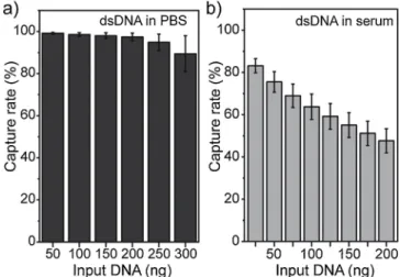
To quantify the capture efficiency of METRO, we perfused different volumes of sample containing fluorescently labeled dsDNA in PBS buffer (200 bp in length, MW = 129,000 g/mol, and [C] = 11 ng/μL). To evaluate the amount of captured DNA in real time, we assessed the level offluorescence intensity from the sample in the outlet channel (Fig. 1a-i): 1) before injecting the beads, and 2) during the DNA capture process (Appendix A Supplementary material, Section1.4). We mea-sured a DNA capture rate higher than 97% for samples with up to 200 ng of DNA, with a slight decrease in capture rate for 300 ng of DNA (89 ± 8%), probably due to saturation effects and steric repulsion from DNA molecules captured on the surface of the beads (Fig. 2a). We ob-served a linear capture rate for DNA inputs in the 0–250 ng range (Fig. S4), demonstrating the capabilities of our system for the efficient ex-traction of dsDNA fragments in ideal conditions (i.e. using an aqueous matrix). We estimated the DNA binding capacity of our system to be at least in the order of 10μg of DNA/mg of beads; placing our system in the higher end of the 5–10 μg of DNA/mg range expected from the supplier, using the batch procedure recommended for the ChargeS-witch®particles, and well above the capacity for other commercially
Fig. 2. Capture rates for METRO measured for different DNA inputs using fluorescently labeled dsDNA spiked in (a) PBS and (b) human serum samples from healthy donors.
available particles [22].
3.1.3. DNA extraction from human serum
To evaluate the effect of the sample matrix on DNA binding affinity, we processed human serum samples from healthy donors, spiked with different amounts of fluorescent dsDNA. We measured a capture rate higher than 63% for samples containing up to 100 ng of DNA and a stabilization of the capture efficiency around 50% for higher amounts of DNA (48 ± 6% for 200 ng) (Fig. 2b). As expected, we observed a lower capture rate when using undiluted serum as a matrix, as com-pared to DNA samples in PBS, which could be related to non-specific adsorption of serum proteins (e.g., albumin) or other nucleic acids (e.g., ccfDNA or miRNA from serum) to the beads surface through electro-static interactions [11,39]. The high molecular content of complex matrices such as human serum or plasma has indeed limited the im-plementation of microfluidic DNA isolation procedures. One approach to avoid potential screening effects or clogging artifacts during the isolation process has been to dilute the samples prior to their injection on chip (10–100-fold) [18,22]. However, such approach is not suitable for the analysis of ctDNA, as it would radically limit sample processing capabilities, as well as the sensitive detection of rare events. In this work, the capture rate for DNA samples in serum is still particularly high considering the use of undiluted human serum, the small size of extracted DNA molecules, and the electrostatic-based (thus non-spe-cific) nature of the DNA binding strategy.
To estimate the global extraction efficiency (including elution) for the spiked serum samples on chip, we collected the eluted DNA for off-chip quantification (Qubit®assay, Fig. S5). For an input of 200 ng of
DNA using METRO, we obtained an average extraction efficiency of 64 ± 9% (n = 5), in the same range as the capture rate for 200 ng measured above using continuousfluorescence measurements on chip. We thus expect that all DNA captured inside thefluidized bed was ef-ficiently eluted (i.e. no detectable DNA remaining at the surface of the beads). In comparison to the batch ChargeSwitch®procedure, the elu-tion volume on chip was reduced by at least 15-fold (the volume of the microfluidic chamber is 0.65 μL), while maintaining a similar extraction efficiency (Fig. S5).
Other groups have demonstrated the interest of electrostatic-based approaches for DNA isolation on chip. Nakagawa et al. used an amino silane-coated microchip to obtain 60% capture and 45% elution rates forλ-DNA (2 ng/μL) [23]. However, their recovery ratio decreased to 27–40% when extracting human genomic DNA from whole blood, due to the competitive binding of negatively charged proteins, as demon-strated here. More recently, Gan et al. developed an original approach combining a chitosan-modified filter paper inside a microfluidic device for DNA extraction [24]. Although they obtained a 62–89% capture efficiency using λ-DNA (with concentrations from 0.05 to 1 ng/μL),
only 8.9–11.5% of the captured DNA could be eluted, thus limiting the possibility of interfacing with downstream analysis modules. Finally, non-commercial chitosan-coated magnetic particles have also been used for efficient DNA isolation, providing high stability, pH tunability and yield (52μg from 3 mL human saliva), but their implementation on chip has not been demonstrated [31]. Our technology provides similar or better DNA extraction efficiencies to those reported in the literature with the added advantages of a higher binding capacity, as well as a higher potential for scalability and integration with DNA detection and quantification techniques, such as droplet-based digital PCR, as shown in the following sections.
3.2. Processing biological samples
3.2.1. Quantification of DNA extraction efficiency by ddPCR
To evaluate the efficiency of METRO using more realistic samples and demonstrate its compatibility with ddPCR analysis (RainDrop Digital PCR System), we processed undiluted serum from healthy do-nors spiked with cancer cell line DNA, with fragment size lengths and concentrations relevant to ctDNA analysis (from 50 to 1,100 bp and [C] = 0–357 ng/mL, Fig. S2) [40]. To differentiate between spiked
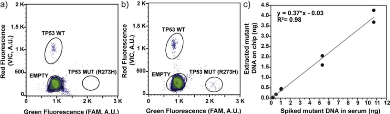
mutant DNA (MUT) and non-mutated ccfDNA (wild type, WT, initially present in serum samples), we used a mutation specific assay containing allele-specific hydrolysis probes (TaqMan™ probes targeting the R273H mutation in the TP53 gene). Wefirst evaluated the levels of WT DNA for non-spiked samples, and detected an average concentration of 23 ± 11 ng/mL (equivalent to 7045 ± 3315 copies in 1 mL of serum, n = 11), in line with concentrations of ccfDNA previously described for healthy donors [11]. We observed no false positives or MUT events in the healthy test samples (Fig. 3a). We then extracted the different
spiked samples, and used the digital quantification to compare the number of expected vs. extracted MUT and WT genomes for each sample (Fig. 3b). We established a calibration curve for the MUT gen-omes (Fig. 3c) and observed a linear capture/elution relationship for the different input mutant DNA with an overall 37% extraction and detection efficiency. In parallel, we performed the extraction of the same spiked samples using the ChargeSwitch® procedure in batch. However, the analysis of eluted DNA from the bench-top protocol did not generate consistent ddPCR results, probably due to the presence of inhibiting reagents or molecules still present in the eluent (Fig. S6). METRO allowed for a more efficient washing process and buffer ex-change inside the microfluidic device, thus delivering inhibitor free samples compatible with ddPCR analysis. These results demonstrated that our system allows analyzing MUT DNA fragments in concentra-tions as low as 3 ng/mL of serum in a background of WT DNA (leading to a mutational ratio of about 10% when considering the amount of WT ccfDNA in serum samples), using a minute amount of sample (60μL).
Fig. 3. Detection of R273H TP53 mutation in serum samples spiked with mutant HT29 cell line DNA. (a-b) Two dimensional histograms of the digital PCR assay for the analysis of circulating DNA extracted from healthy donors (a) and from spiked samples (b). Calibration curve (c): Extracted R273H TP53 mutant DNA digitally quantified for varying spike in DNA amounts (0.0033 ng/genome copies). MUT probes: FAM, 6- carboxyfluorescein; WT probes: VIC, a proprietary dye of Life Technologies; A.U., arbitrary units.
3.2.2. Analysis of serum samples from cancer patients
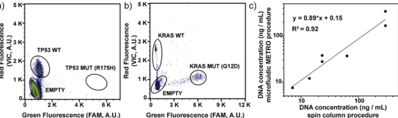
As a proof of concept, we implemented METRO for the detection of DNA in serum samples from breast (n = 5) and colon (n = 2) cancer patients with a known somatic variant in tumor tissue (Table S4). Specifically, we extracted 60 μL of serum from each of these patients on chip and quantified the number of MUT and WT genomes, using ddPCR assays specific to each patient [2,38,41]. We performed two extractions in parallel for each patient sample, using two microfluidic devices with the same operating conditions. The quantification results showed con-centrations of total ccfDNA (WT + MUT) ranging from 0 to 342 ng/mL of serum (Table S4), in line with previously reported concentrations for ccfDNA in the serum of cancer patients [11]. We detected the somatic variant from patient #6 in the serum samples extracted on chip, with a concentration of 15.45 ng/mL of MUT ctDNA (equivalent of a 47.5% mutational ratio for the G12D mutation in the KRAS gene). We con-sidered this sample as positive for ctDNA, since the concentration was higher than the limit of detection determined above for spiked serum samples (Fig. 4b,c and Table S4). We also noticed that patient #1 presented a high concentration of WT DNA but no detectable MUT ctDNA in serum (Fig. 4a,c and Table S4), which could be related to an important background of DNA from non-tumor cells (e.g., leukocytes). We observed a linear relationship when comparing the total ccfDNA concentrations obtained on chip and through a standard column-based protocol, for values above the limit of detection of the microfluidic METRO procedure (Fig. 4c, Table S5, Appendix A Supplementary ma-terial, Section1.5).
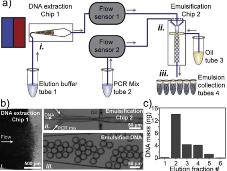
3.3. Integrated DNA extraction and detection procedure on chip
Finally, we developed an integrated device combining METRO, with a microfluidic module enabling the direct emulsification of the eluted DNA, together with PCR reagents, on chip (Fig. 5a,b). The generated droplets were collected unto 6 individual tubes and submitted to thermal cycling for DNA amplification (Appendix A Supplementary material, Section 1.6). We evaluated the number of positive events (droplets containing amplified DNA) on each elution fraction through fluorescence detection on chip (Fig. 5c), and estimated an overall DNA extraction efficiency of 64%. We observed that most eluted DNA was contained in elution fractions from 2 to 4 (with occupation ratios ran-ging from 0.3 to 1.5%). Although the amount of DNA in elution frac-tions 1 and 6 was very low (< 250 pg), it was readily preserved and amplified using this integrated procedure. Considering the operating conditions, the complete DNA extraction and emulsification of 30 μL of sample was performed in less than 2 h (Table S2).
Although a DNA isolation and amplification procedure using con-tinuous flow microfluidics and microchambers for sample compart-mentalization was demonstrated earlier [42], we present in this work
thefirst direct eluent emulsification method for picoliter droplet-based dPCR. This approach enables the generation of millions of individual compartments for single molecule detection. It also allows for the ef-ficient elution and amplification of all DNA captured at the beads sur-face and offers the possibility to rationally modify the PCR amplifica-tion environment for each eluamplifica-tion fracamplifica-tion (e.g. emulsification with a variety of primers and probes or using different thermal cycling con-ditions), thus opening the route for multiplex analyses.
4. Conclusion
We have combined on-chip, for thefirst time, a miniaturized DNA extraction platform with ddPCR. We applied this method for the de-tection and quantification of ctDNA from biological samples. Specifically, we isolated fragmented DNA in low concentrations directly from serum samples from cancer patients and demonstrated the com-patibility of our system with sensitive DNA amplification methods, such as ddPCR. As compared to conventional column-based methods, this approach has the ability to perform both sample preparation and DNA detection in a single device, providing comparable results but using only a few microliters of sample, which could be of great interest for the implementation of minimally invasive cancer diagnostic procedures. Furthermore, the METRO procedure offers a high degree of flexibility in terms of size, number and surface functionalities of the magnetic par-ticles that can be implemented (e.g. to selectively capture mRNA, microRNA, methylated DNA sequences, as well as bacterial or viral DNA).
Acknowledgments
This work was supported by the SIRIC CARPEM, the Ministère de l’Enseignement Supérieur et de la Recherche, the Université Paris-Descartes, the Centre National de la Recherche Scientifique (CNRS), the Institut National de la Santé et de la Recherche Médicale (INSERM), the French Ile-de-France canceropôle, by ANR“Investissements d'Avenir” for Digidiag project (ANR Nanobiotechnologies; no. ANR-10-NANO-0002-09), the ligue nationale contre le cancer (LNCC, Program“Equipe labelisée LIGUE”; no. EL2016.LNCC/VaT) as well as Labex and Equipex IPGG, and by European FP7 programs (LOVEFOOD FP7-ICT- 2011-317742, NAPES FP7-NMP-2013-604241, CATCH-U-DNA, FETOPEN H2020 and ERC Advanced Grant CellO (FP7-IDEAS-ERC-321107). The authors acknowledge the Plateforme Technologique of the Institut Universitaire d’Hématologie (IUH, Paris, France) and the biochemistry and molecular biology service (BMB168, Institut Curie, UMR168) for help with DNA analysis.
Fig. 4. Detection of ccfDNA in the serum of breast and colon cancer patients. (a-b) Two dimensional histograms of the digital PCR assay for the analysis of circulating DNA extracted on chip from two patients bearing TP53 (a) and KRAS mutations (b). (c) Digital quantification for total ccfDNA concentration (MUT + WT) following extraction using the microfluidic METRO procedure and standard column-base protocol. MUT probes: FAM, 6- carboxyfluorescein; WT probes: VIC, a proprietary dye of Life Technologies; A.U., arbitrary units.
Appendix A. Supplementary data
Supplementary material related to this article can be found, in the online version, at doi:https://doi.org/10.1016/j.snb.2019.01.159. References
[1] J.C.M. Wan, C. Massie, J. Garcia-Corbacho, F. Mouliere, J.D. Brenton, C. Caldas, et al., Liquid biopsies come of age: towards implementation of circulating tumour DNA, Nat. Rev. Cancer 17 (2017) 223–238.
[2] S. Garrigou, G. Perkins, F. Garlan, C. Normand, A. Didelot, D. Le Corre, et al., A study of hypermethylated circulating tumor DNA as a universal colorectal cancer biomarker, Clin. Chem. 62 (2016) 1129–1139.
[3] L.A. Diaz, A. Bardelli, Liquid biopsies: genotyping circulating tumor DNA, J. Clin. Oncol. 32 (2014) 579–586.
[4] C. Alix-Panabieres, K. Pantel, Clinical applications of circulating tumor cells and circulating tumor DNA as liquid biopsy, Cancer Discov. 6 (2016) 479–491. [5] S.J. Dawson, D.W.Y. Tsui, M. Murtaza, H. Biggs, O.M. Rueda, S.F. Chin, et al.,
Analysis of circulating tumor DNA to monitor metastatic breast cancer, N. Engl. J. Med. 368 (2013) 1199–1209.
[6] R. Lebofsky, C. Decraene, V. Bernard, M. Kamal, A. Blin, Q. Leroy, et al., Circulating tumor DNA as a non-invasive substitute to metastasis biopsy for tumor genotyping and personalized medicine in a prospective trial across all tumor types, Mol. Oncol. 9 (2015) 783–790.
[7] C. Decraene, A. Silveira, F.-C. Bidard, A. Vallee, M. Michel, S. Melaabi, et al., Multiple hotspot mutations scanning by single droplet digital PCR, Clin. Chem. (2017).
[8] U. Malapelle, C. Mayo de-Las-Casas, D. Rocco, M. Garzon, P. Pisapia, N. Jordana-Ariza, et al., Development of a gene panel for next-generation sequencing of clinically relevant mutations in cell-free DNA from cancer patients, Br. J. Cancer 116 (2017) 802.
[9] F. Garlan, P. Laurent-Puig, D. Sefrioui, N. Siauve, A. Didelot, N. Sarafan-Vasseur, et al., Early evaluation of circulating tumor DNA as marker of therapeutic efficacy in metastatic colorectal cancer patients (PLACOL study), Clin. Cancer Res. 23 (2017) 5416–5425.
[10] N. Pecuchet, Y. Rozenholc, E. Zonta, D. Pietraz, A. Didelot, P. Combe, et al., Analysis of base-position error rate of next-generation sequencing to detect tumor mutations in circulating DNA, Clin. Chem. 62 (2016) 1492–1503.
[11] S. El Messaoudi, F. Rolet, F. Mouliere, A.R. Thierry, Circulating cell free DNA: preanalytical considerations, Clin. Chim. Acta 424 (2013) 222–230. [12] J.H. van Ginkel, D.A. van den Broek, J. van Kuik, D. Linders, R. de Weger,
S.M. Willems, et al., Preanalytical blood sample workup for cell-free DNA analysis using droplet digital PCR for future molecular cancer diagnostics, Cancer Med. 6 (2017) 2297–2307.
[13] S.J. Reinholt, A.J. Baeumner, Microfluidic isolation of nucleic acids, Angew. Chem, Int. Ed. 53 (2014) 13988–14001.
[14] L.A. Christel, K. Petersen, W. McMillan, M.A. Northrup, Rapid, automated nucleic acid probe assays using silicon microstructures for nucleic acid concentration, J. Biomech. Eng. 121 (1999) 22–27.
[15] A. Sonnenberg, J.Y. Marciniak, R. Krishnan, M.J. Heller, Dielectrophoretic isolation of DNA and nanoparticles from blood, Electrophoresis 33 (2012) 2482–2490.
[16] A. Sonnenberg, J.Y. Marciniak, L. Rassenti, E.M. Ghia, E.A. Skowronski, S. Manouchehri, et al., Rapid electrokinetic isolation of cancer-related circulating cell-free DNA directly from blood, Clin. Chem. 60 (2014) 500–509.
[17] T. Hahn, K.S. Drese, C.K. O’Sullivan, Microsystem for isolation of fetal DNA from maternal plasma by preparative size separation, Clin. Chem. 55 (2009) 2144–2152. [18] H.J. Tian, A.F.R. Huhmer, J.P. Landers, Evaluation of silica resins for direct and
efficient extraction of DNA from complex biological matrices in a miniaturized format, Anal. Biochem. 283 (2000) 175–191.
[19] N.C. Cady, S. Stelick, C.A. Batt, Nucleic acid purification using microfabricated si-licon structures, Biosens. Bioelectron. 19 (2003) 59–66.
[20] M.C. Breadmore, K.A. Wolfe, I.G. Arcibal, W.K. Leung, D. Dickson, B.C. Giordano, et al., Microchip-based purification of DNA from biological samples, Anal. Chem. 75 (2003) 1880–1886.
[21] G.R.M. Duarte, C.W. Price, J.L. Littlewood, D.M. Haverstick, J.P. Ferrance, E. Carrilho, et al., Characterization of dynamic solid phase DNA extraction from blood with magnetically controlled silica beads, Analyst 135 (2010) 531–537. [22] W.D. Cao, C.J. Easley, J.P. Ferrance, J.P. Landers, Chitosan as a polymer for
pH-induced DNA capture in a totally aqueous system, Anal. Chem. 78 (2006) 7222–7228.
[23] T. Nakagawa, T. Tanaka, D. Niwa, T. Osaka, H. Takeyama, T. Matsunaga, Fabrication of amino silane-coated microchip for DNA extraction from whole blood, J. Biotechnol. 116 (2005) 105–111.
[24] W.P. Gan, Y. Gu, J.P. Han, C.X. Li, J. Sun, P. Liu, Chitosan-modified filter paper for nucleic acid extraction and“in situ PCR” on a thermoplastic microchip, Anal. Chem. 89 (2017) 3568–3575.
[25] G.R.M. Duarte, C.W. Price, B.H. Augustine, E. Carrilho, J.P. Landers, Dynamic solid phase DNA extraction and PCR amplification in polyester-toner based microchip, Anal. Chem. 83 (2011) 5182–5189.
[26] K.Y. Lien, C.J. Liu, Y.C. Lin, P.L. Kuo, G.B. Lee, Extraction of genomic DNA and detection of single nucleotide polymorphism genotyping utilizing an integrated magnetic bead-based microfluidic platform, Microfluid Nanofluid 6 (2009) 539–555.
[27] D.Y. Liu, G.T. Liang, Q. Zhang, B. Chen, Detection of Mycobacterium tuberculosis using a capillary-array microsystem with integrated DNA extraction, loop-mediated isothermal amplification, and fluorescence detection, Anal. Chem. 85 (2013) 4698–4704.
[28] D. Ferraro, J. Champ, B. Teste, M. Serra, L. Malaquin, J.L. Viovy, et al., Microfluidic platform combining droplets and magnetic tweezers: application to HER2 expres-sion in cancer diagnosis, Sci. Rep. 6 (2016).
[29] B. Teste, N. Jamond, D. Ferraro, J.L. Viovy, L. Malaquin, Selective handling of droplets in a microfluidic device using magnetic rails, Microfluid Nanofluid 19 (2015) 141–153.
[30] K.A. Hagan, W.L. Meier, J.P. Ferrance, J.P. Landers, Chitosan-coated silica as a solid phase for RNA purification in a microfluidic device, Anal. Chem. 81 (2009) 5249–5256.
[31] A.P. Tiwari, R.K. Satvekar, S.S. Rohiwal, V.A. Karande, A.V. Raut, P.G. Patil, et al., Magneto-separation of genomic deoxyribose nucleic acid using pH responsive Fe3O4@silica@chitosan nanoparticles in biological samples, RSC Adv. 5 (2015) 8463–8470.
[32] I. Pereiro, A. Bendali, S. Tabnaoui, L. Alexandre, J. Srbova, Z. Bilkova, et al., A new microfluidic approach for the one-step capture, amplification and label-free quan-tification of bacteria from raw samples, Chem. Sci. 8 (2017) 1329–1336. [33] I. Pereiro, S. Tabnaoui, M. Fermigier, O. du Roure, S. Descroix, J.L. Viovy, et al.,
Magneticfluidized bed for solid phase extraction in microfluidic systems, Lab Chip
Fig. 5. a) Schematic of the integrated procedure for DNA ex-traction and emulsification for ddPCR analysis. b) Micrograph images from thefluidized bed (bi) and the droplet generator module (bii-iii) during elution and encapsulation of DNA. c) Amount of extracted and amplified DNA by ddPCR for the different elution fractions, collected in PCR tubes (emulsion collection tubes inFig. 5a-iii).
17 (2017) 1603–1615.
[34] T.D. Mai, D. Ferraro, N. Aboud, R. Renault, M. Serra, N.T. Tran, et al., Single-step immunoassays and microfluidic droplet operation: towards a versatile approach for detection of amyloid-beta peptide-based biomarkers of Alzheimer’s disease, Sens. Actuators B 255 (2018) 2126–2135.
[35] K. Perez-Toralla, J. Champ, M.R. Mohamadi, O. Braun, L. Malaquin, J.L. Viovy, et al., New non-covalent strategies for stable surface treatment of thermoplastic chips, Lab Chip 13 (2013) 4409–4418.
[36] A. Bruchet, V. Taniga, S. Descroix, L. Malaquin, F. Goutelard, C. Mariet, Centrifugal microfluidic platform for radiochemistry: potentialities for the chemical analysis of nuclear spent fuels, Talanta 116 (2013) 488–494.
[37] C.A. Milbury, Q. Zhong, J. Lin, M. Williams, J. Olson, D.R. Link, et al., Determining lower limits of detection of digital PCR assays for cancer-related gene mutations, Biomol. Detect. Quantif. 1 (2014) 8–22.
[38] E. Zonta, F. Garlan, N. Pecuchet, K. Perez-Toralla, O. Caen, C. Milbury, et al., Multiplex detection of rare mutations by picoliter droplet based digital PCR: sen-sitivity and specificity considerations, PLoS One 11 (2016).
[39] K.D. Clark, O. Nacham, H.L. Yu, T.H. Li, M.M. Yamsek, D.R. Ronning, et al., Extraction of DNA by magnetic ionic liquids: tunable solvents for rapid and selec-tive DNA analysis, Anal. Chem. 87 (2015) 1552–1559.
[40] F. Mouliere, B. Robert, E.A. Peyrotte, M. Del Rio, M. Ychou, F. Molina, et al., High fragmentation characterizes tumour-derived circulating DNA, PLoS One 6 (2011). [41] P. Laurent-Puig, D. Pekin, C. Normand, S.K. Kotsopoulos, P. Nizard, K.
Perez-Toralla, et al., Clinical relevance of KRAS-mutated subclones detected with pico-droplet digital PCR in advanced colorectal cancer treated with anti-EGFR therapy, Clin. Cancer Res. 21 (2015) 1087–1097.
[42] Q.C. Tian, B.D. Yu, Y. Mu, Y.A. Xu, C.C. Ma, T. Zhang, et al., An integrated tem-porary negative pressure assisted microfluidic chip for DNA isolation and digital PCR detection, RSC Adv. 5 (2015) 81889–81896.
Karla Perez-Toralla was a postdoctoral researcher at Université Paris Descartes and Institut Curie, France (currently postdoctoral researcher at University of Nebraska-Lincoln). Her main research interest regards microfluidic devices for biomedical appli-cations, functional materials and microfabrication.
Iago Pereiro was a PhD student at Institut Curie and Institut Pierre-Gilles de Gennes, France (currently postdoctoral researcher at IBM-Research, Zurich). His main research interests are the development of microfluidic-based strategies for biochemical analysis. Sonia Garrigou is a molecular biologist engineer at Université Paris Descartes, France. Her research interest is the development of droplet-based digital PCR technologies for the analysis cancer biomarkers.
Fahima Di Federico is a molecular biologist engineer at Institut Curie, France. Her re-search interest is the technological development of Molecular Biology tools, Biochemistry and Bacteriology.
Charlotte Proudhon is Group Leader of the Circulating Cancer Biomarkers team at Institut Curie, France. Her research focuses on the study of circulating biomarkers in cancer patients to monitor minimal residual disease.
François-Clément Bidard is Professor of Medicine at the University of Paris-Saclay and research physician in medical oncology at Institut Curie, France. His research activity concerns the characterization of mechanisms involved in the metastatic process, parti-cularly the detection of tumor biomarkers found in the blood.
Jean-Louis Viovy is a research director at Institut Curie and Institut Pierre-Gilles de Gennes, France. He is the leader of the group“Macromolecules and Microsystems in Biology and Medicine” at the Institute Curie. His main research interests are organs on chips and lab-on-chip technologies.
Valérie Taly is a research director at Université Paris Descartes, France. Her research is dedicated to the clinical validation of the developed droplet-based procedures for the non-invasive detection of Cancer biomarkers.
Stéphanie Descroix is a research director at Institut Curie and Institut Pierre-Gilles de Gennes, France. Her main research interests are organs on chips and lab-on-chip tech-nologies.