HAL Id: hal-00392552
https://hal.archives-ouvertes.fr/hal-00392552
Preprint submitted on 8 Jun 2009
HAL is a multi-disciplinary open access
archive for the deposit and dissemination of sci-entific research documents, whether they are pub-lished or not. The documents may come from teaching and research institutions in France or abroad, or from public or private research centers.
L’archive ouverte pluridisciplinaire HAL, est destinée au dépôt et à la diffusion de documents scientifiques de niveau recherche, publiés ou non, émanant des établissements d’enseignement et de recherche français ou étrangers, des laboratoires publics ou privés.
Defect in lung growth Comparative study of three
diagnostic criteria.
Pierre Dechelotte, André Labbé, O. Caux, Ph. Vanlieferinghen, J.E. Raynaud
To cite this version:
Pierre Dechelotte, André Labbé, O. Caux, Ph. Vanlieferinghen, J.E. Raynaud. Defect in lung growth Comparative study of three diagnostic criteria.. 2009. �hal-00392552�
Defect in lung growth
*
Comparative study of three diagnostic criteria.
P. DECHELOTTE, A. LABBE, O. CAUX, Ph. VANLIEFERINGHEN, E. J. RAYNAUD
* English translation of an article first published in French in 1987 in the Archives Françaises de Pédiatrie.
Arch. Fr. Pediatr. 1987 ; 44 : 255-61.
Défaut de croissance pulmonaire. Etude compararative de trois critères de diagnostic. CHU Clermont-Ferrand, Hôtel-Dieu, Services d’Anatomie Pathologique et de Pédiatrie A
Univ Clermont 1, UFR Médecine, EA 3843, Laboratoires d’Anatomie pathologique et de Physiologie, Faculté de Médecine, Place Henri Dunant, Clermont-Ferrand, accepté pour publication le 24 novembre 1986.
Abstract.
A systematic analysis was made of the autopsies of 74 newborns and fetuses (49 pathological cases and 25 controls) to detect defects in lung growth. In each case lung/body (L/B) weight ratio was calculated, and radial alveolar (RA) count and histological assessment were performed. The L/B ratio is of diagnostic value when lower than 0.012 but not when there is intercurrent disease. RA count is low in lung hypoplasia but is not an entirely reliable diagnostic criterion since it change throughout pregnancy and the earlier the gestational age the wider the range of variation. Histological assessment showed an abnormally high number of bronchi and bronchi in distal location with in some cases delayed differentiation of distal airways. If any one of the above three critera fails to determine lung hypoplasia the other two can be used to arrive at diagnosis.
Key-words :
Lung diseases, infant, newborn, diseases, pulmonary fibrosis. Lung growth assessment, pulmonary hypoplasia, l u n g h y p o p l a s i a , r a d i a l a l v e o l a r c o u n t , l u n g morphometry.
Several conditions are required to achieve a functional lung. Lung growth is one aspect of this process of specialization. It is a complex, dynamic process that involves various interdependent factors. Growth takes place throughout gestation and continues after birth. It is not surprising, therefore, that lung hypoplasia (LH) is seen in numerous malformation syndromes and/or as the result of various insults. Underdevelopment is sometimes clearly evidenced by clinical and radiographic findings or by macroscopy. In other cases, however, diagnosis is more difficult and requires a more accurate, discriminatory analysis. We compared the validity of three criteria commonly used in the diagnosis of LH : the ratio of lung weight to total body weight (L/B ratio) ;
radial alveolar count (RA count) ; and histological assessment.
MATERIAL AND METHODS
Between 1 January 1982 and 30 June 1985, 435 autopsies were carried out on fetuses, newborns and infants in the Department of Pathology of the Hôtel-Dieu hospital, Clermont-Ferrand. We selected 66 cases in which there was a likelihood of LH. Histological assessment was possible in only 49 of the 66 cases (group 2), of which 24 presented malformations classically associated with LH and 25 a disease that could be accompanied by a growth defect. A control group of 25 lung specimens (group 1) was created on the basis of four criteria : absence of disease classically associated with LH ; absence of intercurrent disease likely to modify L/B ratio ; absence of postmortem autolysis, so as to be able to perform RA count ; and histological assessment.
L/B ratio was determined from autopsy reports. Body weight and combined lung weight were measured no later than 18 h post mortem. Alveolar counts (fig. 1) were done according to the method of Emery and Mithal (2). For cases of prealveolar stage we followed the recommendations of Cooney and Thurlbeck (3) : a perpendicular line to the nearest fibrous septum was drawn from the most distal tube containing both bronchiolar and alveolar lining and the number of alveoli cut by this line was then counted. The counts were done by two physicians, neither of whom had details of the clinical history.
FIG. 1. – Calculation of RA count : a perpendicular line to the nearest fibrous septum was drawn from the most distal tube containing both bronchiolar and alveolar lining. (Gr. X 21, réd. 0,76).
Results were expressed as the mean of 10 counts per lung, and, hence, of 20 counts for each case. Histological investigation was performed to look for evidence of an excess number of bronchi or distal location bronchi (DB), that is, bronchial airways occupying an abnormally large area in relation to distal exchange areas. These bronchial airways, with or without cartilage, were sometimes located considerably distal to the hilum and sometimes were subpleural. Investigation was also made of anomalies in the differentiation of distal airways (DA) related to gestational age(1 ) and also of possible
associated disease.
Three categories were defined on the basis of
these three parameters : normal lungs (NL), in which L/ B ratio and RA count were not low and which histologically had no structural anomaly ; miniature
type lungs (ML), in which L/B ratio (in varying degrees)
and RA count were low, and which presented evidence of an excess number of bronchi but no anomaly in the differentiation of DA ; and fetal type lungs (FL), in which both L/B ratio and RA count were very low and which presented evidence of an excess number of bronchi and distal bronchi and delayed differentiation of DA.
Group 2 was subdivided as shown in Table 1. Mean standard deviation and standard error of mean were calculated for each group. Student’s test and/or variance analysis (Snedecor's F Test) were used to compare quantitative variables. Qualitative variables were compared using Chi-squared test, corrected if the number of specimens was too low according to the method of Yates, with a level of significance of 0.05.
RESULTS
The average gestational age at birth was 29.6 ± 6.65 weeks in the pathological group (group 2) and 31.6 ± 7.10 weeks in the control group (group 1).
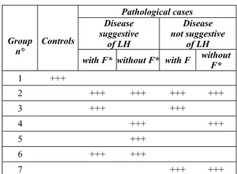
TABLE I. – Definition of groups studied.
Group n° Controls Pathological cases Disease suggestive of LH Disease not suggestive of LH
with F* without F* with F without
F* 1 +++ 2 +++ +++ +++ +++ 3 +++ +++ 4 +++ +++ 5 +++ 6 +++ +++ 7 +++ +++
* Factor modifying lung /body (L/B) weight ratio ; LH : lung hypoplasia.
L/B ratio (Table IV)
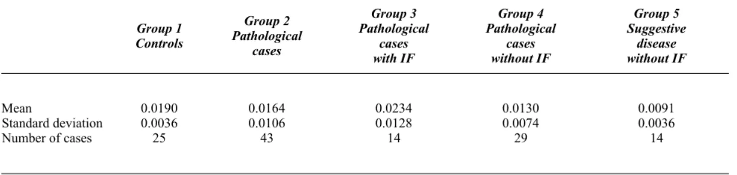
The mean of the L/B ratios (0.0164) in the pathological cases (group 2) was lower than that in the control group (group 1) but not to a level of significance. Among the cases with suspected LH, 14 presented an intercurrent factor, such as haemorrhage, hyaline membrane disease or anencephalia, that could have artificially modified the ratio. The mean of this group’s ratios (groupe 3 ; 0.0234) was geater than that of the controls (0.019), which speaks in favour of an effect of the intercurrent factors. If the 14 cases are excluded, the difference in ratios between the control and disease (group 4) groups is significant (p<0.001). It is even more so if comparison is made with the sub-group (group 5), in which accompanying disease, such as Potter’s syndrome or diaphragmatic hernia, was highly suggestive of LH .
RA count study (Table V)
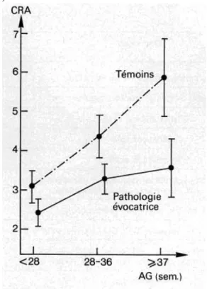
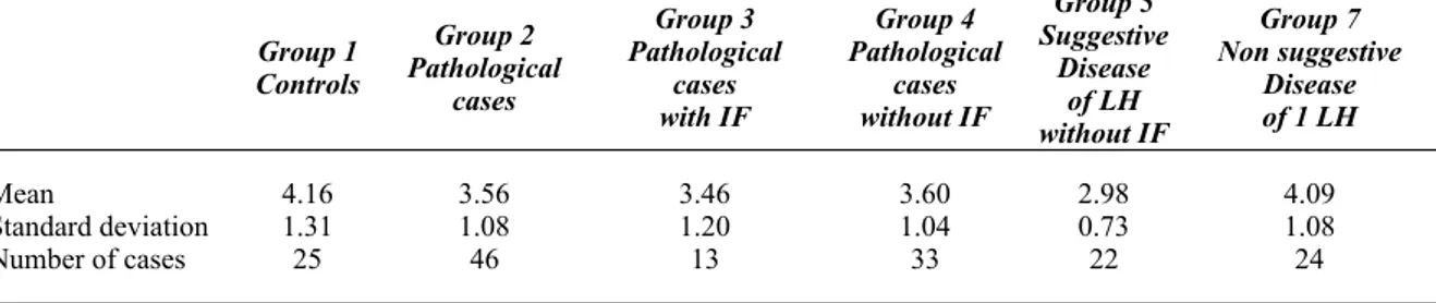
The mean RA count of disease group 2 (3.56) was significanlly lower (p<0.001) than that of control group 1 (mean = 4.16) : counts in group 3 were close to those in group 2. The mean RA count of group 5 (2.98) was distinctly lower than that of group 7 (4.09), in which there was no indicative disease. The RA count of controls increased with gestational age (fig. 2). The differences between the two groups remained distinct and became greater with increasing gestational age.
(1 ) Stages in lung growth (according to Langston C. and Thurlbeck W. M. [10]) :
- embryonic stage : days 22-26 to week 5 - pseudoglandular stage : weeks 6 to 16 - canilicular stage : weeks 17 to 24 - terminal sac stage : from week 25 - alveolar stage : after birth
Histological assessment
Assessments showed a highly significant (χ2 = 12.9,
p<0.005) relation between the type of disease, suggestive or not of LH, and the existence of specific anomalies (table VI).
FIG. 2. – Variation in radial alveolar RA counts (CRA) according to
gestational age (AG).
There was also a highly significant relation between these histological anomalies and L/B ratio once those cases presenting an intercurrent factor were eliminated (table VII). The L/B ratio of fetal type lungs (FL) were significantly lower than those of normal lungs (p<0.001). The L/B ratio of FL was lower than that of ML but not to a level of significance. There was a highly significant relation between histological anomalies and the mean value of RA counts (F = 16.01, DOF * = 2,43, p<0.001). The RA count of PM (3.90±0.94) were lower than those of NL (4.56±1.01) not significant (NS). The difference is greater when the counts of NL and FL are compared. NL counts : (4.56±1.01) are slightly higher than those of controls (4.16±1.31).
DISCUSSION
Except for cases in which antenatal or neonatal clinical findings or macroscopy provide clear evidence, diagnosis of LH is based on a number of variably reliable criteria. The easiest way of predicting LH is to use L/B ratio. Opinion differs between authors as to what the threshold value of this index should be. Emery and Mithal (2) suggest 0.013 as the lower limit of normal. Askenazi and Perlman (4) found a mean ratio of 0.018 ± 0.03 in 17 newborns of 38 weeks gestational age and suggested a value of 0.012, based on the ratio of control cases minus
two standard deviations. Wigglesworth et al. (5) consider 0.012 to be reliable from 28 weeks onwards and 0.015 before. In our case series, we observed that the ratio is only of diagnostic value when it is low.
* F : F Fisher-Snedecor ; DOF : Degrees of Freedom
A normal ratio in no way rules out LH since it may be distorted by a local disorder such as lung haemorrhage or by general disease. To make results more reliable, Wigglesworth and Desai (6) suggested making a concomitant measurement of lung DNA content. They showed a good correlation between the ratio of DNA content to body weight and L/B ratio. They posited that the presence of superimposed diseases does not alter lung weight in cases of severe hypoplasia because hypoplastic lungs cannot contain a great amount of liquid. In one of our cases (n° 17), in which Potter’s syndrome was associated with lung haemorrhage, the ratio was 0.017. RA count indicates the number of alveoli in a lobule. The number increases rapidly until the age of 1 year and continues to rise, but at a slower rate, until the age of 2 (7). We recorded higher numbers than Emery and Mithal (2) but we observed a similar increase in RA count with gestational age. Thus, the difference between our two groups increased according to fetal age.
Like other authors, we had difficulty in assessing RA count. When there is major superimposed disease, it is more difficult to identify the bronchioli if the lining is damaged or if the distal airways are not clearly differentiated. We observed a wide range in values, with 9 cases having a count higher than the mean and 15 a count between the mean and standard deviation. These results are consistent with those reported elsewhere, in particular in the studies of Hislop et all. (8), Chamberlain et al. (9), and Emery and Mithal (2). As one possible explanation of the discrepancy in values, these authors point to their observation that there were fewer divisions of the bronchi and bronchioli. Thus, while the number of alveoli counted was normal, with a normal or high RA count, the total number of alveoli was actually low.
FIG. 3. – Fetal lung : picture of excessive number of bronchi with peripheral
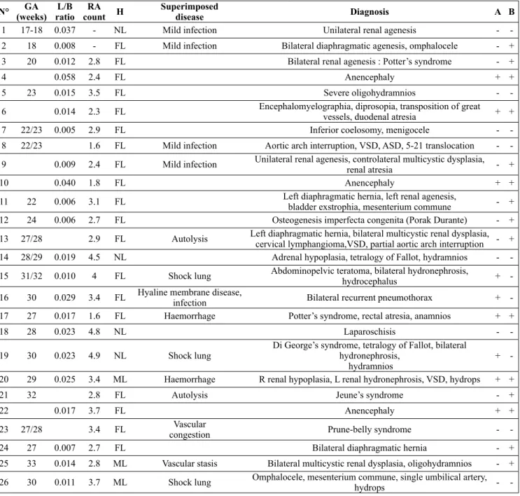
TABLE II. – N° GA (weeks) L/B ratio RA count H Superimposed disease Diagnosis A B
1 17-18 0.037 - NL Mild infection Unilateral renal agenesis -
-2 18 0.008 - FL Mild infection Bilateral diaphragmatic agenesis, omphalocele - +
3 20 0.012 2.8 FL Bilateral renal agenesis : Potter’s syndrome - +
4 0.058 2.4 FL Anencephaly + +
5 23 0.015 3.5 FL Severe oligohydramnios -
-6 0.014 2.3 FL Encephalomyelographia, diprosopia, transposition of great
vessels, duodenal atresia + +
7 22/23 0.005 2.9 FL Inferior coelosomy, menigocele -
-8 22/23 1.6 FL Mild infection Aortic arch interruption, VSD, ASD, 5-21 translocation - -9 0.009 2.4 FL Mild infection Unilateral renal agenesis, controlateral multicystic dysplasia,
renal atresia - +
10 0.040 1.8 FL Anencephaly + +
11 22 0.006 3.1 FL Left diaphragmatic hernia, left renal agenesis,
bladder exstrophia, mesenterium commune - +
12 24 0.006 2.7 FL Osteogenesis imperfecta congenita (Porak Durante) - +
13 27/28 2.9 FL Autolysis Left diaphragmatic hernia, bilateral multicystic renal dysplasia, cervical lymphangioma,VSD, partial aortic arch interruption - +
14 28/29 0.019 4.5 NL Adrenal hypoplasia, tetralogy of Fallot, hydramnios -
-15 31/32 0.010 4 FL Shock lung Abdominopelvic teratoma, bilateral hydronephrosis,
hydrocephalus +
-16 30 0.029 3.4 FL Hyaline membrane disease,
infection Bilateral recurrent pneumothorax +
-17 27 0.017 1.6 FL Haemorrhage Potter’s syndrome, rectal atresia, anamnios + +
18 28 0.023 4.8 NL Laparoschisis -
-19 30 0.023 4.9 NL Shock lung
Di George’s syndrome, tetralogy of Fallot, bilateral hydronephrosis,
hydramnios
+ -20 29 0.025 3.4 ML Haemorrhage R renal hypoplasia, L renal hydronephrosis, VSD, hydrops + +
21 32 2.8 FL Autolysis Jeune’s syndrome - +
22 0.017 3.7 FL Anencephaly + +
23 27/28 3.4 FL Vascular
congestion Prune-belly syndrome -
-24 27 0.007 2.7 FL Bilateral diaphragmatic hernia - +
25 33 0.014 2.8 ML Vascular stasis Bilateral multicystic renal dysplasia, oligohydramnios - + 26 30 0.011 3.7 ML Shock lung Omphalocele, mesenterium commune, single umbilical artery,
hydrops -
-The first aim of histological assessment was to evidence a delay in alveolar differentiation on the basis of the different stages of fetal lung development (10, 11).
The diagnosis was easy when there was a discrepancy between the stage of differentiation and gestational age. In 17 cases of LH, Bouley et al. (12) reported an arrest of differentiation during the canalicular phase (weeks 16-20). In 3 of our cases, we observed such an arrest at gestational ages ranging from 27 to 32 weeks and in the 14 others no arrest.
This suggests that in a certain number of cases growth is not arrested but simply slowed. The second aim of histological assessment was to evidence numbers of bronchi and distal bronchi that appeared to be high in relation to those of DA (fig. 3).
Several studies have reported anomalies of the bronchi during LH (13-16), manifested mainly in the undervelopment of distal airways. With the exception of the study of Finegold et al. (17), who confirmed the visual impression by use of a reliable morphometric method, all other observations were simply qualitative.
Pathological cases. N° GA (weeks) L/B ratio RA count H Superimposed disease Diagnosis A B
27 35 0.005 3.3 FL Hydramnios, hydrops fetalis - +
28 36 0.006 3.2 ML Thanatophore dwarfism, hydramnios - +
29 36/37 0.011 4.5 ML Infection, aspiration,
shock lung Bilateral multicystic renal dysplasia, posterior valve of the urethra + +
30 35/36 0.006 2.9 FL Aspiration Hydrops, hydramnios, AVSD - +
31 32 0.015 4.6 ML Spondylocostal dysostosis, tricuspid atresia,
Single ventricle, duodenal atresia - +
32 36/37 0.011 3.6 ML Autolysis ARPKD - +
33 37 0.028 4.8 NL Bilateral hydronephrosis &
Bilateral anomalous ureterovesical junction, hydrops - -34 38/39 0.015 2.7 ML Autolysis, shock lung Bilateral multicystic renal dysplasia,
megadolicho-urethra, trisomy 21 - +
35 40 0.029 5.6 NL Septicemia VSD, œsophageal atresia, hydrops fetalis +
-36 37 0.009 3.3 FL Focal emphysema Respiratory distress -
-37 38 0.016 3.9 FL Focal emphysema Dandy Walker syndrome, L ventricle hypoplasia,
mitral and aortic atresia, VSD, ASD -
-38 38 2.4 NL Massive infection Oesophageal atresia, R lung hypoplasia -
-39 38 0.007 3.5 FL Thanatophore dwarfism - +
40 39/40 0.017 NL Autolysis, shock lung Partial AVSD, hydrops + +
41 31 0.007 3.8 ML Autoysis Atresia of pulmonary artery, ASD, mild hydrops - -42 36/37 0.019 4.7 ML Vascular stasis Idiopathic hypertrophic cardiomyopathy - -43 37/38 0.012 3.8 ML Mild infection Bilateral hydronephrosis,
L renal hypoplasia, double aortic arch -
-44 38/39 0.017 4.2 NL Aspiration Anomalous ureterovesical junction +
-45 40 0.013 6.1 NL Elis Van Crefeld syndrome, AVSD -
-46 42 0.020 3.3 NL Shock lung Drash’s syndrome +
-47 42 4.7 NL Mild infection CHARGE association -
-48 42 0.019 6.1 ML Oesophageal atresia, anomalous ureterovesical junction -
-49 42 0.017 5 NL Shock lung Bilateral adrenal hypoplasia -
-GA : gestational age (in weeks) ; L/B : lung/body weight ratio ; RA count : radial alveolar counts ; H : histological investigation ; NL : normal lung ; FL : fetal type lung ; ML : miniature type lung ; A : case in which disease modified RAC; B : case in which disease was suggestive of LH ; ARPKD : autosomal recessive polycystic kidney disease ; AVSD : atrioventricular septal defect ; ASD : atrio septal defect ; VSD : ventricular septal defect ; L : left ; D : right.
In our series, there was a significant correlation between the clinical presentation and the results of histological assessment. L/B ratio and RA count were lower in fetal type than in miniature type lungs, which
indicates that LH was more severe in the former. However, as noted by Bouley et al. (12), this clear distinction between fetal and miniature lungs is not found in other published reports.
TABLE III. – Controls. N° GA (weeks) L/B ratio RA count H Diagnosis
1 19-20 0.0015 2.3 NL TOP maternal indication, NM
2 18 0.0015 2.3 NL TOP maternal indication, NM
3 21-22 0.023 2.9 NL TOP maternal indication, NM
4 22-23 0.017 3.7 NL TOP maternal indication, NM
5 23-24 0.018 2.8 NL SA, PROM, mild amniotic aspiration
6 24-25 0.022 3 NL SA, maternal cause, NM
7 25-26 0.026 2.7 NL SA, placental abruption, NM
8 24-25 0.018 4.4 NL SA, PROM, ventricular haemorrhage
9 29 0.021 3.6 NL TOP, retroplacental haematoma, NM
10 27 0.015 3.2 NL SA maternal cause, NM
11 27-28 0.025 3.3 NL TOP, preeclampsia, ventricular haemorrhage
12 27-28 0.024 3.7 NL TOP, amniotic aspiration, NM
13 29-30 0.017 3.2 NL TOP, stillbirth, ventricular haemorrage
14 29-30 0.021 4.7 NL TOP, dysgravidia, aspiration, NM
15 32 0.022 5 NL TOP, collodion baby, NM internal
16 33-34 0.017 4.5 NL TOP, maternal indication, NM
17 33-34 0.017 4.4 NL TOP, stillbirth, PROM
18 29-30 0.014 5.8 NL TOP, stillbirth, VSD, mild amniotic aspiration
19 37-38 0.021 4.4 NL Stillbirth, mild amniotic aspiration, NM
20 32-33 0.024 4.4 NL TOP, retroplacental haematoma, NM
21 41 0.018 5.8 NL Sudden death at H24, maternal infection, NM
22 0.014 6.2 NL Sudden death at H48, NM
23 42 0.017 5.3 NL Fetal death, diabetes melitus
24 42 0.016 7.5 NL Acute major anaemia, subgaleal hemorrhage,
25 3m 0.019 5.1 NL Cot death
GA : gestational age (in weeks) ; L/B : lung/body weight ratio ; RA count : radial alveolar counts ; H : histological investigation ; NL : normal lung ; FL : foetal lung ; ML : miniature lung ; NM : No Malformation ; TOP : termination of pregmnancy ; PROM : premature rupture of membranes ; SA : spontaneous abortion; A : case in which disease modified RA count ; B : case in which disease was suggestive of LH ; AVSD : atrioventricular septal defect ; ASD : atrio septal defect ; VSD : ventricular septal defect; L : left ; D : right.
Group 1 Controls Group 2 Pathological cases Group 3 Pathological cases with IF Group 4 Pathological cases without IF Group 5 Suggestive disease without IF Mean Standard deviation Number of cases 0.0190 0.0036 25 0.0164 0.0106 43 0.0234 0.0128 14 0.0130 0.0074 29 0.0091 0.0036 14
TABLE IV. – Value of lung/body (L/B) weight ratio in the different groups.
Group 1 Controls Group 2 Pathological cases Group 3 Pathological cases with IF Group 4 Pathological cases without IF Group 5 Suggestive Disease of LH without IF Group 7 Non suggestive Disease of 1 LH Mean Standard deviation Number of cases 4.16 1.31 25 3.56 1.08 46 3.46 1.20 13 3.60 1.04 33 2.98 0.73 22 4.09 1.08 24
TABLE V. – Value of radial alveolar count.
*IF : intercurrent factor. LH : lung hypoplasia.
TABLE VI. – Result of the histological study according to whether the
initial clinical picture was suggestive or not of the existence of associated lung hypoplasia.
Clinical picture Histology Total NL ML FL Suggestive disease 1 7 16 24 Non-suggestive disease 12 5 8 25
NL : normal lungs ; ML : miniature type lungs ; FL : fetal type lungs.
TABLE VII. – Relation between results of histological study and the
value of L/B weight ratio in group 4.
Clinical picture Histology Normal lung Miniature lung Fetal lung Number of cases 6 10 13 CONCLUSION
This study shows that the diagnosis of anomalies in lung growth requires the analysis of several factors that may be misleading, particularly L/B ratio, if taken in isolation. Although RA count and histological assessment are more difficult to perform they provide indispensable diagnostic information especially if pathological and clinical findings are not suggestive of a defect in lung growth.