Publisher’s version / Version de l'éditeur:
Optics Letters, 32, 21, pp. 3092-3094, 2007-11-01
READ THESE TERMS AND CONDITIONS CAREFULLY BEFORE USING THIS WEBSITE. https://nrc-publications.canada.ca/eng/copyright
Vous avez des questions? Nous pouvons vous aider. Pour communiquer directement avec un auteur, consultez la première page de la revue dans laquelle son article a été publié afin de trouver ses coordonnées. Si vous n’arrivez pas à les repérer, communiquez avec nous à PublicationsArchive-ArchivesPublications@nrc-cnrc.gc.ca.
Questions? Contact the NRC Publications Archive team at
PublicationsArchive-ArchivesPublications@nrc-cnrc.gc.ca. If you wish to email the authors directly, please see the first page of the publication for their contact information.
NRC Publications Archive
Archives des publications du CNRC
This publication could be one of several versions: author’s original, accepted manuscript or the publisher’s version. / La version de cette publication peut être l’une des suivantes : la version prépublication de l’auteur, la version acceptée du manuscrit ou la version de l’éditeur.
For the publisher’s version, please access the DOI link below./ Pour consulter la version de l’éditeur, utilisez le lien DOI ci-dessous.
https://doi.org/10.1364/OL.32.003092
Access and use of this website and the material on it are subject to the Terms and Conditions set forth at
Enhanced surface plasmon resonance imaging detection of DNA
hybridization on periodic gold nanoposts
Malic, L.; Cui, B.; Veres, T.; Tabrizian, M.
https://publications-cnrc.canada.ca/fra/droits
L’accès à ce site Web et l’utilisation de son contenu sont assujettis aux conditions présentées dans le site LISEZ CES CONDITIONS ATTENTIVEMENT AVANT D’UTILISER CE SITE WEB.
NRC Publications Record / Notice d'Archives des publications de CNRC:
https://nrc-publications.canada.ca/eng/view/object/?id=0b723396-1f7d-49b1-92b3-aa53ac685cec https://publications-cnrc.canada.ca/fra/voir/objet/?id=0b723396-1f7d-49b1-92b3-aa53ac685cec
Enhanced surface plasmon resonance imaging
detection of DNA hybridization on periodic
gold nanoposts
L. Malic,1B. Cui,2T. Veres,2and M. Tabrizian1,*
1
Biomedical Engineering Department, McGill University, Montreal, Quebec H3A 2T5, Canada
2
Industrial Materials Institute, Boucherville, Quebec J4B 6Y4, Canada
*Corresponding author: maryam.tabrizian@mcgill.ca
Received June 25, 2007; revised August 22, 2007; accepted August 29, 2007; posted September 13, 2007 (Doc. ID 84405); published October 18, 2007
We explore periodic gold nanoposts as substrates for the enhanced surface plasmon resonance imaging (SPRi) detection of DNA hybridization. Rigorous coupled-wave analysis was used to model and design the nanopost-based SPRi biosensor. Arrayed gold nanoposts on gold-coated glass substrate, with various widths and periodicity, were fabricated using electron-beam lithography and characterized with scanning electron and atomic force microscopy. A scanning-angle SPRi apparatus was used to conduct the kinetic analysis of DNA hybridization on nanopost-based sensor surface and assess the corresponding SPR signal amplifica-tion. Experimental results showed that both the nanostructure size and period influenced the SPR signal enhancement; the optimized30 nm height, 50 nm size, and 110 nm period nanoposts provided a fivefold SPR signal amplification compared with the plain50 nm thick gold film used as control. © 2007 Optical Society of America
OCIS codes: 240.5420, 240.6680, 310.6860, 130.6010, 120.1880.
In the past decade, the surface-sensitive optical tech-nique of surface plasmon resonance imaging (SPRi) has emerged as an attractive alternative to tradi-tional fluorescence-based microarray detection meth-ods for real-time, label-free detection of DNA hybrid-ization. However, the sensitivity of SPRi is limited by a small angular shift of the SPR spectrum dip and a small fractional reflectivity change. To overcome the sensitivity limitation, nanoparticle-based SPR bio-sensors have drawn tremendous interest in recent years. The exploitation of nanoparticles allows strong optical coupling of incident light to plasmon reso-nances, so-called localized surface plasmons (LSPs). This phenomenon has given rise to a whole new host of biosensors based on localized SPR spectroscopy and has also been employed to enhance the signals of conventional SPR [1–4]. It has been empirically shown that colloidal gold (Au) nanoparticles attached to thin film of an SPR biosensor exhibit more than tenfold signal amplification [5]. The sensitivity en-hancement is attributed to strong interactions be-tween LSPs, SPs, and binding biomolecules in the presence of nanoparticles, leading to different nance properties with an additional shift of reso-nance angle. However, while the nanoparticles gener-ate pronounced SPR signals, they essentially transform an advantageous label-free sensing tech-nique into a labeled one [6]. On the other hand, noble metal nanostructures fabricated on SPR active thin film can also be used to amplify the SPR signal. In comparison with colloidal Au nanoparticles, the use of periodic Au nanostructures provides the advantage of spatial uniformity and performance reproducibil-ity, while retaining the benefit of SPR label-free de-tection. Recent theoretical studies have shown that
the optimized design of periodic one-dimensional Au nanowires provides an order of magnitude sensitivity enhancement compared with conventional flat-surface SPR sensors [7–9]. In this context, periodic two-dimensional Au nanostructures should thus sig-nificantly improve the SPR signal response. However, unlike massive experimental research on SPR signal enhancement via conjugated Au nanoparticles, to our knowledge no experimental studies have been re-ported using periodic two-dimensional Au nanostruc-tures. We demonstrate here both numerically and ex-perimentally that periodic gold nanoposts fabricated on thin Au film can enhance the sensitivity of conven-tional SPRi. Experimental results indicate that both the nanostructure size and period play a role in SPR signal amplification.
The well-established rigorous coupled-wave analy-sis (RCWA) is successfully employed to corroborate the nanostructure size, period, and the initial Au film thickness to the experimental results [8,10]. A nanopost-based SPR biosensor is represented as an array of rectangular Au nanostructures residing on a gold thin film. The DNA hybridization event is mod-eled with a homogeneous single-stranded DNA (ss-DNA) monolayer that changes its refractive index and thickness by 5% and 3.5 nm, respectively, to form a double-stranded DNA (dsDNA) [11]. The simulation is performed by scanning the incidence angle of a TM-polarized monochromatic plane wave at 800 nm wavelength with an angular resolution of 0.01°. The optimized nanostructures are selected based on the maximum shift of the SPR dip for the given refractive index change. The angular sensitivity enhancement factor ASEF is then defined as the ratio of resonant angle shift due to DNA hybridization on a
nanostruc-3092 OPTICS LETTERS / Vol. 32, No. 21 / November 1, 2007
tured surface to that of a plain 50 nm thick gold film conventional SPR structure, used and referred to hereafter as the control:
ASEF =
冏
⌬⌰NSPR ⌬⌰SPR冏
=冏
⌰NSPR共dsDNA兲− ⌰NSPR共ssDNA兲 ⌰SPR共dsDNA兲− ⌰SPR共ssDNA兲
冏
, where ⌰NSPRand ⌰SPR represent the resonant anglewith and without the nanoposts, respectively [7]. To determine the optimum ratio of initial Au film thick-ness to that of nanopost height, the two parameters are changed from 0 to 60 nm and 10 to 100 nm, re-spectively, at a constant width and period. From the results obtained (not shown), the enhancement is ob-served for nanostructures not exceeding 40 nm in height for all Au film thicknesses, above which the control provides better sensitivity. Maximum angular shift is obtained for 30 nm high nanostructures resid-ing on a 20 nm Au film. For optimization of nano-structure size and period, the two values were con-stant, while the nanopost width and spacing (periodicity) were varied from 30 to 100 nm and 30 to 170 nm, respectively (Fig. 1). The nanopost spacing that yields the highest ASEF is between 30 and 50 nm for differently sized nanostructures (Fig. 1). For instance, nanostructures that are 50 or 60 nm in size have maximum ASEF at a period that corre-sponds to 90 and 110 nm, respectively.
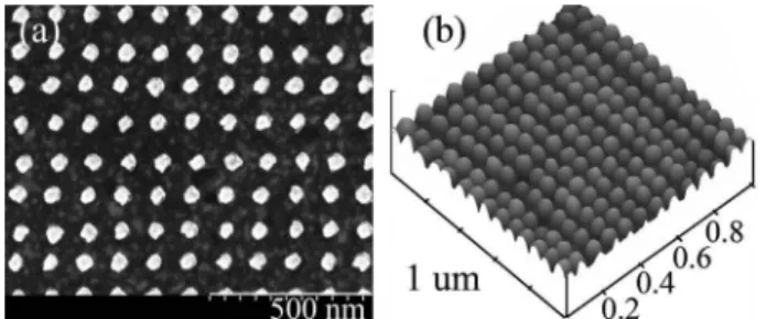
Following the simulation results, three substrates, each having four 400⫻ 400m arrays of differently sized nanostructures 30 nm in height with 80, 110, and 200 nm periods, are fabricated using electron beam lithography (EBL) on a 20 nm thick Au-coated SF-11 glass. Additionally, to investigate the effect of initial Au film thickness on the SPR response, two substrates with 30 nm high, 110 nm period nanoposts of various widths are fabricated on bare and 50 nm thick Au-coated SF-11 glass. Due to fabrication con-straints, the nanostructure shape is simply defined by the shape of the beam, while the exposure dose is adjusted from 10 to 45 fC at 160 pA and 30 kV to ob-tain structures of different sizes. Figure2(a)shows a sample scanning electron microscope (SEM) image of nanoposts of nominally circular shape with 110 nm period having a diameter of 50± 3.5 nm. Atomic force microscopy (AFM) characterization shows dome-shaped fabricated nanoposts [Fig. 2(b)], as expected
from anisotropic gold evaporation in the narrow PMMA trenches defining the nanostructures. The height and the period of the nanoposts were found to be of excellent uniformity with 3.73% and 1.21% variation, respectively.
Surface functionalization of substrates is done us-ing 1M thiol-modified 20 mer oligonucleotide probe sequence in 1 M KH2PO4for 120 min. Following im-mobilization, substrates are treated with 1 mM mer-captohexanol for 90 min to render the probes highly accessible to the target while preventing unspecific target binding to the gold surface [12].
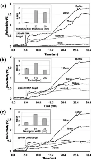
All DNA hybridization experiments are carried out using 20 mer oligonucleotide target sequence comple-mentary to the immobilized probe. Hybridization ki-netic curves are monitored using SPRi Lab+ appara-tus equipped with 800 nm LED source and a CCD camera (Genoptique, France). A baseline signal is ob-tained first for hybridization buffer (1 M NaCl in TE buffer), followed by hybridization signal for which 250 nM target is injected into the flow cell, allowing the target to bind to the immobilized probe for 20 min to yield sufficient refractive index change (high hy-bridization efficiency) while keeping the reaction time low. Finally, the substrate is washed with buffer, and the difference in the reflected intensity is com-puted by the difference between the initial and final buffer injections. The kinetic curves of the reaction on the nanostructured surface are shown in Fig.3for (a) different initial gold film thickness, (b) nanopost period, and (c) nanopost size. To enable qualitative assessment of the effect of nanostructured surface for SPR signal amplification, similar to the ASEF, the re-flectivity sensitivity enhancement factor RSEF = ⌬RNSPR/ ⌬RSPR (inset of Fig. 3) is included to de-scribe the ratio of the change in reflected intensity due to DNA hybridization on the nanostructured sur-face 共⌬RNSPR兲, to that of the control 共⌬RSPR兲. The
RSEF is important for real-time SPR sensing that re-lies on continuous monitoring of reflected light inten-sity at a specific angle, used in SPR imaging.
From the curves of Fig. 3(a), as the nanostructure size and period are kept constant at 50 and 110 nm respectively, the initial Au film thickness has little ef-fect on the SPR signal amplification. However, in the absence of underlying SPR active thin Au film, the SPRi apparatus is almost incapable of tracking the kinetic changes of the reaction occurring on the nano-structured biosensor surface. For the optimum initial 20 nm thick Au film, the kinetic response for nano-structures 50 nm in size having 80, 110 and 200 nm
Fig. 1. Numerically obtained ASEF for SPR substrate
with nanoposts of different size and periodicity.
Fig. 2. Sample images of fabricated Au nanoposts 50 nm in size, 110 nm period (a) SEM image; (b) AFM surface plot.
periods are shown in Fig.3(b). From the curves it can be seen that the nanoposts having 80 and 110 nm pe-riods exhibit higher reflectivity change compared with control than those with 200 nm period. The simulation predicted that smaller spacing between the neighboring structures allows for more pro-nounced coupling of localized and propagated surface plasmons in the presence of the dielectric layer re-sulting in enhanced electromagnetic fields. Although the periodicity plays the main role in the SPR signal amplification, the sensitivity enhancement can be further fine-tuned by nanostructure size. This is shown in Fig. 3(c), where nanoposts with 50 nm di-ameter and 110 nm period yield the highest signal amplification 共RSEF=5兲, while the SPR response for 30 nm structures was comparable with the control. Similar size-dependent SPR signal results have been obtained for 80 and 200 nm period structures (results
not shown). In particular, 50 nm wide nanoposts pro-vided the highest signal amplification for all exam-ined periods. However, it should be noted that for the same nanostructure size, the 110 nm period always provided superior performance. For instance, 75 nm size, the 110 nm period nanostructures amplified 2.5 ⫻ the signal, while the 200 nm period was inferior to the control. This is mostly due to weaker LSP–SPP coupling at larger nanopost spacing and more pro-nounced SPR curve broadening at larger nanostruc-ture periods. These findings suggest that the fabrica-tion process should be directed toward optimizing the period rather than the size of the nanostructures, given that the period is more easily controlled during EBL.
In this Letter, we have shown that a nanostructure-based SPR biosensor yields an en-hancement in the sensitivity of conventional SPR im-aging. We have demonstrated both numerically and experimentally that due to the increased surface binding area and the excitation and coupling of local-ized and bulk surface plasmons provided by the na-noposts, the SPR signal can be amplified up to five times in only 20 min, as was the case for 50 nm wide structures spaced 60 nm. This is significant sensitiv-ity improvement in comparison with a conventional SPRi biosensor, with potential for future applications in rapid, ultrasensitive DNA detection and DNA mi-croarray analysis.
The authors thank le Fonds québécois de la recher-che sur la nature et les technologies (FQRNT) for the scholarship, FQRNT-team grant, FQRNT-Centre for Biorecognition and Biosensors, and the National Research Council of Canada for their financial support.
References
1. C. D. Chen, S. F. Cheng, L. K. Chau, and C. R. C. Wang, Biosens. Bioelectron. 22, 926 (2007).
2. A. B. Dahlin, J. O. Tegenfeldt, and F. Hook, Anal. Chem. 78, 4416 (2006).
3. A. J. Haes and R. P. Van Duyne, Anal. Bioanal. Chem.
379, 920–930 (2004).
4. E. Hutter and J. H. Fendler, Adv. Mater. (Weinheim, Germany) 16, 1685 (2004).
5. L. He, M. D. Musick, S. R. Nicewarner, F. G. Salinas, S. J. Benkovic, M. J. Natan, and C. D. Keating, J. Am. Chem. Soc. 122, 9071 (2000).
6. S. J. Chen, F. C. Chien, G. Y. Lin, and K. C. Lee, Opt. Lett. 29, 1390 (2004).
7. K. M. Byun, S. J. Kim, and D. Kim, Opt. Express 13, 3737 (2005).
8. K. M. Byun, D. Kim, and S. J. Kim, Sens. Actuators B
117, 401 (2006).
9. D. Kim, J. Opt. Soc. Am. A 23, 2307 (2006).
10. S. Park, G. Lee, S. H. Song, C. H. Oh, and P. S. Kim, Opt. Lett. 28, 1870 (2003).
11. S. Elhadj, G. Singh, and R. F. Saraf, Langmuir 20, 5539 (2004).
12. A. W. Peterson, R. J. Heaton, and R. M. Georgiadis, Nucl. Acid Res. 29, 5163 (2001).
Fig. 3. Kinetic curves of 250 nM DNA target hybridization on nanostructured SPR substrate having nanoposts 30 nm in height with (a) 110 nm period, 50 nm width and different initial Au film thickness; (b) 50 nm width, underlying 20 nm thick Au film and different period; (c) 110 nm period, underlying 20 nm thick Au film and different size.