1
Discovery and Characterization of Prions
in Saccharomyces cerevisiae
by
Randal A. Halfmann
B.S. Genetics
Texas A&M University, 2004
Submitted to the Department of Biology in partial fulfillment of the requirements for the
degree of
DOCTOR OF PHILOSOPHY IN BIOLOGY
at the
MASSACHUSETTS INSTITUTE OF TECHNOLOGY
FEBRUARY 2011
© 2011 Randal A. Halfmann. All rights reserved.
The author hereby grants to MIT permission to reproduce and to distribute publicly paper
and electronic copies of this thesis document in whole or in part in any medium now known or
hereafter created
Signature of Author:_________________________________________________________________________________
Department of Biology
December 3, 2010
Certified by:___________________________________________________________________________________________
Susan L. Lindquist
Professor of Biology
Thesis Supervisor
Accepted by:__________________________________________________________________________________________
Stephen P. Bell
Professor of Biology
Co-Chair, Biology Graduate Committee
3
Discovery and Characterization of Prions
in Saccharomyces cerevisiae
by
Randal A. Halfmann
Submitted to the Department of Biology
in partial fulfillment of the requirements for the Degree of Doctor of Philosophy in Biology
ABSTRACTSome protein aggregates can perpetuate themselves in a self-templating protein-misfolding reaction. These aggregates, or prions, are the infectious agents behind diseases like Kuru and mad-cow disease. In yeast, however, prions act as epigenetic elements that confer heritable alternative phenotypes. Prion-forming proteins create bistable molecular systems whose semi-stochastic switching between functional states increases phenotypic diversity within cell populations. My thesis work explores the idea that rather than being detrimental, prions may commonly act to their host’s advantage.
To broaden the known range of prion phenomena in S. cerevisiae, I, together with a postdoctoral fellow in our lab, systematically surveyed the yeast proteome for prion-forming proteins. Using a combination of computational, cell biological, and biochemical approaches, we ultimately identified 18 novel prion domains capable of driving phenotypic switching, and an additional 6 domains that were highly positive for prion-like aggregation in other assays. These results establish the critical importance of intrinsic amyloid-forming tendencies for prion behavior by Q/N-rich proteins. We further confirmed that one of these proteins, the transcription factor Mot3, forms a novel prion in its endogenous context.
An analysis of these findings revealed a strong and unexpected amino acid bias in prionogenic proteins: prions were strongly enriched for asparagine (N), but not the chemically related amino acid glutamine (Q). We validated this finding using molecular simulations and experimental analyses of Q-to-N and N-to-Q variants of prion domains. N-rich sequences had an intrinsic tendency to both nucleate and propagate amyloid conformers. Q-rich proteins tended instead to make structurally constrained interactions leading to proteotoxic soluble and non-amyloid aggregated conformers.
The appendices include works in progress. Each explores a different aspect of prion biology. Appendix A confirms a theoretical prediction that prions, if functional, should preferentially
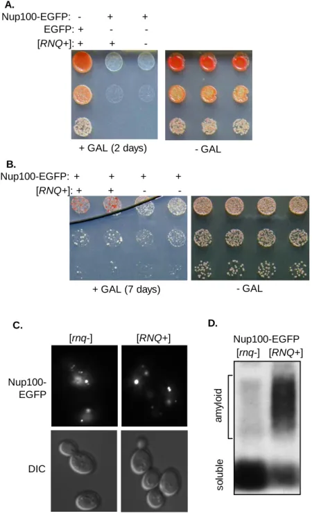
regulate certain rapidly evolving genes. I demonstrate with the newly discovered prion protein, Mot3, that prions accelerate the appearance of new phenotypes in important traits like mating behavior and cell-adhesion. I further identify naturally occurring prion states of Mot3 and other prion proteins in wild yeast isolates, and show that elimination of these prions has strong phenotypic effects in these strains. Appendix B, work done in collaboration with another lab, establishes that Nup100, a GLFG nucleoporin, is a prion. The conformational flexibility of GLFG nucleoporins is critical for the function of the nuclear pore complex, a molecular sieve that regulates all macromolecular transport between the nucleus and cytoplasm.
Thesis supervisor: Susan Lindquist Title: Professor of Biology
5
Acknowledgements
I will fondly remember my time here at MIT, and the people who helped me make the most of it. I am forever indebted to my advisor, Susan Lindquist. Quite simply, none of my work would have been possible without her guidance, enthusiasm and support. I hope that at least a fraction of the lessons I’ve learned with each trip to her office, whether to talk science or rewrite an abstract for the 42nd time, stay with me as I leave the lab. I will be a much better scientist for it.
I want to thank my thesis committee members, Thomas Schwartz and Jonathan King, for sticking with me from beginning to end. My other interactions with Jon, first as his student and later as his teaching assistant, added valuable perspective to my research-driven graduate education. Teaching, as he explained, ensures that the next generation can pick up where we leave off. Finally, I thank Leona Samson and Eugene Shakhnovich for serving on my defense committee.
The Lindquist lab has been a very supportive environment full of genuinely friendly and approachable people. Their extraordinary collective expertise, and willingness to share it with me, ensured that I never had to go far to find someone who could help.
I want to especially thank Brooke Bevis for doing her job so well. She kept everything, and every one, running smoothly. I could always count on her insightful psychoanalyses to make sense of the few times when things did not go smoothly. I thank my baymate Ben Vincent for his quick and entertaining wit. I thank Kent Matlack for saving me the hassle of having to use a real thesaurus, and for showing me from time to time how to find beauty in the most unexpected places. I thank Dubi Azubuine for a constant supply of media no matter how frequent or unusual my requests.
I am very fortunate to have had many talented collaborators both inside and outside the lab. Most importantly, I have to thank Simon Alberti. He has been a great friend and a valuable intellectual resource. I also thank my first postdoc mentor, Pete Tessier, for his limitless patience during my early and hectic days in the lab. Thanks to Oliver King, Charlie O’Donnell, and Alex Lancaster for doing insightful and magical things with computers. Similarly, thanks to Rohit Pappu and Nick Lyle for lending their expertise with molecular simulations. Finally, thanks to Jessica Wright and Michael Rexach for their contributions to the never-ending Nup100 saga.
I want to thank the people who gave me my start. First, my parents for constant encouragement and for providing a sounding board no matter how bored I know they must have been. The lessons they’ve taught me from an early age, like the universal value of hard work, will always serve me well. I also thank my undergraduate advisor, David Stelly, who took me under his wing to get me started in research.
Finally, I can’t imagine where I would be without Megan. Her constant love and companionship reminds me every day what is truly important.
7
Table of Contents
Abstract 3
Acknowledgements 5
Table of Contents 7
Chapter One: Prions, Protein Homeostasis, and Phenotypic Diversity 9 Chapter Two: A Systematic Survey Identifies Prions and Illuminates 29
Sequence Features of Prionogenic Proteins
Abstract 30 Introduction 30 Results 32 Discussion 40 Experimental Procedures 42 References 48 Figures 55
Chapter Three: Opposing Effects of Glutamine and Asparagine Dictate 83 Prion Formation by Intrinsically Disordered Proteins
Summary 84
Introduction 84
Results 86
Discussion 92
Experimental Procedures 95
Supplemental Results and Discussion 100
References 102
Figures 107
Chapter Four: Epigenetics in the Extreme: Prions and the Inheritance 125 of Environmentally Acquired Traits
Appendix A: Evolutionary Capacitance by Yeast Prions 135
Abstract 136
Introduction 136
Results 138
Discussion 142
Materials and Methods 144
References 145
Figures 147
Appendix B: Prion Formation by the GLFG Nucleoporin, Nup100 155
Abstract 156
Introduction 156
Results 158
Discussion 162
Materials and Methods 163
References 166
Figures 178
Appendix C: Specific Author Contributions 185
9
Chapter One
Prions, Protein Homeostasis, and Phenotypic
Diversity
This chapter was published previously: Halfmann, R., Alberti, S., and Lindquist, S. (2010). Trends in Cell Biology 20, 125-33
10
Introduction
Prions are self-replicating protein entities that underlie the spread of a mammalian neurodegenerative disease, variously known as Kuru, scrapie, and bovine spongiform encephalopathy, in humans, sheep and cows, respectively (Aguzzi et al., 2008). However, most prions have been discovered in lower organisms and in particular, the yeast Saccharomyces cerevisiae. Despite assertions that these prions, too, are diseases (Wickner et al., 2007a) (Box 1), many lines of evidence suggest that these mysterious elements are generally benign and, in fact, in some cases beneficial. In fungi, prions act as epigenetic elements that increase phenotypic diversity in a heritable way and can also increase survival in diverse environmental conditions (True and Lindquist, 2000; True et al., 2004; Shorter and Lindquist, 2005; Alberti et al., 2009). In higher organisms, prions may even be a mechanism to maintain long-term physiological states, as suggested for the Aplysia californica (sea slug) neuronal isoform of CPEB, cytoplasmic polyadenylation element binding protein. The prion form of this protein appears to be responsible for creating stable synapses in the brain (Si et al., 2003). CPEB is the prominent first example of what may be a large group of prion-like physiological switches, the potential scope of which cannot be given adequate coverage here. Instead, this piece will focus on prions as protein-based genetic elements – their ability to drive reversible switching in diverse phenotypes, and the way that such switching can promote the evolution of phenotypic novelty.
The self-templating replicative state of most biochemically characterized prions is amyloid (Glover et al., 1997; Alberti et al., 2009) (Figure 1), although other types of self-propagating protein conformations may also give rise to prion phenomena (Wickner et al., 2007b; Brown and Lindquist, 2009). Amyloid is a highly ordered, fibrillar protein aggregate with a unique set of biophysical characteristics that facilitate prion propagation: extreme stability, assembly by nucleated polymerization, and a high degree of templating specificity. Prion propagation proceeds from a single nucleating event that occurs within an otherwise stable intracellular population of non-prion conformers. The nucleus is then elongated into a fibrillar species by templating the conformational conversion of non-prion conformers (Serio et al., 2000; Tessier and Lindquist, 2009) (Figure 1). Finally, the growing protein fiber fragments into smaller propagating entities, which are ultimately disseminated to daughter cells (Shorter and Lindquist, 2005). Because the change in protein conformation causes a change in function, these self-perpetuating conformational changes create heritable phenotypes unique to the determinant protein and its genetic background (Figure 1). The genetic properties that arise are distinct from those of most nuclear-encoded mutations: prion
11 phenotypes are dominant in genetic crosses and exhibit non-Mendelian inheritance patterns. Hence prion-based genetic elements are denoted with capital letters and brackets – “[PRION]”.
Protein remodeling factors, chaperones, and other protein quality control mechanisms interact with prions at every step in their propagation. Further, prion-driven phenotypic switches are modulated by environmental conditions that perturb protein homeostasis (Tyedmers et al., 2008) – the proteome-wide balance of protein synthesis, folding, trafficking, and degradation processes (Balch et al., 2008). Prions could thereby constitute an intrinsic part of the biological response to stress. We postulate that the relationship between prions and protein homeostasis, as well as the dynamic nature of prion propagation, render prions into sophisticated evolutionary bet-hedging devices. Herein, we explore multiple intriguing features of prion biology that together argue for a general role for prions in adaptation to new environments, and thereby the evolution of new traits.
Prions as bet-hedging devices
Prions can allow simple organisms to switch spontaneously between distinct phenotypic states (True and Lindquist, 2000). For this reason, prions can be regarded as bet-hedging devices. Bet-hedging devices increase the reproductive fitness of organisms living in fluctuating environments by creating variant subpopulations with distinct phenotypic states (Seger and Brockmann, 1987) (Box 2).
The first prion protein proposed to increase survival in fluctuating environments is the translation termination factor Sup35, which forms a prion state called [PSI+] (True and Lindquist, 2000). This prion reduces Sup35 activity relative to the non-prion, or [psi-] state, thereby creating a
variety of phenotypes related to alterations in translation fidelity (Liebman and Sherman, 1979; Patino et al., 1996; True et al., 2004; Namy et al., 2008b). A surprisingly large fraction of the phenotypes (~25% in one study (Tyedmers et al., 2008)) are advantageous under particular growth conditions. While reduced translational fidelity cannot, in the long run, be advantageous for growth, in the short run changes in gene expression brought about by [PSI+] can allow cells to grow in the presence of antibiotics, metals and other toxic conditions, or with different carbon or nitrogen sources, depending on the genetic background. Because cells spontaneously gain the prion at an appreciable frequency (10-7 to 10-6) (Liu and Lindquist, 1999; Tank et al., 2007; Lancaster et
al., 2009), at any one time a sizable population of yeast cells will contain a few that have already switched states. If the environment is such that [PSI+] is beneficial, these cells would then have a greater chance to survive in that environment. Importantly, the prion state can be reversed by its
12 occasional loss during cell division (Cox et al., 1980) (with as yet undetermined frequencies), resulting in progenitors with the original [psi-] phenotype. If after a period of growth, the
environment changes to a state where [PSI+] is not advantageous, those few cells that have spontaneously lost the prion then have a survival advantage. From a gene-centric point of view, the net effect of this phenotype switching is that the common genotype shared by both [PSI+] and [psi-]
cells survives through the strenuous series of environmental transitions. Even if the rare switches to the [PSI+] state are commonly disadvantageous, [PSI+] could dramatically improve the long-term fitness of a genotype if it is advantageous on occasion. Related phenotypic switching phenomena, like the reversible appearance of antibiotic-resistant “persister” bacteria, appear to constitute environmentally-optimized risk-reduction strategies (Kussell et al., 2005) (Box 2).
Other than Sup35, the best characterized yeast prion is the Ure2 nitrogen catabolite repressor. Its prion state, [URE3], causes cells to constitutively utilize poor nitrogen sources (Shorter and Lindquist, 2005). This same phenotype, when conferred by URE2 loss-of-function mutants, has been shown to confer a proliferative advantage to cells in fermenting grape must (Shorter and Lindquist, 2005), strongly suggesting that this prion, too, may have a functional role in coping with yeast’s diverse ecological niches.
Until recently, the prion field has been confined to a small handful of proteins, and for this reason, conjectures about their potential roles in adaptation and evolution have been limited. However, a wave of recent discoveries in yeast has dramatically expanded the prion world as we know it (Table 1). The newly discovered prions include functionally diverse proteins: multiple chromatin remodeling and transcription factors (Du et al., 2008; Alberti et al., 2009; Patel et al., 2009b), a metacaspase (Nemecek et al., 2009), and a range of additional prionogenic proteins whose putative endogenous prion states are yet to be examined (Alberti et al., 2009). We suggest that the existence of these prions and the phenotypic heterogeneity they produce contributes to a general bet-hedging strategy that arms yeast populations against environmental fluctuations. Recent analyses of some of these novel prions lend support to this idea (Alberti et al., 2009; Patel et al., 2009b).
[MOT3+] is a prion formed by the transcription factor Mot3, an environmentally responsive regulator of yeast cell wall composition and pheromone signaling (Grishin et al., 1998; Abramova et al., 2001). In general, the cell surface of yeast determines the communication and interaction of yeast cells with the environment, yet it is also involved in a host of morphological and behavioral phenotypes, such as cell growth, cell division, mating, filamentation, and flocculation. Whether the phenotypic variation introduced by [MOT3+] affects all of these processes remains to be explored,
13 but [MOT3+] does confer increased resistance to certain cell wall stressors (Alberti et al., 2009). Therefore, the phenotypes produced by [MOT3+] should be advantageous in many microbial environments. The biological significance of Mot3 prion formation is supported by its high frequency of appearance – approximately 1 in 10,000 cells ((Alberti et al., 2009) and Halfmann and Lindquist, in preparation).
[SWI+] and [OCT+] are formed by the globally acting transcriptional regulators, Swi1 and Cyc8, respectively (Du et al., 2008; Patel et al., 2009b). [SWI+] cells are resistant to the microtubule disruptor, benomyl (Alberti et al., 2009); and [OCT+] induces flocculation (Patel et al., 2009b), a growth form that has been shown to protect cells from diverse stresses (Smukalla et al., 2008). Given the large size and complexity of the gene networks regulated by each of these prion transcription factors, it is likely that many more phenotypes are yet to be linked with prions.
Finally, for the well-characterized prions, it has been established that the presence of one protein in its prion state can influence the prion switching of other proteins. The [RNQ+] prion, for instance, strongly increases the rate of appearance of other prions (Shorter and Lindquist, 2005; Alberti et al., 2009). Conversely, some prions destabilize each other when both exist in the same cell (Bradley et al., 2002). Such prion cross-talk is influenced both by the sequence similarity between the proteins and the degree to which they share common components of the cellular prion-propagating machinery (Schwimmer and Masison, 2002; Mathur et al., 2009). The likely existence of over twenty interconnected prion switches (Alberti et al., 2009), all contributing to phenotypic heterogeneity, would greatly increase a genetic lineage’s potential to explore phenotypic space. Prions are being discovered at an increasingly rapid pace, suggesting that many exciting possibilities remain to be discovered en route to a deeper understanding of the prevalence and functionality of prions in biology.
Prions as evolutionary capacitors
In addition to “normal” bet-hedging, prions may have an even deeper and more sophisticated role in microbial evolution. Specifically, prions have been proposed to be capable of evolutionary capacitance (Shorter and Lindquist, 2005). An evolutionary capacitor is any entity that normally hides the effects of genetic polymorphisms, allowing for their storage in a silent form, and releases them in a sudden stepwise fashion (Masel and Siegal, 2009). The complex phenotypes produced by the sudden expression of accumulated genetic variation on occasion will prove beneficial to the organism. As the organism proliferates, further genetic and epigenetic variations will accumulate that stabilize the beneficial phenotype. The extent to which evolutionary capacitors
14 impact the evolution of natural populations is highly debated, and even more so the notion that capacitance itself can be subject to natural selection (Pigliucci, 2008).
However, the accumulated evidence that at least one prion protein, Sup35, acts in this manner is exceedingly difficult to dismiss. Sup35 can act as an evolutionary capacitor by connecting protein folding to the relationship between genotype and phenotype in a remarkable way. The reduced translation fidelity brought about by Sup35’s prion state, [PSI+], results in the translation of previously silent genetic information through a variety of mechanisms including stop-codon readthrough and ribosome frameshifting (Liebman and Sherman, 1979; True et al., 2004; Wilson et al., 2005; Namy et al., 2008a). Stop-codon readthrough can also affect genetic expression by changing mRNA stabilities. Untranslated regions and cryptic RNA transcripts experience relaxed selection under normal ([psi-]) conditions, and consequently, are free to accumulate genetic variation. Upon the appearance of [PSI+], these polymorphisms become phenotypically expressed. Because [PSI+] operates on genetic variation in a genome-wide fashion, it allows for the sudden acquisition of heritable traits that are genetically complex (True et al., 2004). Such traits are initially unlikely to become [PSI+]-independent because they involve multiple genetic loci and cells will revert to their normal phenotype when they lose the prion. But if the environment that favors the changes in gene expression brought about by [PSI+] occurs frequently or lasts for a very long time, as the population expands, mutations will accumulate that allow cells to maintain the traits even when they revert to normal translational fidelity through the spontaneous loss of [PSI+]. Arguing that Sup35 is under selective pressure to maintain the ability to reveal such variation, Sup35 homologs from other yeasts have conserved prion-forming capabilities, despite their sequences having diverged extensively over hundreds of millions of years (Chernoff et al., 2000; Kushnirov et al., 2000; Nakayashiki et al., 2001). Mathematical modeling confirms that the complexity of [PSI+ ]-revealed phenotypes can theoretically account for the evolution of its prion properties in yeast (Griswold and Masel, 2009). Finally, a phylogenetic analysis of the incorporation of 3’ untranslated regions (UTRs) into coding sequences provides compelling evidence for [PSI+]-mediated evolution in natural yeast populations. When comparing yeast and mammalian genomes, yeast displayed a strong bias for mutation events leading to in-frame, rather than out-of-frame incorporation of 3’ UTRs (Giacomelli et al., 2007). Thus, yeast 3’ UTRs are translated at a relatively high frequency, consistent with the occasional appearance of [PSI+] in natural populations.
Buffering of phenotypic variation is an inherent property of regulatory networks, such that the conditional reduction of network integrity may be a common mode of evolutionary capacitance (Masel and Siegal, 2009). The distinction between this type of capacitance and prions is that the
15 latter are necessarily epigenetic, and therefore provide a mechanism for the persistence, and ultimately, genetic assimilation, of the revealed phenotypes (Masel and Siegal, 2009). Prion-associated phenotypes can appear spontaneously and persist for multiple generations, whereas the revelation of variant phenotypes by other capacitors is generally contingent on stress, and consequently, relatively transient.
Is prion-driven evolutionary capacitance unique to Sup35, or might prion formation within any number of proteins also promote the expression of hidden genetic variation? Intriguingly, many of the newly identified prions are situated to function as genetic capacitors in their own right. Conspicuously overrepresented among these prionogenic proteins are gene products that control gene expression, cell signaling and the response to stimuli such as stress ((Alberti et al., 2009) and Table 1). Many of them represent highly connected nodes in the yeast genetic network. The Swi1 chromatin remodeler, for instance, regulates the expression of 6% of the yeast genome (Du et al., 2008). Likewise, Cyc8 represses 7% of the yeast gene complement (Green and Johnson, 2004). The prion candidates Pub1, Ptr69 and Puf2 are members of a family of RNA-binding proteins that regulate the stability of hundreds of mRNAs encoding functionally related proteins (Hogan et al., 2008). The strong enrichment of putative prions among proteins that regulate and transact genetic information suggests that prion-based switches evolve preferentially among proteins whose functions impinge on multiple downstream biological processes. Pre-existing genetic polymorphisms whose expression is altered by these prions would create different phenotypes in different genetic backgrounds. Thus, many prions are quite likely to create strong and complex phenotypes upon which natural selection can act.
Prion formation as an environmentally responsive adaptation
Many bet-hedging devices are environmentally responsive (Avery, 2006) (Box 2). That is, in addition to entirely stochastic switches, organisms may also make what, in effect, amounts to “educated guesses” by integrating environmental cues to modulate the frequency of phenotypic switching. Indeed, the frequency of prion switching is affected by environmental factors. The appearance of [PSI+] is strongly increased by diverse environmental stresses (Eaglestone et al., 1999; Tyedmers et al., 2008). Incidentally, this property is necessary and sufficient for [PSI+] formation to have been favored by natural selection for evolvability (Lancaster et al., 2009). Other well-characterized prions are also known to be induced by prolonged refrigeration and/or deep stationary phase (Chernoff, 2007). Because prions are a special type of protein misfolding process, logically their induction is intrinsically tied to environmental stresses that perturb protein stability.
16 Many if not most polypeptides have a generic capacity to form amyloid (Chiti and Dobson, 2006). Situations that alter native protein stability, like thermal stress, altered pH, or metal ion imbalances, are therefore likely to facilitate polypeptides’ access to prion or prion-like amyloid conformations (Chiti and Dobson, 2006) with the potential to perpetuate phenotypic changes even after the stress subsides.
The connection to environmental stresses is much deeper than that, however. Protein quality control machinery is ubiquitous throughout all kingdoms of life and is essential for both normal protein folding and for coping with stress. Components of the ubiquitin-proteasome system strongly impact prion formation (Chernoff, 2007). And prion propagation requires the actions of members of the Hsp40, Hsp70, and Hsp110 chaperone families as well as the AAA+ protein disaggregase Hsp104 (Chernoff, 2007; Sweeny and Shorter, 2008). Hsp104 is a member of ClpA/ClpB family of chaperones whose members are found throughout bacteria, fungi, plants and eukaryotic mitochondria. Hsp104 provides thermotolerance by resolubilizing stress-induced protein aggregates, and also has the unique ability to sever amyloid fibers into new prion propagons. This property has been conserved for hundreds of millions of years of fungal evolution (Zenthon et al., 2006). On the other hand, the Hsp104 protein of fission yeast appears incapable of propagating amyloid-based prions, despite maintaining its important ability to solubilize non-amyloid stress-induced protein aggregates (Senechal et al., 2009). We note that fission yeast also has a relative paucity of computationally predicted prions (Harrison and Gerstein, 2003), consistent with the suggestion that Hsp104’s amyloid shearing capability coevolved with prions to promote their propagation. Indeed, at least 25 of the 26 known amyloidogenic yeast prion domains require Hsp104 for their propagation as prions (Osherovich and Weissman, 2001; Alberti et al., 2009; Nemecek et al., 2009).
Perhaps the dominant force, then, for stress-induced prion formation involves perturbations in the interactions of prion proteins with chaperones and the cellular environment. The distribution of proteins between soluble and aggregated states is exquisitely sensitive to the status of the protein homeostasis network, which comprises protein synthesis, folding, sorting, and degradation machinery (Morimoto, 2008). Chaperones are highly connected in protein interaction networks and serve an important role as transducers of the stress response (Morimoto, 2008). Prion proteins, in turn, are highly connected to chaperones and thus to the protein homeostasis network at large. Prion conformational switching may therefore respond to stress indirectly through, for example, alterations in the abundance, availability, and connectivity of chaperones like Hsp104 and Hsp70s (Shorter and Lindquist, 2008). The induction of prions by diverse proteostatic
17 stresses, and their dependence on chaperones for propagation, may reflect the long history of chaperone involvement in the relationship between environment and phenotype.
Phenotypic diversity further enhanced by prion conformational and temporal diversity
The morphological adaptive radiation of organisms appears to result predominantly from genetic changes that have quantitative rather than qualitative effects (Stern and Orgogozo, 2009). In yeast and other microbes, social behaviors like mating, flocculation, and colony formation are subject to frequent stochastic changes in the expression of extracellular adhesins, leading to the rapid divergence of variant subpopulations (Verstrepen and Fink, 2009). These changes facilitate their expansion into diverse and highly dynamic ecological niches. The mechanisms for such changes are both genetic and epigenetic in nature (Masel and Siegal, 2009; Verstrepen and Fink, 2009), and include nucleotide repeat expansions and contractions, chromatin remodeling, and as recently discovered, prion formation (Patel et al., 2009b). Importantly, all of these mechanisms tend to modulate the activity levels, rather than the functional nature of, the affected gene products.
The ability of organisms to explore such modulations of gene activity, either as individuals (e.g. phenotypic plasticity), or as members of a genetic lineage (e.g. bet-hedging), enhances their survival under adverse conditions and is thought to facilitate the subsequent genetic assimilation of beneficial phenotypic variations (Masel and Siegal, 2009). Molecular mechanisms that allow for the rapid stabilization or amplification of initially non-genetic adaptive phenotypes within a lineage could greatly accelerate this process. Indeed, epigenetic processes are likely to play an important role in adaptive diversification (Bossdorf et al., 2008). As examined below, prions may represent an ideal epigenetic mechanism for the heritable modulation of gene activity.
Prions have a unique capacity to stratify protein functionality into multiple semi-stable levels, which greatly increases the phenotypic diversity created by prion-driven switches. It derives from the unusual and variable way in which prion conformers nucleate and propagate, and has both static and temporal components. For a given prion, multiple distinct yet related protein conformations can each self-perpetuate (Figure 2a). These prion “strains” differ in phenotypic strength and heritability. Strain multiplicity has been observed with both mammalian and yeast prions (Tessier and Lindquist, 2009), and is a common feature of diverse amyloids when polymerized in vitro (Pedersen and Otzen, 2008). The nature of the conformational differences between strains is still poorly understood, although progress has been made in elucidating how physical differences between amyloid strains – such as the extent of sequence involved with the fibril core of the amyloid – translate into differences in amyloid growth and division rates, and in
18 turn the phenotypic strength of the prion (Tessier and Lindquist, 2009). Importantly for the bet-hedging aspect of prion biology, the conformational plasticity of the prion nucleation process further increases the phenotypic “coding potential” of a single prion gene.
Several observations also demonstrate a temporal component to the strength and stability of prion phenotypes. For example, the mitotic stability of newly induced prion states increases with repeated cell passaging (Chernoff et al., 2000; Derkatch et al., 2000; Fernandez-Bellot et al., 2000; Santoso et al., 2000; Taneja et al., 2007). Additionally, selection for incipient prions using mild selective conditions creates a much larger population of strong prion states than would be expected from the numbers obtained by immediate stringent selection (Brachmann et al., 2005; Brachmann et al., 2006; Edskes et al., 2006; Tyedmers et al., 2008). Recent observations that even “non-prion” amyloids, such as polyglutamine-based aggregates, can become mitotically stable (Alexandrov et al., 2008), suggest that a capacity for the maturation of propagating states may be a generic feature of amyloid-like aggregates. The rate of prion maturation is strongly influenced by Hsp104 activity (Alexandrov et al., 2008), indicating an additional mode by which the protein homeostasis network connects the environment to epigenetic changes.
Multiple mechanisms for generating prion diversity temporally can be envisioned (Figure 2), including amyloid strain-like conformational transitions (Alexandrov et al., 2008), the mass-action population dynamics of prion particles, the variable association of prion particles with specific cellular structures, and the participation in early stages of prion propagation by an array of oligomeric species that have been increasingly observed en route to amyloid fibrillation (Serio et al., 2000; Kodali and Wetzel, 2007; Tessier and Lindquist, 2009). It is plausible that some pre-amyloid species have rudimentary self-propagating activities themselves.
Regardless of the mechanisms involved, what is clear is that incipient prion states represent dynamic molecular populations, a view that challenges the prevailing assumption that prions increase phenotypic heterogeneity solely by acting as simple binary switches. Prion nucleation allows for a single protein species to create a dynamic continuum of semi-stable phenotypes (Figure 2c) that do not require genetic, expression-level, or posttranslational regulatory changes to that protein. For each prion protein, natural selection could operate at any point in this continuum to favor prion-containing cells, resulting in their clonal expansion relative to other cells and acting to shift the distribution of phenotypes within the continuum. Stress-induced formation of prions followed by their iterative maturation offers a rapid route to tunable, advantageous phenotypes. Ultimately, the beneficial phenotypes conferred by prions can become hard-wired by the accumulation of genetic and further epigenetic modifications (True and Lindquist, 2000). In this
19 way, semi-stable phenotypic heterogeneity conferred by the diversity of prion conformations and maturation states would greatly improve the odds of organismal survival in unpredictable or fluctuating environments, and thereby facilitate subsequent adaptive genetic changes.
Concluding remarks
The ability of prions to create heritable phenotypic diversity that is inducible by stress, coupled with the conformational and temporal diversity of prion states, suggests a prominent role for prions in allowing microorganisms to survive in fluctuating environments. However, broader validation is needed, and many questions remain (Box 3). The field of prion biology is now poised to answer these questions, and in so doing, make important contributions to our understanding of evolutionary processes. In particular, we may more fully realize that organisms have specific mechanisms to enhance the evolution of phenotypic novelty.
References
Abramova, N., Sertil, O., Mehta, S., and Lowry, C.V. (2001). Reciprocal regulation of anaerobic and aerobic cell wall mannoprotein gene expression in Saccharomyces cerevisiae. Journal of
bacteriology 183, 2881-2887.
Aguzzi, A., Baumann, F., and Bremer, J. (2008). The prion's elusive reason for being. Annual review of neuroscience 31, 439-477.
Alberti, S., Halfmann, R., King, O., Kapila, A., and Lindquist, S. (2009). A systematic survey identifies prions and illuminates sequence features of prionogenic proteins. Cell 137, 146-158.
Alexandrov, I.M., Vishnevskaya, A.B., Ter-Avanesyan, M.D., and Kushnirov, V.V. (2008). Appearance and propagation of polyglutamine-based amyloids in yeast: tyrosine residues enable polymer fragmentation. J Biol Chem 283, 15185-15192.
Avery, S.V. (2006). Microbial cell individuality and the underlying sources of heterogeneity. Nature reviews 4, 577-587.
Balch, W.E., Morimoto, R.I., Dillin, A., and Kelly, J.W. (2008). Adapting proteostasis for disease intervention. Science 319, 916-919.
Bossdorf, O., Richards, C.L., and Pigliucci, M. (2008). Epigenetics for ecologists. Ecol Lett 11, 106-115.
Brachmann, A., Baxa, U., and Wickner, R.B. (2005). Prion generation in vitro: amyloid of Ure2p is infectious. Embo J 24, 3082-3092.
Brachmann, A., Toombs, J.A., and Ross, E.D. (2006). Reporter assay systems for [URE3] detection and analysis. Methods (San Diego, Calif 39, 35-42.
Bradley, M.E., Edskes, H.K., Hong, J.Y., Wickner, R.B., and Liebman, S.W. (2002). Interactions among prions and prion "strains" in yeast. Proc Natl Acad Sci U S A 99 Suppl 4, 16392-16399.
Brown, J.C., and Lindquist, S. (2009). A heritable switch in carbon source utilization driven by an unusual yeast prion. Genes Dev 23, 2320-2332.
Chernoff, Y.O. (2007). Stress and prions: lessons from the yeast model. FEBS Lett 581, 3695-3701. Chernoff, Y.O., Galkin, A.P., Lewitin, E., Chernova, T.A., Newnam, G.P., and Belenkiy, S.M. (2000). Evolutionary conservation of prion-forming abilities of the yeast Sup35 protein. Mol Microbiol 35, 865-876.
20 Chiti, F., and Dobson, C.M. (2006). Protein misfolding, functional amyloid, and human disease. Annu Rev Biochem 75, 333-366.
Cox, B.S., Tuite, M.F., and Mundy, C.J. (1980). Reversion from suppression to nonsuppression in SUQ5 [psi+] strains of yeast: the classificaion of mutations. Genetics 95, 589-609.
Derkatch, I.L., Bradley, M.E., Masse, S.V., Zadorsky, S.P., Polozkov, G.V., Inge-Vechtomov, S.G., and Liebman, S.W. (2000). Dependence and independence of [PSI(+)] and [PIN(+)]: a two-prion system in yeast? Embo J 19, 1942-1952.
Du, Z., Park, K.W., Yu, H., Fan, Q., and Li, L. (2008). Newly identified prion linked to the chromatin-remodeling factor Swi1 in Saccharomyces cerevisiae. Nat Genet 40, 460-465.
Eaglestone, S.S., Cox, B.S., and Tuite, M.F. (1999). Translation termination efficiency can be regulated in Saccharomyces cerevisiae by environmental stress through a prion-mediated mechanism. Embo J 18, 1974-1981.
Edskes, H.K., Naglieri, B.M., and Wickner, R.B. (2006). Nitrogen source and the retrograde signalling pathway affect detection, not generation, of the [URE3] prion. Yeast (Chichester, England) 23, 833-840.
Fernandez-Bellot, E., Guillemet, E., and Cullin, C. (2000). The yeast prion [URE3] can be greatly induced by a functional mutated URE2 allele. The EMBO journal 19, 3215-3222.
Giacomelli, M.G., Hancock, A.S., and Masel, J. (2007). The conversion of 3' UTRs into coding regions. Mol Biol Evol 24, 457-464.
Glover, J.R., Kowal, A.S., Schirmer, E.C., Patino, M.M., Liu, J.J., and Lindquist, S. (1997). Self-seeded fibers formed by Sup35, the protein determinant of [PSI+], a heritable prion-like factor of S. cerevisiae. Cell 89, 811-819.
Green, S.R., and Johnson, A.D. (2004). Promoter-dependent roles for the Srb10 cyclin-dependent kinase and the Hda1 deacetylase in Tup1-mediated repression in Saccharomyces cerevisiae. Molecular biology of the cell 15, 4191-4202.
Grishin, A.V., Rothenberg, M., Downs, M.A., and Blumer, K.J. (1998). Mot3, a Zn finger transcription factor that modulates gene expression and attenuates mating pheromone signaling in
Saccharomyces cerevisiae. Genetics 149, 879-892.
Griswold, C.K., and Masel, J. (2009). Complex adaptations can drive the evolution of the capacitor [PSI], even with realistic rates of yeast sex. PLoS Genet 5, e1000517.
Harrison, P.M., and Gerstein, M. (2003). A method to assess compositional bias in biological sequences and its application to prion-like glutamine/asparagine-rich domains in eukaryotic proteomes. Genome Biol 4, R40.
Hogan, D.J., Riordan, D.P., Gerber, A.P., Herschlag, D., and Brown, P.O. (2008). Diverse RNA-binding proteins interact with functionally related sets of RNAs, suggesting an extensive regulatory system. PLoS biology 6, e255.
Kodali, R., and Wetzel, R. (2007). Polymorphism in the intermediates and products of amyloid assembly. Curr Opin Struct Biol 17, 48-57.
Kushnirov, V.V., Kochneva-Pervukhova, N.V., Chechenova, M.B., Frolova, N.S., and Ter-Avanesyan, M.D. (2000). Prion properties of the Sup35 protein of yeast Pichia methanolica. The EMBO journal 19, 324-331.
Kussell, E., Kishony, R., Balaban, N.Q., and Leibler, S. (2005). Bacterial persistence: a model of survival in changing environments. Genetics 169, 1807-1814.
Lancaster, A.K., Bardill, J.P., True, H.L., and Masel, J. (2009). The Spontaneous Appearance Rate of the Yeast Prion [PSI+] and Its Implications For the Evolution of the Evolvability Properties of the [PSI+] System. Genetics, DOI: 10.1534/genetics.1109.110213 (http://www.genetics.org).
Liebman, S.W., and Sherman, F. (1979). Extrachromosomal psi+ determinant suppresses nonsense mutations in yeast. J Bacteriol 139, 1068-1071.
Liu, J.J., and Lindquist, S. (1999). Oligopeptide-repeat expansions modulate 'protein-only' inheritance in yeast. Nature 400, 573-576.
21 Masel, J., and Siegal, M.L. (2009). Robustness: mechanisms and consequences. Trends Genet 25, 395-403.
Mathur, V., Hong, J.Y., and Liebman, S.W. (2009). Ssa1 overexpression and [PIN(+)] variants cure [PSI(+)] by dilution of aggregates. Journal of molecular biology 390, 155-167.
Morimoto, R.I. (2008). Proteotoxic stress and inducible chaperone networks in neurodegenerative disease and aging. Genes & development 22, 1427-1438.
Nakayashiki, T., Ebihara, K., Bannai, H., and Nakamura, Y. (2001). Yeast [PSI+] "prions" that are crosstransmissible and susceptible beyond a species barrier through a quasi-prion state. Molecular cell 7, 1121-1130.
Namy, O., Galopier, A., Martini, C., Matsufuji, S., Fabret, C., and Rousset, J.P. (2008a). Epigenetic control of polyamines by the prion [PSI(+)]. Nat Cell Biol.
Namy, O., Galopier, A., Martini, C., Matsufuji, S., Fabret, C., and Rousset, J.P. (2008b). Epigenetic control of polyamines by the prion [PSI+]. Nat Cell Biol 10, 1069-1075.
Nemecek, J., Nakayashiki, T., and Wickner, R.B. (2009). A prion of yeast metacaspase homolog (Mca1p) detected by a genetic screen. Proceedings of the National Academy of Sciences of the United States of America 106, 1892-1896.
Osherovich, L.Z., and Weissman, J.S. (2001). Multiple Gln/Asn-rich prion domains confer susceptibility to induction of the yeast [PSI(+)] prion. Cell 106, 183-194.
Patel, B.K., Gavin-Smyth, J., and Liebman, S.W. (2009). The yeast global transcriptional co-repressor protein Cyc8 can propagate as a prion. Nature cell biology 11, 344-349.
Patino, M.M., Liu, J.J., Glover, J.R., and Lindquist, S. (1996). Support for the prion hypothesis for inheritance of a phenotypic trait in yeast. Science 273, 622-626.
Pedersen, J.S., and Otzen, D.E. (2008). Amyloid-a state in many guises: survival of the fittest fibril fold. Protein Sci 17, 2-10.
Pigliucci, M. (2008). Is evolvability evolvable? Nat Rev Genet 9, 75-82.
Santoso, A., Chien, P., Osherovich, L.Z., and Weissman, J.S. (2000). Molecular basis of a yeast prion species barrier. Cell 100, 277-288.
Schwimmer, C., and Masison, D.C. (2002). Antagonistic interactions between yeast [PSI(+)] and [URE3] prions and curing of [URE3] by Hsp70 protein chaperone Ssa1p but not by Ssa2p. Molecular and cellular biology 22, 3590-3598.
Seger, J., and Brockmann, H.J. (1987). What is bet-hedging? Oxford Surv Evol Biol 4, 192-211. Senechal, P., Arseneault, G., Leroux, A., Lindquist, S., and Rokeach, L.A. (2009). The
Schizosaccharomyces pombe Hsp104 disaggregase is unable to propagate the [PSI] prion. PLoS One 4, e6939.
Serio, T.R., Cashikar, A.G., Kowal, A.S., Sawicki, G.J., Moslehi, J.J., Serpell, L., Arnsdorf, M.F., and Lindquist, S.L. (2000). Nucleated conformational conversion and the replication of conformational information by a prion determinant. Science (New York, NY 289, 1317-1321.
Shorter, J., and Lindquist, S. (2005). Prions as adaptive conduits of memory and inheritance. Nat Rev Genet 6, 435-450.
Shorter, J., and Lindquist, S. (2008). Hsp104, Hsp70 and Hsp40 interplay regulates formation, growth and elimination of Sup35 prions. The EMBO journal 27, 2712-2724.
Si, K., Lindquist, S., and Kandel, E.R. (2003). A neuronal isoform of the aplysia CPEB has prion-like properties. Cell 115, 879-891.
Smukalla, S., Caldara, M., Pochet, N., Beauvais, A., Guadagnini, S., Yan, C., Vinces, M.D., Jansen, A., Prevost, M.C., Latge, J.P., et al. (2008). FLO1 is a variable green beard gene that drives biofilm-like cooperation in budding yeast. Cell 135, 726-737.
Stern, D.L., and Orgogozo, V. (2009). Is genetic evolution predictable? Science 323, 746-751. Sweeny, E.A., and Shorter, J. (2008). Prion proteostasis: Hsp104 meets its supporting cast. Prion 2, 135-140.
22 Taneja, V., Maddelein, M.L., Talarek, N., Saupe, S.J., and Liebman, S.W. (2007). A non-Q/N-rich prion domain of a foreign prion, [Het-s], can propagate as a prion in yeast. Molecular cell 27, 67-77. Tank, E.M., Harris, D.A., Desai, A.A., and True, H.L. (2007). Prion protein repeat expansion results in increased aggregation and reveals phenotypic variability. Molecular and cellular biology 27, 5445-5455.
Tessier, P.M., and Lindquist, S. (2009). Unraveling infectious structures, strain variants and species barriers for the yeast prion [PSI+]. Nat Struct Mol Biol 16, 598-605.
True, H.L., Berlin, I., and Lindquist, S.L. (2004). Epigenetic regulation of translation reveals hidden genetic variation to produce complex traits. Nature 431, 184-187.
True, H.L., and Lindquist, S.L. (2000). A yeast prion provides a mechanism for genetic variation and phenotypic diversity. Nature 407, 477-483.
Tyedmers, J., Madariaga, M.L., and Lindquist, S. (2008). Prion switching in response to environmental stress. PLoS biology 6, e294.
Verstrepen, K.J., and Fink, G.R. (2009). Genetic and epigenetic mechanisms underlying cell-surface variability in protozoa and fungi. Annu Rev Genet 43, 1-24.
Wickner, R.B., Edskes, H.K., Shewmaker, F., and Nakayashiki, T. (2007a). Prions of fungi: inherited structures and biological roles. Nature reviews 5, 611-618.
Wickner, R.B., Edskes, H.K., Shewmaker, F., Nakayashiki, T., Engel, A., McCann, L., and Kryndushkin, D. (2007b). Yeast prions: evolution of the prion concept. Prion 1, 94-100.
Wilson, M.A., Meaux, S., Parker, R., and van Hoof, A. (2005). Genetic interactions between [PSI+] and nonstop mRNA decay affect phenotypic variation. Proc Natl Acad Sci U S A 102, 10244-10249. Zenthon, J.F., Ness, F., Cox, B., and Tuite, M.F. (2006). The [PSI+] prion of Saccharomyces cerevisiae can be propagated by an Hsp104 orthologue from Candida albicans. Eukaryot Cell 5, 217-225.
23 Figure 1: Prions as self-templating aggregates
(a) Prions of S. cerevisiae cause heritable changes in phenotype. In this particular genetic background, the prion [PSI+] can be observed by white coloration and adenine prototrophy due to translational
readthrough of a nonsense mutation in the ADE1 gene. However, the cryptic genetic variation that can be revealed by [PSI+] is inherently polymorphic resulting in a wide variety of strain-specific [PSI+]
phenotypes (True et al., 2004).
(b) Prion phenotypes are generally caused by a reduction of the prion protein’s normal cellular activity.
In vivo, the aggregation and partial loss-of-function of the prion protein , can be observed by the
presence of Sup35-GFP foci in [PSI+] cells. These foci are composed of self-templating prion
aggregates that are cytoplasmically transmitted during cell division.
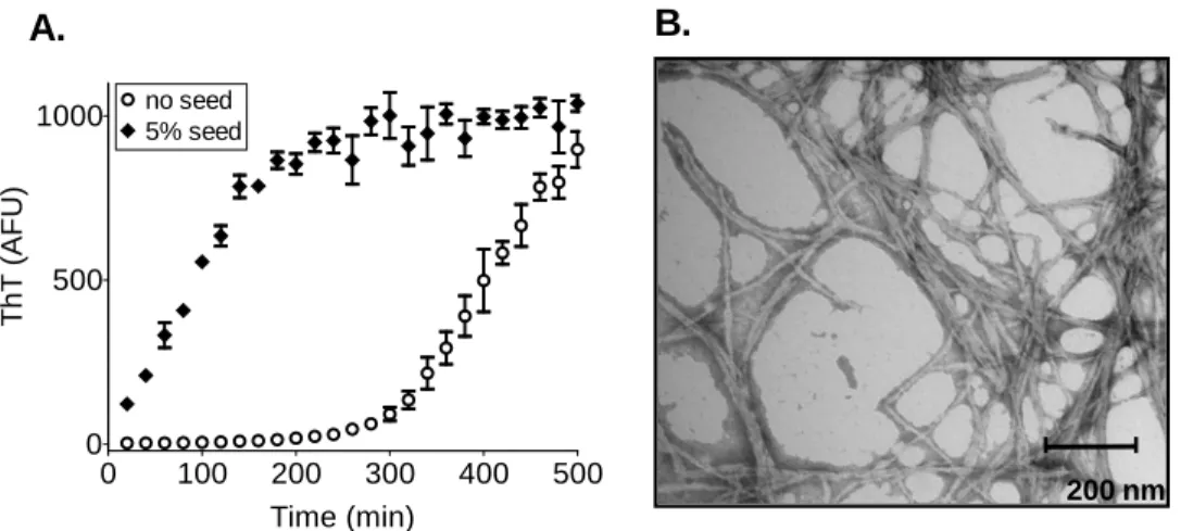
(c) Nucleated aggregation of a prion protein. Purified prion protein populates a soluble state for an extended period of time, then polymerizes exponentially after the appearance of amyloid nuclei (blue trace). The lag phase can be eliminated by the addition of small quantities of preformed aggregates (red trace), demonstrating the biochemical property underlying the self-propagating prion state (Serio et al., 2000).
(d) The self-propagating prion conformation is amyloid-like, as seen by the highly ordered, fibrillar appearance of prion domain aggregates visualized by transmission electron microscopy. Amyloid is a one-dimensional protein polymer. Its free ends template a protein folding reaction that incorporate new subunits while regenerating the active template with each addition.
25 Figure 2: Conformational and temporal diversity of prion states
(a) Prions create multiple stable phenotypic states, or “strains”. [PSI+] strains differ by their levels of
nonsense suppression, with stronger strains having less functional Sup35 available to fulfill its role in translation termination, giving rise to a whiter coloration in a particular genetic background (top). At the molecular level, strains are determined by amyloid conformational variants (bottom) that arise during nucleation but then stably propagate themselves.
(b) Along with the conformational diversity apparent in the end products of amyloid formation, multiple conformational variants are also transiently populated during the early stages of amyloid assembly, and may constitute integral on-pathway species (Chiti and Dobson, 2006). These oligomeric intermediates likely have limited self-templating capacity, but nevertheless may contribute to the weak phenotypes associated with incipient prion states.
(c) Incipient prion states acquire progressively stronger phenotypes and stabilities, possibly via mass-action population dynamics of prion particles. A number of elegant studies have correlated the phenotypic strength of the prion state with the intracellular number of prion particles (Cox et al., 2003; Tanaka et al., 2006). Upon de novo nucleation within a prion-free cell, prion polymerization onto limiting fiber ends proceeds during the “maturation” phase under pre-steady state conditions. Upon each cell division, prion particles are distributed passively and asymmetrically to daughter cells (Cox et al., 1980). Progeny that inherit more particles will have faster total prion polymerization rates and correspondingly stronger phenotypes, and will tend to accumulate more prion particles that will in turn strengthen the prion phenotype in subsequent generations (light pink and white cells). Conversely, cells that inherit fewer particles will have slower polymerization rates and weaker phenotypes (red and pink prion-containing cells), and themselves will tend to accumulate fewer particles to pass on to their progeny. Such noise in prion distribution may allow prions to stratify protein functionality along a continuum of semi-stable phenotypes (e.g. red cells, pink cells, and white cells) within a small number of cell generations.
27
29
Chapter Two
A Systematic Survey Identifies Prions and
Illuminates Sequence Features of Prionogenic
Proteins
This chapter was published previously: Alberti, S.*, Halfmann, R.*, King, O., Kapila, A., and Lindquist, S. (2009). Cell 137(1), 136-158
30
ABSTRACT
The prion hypothesis posits that biological information can be replicated solely through self-propagating conformations of proteins. Though it was initially conceived to explain baffling neurodegenerative diseases in mammals (Griffith, 1967; Prusiner, 1982), it has since grown to encompass a number of non-Mendelian traits in fungi (Ross et al., 2005b; Shorter and Lindquist, 2005; Shkundina and Ter-Avanesyan, 2007). All known prions, except for the initially discovered disease-causing prion PrP, are benign, and in some cases can confer selectable advantages (Saupe et al., 2000; True and Lindquist, 2000; True et al., 2004). The self-templating property of prions makes them both conformationally and epigenetically dominant, and positions prion-forming proteins as metastable cellular switches of protein function.
INTRODUCTION
The realization that protein conformational switches could provide a means for inheritance of phenotypes dates back 15 years (Wickner, 1994), yet only a few proteins with this capacity have been discovered (Shorter and Lindquist, 2005; Du et al., 2008). Most of these have been found in the yeast S. cerevisiae, with the [PSI+] element being the best understood.
[PSI+] is caused by an amyloid-like aggregated state of the translation-termination factor Sup35p. In the prion state, the majority of Sup35p molecules are inactive, resulting in increased levels of nonsense suppression (Liebman and Sherman, 1979; Patino et al., 1996) and programmed frameshifting (Namy et al., 2008a). This gives rise to RNA stability changes and functionally altered polypeptides and consequently to phenotypes that can be advantageous under diverse conditions (Eaglestone et al., 1999; True et al., 2004). Remarkably, the ability of Sup35p to switch into a prion conformation, and the regulation of that switch by the protein remodeling factor Hsp104p, have been conserved for over 800 million years of fungal evolution (Chernoff et al., 2000; Zenthon et al., 2006).
Three other amyloid-based prions, formed by the functionally diverse proteins Ure2p, Rnq1p, and Swi1p, have been described in S. cerevisiae. Ure2p regulates nitrogen catabolism; its prion state, [URE3], attenuates this activity resulting in the constitutive utilization of poor nitrogen sources (Aigle and Lacroute, 1975; Wickner, 1994). The Rnq1p protein in its prion state, [RNQ+] (also called [PIN+]), enhances the inducibility of other prions (Derkatch et al., 2000; Bradley et al., 2002). [SWI+], the most recently discovered prion, is caused by an inactive state of the chromatin remodeling factor Swi1p (Du et al., 2008). Intriguingly, [SWI+] represents the first established link between chromatin-based and prion-based epigenetics, although a biological relevance of this
31 connection remains to be elucidated. Indeed, for all of these prion proteins, the putative functionality of their prion forms is highly debated (Nakayashiki et al., 2005).
The conformational duality of amyloid-based prions resides in structurally independent prion-forming domains (PrDs) (Edskes et al., 1999; Li and Lindquist, 2000; Santoso et al., 2000; Sondheimer and Lindquist, 2000). These PrDs are modular and can be transferred to other proteins to create novel prions (Li and Lindquist, 2000). They have a very unusual amino acid composition: enriched for polar residues such as glutamine (Q) and asparagine (N) and depleted of hydrophobic and charged residues. This composition promotes a disordered molten-globule-like conformational ensemble, within which amyloid-nucleating contacts can be made (Serio et al., 2000; Wang et al., 2006; Mukhopadhyay et al., 2007).
The ability of prions to propagate is afforded by the inherent and extremely efficient self-templating capacity of amyloid, a highly ordered β-sheet-rich protein aggregate. The amyloid-like prion state nucleates in cells at a low frequency de novo, but once formed, efficiently propagates this change to soluble conformers (Patino et al., 1996; Glover et al., 1997). Prion domains also form self-propagating amyloid in vitro under physiological conditions (Glover et al., 1997; Taylor et al., 1999), and remarkably, the resulting fibers alone can transform cells to the corresponding prion state (Sparrer et al., 2000; Maddelein et al., 2002; Brachmann et al., 2005; Patel and Liebman, 2007), firmly establishing the protein-only nature of prion inheritance.
However, the relationship between amyloid polymerization and prion propagation is still poorly understood. In fact, most known amyloid-forming proteins are not prions, and even amyloids of prion proteins are not always transmissible (Diaz-Avalos et al., 2005; Salnikova et al., 2005; Baskakov and Breydo, 2007; Sabate et al., 2007). While specific sequence elements ultimately determine the intrinsic amyloidogenic properties of polypeptides (Liu and Lindquist, 1999; Lopez de la Paz and Serrano, 2004; Alexandrov et al., 2008), there are multiple trans-acting factors within the cell, including molecular chaperones, the cytoskeletal machinery, and nucleating factors such as [PIN+], that interact with amyloid prions at every stage of propagation (Chernoff, 2007; Perrett and Jones, 2008). These observations, and our lack of knowledge of the pervasiveness of amyloid-based biological phenomena, create a need to elucidate the amyloid-prion relationship on a comprehensive, genome-wide level.
Such a genome-wide analysis could also reveal new prion-based phenotypes, which would support the idea that prion-mediated phenotypic variation is functionally significant. Prion-like Q/N-rich proteins are abundant in the proteomes of lower eukaryotes, with 100 to 170 such sequences in S. cerevisiae (Michelitsch and Weissman, 2000; Harrison and Gerstein, 2003).
32 However, the experimental tools needed to determine the prion properties of these proteins in a systematic manner have been lacking. Consequently, other than the four aforementioned prions, only one additional yeast protein, New1p, has been shown to harbor a domain capable of forming a prion, albeit in an artificial context (Osherovich and Weissman, 2001).
We bioinformatically scanned the yeast genome for proteins with prion-like character. We then subjected the highest-scoring candidates to genetic, cell biological, and biochemical assays to discern their prion-forming capacity, ultimately determining that at least 24 yeast proteins contain a prion-forming domain. We further evaluated one of these, Mot3p, confirming that it is a bona fide prion with a phenotype that is likely to be advantageous under certain environmental conditions.
RESULTS
A bioinformatics screen reveals multiple prion candidates in yeast
We developed a hidden Markov Model (HMM)-based approach for predicting prions, using the experimentally determined prion domains (PrDs) of Sup35p, Ure2p and Rnq1p, and the prion candidate New1p as positive training examples (at the time, Swi1p had not yet been shown to be a prion). We did not incorporate the other known fungal prion protein, HET-s, nor the mammalian prion protein, PrP, into our model because these proteins have unique sequences that are dissimilar in amino acid composition from the other prion proteins and thus would decrease the predictive power of the model. We acknowledge that our approach is thus necessarily biased towards a particular class of prions, but is nevertheless merited by the large number of Q/N-rich yeast proteins with unknown prion potential. All yeast protein sequences were parsed into prion-like regions and non-prion (background) regions. Proteins with prion-like regions at least 60 amino acids long (denoted “cores”) were considered to be prion candidates, based on the lower size limit of previously characterized yeast prion domains (Masison and Wickner, 1995; King and Diaz-Avalos, 2004). These proteins were then ranked by their core scores. Figure 1A shows an example of the output format of our prediction for the PrD of Sup35p.
Our query revealed ~ 200 proteins that have candidate PrDs (cPrDs) in the S. cerevisiae genome (see Table S1 for the complete set of predictions). The list of candidate prions includes Sup35p and Rnq1p as well as the prion candidate New1p in the group of the top 20 candidates. Although the recently discovered Swi1p prion (Du et al., 2008) was not used for training of the algorithm, it ranks at position 21, indicating that our prediction is a valuable tool to uncover new
33 prions. To evaluate these candidates, we tested the amyloid and prion-forming properties of the 100 highest-scoring cPrDs (Figure 1).
Many cPrDs form foci in the cytosol
As a first step to experimentally characterize the cPrDs, we investigated their propensity to form foci in living yeast cells by transiently expressing them as chimeras with yellow fluorescent protein (cPrD-EYFP). Several studies have described two morphologically distinct forms of protein aggregation in the yeast cytosol (Derkatch et al., 2001; Ganusova et al., 2006; Taneja et al., 2007). One of these structures has a ring- or ribbon-like appearance and is localized around the vacuole and/or adjacent to the plasma membrane, whereas the other is more punctate and preferentially resides close to the vacuole. We performed pilot experiments with known prions and amyloidogenic proteins to gain a better understanding of the aggregation structures that can be visualized by fluorescence microscopy. We transiently expressed these proteins as EYFP fusions using a galactose-regulatable promoter (Figure 2A). The PrDs of Sup35p and Ure2p proceeded through a characteristic maturation pathway that included an early stage with ribbon-like aggregation patterns and a later stage with puncta (Tyedmers and Lindquist, manuscript in preparation; Derkatch et al., 2001; Ganusova et al., 2006). In contrast, the PrD of Rnq1p and an expanded Q-rich region of the huntingtin protein almost exclusively formed puncta, when expressed from the same promoter.
We transiently expressed the cPrD-EYFP fusions in yeast cells using a galactose-regulatable plasmid. Despite the intrinsically unfolded nature of the cPrDs, the expression levels of most of the fusions were robust, with a few outliers that expressed at only low levels (Figure S1). A large fraction (69%) of the cPrD-EYFP fusions formed fluorescent foci (Figure 2B and S2). The proteins produced a surprising diversity of patterns with a large variety of ribbon-like structures and punctate foci. Overall, punctate patterns were much more abundant than ribbon structures and ranged from one large focus to multiple small foci distributed all over the cytoplasm. Since foci formation is also a feature of many non-prion proteins, we conducted additional experiments to assess the biochemical properties of the cPrDs.
Multiple cPrDs form highly stable aggregates
Protein aggregates can differ substantially in terms of detergent stability and can adopt either highly ordered (amyloid fibril) or disordered (amorphous) superstructures. To determine whether the cPrDs formed aggregates, and if so, whether they formed prion-like structures, we
34 used semi-denaturing detergent-agarose gel electrophoresis (SDD-AGE). SDD-AGE allows for the resolution of a wide size-range of SDS-resistant (amyloid-like) aggregates, ranging from oligomeric species to polymers assembled from hundreds of individual polypeptides (Bagriantsev et al., 2006; Halfmann and Lindquist, 2008).
Lysates from cells expressing cPrD-EYFP chimeras were analyzed by SDD-AGE after 24 hours of expression (Figure 2C). Remarkably, about one third of the cPrD-EYFP fusions had the SDS-resistance properties of known amyloidogenic proteins (the N and NM domains of Sup35p and the 72Q and 103Q fragments of huntingtin), including all four experimentally verified prions and the prion candidate New1p.
Since amyloid formation is time- and concentration-dependent, we increased the time of transient expression to 48 hours. Despite only modest increases in the cellular levels of the cPrDs (~ 2-3 fold), additional proteins became SDS-resistant, now including almost half the set of investigated cPrDs, with an enrichment for those that scored highly in our algorithm. The ratio of aggregated to soluble protein differed in a time-dependent manner for each candidate and was strongly reproducible. Some cPrD-EYFP fusions were completely aggregated after 24 hours, whereas others initially exhibited a small fraction of aggregated protein that increased after 48 hours of expression (see Table S2 for a classification). In addition, we observed substantial variation in the range of particle sizes, and again these were reproducible for individual cPrDs.
Multiple cPrDs form amyloid in vitro
Purified PrDs of known prions form amyloid in vitro. To assess amyloid propensities of our candidate prion domains in the absence of cellular factors, we purified bacterially expressed cPrDs under denaturing conditions, and then analyzed amyloid formation following dilution into a physiological buffer containing thioflavin-T (ThT). ThT is a dye that does not interfere with amyloid assembly, but changes its fluorescence properties upon amyloid binding (LeVine, 1993, 1997).
Overall, the cPrDs displayed tremendous diversity in amyloid propensities (Figure 3). Many, including the four known prions, largely completed amyloid formation within 12 hours. Others did not acquire ThT fluorescence until well over 24 hours after dilution from denaturant (e.g. Pan1p and Yap1801p). Most positive samples initially had very little fluorescence, consistent with nucleated polymerization that is a hallmark of amyloid assembly. Finally, roughly half of the proteins were unable to nucleate within the time-frame examined.
PrDs can follow both amyloid and non-amyloid aggregation pathways (Liu et al., 2002; Vitrenko et al., 2007; Douglas et al., 2008), and some non-amyloid β-sheet structures can alter ThT
35 fluorescence (LeVine, 1993). As an additional characterization, we therefore assayed the SDS-resistance of samples after 72 hours using a filter retardation assay (Scherzinger et al., 1999). In this assay, a non-binding membrane is used to detect protein aggregates, which, due to their size, are specifically retained on the membrane surface. Treating the impeded aggregates with SDS then distinguishes amyloid from non-amyloid, since non-amyloid aggregates become solubilized and flow through.
We found remarkable agreement between the ThT and filter retardation amyloid assays (Figure 3). Acquisition of moderate to strong ThT fluorescence (>100 AFU) coincided with SDS-stable aggregation in every case. Several cPrDs (e.g. those of Snf5p and Nab3p) formed aggregates that were retained by the filter in non-denaturing conditions (0.1% Tween 20) but were eliminated by SDS. Interestingly, this type of aggregate was only observed when ThT fluorescence was absent or greatly delayed, and only with proteins that were more enriched for glutamines than asparagines. In total, the amyloid propensities of isolated cPrDs were in good agreement with the aggregation of these cPrDs in vivo (see Figure S3 and Table S2 for a comparison).
A Sup35p-based prion assay identifies phenotypic switching behavior
To determine whether the cPrDs can confer a heritable switch in the function of the protein to which they are attached, we employed an assay based on the well-characterized prion phenotypes of the translation termination factor Sup35p. The Sup35p protein consists of an N-terminal PrD (N), a highly charged middle domain (M) and C-N-terminal domain (C), which provides the translation termination function. The PrDs of Sup35p and other prions are modular and can be transferred to non-prion proteins, thereby creating new protein-based elements of inheritance (Li and Lindquist, 2000). This property allowed us to generate cPrD-SUP35C chimeras (under the control of the constitutive ADH1 promoter) that could be tested for their ability to generate [PSI+]-like states, as previously reported for Rnq1p and New1p (Sondheimer and Lindquist, 2000; Osherovich and Weissman, 2001).
Because Sup35p is an essential protein, we first generated a strain in which a deletion of the chromosomal SUP35 was covered by a Sup35p-expressing plasmid (Figure S4). When these cells were transformed with cPrD-SUP35C expression plasmids, a URA3 marker on the covering SUP35 plasmid allowed it to be selected against in 5-FOA-containing medium (plasmid shuffle). The resulting strains contained cPrD-Sup35C fusions as their only source of functional Sup35p. Each was tested for the ability to form a heritable [PSI+] state, using an ADE1 allele with a premature stop codon. Read-through of this allele in [PSI+] cells creates two easily monitored phenotypes, the


![Figure 6- Nup100 forms a cytoprotective prion state. (A) A spontaneous suppressor of Nup100-EGFP toxicity ([ NUP100+ ]) was passaged 6 times on glucose media with or without GdnHCl, and then retested for toxicity by replating to galactose (+GAL) or gl](https://thumb-eu.123doks.com/thumbv2/123doknet/14747883.578870/184.918.215.694.131.391/cytoprotective-spontaneous-suppressor-toxicity-passaged-retested-replating-galactose.webp)
