DNA-damage-mediated remodeling of normal and
tumor microenvironments modulates cell survival
By
Luke A. Gilbert
B.S. Microbiology, Immunology and Molecular Genetics University of California Los Angeles, 2006
SUBMITTED TO THE DEPARTMENT OF BIOLOGY IN PARTIAL FULFILLMENT OF THE REQUIREMENTS FOR THE DEGREE OF:
DOCTOR OF PHILOSOPHY IN BIOLOGY
AT THE
MASSACHUSETTS INSTITUTE OF TECHNOLOGY
June 2012
ARCHIVES
f ACSjFTS INSTITUT-rE OF ThF0,-#';OL0C'y r 1RAYR ES
A I ES@ 2012 Massachusetts Institute of Technology. All Rights Reserved.
Signature of Authoi April 24, 2012 Certified by: Dr. Michael Hemann Thesis Advisor
If0-Accepted by: Dr. Robert Sauer Chairperson, Graduate Committee
Table of contents:
Chapter 1: Introduction
5-34
Chapter 2: DNA damage mediated induction of a chemoresistant niche
35 -82
Chapter 3: DNA damage induces distinct acute and senescence associated secretory phenotypes in a context specific manner
83 - 104
Chapter 4: BCL-2 family genetic profiling reveals microenvironment-specific determinants of chemotherapeutic response
105 - 132
Chapter 5: Developmentally specified lymphomagenesis
roles for paracrine IL-6 in
133 - 164
Chapter 6: Discussion
165 - 175
References:
DNA-damage-mediated remodeling of normal and tumor
microenvironments modulates cell survival
By Luke A. Gilbert
Submitted to the Department of Biology on May 11, 2012 in Partial Fulfillment of the requirements for the degree of Doctor of Philosophy in Biology
Abstract:
Chemotherapeutic regimens involve the systemic administration of genotoxic compounds that induce cancer cell death via well-established DNA damage response signaling networks. While modern chemotherapeutic regimens can be curative, chemotherapeutic drug resistance remains a major clinical problem. This drug resistance can be cancer cell intrinsic or extrinsic. Mechanisms of cancer cell intrinsic drug resistance include apoptotic defects, DNA repair mechanisms, drug efflux pumps, and cell cycle defects. Less well understood is how cancer cell extrinsic drug resistance occurs and whether this process is modulated by DNA damage
associated with chemotherapy.
Here, I have used the Ep-myc lymphoma model to study cancer cell extrinsic drug resistance. In this model, I see that certain tumor microenvironments such as the thymus are chemoresistant and that DNA damage in thymic endothelial cells induces an acute secretory response that promotes lymphoma cell chemotherapeutic resistance. Mechanistically, DNA damage induces the rapid activation of a p38-dependent stress response in endothelial cells resulting in the acute release of many proteins including IL-6 and Timp-1. Together these two proteins promote lymphoma cell resistance to apoptosis through the induction of Bcl-XL. While this acute secretory response includes some of the same secreted proteins as the senescence-associated secretory phenotype it differs substantially in both kinetics and mechanism suggesting the two are distinct cellular processes. Furthermore, we see in these chemoresistant
microenvironments that drug response requires activation of death-receptor-activated apoptosis suggesting an unexpected complexity to therapeutic response in drug-resistant tumor
microenvironments. Thus, local pro-survival signaling may present a fundamental barrier to tumor clearance by genotoxic agents, suggesting that effective treatments need to target both cancer cells and the tumor microenvironment.
Long-lived metazoans have evolved complex mechanisms of tissue protection and repair. To better understand the physiological importance of secretory phenotypes in response to sterile
injuries such as DNA damage, we investigated whether IL-6 promotes progenitor cell survival and tissue repair. Here, I have identified a role for the acute DNA-damage-mediated secretory
phenotype in the protection of hematopoietic stem cells and in thymic regeneration. Together these observations suggest that tissue repair and response to chemotherapy can be similar processes with different therapeutic windows.
Thesis Supervisor: Michael Hemann Title: Associate Professor of Biology
Acknowledgements
I would first like to thank my thesis advisor Dr. Michael Hemann. He has
given me the intellectual freedom to pursue the research I was most interested in. He has also provided much needed focus, a lesson that has helped me to
become a better scientist. Thank you for your constant support and friendship.
I also thank my thesis committee members - Dr. Richard Hynes and Dr.
Dennis Kim - for their invaluable advice and suggestions along the way. I continue to appreciate all of your thoughtful guidance. I owe a special thanks to Dr. Karen Cichowski for graciously being willing to serve on my thesis defense committee.
This work would not have possible without the contributions of Dr. Michael Yaffe and Dr. Douglas Lauffenburger. I would also like to thank Dr. Hemann's former associates at Cold Spring Harbor Laboratories for laying down much of the intellectual and technical groundwork on which this thesis is based.
Additionally, I would also like to thank all the wonderful Koch Institute core facilities, with special thanks to the Flow Cytometry core facility. Their support and dedication to quality has been instrumental to obtaining the results presented in this thesis.
To all my current and former colleagues in the Hemann lab: you have all been incredibly influential in this research and I am truly in your debt. You will all be missed. I would specifically like to thank Dr. Jason Doles, Dr. Corbin
Meacham and especially Dr. Justin Pritchard: my collaborator and bay mate for the last 4 years. I appreciate all the time spent together both at work and
elsewhere. I have learned a lot from you all.
Lastly, and most importantly, I want to express my deep appreciation to my family, without whom this work would not have been possible. I would like to thank my parents David and Ellen, and my sisters Anne, Alisha and Laura whose
love and encouragement I treasure. To my all my Grandparents, Aunts, Uncles and Cousins, thank you for all your continued support and interest. It means the world to me. Finally, to LL, thank you for everything. I cannot wait for everything still to come.
Chapter 1:
Introduction
Tumor development and treatment occur in the context of an endogenous tissue, with neoplastic cells surrounded by a diverse set of non-transformed cells (1). In fact, for some tumors, the stromal tissue constitutes the majority of the overall tumor mass (2,3). Tumor cells interact with normal cells in the tumor microenvironment through secreted and surface-bound proteins, and these interactions are critical for tumor progression. For example, tumor-stromal interaction is essential for numerous processes that occur during tumor
development, including neovascularization, immune surveillance and evasion, and metastasis. Furthermore, it is well established that normal cells in the tumor microenvironment secrete a variety of factors that promote tumor cell survival and growth during various stages of tumor development. Less well understood is how the tumor microenvironment modulates the response to genotoxic
chemotherapy. This thesis describes how the tumor microenvironment modulates tumorigenesis and the response to frontline cancer therapy in the Ep-myc
lymphoma model.
Chemotherapeutic efficacy, resistance and relapse in the clinic
The use of chemotherapeutic agents to treat cancer is a mainstay of cancer therapy. Modern combinatorial chemotherapeutic regimens are the product of decades of research. The idea of using a specific chemical to treat
human disease was described in the early 1900's by Paul Ehrlich who coined the term chemotherapy and performed some of the first treatments of animal models of disease using specific chemical compounds (4). This seminal work also
described important concepts such as a therapeutic window for toxic chemicals based on disease-intrinsic differences between normal cells and target cells.
Several decades later, it was accidentally discovered that nitrogen mustards potently ablate the bone marrow and lymphatic tissues of humans suggesting that perhaps they could be of use in the treatment of blood cancers
(5,6). In 1946, Goodman, Gilman and Linskog were the first to use a
chemotherapeutic to treat a cancer patient (7,8). Here they administered the nitrogen mustard, mustine, to a non-Hodgkins lymphoma patient. In this patient they observed significant disease remission, suggesting that systemic
administration of genotoxic compounds could be used to kill cancer cells and improve patient prognosis. By 1948, Farber and colleagues used the anti-folate, aminopterin, to achieve near complete remission in children with advanced acute leukemias. Here for the first time they described the clinical phenomenon of minimal residual disease in which a patient in complete remission following the cessation of therapy exhibited leukemic nodules in the scalp (9).
While chemotherapy was initially used primarily in unresectable cancers such as blood cancer by the 1960's, it became apparent to oncologists that local radiation and surgery could only achieve partial cure rates (10). This realization
that micrometastatic disease could be disseminated throughout the body at the time of treatment suggested that systemic chemotherapy would be an important adjuvant therapy in resectable solid tumors (11). Excitingly, the introduction of combinatorial chemotherapeutic regimes quickly resulted in high long-term cure
rates in 10 types of tumors by 1978 (10,12,13).
Currently, nearly all tumors are treated with chemotherapy. This therapy can take the form of conventional genotoxic chemotherapeutics or newer targeted therapeutics. Each year, new progress is made utilizing new
chemotherapeutics or new combinations of existing chemotherapeutics. For example, bendamustine, a new nitrogen-mustard derivative, was recently approved for the treatment of chronic lymphocytic leukemia (14). Alternatively, using existing chemotherapeutics, a novel combinatorial chemotherapeutic
regime termed FOLFIRINOX was recently developed for the treatment of metastatic pancreatic adenocarcinoma (15).
Modern molecular biology has created many new targeted
chemotherapies that attempt to fulfill the "magic bullet" hypothesis described by Paul Ehrlich over 100 years ago. He hypothesized that the perfect therapeutic agent would be a chemical targeting a process in diseased cells not required for the survival of normal cells (4). New chemotherapeutics that inhibit oncogenic Bcr-Abl, ErbB-1, Eml4-Alk and B-Raf proteins fulfill this requirement (16-19). Due
in part to the success of targeted and conventional chemotherapeutics, mortality from cancer has decreased every year since 1990 (11).
Drug resistance and minimal residual disease in cancer therapy
Despite the many successes using chemotherapy, intrinsic and acquired resistance to chemotherapy remains a major clinical problem. In intrinsically drug-resistant tumor types, such as glioblastoma, hepatocellular carcinoma and pancreatic adenocarcinoma, the current standard of care confers a modest survival advantage of only weeks to months (15,20,21). Two major subtypes of intrinsic drug resistance occur in the clinic. Drug resistance can arise from
apoptotic defects, low mitochondrial priming, detoxifying enzymes, or high levels of multidrug efflux pumps, as is the case in hepatocellular carcinoma (22,23). Alternatively, resistance may arise due to physical barriers, which limit drug delivery. Tumor cells in the brain are highly resistant to chemotherapy, as the blood brain barrier is a physical barrier to drug delivery (24). In other tumors types, such as pancreatic adenocarcinomas, the tumors are poorly vascularized,
highly fibrotic and have very high levels of negative interstitial fluid pressure,
presenting a physical barrier, which limits drug delivery (25).
Acquired drug resistance is a clinical phenomenon seen in most tumor types in which a tumor initially regresses in response to chemotherapy but at relapse is insensitive. Here, many genetic alterations, which confer resistance to traditional chemotherapeutics, have been described. These include up-regulation
of detoxifying enzymes or drug transporters, alterations to DNA repair processes, apoptotic defects, or the acquisition of stem-cell-like characteristics (26-31). New forms of acquired resistance have emerged with increased use of targeted therapeutics. In this case, resistance most frequently occurs due to mutations in the biochemical target of the therapeutic or in proteins of the downstream
pathway (32-34).
Effective cancer therapy using surgery, radiotherapy, or chemotherapy results in the absence of macroscopic disease either at the site of the primary tumor or at common distal sites of disease dissemination. However, despite this
initial tumor clearance, many patients who have undergone such therapy will relapse (35). Thus, small cohorts of tumor cells can survive in cryptic anatomic loci following therapy. This fundamental problem has been recognized as the major limiting factor in curing patients since the initiation of systemic
chemotherapy (36,37). These surviving cancer cells represent minimal residual disease (MRD) (38). Patients in disease remission can be further sub-classified as MRD-positive or -negative with the use of high-resolution tumor detection techniques, including flow cytometry and PCR (36). Not surprisingly, patients who are MRD-positive have a significantly poorer prognosis than those who are
MRD-negative.
The mechanisms by which MRD survives chemotherapy despite the effective elimination of bulk tumor cell populations remain unclear (39). Tumor
drug resistance at relapse is classically associated with cell-intrinsic processes, including apoptotic defects, up-regulation of multidrug efflux pumps, decreased proliferation rates, and defects in DNA damage recognition (40-42). However it is not clear when during relapse this resistance arises. More recently, it has been suggested that cancer stem or initiating cells are more resistant to conventional chemotherapy, and it is this population of tumor cells that fuels disease relapse (43). However, these putative resistance mechanisms for MRD have not been examined in relevant therapeutic settings, largely due to the absence of
established preclinical models of MRD persistence. Thus, it is unclear whether MRD survives therapy in a stochastic or cell-autonomous manner, or if response to therapy is specific to the tumor microenvironment. Although the persistence of residual disease is a well-established contributor to disease recurrence and treatment failure, preclinical animal models of cancer therapy have generally failed to interrogate how these cancer cells survive and relapse.
Mouse models of cancer therapy
Cancer is a genetic disease in which the normal function and regulation of a cell is lost. Historically, cancer cells have been described as having several hallmark traits, including deregulated growth, resistance to cell death, and immortalization (44). However, this cell-intrinsic view of the genetic alterations leading to the onset of a frank tumor does not account for the complexity of a cell in the context of an organism (1). Therefore, to study tumor development,
cancer models in several model organisms, including D. rerio, D. melanogaster,
M. musculus and R. norvegicus (45-47). Here I will discuss both xenograft and
genetically engineered mouse models of cancer with an emphasis on cancer therapy.
While mice have been widely used to study tumor development,
progression, and metastasis, the use of mice to study cancer therapy has lagged behind. Historically, our understanding of how chemotherapeutic agents kill tumor cells is derived from cell-culture models (48). Cell-culture models of
chemotherapy are important in understanding cancer-cell-intrinsic resistance and sensitivity; however, they fail to account for the complexity of a tumor within an organism. Thus, to study cancer therapy in a relevant microenvironment, therapy studies must be carried out in vivo. Here rodents, as mammals, are particularly
important in the study of cancer therapy as they enable interrogation of the pharmacodynamics, pharmacokinetics and toxicology of novel chemotherapeutic agents (49).
Xenograft mouse models of cancer therapy
Early experimental models of drug treatment in vivo demonstrated that mice xenografted with human tumors could recapitulate the resistance and sensitivity seen in the patients from which these xenografts were derived. In a seminal study, 2-3mm tumor samples from bronchial carcinoma patients were xenografted subcutaneously into immune-suppressed mice. These mice were
then treated with the same chemotherapeutic agents as the patients had received (50). Here, the tumor response in mice was very similar to what was seen in the corresponding patients, suggesting that xenograft models are a useful tool for studying cancer therapy. These data have been recapitulated in a number of types of cancer, suggesting that primary patient samples xenografted into mice have significant pre-clinical value as a model for cancer therapy
(51-53).
These data has also been cited as evidence for the reverse pre-clinical relationship. Here, mice xenografted with human tumors derived from cancer cells lines are treated with a novel therapeutic agent, and partial or complete responses are often sufficient to begin a clinical trial. Disappointingly, it has been observed retrospectively that pre-clinical efficacy for novel therapeutics using only data from cell lines xenografted into immunodeficient mice generally fails to correctly predict efficacy in the clinic (48,52,54). This is most likely due to the common practice of using human tumor cell lines rather than primary patient samples in mice. Improvements to xenografts have been implemented, including the addition of human stromal cells, human growth factors/extracellular matrix, and the use of mice with a human immune system; however, it remains unclear whether these improvements will translate to improved clinical transition of novel therapeutics (55). While these improvements do more closely mimic an
autochthonous tumor, they fail to account for autochthonous vascular and immune contributions to the tumor microenvironment. Thus, it has been
suggested that improved pre-clinical data will be derived using genetically engineered mouse models of cancer.
Increasing evidence suggests that, when compared directly, genetically engineered mouse models (GEM) are indeed a better pre-clinical model than either cell culture or xenografts for the evaluation of novel therapeutics. In one widely cited example, a class of drugs known as thiazolidinediones, which are agonists of peroxisome proliferator-activated receptor-y, showed strong anti-tumor activity against colon cancer cell lines and xenografted anti-tumors but no efficacy in a genetically engineered model of colon cancer and subsequently showed no activity in a Phase Il clinical trial (56-58). In another example, a class of drugs designed to treat Ras-driven tumors, the farnesyltransferase inhibitors, were extremely effective in vitro and in xenografts. However, they were shown not to be k- or n-Ras specific in vivo in a GEM model of breast cancer,
suggesting the efficacy was not related to the inhibition of Ras signaling (59). Unfortunately, this proved to be correct, as this class of drugs failed to
significantly improve patient prognosis in clinical trials (60). In both of these examples, while it is not entirely clear why the GEM models provided the more accurate data, the results support the idea that studying cancer therapy in autochthonous tumors provides better pre-clinical data.
Genetically engineered mouse models have been fundamental to the progress made in all areas of cancer research. Genetically engineered mouse models of cancer have been essential in functionally validating putative
oncogenes and tumor suppressors in vivo. Here, hypothesis-driven genetic experiments have proven much of the theory behind tumor development. These models also enable the dissection of how tumor cells interact with untransformed cells of the immune system, the vasculature, stromal cells, and the extracellular matrix within the tumor microenvironment (61). Evidence for the relevance of these mice as tools comes from the observation that primary autochthonous and syngeneic transplantable tumors models more faithfully recapitulate the complex histopathology seen in human tumors than do cell line xenografts (62). These models arise in the correct tumor microenvironment with an appropriate complement of stromal, endothelial and immune cells (63). The creation of models syngeneic with the host has also enabled the study of how the immune system modulates tumorigenesis in ways not possible with human tumors
(64,65). Thus, for many types of human cancer, mouse models have been
constructed that model the genetics, histopathology, and disease progression seen in the clinic.
Mice are the most tractable mammalian organism for modeling human disease due to their small size, short generation time and large litter sizes (66).
Even more important is the ability to easily generate transgenic mice, thus creating a genetic model organism for rapidly testing hypotheses. In the 1980's,
the ability to generate transgenic and gene-targeted mice by genetic
manipulation of mouse embryonic stem cells resulted in the creation of the first genetic models of cancer (67-70). More recently, mouse models have become increasingly sophisticated due to the development of conditional and inducible alleles in which genes are manipulated in a developmentally and temporally
restricted manner (71,72). This has enabled the construction of models that more closely mimic the penetrance and timing of human disease.
The ability to create tumors in mice from a specific cell of origin with complex or inducible genotypes has enabled the causal interrogation of correlative genetic data from patient cohorts. Many studies have stratified patients by outcome following therapy and performed expression profiling, genomic copy number analysis, or sequencing to identify alterations associated with prognosis (73). Every year new transgenic mouse models are created to functionally test the causality of these genetic associations (74-77).
Broadly, genetically engineered mouse models can be divided into two groups. In the more stringent and classic class of cancer models, mice are born with germ-line genetic alterations that predispose them to cancer. Here, tumors develop in a truly autochthonous manner and mimic the variability in latency and metastasis seen in human tumors (61). As all such GEM tumor models have variable latency, mice must be staged using advanced imaging techniques such as PET or CT scans prior to therapy. Such GEM mice are considered a gold
standard for therapy studies, but it is difficult and expensive to generate sufficient numbers of appropriately staged mice for therapy studies.
In a second approach, transplantable genetically engineered mouse models of cancer have been developed. Here, tumor cells or progenitor cells are transplanted into multiple recipient mice such that tumors arise in a more
synchronous manner for therapy studies (78). These tumors, while not truly autochthonous, can give rise to tumors in "autochthonous" tumor
microenvironments with the correct complement of stromal, endothelial and immune cells. The main limitation in these models is whether transplanted cells form a tumor in tumor microenvironments that resembles autochthonous disease. Leukemia and lymphoma are particularly amenable to transplantation, although lung, breast, pancreatic, and liver cancer models have also been constructed
(79-85). For solid tumors, injection of tumor cells into orthotopic sites or into the
spleen or portal vein may be required for proper tumor seeding.
Recently, RNAi has enabled, transplantable models of cancer to be increasingly used as tools for the rapid validation of pools of genes putatively involved in cancer. Here, tumor cells or progenitor cells can be genetically modified ex-vivo using viral transduction with vectors expressing stable shRNAs prior to transplantation into recipient mice (86-88). This process of functionally validating large numbers of genes correlated with outcome following cancer therapy is a crucial step in drug discovery, as it provides a biochemical target for
novel therapeutics. Unfortunately, the use of GEM models of cancer to study therapeutic response has lagged behind the study of primary tumor initiation, growth, and metastasis (49). Here, I will discuss in detail seminal work on cancer therapy in two cancer models: the Ey-myc lymphoma model and the k-RasG1
2D/+
model of non-small-cell lung cancer.
Understanding the genetics of therapeutic response using the Ep-myc mouse
One of the first pre-clinical GEM models used to study the genetics of therapeutic response to conventional chemotherapeutic agents was the Ep-myc mouse, a model of human Burkitt's lymphoma. In this transgenic mouse model, the Ig heavy chain enhancer drives expression of high levels of c-myc during B cell development (89). This models the t(8;14)(q24;q32) translocation most commonly associated with human Burkitt's lymphoma. Ep-myc mice develop
spontaneous pre/pro- and immature B cell lymphomas with complete penetrance.
Seminal studies by Schmitt and colleagues utilized the Ep-myc mouse to dissect how the p53 pathway modulates response to therapy in vivo (78,90). Here, they saw that loss of p16/p19 or p53, or overexpression of Bcl-2, promoted resistance to the conventional chemotherapeutic cyclophosphamide. Using this model, they showed that all of these mutations, which disable the apoptotic program, promote both tumorigenesis and resistance to therapy, suggesting that these processes are linked. They also were able to uncouple the role for p53 in
cell-cycle arrest and genomic stability from its role in apoptosis by comparing Ey-myc p53*1 to Ep-Ey-myc p53*1 BcI-2 or Ep-Ey-myc p53*1 Caspase 9DN mice. Here, prior
to transformation, Ep-myc p53*1 Bcl-2 and Ep-myc p53*' Caspase 9DN
pre_
malignant B cells are less apoptotic and the resulting tumors retain the wild-type
p53 allele and a normal ploidy (91). This result suggests that apoptosis is the
only function of p53 that is selected against during lymphomagenesis. In a crucial validation of GEMs as preclinical models, Schmitt and colleagues also showed that in the Ep-myc tumor model, Bcl-2 overexpression promotes resistance to a variety of conventional chemotherapeutic agents, including doxorubicin,
cyclophosphamide and docetaxel, in vivo. In contrast, a long-term, lymphoma-cell-culture model of this same Ep-myc Bc/-2 tumor showed no chemoresistance in a standard clonogenic survival assay (92). These experiments provided much of the early proof that GEM models are important pre-clinical therapeutic models for dissecting the genetics of cancer therapy.
Using the Ep-myc lymphoma model, it was shown that constitutive activation of survival signaling also promotes both lymphomagenesis and chemoresistance. Here Ep-myc Akt lymphomas are quantitatively and
pathologically indistinguishable from Ep-myc Bc/2 or Ep-myc p53-' lymphomas. In the Ep-myc Akt lymphomas, the authors saw no p53 pathway mutations, suggesting that activation of pro-survival pathways during tumorigenesis eliminates the selective pressure to inactivate apoptosis. In these Ep-myc Akt lymphomas the combination of doxorubicin and rapamycin was shown to be an
effective combination therapy, while neither single agent provided a survival advantage (93). In that study and a later study it was shown that activation of eIF4E downstream of Akt overexpression or Pten haploinsufficiency was sufficient to induce resistance to therapy, and a proof-of-principle experiment showed that a novel translation inhibitor silvestrol sensitized Ep-myc Pten*' lymphomas to doxorubicin-induced apoptosis (93,94).
The genetics of resistance to other novel therapeutic agents such as the histone deacetylase inhibitor vorinostat have also been studied in the Ep-myc
model. Here, loss of the pro-apoptotic Bcl-2 family members Bid and Bim, but not
p53, promote resistance to vorinostat (95). This model has also been used to
identify spontaneous recurrent chromosomal aberrations that confer resistance or sensitivity in lymphomas (96) The Ep-myc model has also proven to be an excellent pre-clinical model for predicting cell-intrinsic genetic determinants of therapeutic response in other tumor types. In one seminal example of this, Jiang and colleagues used the Ep-myc lymphoma model to show that loss of the tumor suppressor ATM promotes resistance to therapy if the cells express functional
p53, while it promotes drug sensitivity if cells are p53-defective (97). This initial
observation was then retrospectively confirmed in human breast, lung and colon-cancer patients.
Cancer therapy in the k-RasG12DI+ mouse model of non-small cell lung
Another mouse model that has been used extensively to study response to chemotherapy is the k-RasG12D/+ mouse model of non-small-cell lung cancer
(98). Using this model, Oliver and colleagues demonstrated that lung cancers
are initially sensitive to cisplatin independent of p53 or p21 status but rapidly develop resistance to cisplatin. Here, these chemoresistant tumors have
increased genomic alterations, a more aggressive tumor pathology, and an up-regulation of genes involved in DNA repair and cell-cycle control (28). This theme of acquired resistance is not unique to conventional chemotherapeutics. Using the same model of non-small-cell lung cancer, Xue and colleagues showed that k-RasG12
D/+ p53-'- but not k-RasG12D/+ p53*'* tumors respond to
multiple different NFKB inhibitors, but over time they develop resistance (99). In this lung-cancer model, both cisplatin and NFKB inhibitors induce apoptosis in responsive tumors but not in resistant tumors, suggesting that resistant tumors have evolved to avoid apoptosis associated with DNA damage or the deprivation of survival signals. Interestingly, they also suggest that basal NFKB activity is predictive of response and should be used as a clinical biomarker. An interesting approach to circumventing the problem of acquired resistance to therapy was proposed by Doles and colleagues. They showed using the k-RasG12D/+ p53-"
model of non-small-cell lung cancer and the Ep-myc p19w-/- lymphoma model that the error-prone DNA polymerases Rev 1 and Rev 3 promote the acquisition of therapy-associated mutations and both intrinsic and acquired resistance to cyclophosphamide and cisplatin (84,100).
The k-RasG12
D/+ lung-cancer model has also been used as a model to
identify new therapeutic strategies for Ras-driven tumors. In two GEM examples, the k-RaSG12
D/+ p53-- lung-cancer model and a Nf1*'p53*' malignant
peripheral-nerve-sheath tumor model, De Raedt and colleagues described an exciting new therapeutic strategy that takes advantage of the Ras-driven endoplasmic-reticulum stress in these tumors. Tumor-bearing mice from both GEM models were treated with a combination of HSP90 inhibitors and rapamycin, resulting in irreversible damage to the endoplasmic reticulum and mitochondria and tumor regression (101). Here, they see that normal cells were largely unaffected by this combination, suggesting a novel therapeutic window based on Ras-induced cell stress. Recently, Chen and colleagues have used the k-RasG12
D/+ lung-cancer model to attempt to predict the outcome of an ongoing clinical trial assessing the efficacy of combining the MEK inhibitor selumetinib with docetaxel. Here they see that mice with k-RasG12D/+ and k-RasG12
D/+ p53-/- tumors have a significant
survival advantage when treated with a combination of selumetinib and docetaxel (102). In contrast, mice with k-RasG12D/+ Lkbf-/- tumors show no response. This
approach predicts that with sufficient pre-clinical mouse-model data, clinicians may be able to selectively enroll only patients whom they would predict should see a survival benefit from new therapeutic agents and reduce the incidence of clinical trials enrolling all patients with a particular disease, which can result in false-negative reports.
The k-RasG12
D/+ model of non-small-cell lung cancer and the Ep-myc
lymphoma model have revolutionized how GEM models are used to study chemotherapy. This success has spurred construction or use of other GEM
models of cancer to study resistance to conventional chemotherapeutics and to develop novel therapeutic strategies. In all genetically engineered mouse models of cancer, durable response to therapy is rarely observed, and mice are almost
never cured. This models what is seen in the clinic. Thus, it has been suggested that a better understanding of where and how tumor cells survive in the various microenvironments could inform new clinical advances.
A role for organ physiology and normal cells in cancer: discovery of the tumor microenvironment
A better understanding of how the tumor microenvironment modulates
tumor growth, metastasis and resistance to therapy is a major goal in oncology. The idea that a tumor is not an autonomous entity originated as early as the
1880's when surgeons realized that patterns of metastasis were not random. Paget and colleagues interpreted this as meaning that normal tissue
microenvironments modulate tumor cell seeding and growth during metastasis.
(103). Extensive later work used a variety of models to show that tumor cells
have an inherent propensity to metastasize to select tissues independent of vascular physiology (104-106). This work highlights how tissue-specific physical or genetic factors, which create a local microenvironment, dictate the ability for a cancer cell to form a metastasis. This seminal work also proved that there are
specific interactions between cancer cells and normal cells required for tumor progression (107). Thus, normal cells are critical effectors of tumor metastasis and tumor initiation. In contrast, a frank tumor is not a normal tissue and
untransformed cells within a tumor are thought to be altered (108,109). Thus, the microenvironment surrounding a cancer cell may actually represent a continuum, with neoplastic cells in contact with physiologic tissue or within a tumor in a range of malignant transformation.
Dissection of the soluble microenvironment: growth and survival factors
Soluble paracrine signals emanating from many cell types in the tumor microenvironment can promote tumor progression, metastasis, and drug resistance in various tumor types (110). The establishment of cell culture techniques initially proved that both normal and tumor cells can have diverse paracrine growth or survival requirements. For example, most cancer cell lines proliferate robustly in response to soluble factors present in serum (111). Early research also showed that paracrine factors act in a context-specific manner. Here, normal human fibroblasts proliferate robustly in response to human serum combined with chick embryo extract, while human epithelial cell growth is
inhibited by the addition of chick-embryo extract (112). In 1954, Cohen and colleagues identified the first specific growth factor, Nerve Growth Factor (113). They identified a soluble protein secreted by a mouse sarcoma cell line that
induced the growth of chick embryo sympathetic ganglia in vivo and in vitro. Importantly, they also showed that this effect was context-specific, as the
sarcoma failed to induce chick ganglion outgrowth in vitro if the sarcoma cells were first grown in a mouse rather than a chick embryo.
In the 1980's many paracrine factors, including interleukins 1-6, were identified and later cloned as growth factors secreted by non-transformed cells that induced proliferation of cancer cells (114-118). Work by Orin and colleagues also showed that in other cell lines these same growth factors, such as IL-6, were required not for growth but for survival following induction of apoptosis. This early work suggests that a single protein can have context-specific functions in cancer cell growth and survival (119). In contrast, other inducible molecules, such as
IFN-A or TRAIL, can strongly inhibit proliferation or induce apoptosis in cells
(120). Thus in vitro, both normal and cancer cells respond to paracrine signaling with specific and diverse phenotypes depending on the combination of
extracellular cues.
The tumor microenvironment in cancer therapy and tumor development
While seminal in vitro work deconstructing how paracrine and autocrine signals control cell cycle and apoptosis have advanced our understanding of cancer, it is important to study paracrine signaling in vivo in an actual tumor microenvironment. Soluble paracrine factors have been shown to promote resistance to both conventional cytotoxic chemotherapeutics and to targeted therapeutics. One type of resistance occurs when non-transformed cells within the tumor microenvironment secrete factors that prevent tumor cell apoptosis.
One example of this is multiple myeloma, in which IL-6 secreted by bone marrow stromal cells promotes tumor cell resistance to dexamethasone, bortezomib and lenalidomide in the bone marrow microenvironment (121). Multiple myeloma cells are also reliant on stromal IGF-1, which activates both AKT and NFKB,
suggesting that bone-marrow-stromal cells secrete multiple factors that modulate tumor-cell survival (122). Paracrine factors can also promote resistance to
targeted therapeutics. In a GEM model of BCR-ABL-positive acute lymphoblastic leukemia, Williams and colleagues showed that paracrine IL-7 promotes
resistance to imatinib in the bone marrow of mice (123).
An emerging theme in cancer therapy is that damage associated with conventional chemotherapeutics acutely remodels the tumor microenvironment.
Using the MMTV breast-cancer model, Denardo and colleagues recently showed that paclitaxel acutely induces tumor cells to secrete Csf1 and IL-34, resulting in the rapid recruitment of macrophages to the tumor (124). They showed paclitaxel combined with a Csf1 R antagonist reduced the growth and metastasis of
surviving tumor cells and promoted the anti-tumor activity of CD8' T cells. In another example, Shaked and colleagues showed that treating mice with paclitaxel resulted in the rapid mobilization of circulating endothelial progenitor cells. Here, egress of the endothelial progenitor cells is dependent on SDF1a released by platelets. This induction is specific, as paclitaxel treatment acutely increases the serum concentration of SDFla, while it decreases VEGF and
melanoma they also suggested that circulating endothelial progenitor cells were able to seed the tumors and promote both angiogenesis and tumor regrowth.
(125-127). Similarly, in humans and mice, Roodhart and colleagues observed
that cisplatin therapy induces mobilization of mesenchymal stem cells from the bone marrow (128). Here, following administration of cisplatin, mesenchymal stem cells acutely release two unusual fatty acids, that promote COX1-dependent resistance to cisplatin in tumor cells.
Contact-dependent interactions between cancer cells and the extracellular matrix, stromal, immune, or endothelial cells can also promote resistance to apoptosis (129). Adhesion-mediated survival signaling is required for the survival of both untransformed cells and cancer cells within the tumor microenvironment
(130). For example, cell adhesion to the extracellular matrix activates
integrin-mediated survival signaling required for normal cell growth and in some tumor types, such as multiple myeloma, promotes resistance to chemotherapy (131). In one non-therapy-related example of this, Quintana and colleagues showed that
human melanomas have many more tumor-initiating cells than was previously appreciated, suggesting in this tumor type that cancer stem cells do not drive tumor progression (132). To prove this they injected primary human melanoma cells with human extracellular matrix into NOD/SCID/y-chain-deficient mice, and they saw that 27% of mice injected with a single melanoma cell developed tumors.
Juxtacrine or direct cell-cell-contact-mediated signaling also promotes tumorigenesis and drug-resistance. For example, juxtacrine Notch signaling supports tumorigenesis and drug resistance in pancreatic adenocarcinoma, multiple myeloma and T cell acute lymphoblastic leukemia (133-135). Here, y-secretase inhibitors have been used in combination with conventional cytotoxic
chemotherapeutic agents to successfully treat GEM models of multiple myeloma and pancreatic adenocarcinoma.
The tumor microenvironment can also present physical barriers to effective cancer treatment. Perhaps the most well understood physical barrier affecting cancer therapy is the blood-brain barrier (BBB). This structure is composed of endothelial cells joined by extensive tight junctions. Thus, the BBB is physical barrier within the cerebral capillaries that excludes the transfer of polar molecules, proteins and cells into the brain (24). The BBB physically occludes chemotherapeutic agents from brain tumors and brain metastasis,
making tumor cells in the brain highly refractory to systemic chemotherapy.
This classic problem of delivering drugs across the blood-brain barrier is only one example of a physical aspect of a tumor microenvironment that can influence cancer therapy. Other examples of physical barriers include negative interstitial-fluid pressure in the tumor, aberrant tumor vasculature, and fibrosis (43). Using a k-RasG12D/+ p53' mouse model of pancreatic adenocarcinoma, Olive and colleagues demonstrated that physical barriers can be targeted for
therapy. Here the authors showed that inhibition of Hedgehog signaling could decrease stromal fibrosis and increase vascular permeability and intratumoral gemcitabine delivery, resulting in increased tumor cell apoptosis and improved survival (25). This was not a cancer-cell-autonomous effect, as lineage-tracing experiments showed that expression of activated Smoothened in mesenchymal but not epithelial cells promotes tumorigenesis (136). In another proof-of-principle experiment using the same tumor model, enzymatic disruption of this physical barrier was achieved using PH20, a hyaluronidase (2). Here, tumors treated with PH20 showed decreased interstitial fluid pressure, vascular normalization and increased gemcitabine delivery. Thus, the tumor microenvironment dynamically modulates the response to therapy, suggesting that if we understood how the tumor microenvironment affects therapy we could promote the efficacy of
conventional chemotherapeutics using novel targeted therapies.
Secretory phenotypes associated with transformation and DNA damage
It has long been appreciated that secretory processes are tightly
controlled during the immune response, as uncontrolled inflammation can lead to tissue destruction and death (137). More recently, there has been renewed
interest in secretory phenotypes associated with oncogenic transformation and
DNA damage. Early work by Senger and colleagues showed that oncogenic
transformation induces the secretion of specific proteins (138). Here, cells that are virally transformed at a permissive temperature secrete a phosphorylated
protein, osteopontin, are also secreted by normal fibroblasts following DNA damage. Pazolli and colleagues later showed that osteopontin acts as a
paracrine mitogen, inducing tumor formation in a cancer model of transformation
(139). Here, in a xenograft model, osteopontin secreted by senescent fibroblasts
induces the transformation of pre-neoplastic immortalized keratinocytes. This work suggests that both transformation and DNA damage can induce a secretory phenotype that remodels the tumor microenvironment in vitro and in vivo.
This finding has recently been extended with the discovery that such inducible secretory responses are a common feature during cancer development and cancer therapy. During tumor development, oncogene activation can induce an irreversible state of cell-cycle arrest termed cellular senescence. In four
hallmark papers, Acosta, Coppe, Kuilman and Wajapeyee showed in four different in vitro models that oncogene-induced senescence is associated with the induction of a complex secretory phenotype (140-143). Here, senescent cells
secrete very high levels of many growth factors, chemokines and cytokines. In these papers, IL-6 and IL-8 were shown to be important autocrine tumor
suppressors required for the maintenance of senescence. The senescence associated secretory phenotype (SASP) is also induced at replicative senescence and by DNA-damage-mediated senescence associated with
radiation or chemotherapy (144). The question of why IL-6 signaling is activated in many human cancers but also can act as an autocrine tumor suppressor that reinforces senescence is unclear. Interestingly, two earlier reports demonstrated
that IL-6 and IL-8 are rapidly up-regulated during Ras-induced transformation in a variety of cell types and are required for tumorigenesis and angiogenesis
(145,146). Further complicating the matter is the observation that IL-6 inhibits the growth of some fully transformed melanoma and breast cancer cell lines (147).
The SASP is postulated to play a variety of roles but has been shown to promote tumor cell migration, immune infiltration and the clearance of senescent cells (148,149). While the SASP clearly occurs in vitro it has been difficult to pinpoint whether this occurs broadly in vivo. A process similar to the SASP has also been described in a model of liver fibrosis (150). Here chemical insults induce senescence in hepatic stellate cells, resulting in the up-regulation of chemokines, cytokines, and natural killer cell ligands. In this case, senescent cells are rapidly cleared by natural killer cells and macrophages, which may
prevent the accumulation of high levels of these secreted factors. Thus, senescence induced by oncogene activation or systemic chemotherapy may
result in the dynamic remodeling of normal and tumor microenvironments.
Pro-survival signals in tissue homeostasis, development and cancer: IL-6 as a model cytokine
The concept of pro-survival signaling is well described in developmental
biology and occurs during both adult and embryonic development. For example, during B-cell development, IL-7 is critically required for cell survival during the transition of pre-pro to pro B cells (151,152). Other paracrine signals, such as the
Notch, Wnt, and Hedgehog pathways, similarly support self-renewal and repopulation of stem or progenitor populations in the skin, blood, gut, and nervous system (153). In fact, metazoans have developed many evolutionarily conserved processes to modulate and repair tissues, ensuring the survival of the organism even when widespread cell death occurs in a tissue (154). For
example, Notch signaling from endothelial cells within the bone marrow is required for hematopoietic renewal and repopulation of stem cells following irradiation (155).
These processes can be activated by diverse physiologic stresses, including ischemia, wounds, and pathogens. Here, recognition by the innate
immune system of pathogen- or damage-associated molecular patterns can induce the activation of specific inflammasomes that tailor the response to a given insult. Additionally, it has long been appreciated that wounds or infections induce inflammation in which numerous cytokines are secreted locally and systemically (156). Here, IL-6 is a critical pro-survival signal that is induced acutely following tissue injury and acts primarily to activate immune cells.
Recently it was shown that IL-6 is acutely released from endothelial cells during influenza infection and that this precedes and is required for immune-cell
recruitment and activation (157). Indeed, this cytokine release is required for recruitment of immune and stromal cells and activation of processes required for physiologic tissue restoration.
Emerging literature suggests that IL-6 can act as a potent pro-survival signal in many contexts. For example, viral IL-6 encoded by Kaposi sarcoma herpes virus (KSHV) promotes B-cell survival following KSHV infection (158).
IL-6 promotes T cell survival during development and is critically required for T cell
survival and expansion during chronic viral infections (159,160). IL-6 is also required for liver regeneration, as shown by the fact that IL-6-'~ mice die due to massive necrosis following partial hepatectomy (161). Importantly, this survival signaling must be acute, as mortality occurs within 24 hours of liver damage in IL-6-/- animals. In the colon, IL-6 is also required for the survival of colonic epithelial cells and the maintenance of mucosal integrity in mouse models of colitis (162).
In cancer patients, the IL-6/Jak/Stat signaling pathway is frequently
activated by overexpression or activating mutations. In hepatocellular adenomas, lymphomas, and all three myeloproliferative disorders; polycythemia vera,
essential thrombocythemia, and idiopathic myelofibrosis, most patients have activating mutations in gpl 30, MYD88, or Jak2, which induce high levels of Jak2/Stat3 signaling and drive proliferation (163-165). Mechanistically, our data and those of others indicate that IL-6 or activation of Jak2/Stat3 signaling can induce up-regulation of anti-apoptotic Bcl-2 family members. Indeed,
constitutively active Stat3 is an oncogene in mouse models (166). In mouse models, IL-6 promotes the development of pancreatic adenocarcinomas, hepatocellular carcinoma, colitis-associated cancer and glioblastomas,
melanoma and plasmocytomas (145,162,167-171). Thus, IL-6 is a potent pro-survival factor that can affect both tumorigenesis and response to tissue injury.
Advances in cancer therapy and our understanding of the tumor
microenvironment have opened more questions than they have answered. One thing is clear: the interplay between normal and tumor cells is complicated and context-dependent. Efforts to understand these processes will undoubtedly continue to uncover how survival signals govern tissue homeostasis, the immune response, and cancer therapy.
This thesis contains my work attempting to understand how the tumor microenvironment modulates tumorigenesis and response to therapy in the
Ep-myc lymphoma model. It is divided into 6 chapters.
Chapter 1: Introduction
Chapter 2: DNA-damage-mediated induction of a chemoresistant niche This paper details our discovery of chemoresistant microenvironments and how
they promote minimal residual disease following therapy in the Ep-myc lymphoma model. We show that DNA damage induces an acute secretory
response required for normal tissue repair, which is co-opted by lymphoma cells.
Chapter 3: DNA damage induces distinct acute and senescence associated secretory phenotypes in a context-specific manner
This unpublished chapter delves further into the molecular details of the acute secretory response in an attempt to discover how the senescence-associated secretory phenotype is related to the acute secretory-associated phenotype. We
also uncover a role for the unfolded-protein response in the senescence-associated secretory phenotype.
Chapter 4: BCL-2 family genetic profiling reveals microenvironment-specific determinants of chemotherapeutic response
In this paper, Justin Pritchard and I developed a novel bead-based hybridization technique to measure enrichment and depletion of shRNAs within a pool of shRNAs in a semi-high-throughput manner. We proceeded to use this technique to ask how the Bcl-2 family modulates therapeutic response in vitro and in vivo. Here, we uncover a novel role for the extrinsic death pathway in promoting response to doxorubicin treatment.
Chapter 5: Developmentally specified roles for paracrine IL-6 in
lymphomagenesis
This unpublished chapter is under re-review. Here, we describe how IL-6
modulates tumorigenesis in the Ep-myc lymphoma model. We see that IL-6 loss actually promotes lymphomagenesis. In the absence if IL-6, we see widespread changes in the bone-marrow microenvironment which retard B-cell development in IL-6-' and IL-6 Ep-myc. Here, IL-6 and IL-10 promote the transition between pro/pre-B cells and immature B cells. Interestingly, we see that IL-6 acts directly as a survival factor in hematopoietic stem cells but not in developmental
intermediates. Once transformed, the resulting B-cell lymphomas again utilize paracrine IL-6 signaling as a survival signal, highlighting the ability of tumor cells to co-opt pathways utilized for stem-cell protection.
Chapter 2:
DNA-damage-mediated induction of a chemoresistant
niche
Luke A Gilbert' and Michael T Hemannl*
1 The Koch Institute for Integrative Cancer Research at MIT, Massachusetts
Institute of Technology, Cambridge, MA 02139, USA.
Abstract
While numerous cell-intrinsic processes are known to play decisive roles in chemotherapeutic response, relatively little is known about the impact of the tumor microenvironment on therapeutic outcome. Here, we use a
well-established mouse model of Burkitt's lymphoma to show that paracrine factors in the tumor microenvironment modulate lymphoma cell survival following the
administration of genotoxic chemotherapy. Specifically, IL-6 and Timp-1 are released in the thymus in response to DNA damage, creating a "chemo-resistant niche" that promotes the survival of a minimal residual tumor burden and serves as a reservoir for eventual tumor relapse. Notably, IL-6 is released acutely from thymic endothelial cells in a p38-dependent manner following genotoxic stress,
and this acute secretory response precedes the gradual induction of senescence in tumor-associated stromal cells. Thus, conventional chemotherapies can induce tumor regression while simultaneously eliciting stress responses that protect subsets of tumor cells in select anatomical locations from drug action.
Introduction
While significant progress has been made in the application of chemotherapy over the past 40 years, most chemotherapeutic regimens ultimately fail to cure cancer patients (172). Even tumors that show dramatic initial responses to therapy frequently relapse as chemoresistant malignancies. This chemoresistance is thought to arise as a consequence of cell-intrinsic genetic changes including up-regulation of drug-efflux pumps, activation of detoxifying enzymes or apoptotic defects (77,173). However, recent data suggests that resistance to chemotherapy can also result from cell-extrinsic factors such as cytokines and growth factors (123,174). Additionally, other studies have suggested that rare cancer stem cells are the source of eventual tumor relapse following therapy, as these cells are thought to be drug-resistant due to increased genomic stability, decreased oxidative stress or the presence of multiple drug-resistance transporters (175).
Modern combinatorial chemotherapeutic regimes can reduce patient tumor burdens to undetectable levels, yet in many cases these tumors will relapse (176). Thus, even when a patient is classified as being in complete
remission, surviving cancer cells can persist in particular anatomical locations.
This remnant population of cancer cells has been described as Minimal Residual
Disease (MRD). MRD, is generally not macroscopic and may not be at the site of
the primary tumor, making this phenomenon difficult to dissect experimentally
(177). While MRD is a significant clinical problem, few models exist to study
residual tumor burden following therapy. Thus, it remains unclear whether the
cancer cells that compose the MRD burden are surviving following chemotherapy
due to stochastic events, intrinsic drug-resistance, or microenvironmental cues.
Efforts to experimentally recapitulate the response of human tumors in
vivo to chemotherapy have generally relied upon xenografts of human tumors
transplanted into immunodeficient mice (48). These models have proven
ineffective in predicting drug efficacy, likely due to a failure to reproduce the
complexity of a tumor with its associated complement of stromal, immune and
endothelial cells. This autochthonous tumor microenvironment includes a
complex mixture of pro- and anti- neoplastic factors (178). Both malignant and
untransformed cells within a tumor influence the balance of growth factors,
chemokines and cytokines found in the tumor microenvironment. These factors
play key roles in regulating tumor-cell proliferation, and survival through the
activation of diverse signaling pathways, including the Jak/Stat, NFwB, Smad,
While numerous studies have addressed the role of tumor-proximal factors in tumor growth or metastasis, relatively few have addressed the role of the tumor microenvironment in chemotherapeutic outcome (44). Here we show that two cytokines, IL-6 and Timp-1, protect lymphoma cells from cell death induced by genotoxic chemotherapy, such that small-molecule inhibition of cytokine-induced signaling potentiates chemotherapeutic efficacy. We further show that IL-6 release occurs as a result of activation of p38 MAP kinase in tumor-associated endothelial cells acutely following DNA damage. This acute cytokine release also occurs in treated human endothelial and hepatocellular carcinoma cells, suggesting that acute secretory responses may occur in
numerous contexts. In the thymus, rapid cytokine release precedes the induction of senescence - a process recently shown to promote sustained cytokine release in cultured cells (140-143). Thus, genotoxic drugs can, paradoxically, elicit pro-survival signaling in select anatomical sites, providing a reservoir of minimal residual disease that subsequently fuels tumor relapse.
Results
The thymus represents a chemoprotective tumor microenvironment
To investigate the dynamics of lymphoma response and relapse following
chemotherapy, we used a well established preclinical model of human Burkitt's lymphoma - the Ep-myc mouse (70,78). Tumors from these mice can be
transplanted into immunocompetent, syngeneic, recipient mice, and the resulting tumors are pathologically indistinguishable from autochthonous tumors (180). Six
to 8 week-old mice were tail-vein-injected with GFP-tagged Ep-myc p19'- B lymphoma cells. At tumor onset all mice displayed a characteristic disseminated pattern of disease with lymphoma cells in the peripheral lymph nodes, spleen and mediastinum. Mice were treated with the maximum tolerated dose of the front-line chemotherapeutic doxorubicin at the time of lymphoma manifestation. Three days after administration of doxorubicin, all mice displayed tumor
regression and peripheral tumor clearance, measured by lymph-node palpation. These mice were sacrificed at four days post treatment and sites of minimal
residual disease were identified by GFP imaging. Interestingly, the majority of surviving lymphoma cells were in the mediastinal cavity (Figure 1A), a central component of the thoracic cavity that encapsulates the heart, esophagus, trachea and a large amount of lymphatic tissue including the mediastinal lymph nodes and the thymus.
B.
Cervical Nodes A -Axillary/Brachial Nodes Untreated oE C > U Thyrmus A xillary /Brachial Doxorubicin 10mg/kg LvmDh Node 80 7. 5.M 4.m 3 " 20 0 DoxorubicirD.
Doxorubicin p<0 0001FXI
~-~*43~ S I - + Lymph Node Thymus - + + S-$ 41 -y-H2AX p-TubulinLa
0 100 80 60 40 20 0 C57BL/6Rag1-I
- 0---0.0015 5 10 15 20 25 30 Days Following Treatment35
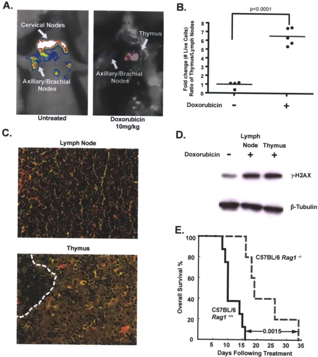
Figure 1. The thymus represents a chemoprotective niche that harbors surviving lymphoma cells following doxorubicin treatment. (A) Lymphoma-bearing mice were imaged for whole-body fluorescence prior to treatment and 4 days following a single dose of 10mg/kg doxorubicin. Representative mice are shown. (B) Ratios of live GFP-tagged Ep-myc p19'- B lymphoma cells in the thymus versus peripheral lymph nodes were quantified by flow cytometry, before (n=4 mice) and 48 hours after (n=5 mice) doxorubicin treatment. Average ratios are indicated with a line. (C) Hematoxylin and eosin (H&E) stained sections of lymph node and thymus from a tumor-bearing mouse 48 hours after doxorubicin treatment. The dotted line in the thymus demarcates a small region of infiltrating
A.
C.
lymphocytes neighboring a larger region of surviving lymphoma cells.
Representative fields are shown at 40x magnification. (D) A western blot showing
y-H2AX levels in FACS-sorted, GFP-positive, lymphoma cells from the thymus
and peripheral lymph nodes following doxorubicin treatment.
P-Tubulin
serves as a loading control. The untreated sample is a lysate from cultured lymphoma cells.(E) A Kaplan-Meier curve showing the overall survival of tumor-bearing C57BL/6
(n=8) or C57BL/6 Rag1-- (n=5) mice following doxorubicin treatment. The p value was calculated using a log rank test. See also Figure S1.
To analyze the effect of drug treatment on specific tumor niches, we harvested all primary lymphoid organs, including peripheral lymph nodes, thymus, spleen and bone marrow, following doxorubicin treatment. All tissues sampled showed extensive lymphoma cell apoptosis and restoration of normal organ architecture. Peripheral lymph nodes, spleen and bone marrow exhibited nearly complete tumor clearance with rare surviving lymphoma cells (Figure 1 C and Supplemental Figure 1A). In contrast, many surviving B lymphoma cells could be seen in the thymus. To quantify this phenotype, cells were harvested from peripheral lymph nodes and the thymus following treatment, and the
number of surviving GFP-positive lymphoma cells was assessed by flow cytometry. The number of viable lymphoma cells in the thymus relative to the lymph nodes increased 6.5 fold following doxorubicin treatment (Figure 1 B). Thus, the thymus represents a chemoprotective niche that protects lymphoma cells from doxorubicin-induced cell death.
To rule out the possibility that the selective survival of tumor cells in the thymus was due to the specific exclusion of doxorubicin from the mediastinum, we sorted live GFP-positive tumor cells from the lymph nodes and thymus 12
hours after doxorubicin treatment and blotted for y-H2AX, a marker of DNA damage (181). Western blot analysis showed that cells in both anatomical locations undergo the same amount of DNA damage (Figure 1 D). Additionally, flow cytometry of mediastinal lymphoma cells failed to identify any sub-population of lymphoma cells with decreased y-H2AX fluorescence (Supplemental Figure
1 B). These data suggest that the thymus offers no physical barrier to drug
A.
Lymph Node Untreated DoxorubicinU.
C.
100 *2 80 60 0 8.0 0 E 20 '~ 0E.
L
i... C57BLI6 Thymectomized 11 0 0 2 4 6 8 10 12 14Days Following Treatment
- ---- i
I
- -1 SL1 C57BU6 F 0.0012-* L.-C57BUJ6 * ag14 5 10 15 20 25 30Days Following Treatment
C57BL6 Rag1
C57B16 I
n.s.
5 10 15 20 25 30 35
Days
Supplemental Figure 1. Athymic mice show improved tumor-free survival following treatment with doxorubicin. (A) Hematoxylin and eosin (H&E) stained sections of lymph node, spleen, bone marrow and thymus from mice bearing Ep-myc p19w4- tumors. Mice were untreated or treated with a single dose of 10mg/kg doxorubicin for 72 hours. Surviving lymphoma cells are absent in the lymph node, spleen and bone marrow but present in the thymus. Scale bars indicate 35 microns on all panels. (B) A histogram showing yH2AX levels across a population of Ep-myc p19^*~' lymphoma cells treated in vitro and in vivo