and Their Reduced Functionality Bypasses the
Requirement for the Essential Pre-60S Factor Nsa1
Dagmar Pratte, Ujjwala Singh, Guillaume Murat, Dieter Kressler
*Unit of Biochemistry, Department of Biology, University of Fribourg, Fribourg, Switzerland
Abstract
Ribosomes are the molecular machines that translate mRNAs into proteins. The synthesis of ribosomes is therefore a fundamental cellular process and consists in the ordered assembly of 79 ribosomal proteins (r-proteins) and four ribosomal RNAs (rRNAs) into a small 40S and a large 60S ribosomal subunit that form the translating 80S ribosomes. Most of our knowledge concerning this dynamic multi-step process comes from studies with the yeast
Saccharomyces cerevisiae, which have shown that assembly and maturation of pre-ribosomal particles, as they
travel from the nucleolus to the cytoplasm, relies on a multitude (>200) of biogenesis factors. Amongst these are many energy-consuming enzymes, including 19 ATP-dependent RNA helicases and three AAA-ATPases. We have previously shown that the AAA-ATPase Rix7 promotes the release of the essential biogenesis factor Nsa1 from late nucleolar pre-60S particles. Here we show that mutant alleles of genes encoding the DEAD-box RNA helicase Mak5, the C/D-box snoRNP component Nop1 and the rRNA-binding protein Nop4 bypass the requirement for Nsa1. Interestingly, dominant-negative alleles of RIX7 retain their phenotype in the absence of Nsa1, suggesting that Rix7 may have additional nuclear substrates besides Nsa1. Mak5 is associated with the Nsa1 pre-60S particle and synthetic lethal screens with mak5 alleles identified the r-protein Rpl14 and the 60S biogenesis factors Ebp2, Nop16 and Rpf1, which are genetically linked amongst each other. We propose that these ’Mak5 cluster’ factors orchestrate the structural arrangement of a eukaryote-specific 60S subunit surface composed of Rpl6, Rpl14 and Rpl16 and rRNA expansion segments ES7L and ES39L. Finally, over-expression of Rix7 negatively affects growth of mak5 and
ebp2 mutant cells both in the absence and presence of Nsa1, suggesting that Rix7, at least when excessively
abundant, may act on structurally defective pre-60S subunits and may subject these to degradation.
Citation: Pratte D, Singh U, Murat G, Kressler D (2013) Mak5 and Ebp2 Act Together on Early Pre-60S Particles and Their Reduced Functionality
Bypasses the Requirement for the Essential Pre-60S Factor Nsa1. PLoS ONE 8(12): e82741. doi:10.1371/journal.pone.0082741
Editor: Sander Granneman, Univ. of Edinburgh, United Kingdom
Received September 12, 2013; Accepted October 28, 2013; Published December 2, 2013
Copyright: © 2013 Pratte et al. This is an open-access article distributed under the terms of the Creative Commons Attribution License, which permits
unrestricted use, distribution, and reproduction in any medium, provided the original author and source are credited.
Funding: This work was supported by a grant from the Swiss National Science Foundation (PP00P3_123341) to DK. The funders had no role in study
design, data collection and analysis, decision to publish, or preparation of the manuscript.
Competing interests: The authors have declared that no competing interests exist.
* E-mail: [email protected]
Introduction
The biogenesis of ribosomes is a fundamental cellular process, which provides cells with the molecular machines that translate the genetic information, contained within mRNAs, into proteins. Basically, the biogenesis of ribosomes consists in the ordered assembly of 33 ribosomal proteins (r-proteins) with the 18S ribosomal RNA (rRNA) and 46 r-proteins with the 25S, 5.8S and 5S rRNA into a small 40S and a large 60S ribosomal subunit (r-subunit), respectively, that form upon subunit joining in the cytoplasm the translating 80S ribosomes. Most of our current knowledge concerning this highly dynamic multi-step process comes from studies with the yeast Saccharomyces cerevisiae, which have shown that the assembly and maturation of pre-ribosomal particles, as they travel from the nucleolus to the cytoplasm, relies on a multitude (>200) of
biogenesis factors [1–3]. Amongst these are, in agreement with the high complexity of this macromolecular assembly process, many energy-consuming enzymes, notably including 19 ATP-dependent RNA helicases and three AAA-type ATPases (ATPases associated with various cellular activities) [2,4–7]. The energy expenditure by these enzymes is thought to be required to trigger irreversible steps of the assembly path and to shape this sophisticated ribonucleoprotein (RNP) complex into its active conformation. Within the nucleolus, the site of rDNA transcription, the emerging precursor rRNA (pre-rRNA) associates with primary binding r-proteins and early biogenesis factors to form the small-subunit (SSU) processome / 90S pre-ribosome [8–10]. Concomitantly, the pre-rRNA associates with 75 small nucleolar RNAs (snoRNAs), with most of these acting, integrated into C/D-box or H/ACA-box snoRNPs, as guides for the modification of pre-rRNA by 2’-O-ribose methylation and
pseudouridylation, respectively [11–14]. These modifications, which are concentrated in conserved regions that are functionally important for ribosome function, could help to fine-tune (pre-)rRNA folding, RNP assembly and translation [11,15]. Still within the nucleolar compartment, a subsequent cleavage step, either co- or post-transcriptional, separates the pre-rRNA and generates the 43S and 66S pre-ribosomal particles that upon further maturation eventually give rise to mature 40S and 60S subunits. Pre-40S subunits exhibit a relatively simple composition and are rapidly exported to the cytoplasm where final maturation steps, including 20S pre-rRNA cleavage at site D by the endonuclease Nob1 [16], confer translation competence [17]. In sharp contrast, proteomic approaches revealed several distinct nucleolar and nucleoplasmic pre-60S intermediates that undergo drastic compositional changes [2,18]. Upon arrival of pre-60S subunits in the cytoplasm, the last biogenesis factors are dis- and re-placed by r-proteins in an ordered series of events, thereby enabling subunit joining [3,19].
Due to the complexity of the process and the importance to build ribosomes that translate with high fidelity, it is not surprising that pre-ribosomal particles and mature r-subunits are subjected to quality control [20–22]. The (pre-)rRNAs of defective nuclear pre-ribosomes undergo polyadenylation by the TRAMP complex and are subsequently degraded by the exosome, with both steps occurring in a sub-nucleolar focus termed No-body [20,23–25]. Recent evidence indicates that the D-site cleavage of 20S pre-rRNA, and hence final maturation of pre-40S subunits, occurs within 80S-like ribosomes, thus representing a proofreading step that probes their suitability to engage in translation [26–28]. Likewise, during cytoplasmic maturation of pre-60S subunits, verification of P-site integrity is coupled to the release of the anti-association factor Tif6 [29]. Finally, stalled or functionally defective r-subunits are subjected to ’non-functional rRNA decay’ (NRD) pathways, which eliminate non-functional 40S and 60S r-subunits via degradation of the rRNA or targeting to the proteasome, respectively [22,30–33].
Most of the DExD/H-box RNA helicases involved in ribosome biogenesis act at early steps of ribosome assembly within the nucleolus where extensive structural rearrangements of (pre-)-rRNA and incorporation of the majority of r-proteins are expected to occur [2,6]. Considering the reported functional and enzymatic properties, DExD/H-box RNA helicases can be generally viewed as energy-consuming chaperones or modulators of RNA and RNP structures [34–36]. In most cases, however, we are far from understanding their exact molecular functions during ribosome assembly (reviewed in [2,5,6]); it will therefore be an enormous future challenge to obtain insight into their pre-rRNA binding sites and to unravel the regulation and timing of their enzymatic activity. On the other hand, GTPases, kinases and the three AAA-type ATPases act predominantly at later stages during ribosome biogenesis [2].
Contrary to the proposed pre-rRNA or RNP remodelling functions of most DExD/H-box RNA helicases involved in ribosome biogenesis, recent evidence revealed that the three AAA-ATPases are required for the release of distinct substrate proteins from nucleolar, nucleoplasmic and cytoplasmic
pre-60S intermediates [37–40]. The type II AAA-ATPases Rix7 and Drg1, which are closely related to the well-known Cdc48 (p97 in mammals) whose diverse functions are largely linked to the recognition of ubiquitinated substrate proteins [41,42], mediate the release of the essential biogenesis factors Nsa1 and Rlp24 from nucleolar or cytoplasmic pre-60S particles, respectively [38,39]. Despite their striking similarity to Cdc48/p97 [4], there is so far no evidence that links Rix7 or Drg1 to the degradation of defective pre-60S ribosomes or parts thereof. On the other hand, Rea1, the largest yeast protein, shares similarity with the dynein heavy chain protein Dyn1 [4]. Rea1 is involved in two successive pre-60S maturation steps and promotes the release of distinct biogenesis modules (Ytm1-Erb1-Nop7 / Rsa4, Rix1-Ipi3-Ipi1), thus mediating nucleolar exit and priming pre-60S subunits for nuclear export [37,40]. In all cases, the removal of these biogenesis factors from pre-60S particles is essential for their recycling and, importantly, triggers major compositional or conformational changes that constitute key maturation events necessary for the progression of ribosome assembly.
We are ultimately interested in understanding the mechanistic details of the Rix7-mediated release of Nsa1 from late nucleolar pre-60S particles and how the entailed structural rearrangements reshape pre-60S intermediates and allow progression of ribosome assembly. Here we reveal that the lethality associated with the absence of Nsa1 can be suppressed by mutant alleles of the biogenesis factors Ebp2, Mak5, Nop1 and Nop4, thus genetically unravelling possible structural changes within pre-60S particles that influence their assembly kinetics and/or path in order to compensate for the lack of Nsa1 recruitment. Moreover, genetic experiments indicate that Rix7 may have other nuclear substrates besides Nsa1 and that it may recognize and target defective pre-60S subunits for degradation. Finally, our analysis of the functional network around the DEAD-box RNA helicase Mak5 suggests that the ’Mak5 cluster’ factors (Mak5, Ebp2, Nop16 and Rpf1) may orchestrate the structural arrangement of a eukaryote-specific 60S subunit surface composed of Rpl6, Rpl14 and Rpl16 and rRNA expansion segments ES7L and ES39L.
Materials and Methods
Yeast strains and genetic methods
The S. cerevisiae strains used in this study are listed in Table S1, all strains are derivatives of W303 [43] or DS1-2b [44]. For yeast two-hybrid analyses the reporter strain PJ69-4A was used [45]. Deletion disruption and C-terminal tagging at the genomic locus were performed as described [46–48]. Preparation of media, yeast transformation, and genetic manipulations were done according to established procedures. Plasmid constructs
All recombinant DNA techniques were according to established procedures using Escherichia coli DH5α for cloning and plasmid propagation. All cloned DNA fragments generated by PCR amplification were verified by sequencing. Human NSA1 was PCR-amplified from a cDNA library and first cloned into pBSKS(-) (Stratagene) and then, after a fusion PCR to
eliminate mutations within the hNSA1 gene, cloned under the control of the ADH1 promoter in a YCplac111-based vector. More information on the plasmids, which are listed in Table S2, is available upon request.
Isolation and cloning of spontaneous ∆nsa1 bypass suppressors
The NSA1 shuffle strain Y3900 (see Table S1) was transformed with pADH111-hNSA1.N328 and restreaked or spotted, after selection of cells having lost the NSA1 shuffle plasmid pHT4467∆-NSA1 (URA3-ADE3 ) on 5-FoA-containing plates, on YPD plates. The gene complementing the suppressor mutation S5 was cloned by transformation of strain YHD60, which is a meiotic segregant of a cross between suppressor strain S5 and Y3274, with a YCplac111-based yeast genomic library [49]. The library plasmid was rescued from one transformant that showed complementation of the slow-growth phenotype but did not grow on 5-FoA-containing plates. The gene complementing the temperature-sensitive (ts) phenotype associated with the suppressor mutation S4 was cloned by transformation of a strain originating from a backcross of suppressor strain S4 with the NSA1 shuffle strain Y3903 with a yeast genomic library in YCplac111. To clone the gene complementing the ts phenotype of the suppressor mutant Sj, the suppressor strain Sj was first transformed with YCplac33-NSA1 and then with the YCplac111-based yeast genomic library. To ascertain that the suppressor strains carried indeed mutations in the genes complementing the growth and suppression phenotypes, mak5-S5, nop4-S4 and nop1-Sj were amplified by PCR from genomic DNA of the respective suppressor strains. Upon cloning of the PCR products, the nature of the mutation within the suppressor alleles was identified by sequencing.
Synthetic lethal screens
Synthetic lethal (sl) screens with the mak5.G218D and mak5.R728* alleles were essentially performed as previously described [49]. The above mak5 alleles were cloned into YCplac22, and the resulting plasmids were transformed into the MAK5 shuffle strains YDP1 and YDP4, which bear MAK5 on the instable URA3 ADE3 marker containing plasmids pHT4467∆. Cells were grown to exponential phase in liquid SC-Ura-Trp medium, plated on SC-Trp plates and then mutagenized by irradiation with UV light in a Stratalinker 1800 (Stratagene). Cloning of the genes complementing the respective sl-mutations was achieved by using a yeast genomic library in YCplac111 or YEplac181 [49,50]. The sl-screen with mak5.R728* yielded the following candidates whose complementing genes could be cloned: six times NOP16 (SL2, SL4, SL63, SL67, SL85 and SL113), two times RPF1 (SL3 and SL33) and one time RPL14A (SL103). The sl-screen with mak5.G218D yielded only one sl-candidate for which it was possible to clone the complementing gene: EBP2 (SL19). To ascertain that the genes cloned by complementation indeed harboured mutations in the sl-mutant strains, genomic DNA was prepared from the sl-strains and the corresponding mutant alleles were amplified by PCR and
directly sequenced and/or cloned into suitable plasmids and then sequenced.
Sucrose gradient analysis and fractionation
Cell extracts for polysome profile analyses were prepared as previously described [51] and layered onto 10-50% sucrose gradients that were centrifuged at 38’000 rpm in a Sorvall TH-641 rotor at 4°C for 2 h 30 min or 2 h 45 min. Sucrose gradients were analysed and fractionated using an ISCO UA-6 system with continuous monitoring at A254.
Preparation of total yeast protein extracts and Western analysis
Total yeast protein extracts were prepared as previously described [52]. Cultures were grown to an OD600 of around 0.8
and protein extracts were prepared from an equivalent of one OD600 of cells. Western blot analysis was carried out according
to standard protocols. The following primary antibodies were used in this study: mouse monoclonal anti-GFP (1:3’000; Roche) and anti-Rpl3 (1:5’000; J. Warner, Albert Einstein College of Medicine, New York, NY); rabbit polyclonal anti-CBP (1:15’000; Open Biosystems), Arc1 (1:20’000; [53]), anti-Ebp2 (1:10’000; J. Woolford, Carnegie Mellon University, Pittsburgh, PA), and anti-Nop7 (1:40’000; B. Stillman, Cold Spring Harbour Laboratory, Cold Spring Harbour, NY). Secondary goat anti-rabbit (Bio-Rad) or anti-mouse (Bio-Rad) horseradish peroxidase-conjugated antibodies were used at a dilution of 1:10’000. For detection of TAP-tagged proteins the Peroxidase-Anti-Peroxidase soluble complex was used at a dilution of 1:15’000 (Sigma). Proteins were visualized using enhanced chemiluminescence detection kits (Immobilon Western, Millipore; Amersham ECL, GE Healthcare).
Tandem affinity purification (TAP)
TAP purifications of TAP-tagged bait proteins were performed in a buffer containing 50 mM Tris-HCl pH 7.5, 100 mM NaCl, 1.5 mM MgCl2, 5% glycerol and 0.1% NP-40
essentially as described previously [44,48]. For TEV cleavage, DTT was added to a final concentration of 1 mM to the above buffer. Elution from Calmodulin-Sepharose beads was performed in the presence of 5 mM EGTA. The EGTA eluates were precipitated by the addition of TCA to a final concentration of 10% and dissolved in SDS sample buffer. The samples were then separated on NuPAGE SDS 4-12% gradient polyacrylamide gels (Invitrogen) and stained with colloidal Coomassie (Sigma) or analyzed by Western blotting. Sequence alignments, secondary structure prediction and analysis of 3D structures
Multiple sequence alignments of orthologous proteins were generated in the ClustalW output format with T-Coffee using the default settings of the EBI website interface [54]. Secondary structure prediction was performed with the PSIPRED v3.3 prediction method available at the PSIPRED website interface [55]. Analysis and image preparation of three-dimensional structures, downloaded from the PDB archive, was carried out with the PyMOL software (PyMOL Molecular
Graphics System). For analysis of the eukaryote-specific 60S subunit surface surrounding Rpl14 (L14e), we used the coordinates for 60S r-subunits from the recent S. cerevisiae 80S crystal structure (PDB 3U5H and 3U5I; [56]) and human 80S cryo-EM structure (PDB 3J3B and 3J3F; [57]).
Results
The essential Nsa1 function can be bypassed by mutation of MAK5, NOP1 and NOP4
We have previously shown that the AAA-type ATPase Rix7 strips the essential biogenesis factor Nsa1 from a distinct nucleolar pre-60S particle (see Introduction and [39]). To address whether Nsa1 fulfils an evolutionarily conserved function in ribosome biogenesis, we tested if human NSA1 complemented the lethality of the ∆nsa1 null mutant. Nsa1 is predicted to form an N-terminal 7-bladed WD β-propeller followed by a non-essential C-terminal extension (D.K., unpublished data). As shown in Figure S1, human NSA1 lacking the C-terminal extension (hNSA1.N328) was capable of conferring weak growth to ∆nsa1 null mutant cells (Figure S1). Interestingly, we observed that faster growing colonies appeared spontaneously at a rather high frequency (Figure S1), indicating that the dysfunctionality or even absence of Nsa1 can be easily suppressed. To facilitate cloning of the genes complementing the suppressor mutations, we selected in a first instance those suppressor strains where the suppressor mutation conferred a clear slow-growth or temperature-sensitive (ts) phenotype. By this approach, we managed to clone the genes complementing the suppressor mutation of three suppressor strains (see Materials and Methods). Subsequent PCR amplification and sequencing of the suppressor alleles revealed that all three strains indeed contained mutations in the genes that we had cloned by complementation. Suppressor strain S5 harbours a mutation in MAK5 [mak5.R728*; Arg728(AGA) to stop (TGA)], which encodes a nucleolar DEAD-box RNA helicase involved in 60S subunit biogenesis [58]. Suppressor strain S4 contains a mutation in NOP4 [nop4.S460L; Ser460(TCG) to Leu(TTG)], encoding a nucleolar protein with four RRM motifs that binds to the pre-rRNA in proximity of the 5’-end of the 5.8S rRNA and is required for the stable formation of pre-60S subunits containing the 27SA and 27SB pre-rRNAs [59–61]. Suppressor strain Sj comprises a mutation in NOP1 [nop1.M232K; Met232(ATG) to Lys(AAG)], coding for the methyltransferase component of C/D-box snoRNPs [62–66]. We first assessed the growth phenotypes of these three mutant alleles when expressed under the control of their cognate promoters from a centromeric plasmid (Figure S2A-C). While the mak5.R728* mutant shows a slow-growth phenotype at all temperatures, especially at 37°C, the nop4.S460L and nop1.M232K mutants display only a very mild growth defect at 30°C and are ts at 37°C. Polysome profile analyses revealed that all three mutations confer a deficiency in 60S subunit production, as evidenced by the reduction in free 60S subunits and the occurrence of half-mer polysomes (Figure S2A-C). Since Nop4 was originally identified in a synthetic lethal (sl) screen with the nop1-5 allele [59], we tested whether the nop1.M232K and nop4.S460L alleles are
also genetically linked. As shown in Figure S3, combination of these two mutations resulted in a synergistic slow-growth phenotype (Figure S3).
We next determined whether the isolated mak5, nop1 and nop4 alleles, when expressed from a centromeric plasmid, had the capacity to suppress the absence of Nsa1 in the setting of clean double shuffle strains. All three alleles bypassed the requirement for Nsa1, as revealed by the growth of ∆nsa1 null mutant cells expressing the mutant variants of Mak5, Nop1 and Nop4 on plates containing 5-fluoroorotic acid (5-FoA) (Figure S4). To better assess the extent of suppression, we compared the growth properties of cells harbouring the suppressor alleles in the presence or absence of NSA1 after plasmid shuffling (Figure 1A-C). This growth analysis showed that the nop4.S460L allele, even though conferring only a very mild growth defect on its own, very efficiently suppressed the lethality of the ∆nsa1 null mutant (Figure 1C), while the mak5.R728* and nop1.M232K alleles exhibited a moderate and weak extent of suppression, respectively (Figure 1A and B). Notably, suppression was for all three alleles more efficient at 30°C than at 23°C; moreover, growth of the mak5.R728* mutant was identical in the presence or absence of Nsa1 at 37°C (Figure 1A-C). We conclude that the absence of the essential, pre-60S associated Nsa1 can be relatively easily bypassed by mutation of genes encoding biogenesis factors that act at different nucleolar stages of pre-60S maturation (see Discussion and also below). Since we did not carry out an in-depth suppressor screen, it is very likely that reduced functionality of other protein trans-acting factors, besides Mak5, Nop1 and Nop4, may also lead to bypass suppression of the lethal ∆nsa1 null phenotype.
Rix7 may have additional nuclear functions besides stripping Nsa1 from pre-60S particles
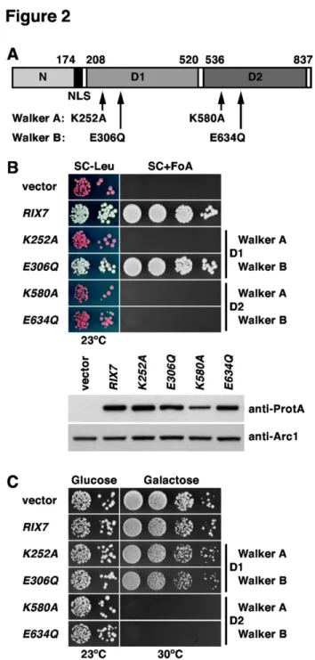
The availability of bypass suppressors of the lethal ∆nsa1 null mutant put us in the unique situation to address whether the AAA-ATPase Rix7 may have additional substrates besides Nsa1 by testing the dominant-negative effect of RIX7 alleles in the ’suppressor’ strains. However, a pre-requisite for this analysis was the construction of dominant-negative alleles of RIX7. Rix7 exhibits the typical domain organization of type II AAA-ATPases, notably containing two central AAA-domains (termed D1 and D2) flanked by a long N-terminal and a very short C-terminal extension (Figure 2A; [4,39]). To define whether ATP binding and ATPase activity are essential features of both D1 and D2 by in vivo experiments, we mutated conserved residues that are implicated in ATP binding (Walker A motif / P-loop) and ATP hydrolysis (Walker B motif / DExx-box) [67], then we assessed the complementation of the lethal ∆rix7 null mutant phenotype by these constructs. This analysis revealed that Rix7 variants with a mutated Walker A motif within AAA-domain D1 (K252A) or mutated Walker A or B motifs within D2 (K580A and E634Q, respectively) are non-functional, while mutation of the Walker B motif within D1 (E306Q) has no significant effect on cell growth (Figure 2B). In conclusion, adenosine nucleotide binding (ATP or ADP) is necessary whereas ATP hydrolysis seems to be dispensable for AAA-domain D1 function; however, AAA-domain D2
function requires both ATP binding and hydrolysis. In agreement with this interpretation, both D2 mutants, but not the D1 Walker A mutant, conferred a dominant-negative phenotype when over-expressed from a galactose-inducible promoter in a wild-type background (Figure 2C). Similarly to Rix7, the main catalytic activity of the closely related AAA-ATPases Cdc48 and Drg1 also resides in their D2 domains [38,68,69].
Having dominant-negative alleles of RIX7 at hand, we next assessed their effect on the growth of the mak5.R728* and nop4.S460L ∆nsa1 null ’suppressor’ strains. Interestingly, galactose-induced over-expression of the dominant-negative RIX7.K580A and E634Q mutants still resulted in a lethal phenotype (Figure 3A and B); thus, suggesting that Rix7 may have additional nuclear substrates besides Nsa1. Intriguingly, we noticed that over-expression of wild-type RIX7 already had a strong negative effect on the growth of mak5.R728* cells lacking Nsa1 (Figure 3A) – this effect was less pronounced when the ∆nsa1 null mutation was suppressed by nop4.S460L (Figure 3B). This observation indicates that Rix7, in order to exert such a negative effect on growth, is capable of recognizing pre-60S particles that lack Nsa1. However, since no negative effect of Rix7 over-expression was observed in a
wild-type background (Figure 2C), Rix7 presumably only acts on damaged pre-60S particles, which likely manifest their structural deficiency in a characteristic manner (see below and Discussion).
Mak5 is associated with the Nsa1 pre-60S particle Since we are ultimately interested in understanding how Rix7 strips Nsa1 from late nucleolar pre-60S particles, we did not consider to further investigate the basis of the suppression of ∆nsa1 by the nop1.M232K and nop4.S460L allele in the framework of this study, because these two factors act substantially upstream of the Nsa1 pre-60S particle: (i) Nop1, as the methyltransferase component of C/D-box snoRNPs [62–66], acts primarily on 90S pre-ribosomes and very early pre-60S particles and (ii) Nop4 is mainly associated with very early pre-60S particles [8,9,39,70–73]. On the other hand, the reduced formation of mature 25S and 5.8S rRNA from 27S pre-rRNAs upon genetic depletion of Mak5 suggested an involvement of Mak5 in a late step during the nucleolar phase of pre-60S maturation [58]. To address whether Mak5 is specifically associated with nucleolar pre-60S particles, we Figure 1. Bypass suppression of ∆nsa1 null lethality by the mak5.R728*, nop1.M232K and nop4.S460L alleles. MAK5/NSA1 (A), NOP1/NSA1 (B) and NOP4/NSA1 (C) double shuffle strains were co-transformed with plasmids harbouring the indicated wild-type and mutant alleles and/or empty vectors (YCplac111 or YCplac22). After plasmid shuffling on plates containing 5-FoA, cells were restreaked on synthetic complete medium lacking leucine and tryptophan (SC-Leu-Trp) and then spotted in 10-fold serial dilution steps onto SC-Leu-Trp plates, which were incubated for 3 d at 30°C, 4 d at 23°C and 4 d at 37°C.
Figure 2. The D2 domain of the AAA-ATPase Rix7 harbors the catalytic activity. (A) Schematic representation of the domain organization of Rix7. N, N-terminal domain (amino acids 1-174); NLS, predicted bipartite nuclear localization signal (amino acids 175-194); D1, AAA-domain D1 (amino acids 208-520); D2, AAA-domain D2 (amino acids 536-823). The amino acid changes within the Walker A and B motifs of D1 or D2, respectively, are indicated. (B) In vivo phenotypes of the Walker A and B mutants generated in D1 and D2. Empty vector (YCplac111) and plasmid-borne wild-type RIX7 or the indicated rix7 mutant alleles under the control of the authentic promoter were transformed into the RIX7 shuffle strain. Transformants were spotted in 10-fold serial dilution steps onto SC-Leu and SC+5-FoA plates, which were incubated for 5 d at 23°C (upper panel). Expression levels of plasmid-borne TAP-tagged wild-type RIX7 and the indicated rix7 Walker A and B mutant alleles, expressed from their cognate promoter in a haploid wild-type strain, were determined in whole cell lysates by Western analysis using anti-ProtA and anti-Arc1 (loading control) antibodies (lower panel). (C) Walker A and B mutations within AAA-domain D2 confer a dominant-lethal phenotype. Empty vector and wild-type RIX7 or the indicated rix7 mutant alleles, expressed under the control of the inducible GAL1-10 promoter, were transformed into a haploid wild-type strain. Transformants were spotted in 10-fold serial dilution steps onto SC-Leu (Glucose) and SGal-Leu (Galactose) plates, which were incubated for 4 d at 23°C and 30°C, respectively.
purified, by applying the tandem-affinity purification (TAP) method, several distinct pre-60S particles from cells expressing chromosomally integrated Mak5-GFP (Figure 4). The TAP-tagged bait proteins subjected to the purifications notably define early nucleolar (Ssf1-TAP), late nucleolar (Nsa1-TAP), nucleoplasmic (Rix1-TAP), nucleoplasmic/cytoplasmic (Arx1-TAP) and late cytoplasmic (Lsg1-(Arx1-TAP) pre-60S particles, while the Nop7-TAP bait purifies a mixture of very early to late nuclear pre-60S particles [19,39,44,74]. As revealed by Western analysis using anti-GFP antibodies, Mak5 is exclusively associated with nucleolar pre-60S particles purified by the Ssf1 and Nsa1 baits (Figure 4). Since Mak5 was previously neither found as a stoichiometric component of Ssf1- nor Nsa1-defined pre-60S particles [39,74], we conclude that Mak5 is only transiently or weakly associated with pre-60S ribosomes.
The C-terminal extension to the DEAD-box core is important for Mak5 function
Mak5 contains N- and C-terminal extensions of around 170 amino acids flanking the central DEAD-box core (Figures S5A and S5B). The identification of mak5.R728* as a ∆nsa1 suppressor allele suggested that the C-terminal extension is important for Mak5 function. Sequence comparison revealed that the C-terminal extension is relatively well conserved both in primary sequence and secondary structure, notably predicted to harbour four conserved α-helices (Figure S5A). On the other hand, the N-terminal extension contains only few conserved regions and there is no clear conservation of the predicted secondary structure elements (data not shown). To better define the contribution of the N- and C-terminal extension to the biological function of Mak5, we undertook a
detailed deletion analysis (Figures S5B and S5C). While a Mak5 variant lacking almost the complete C-terminal extension (mak5.N629; amino acids 1-629) did not support growth (Figure S5B), the N-terminal deletion construct of Mak5 (mak5.166C; amino acids 166-773) exhibited a moderate slow-growth phenotype at 30°C and a pronounced slow-growth defect at 37°C (Figures S5B and S5C). Progressive C-terminal deletion mapping revealed that already a construct expressing amino acids 1-673 of Mak5 (mak5.N673) was not able to confer growth and that the first viable mutant harboured a C-terminal deletion of 50 amino acids (mak5.N723) (Figure S5B). Next, we assessed the growth properties of the viable N- and C-terminal deletion mutants after plasmid shuffling on YPD plates at different temperatures (Figure S5C), revealing in both cases that the growth defects, which were not due to decreased expression levels (data not shown), were more pronounced at 37°C. Finally, polysome profile analysis showed a shortage of 60S subunits both for mutants lacking the complete N-terminal extension (mak5.166C) or the C-terminal 50 amino acids (mak5.N723) (Figure S6). Altogether, we conclude that the C-terminal extension of Mak5, in agreement with its higher degree of evolutionary conservation, is functionally more relevant than the N-terminal extension. Moreover, due to its essential nature, we predict that the C-terminal extension may mediate recognition of the cognate RNA or RNA:protein substrate on pre-60S particles.
Mak5 functionally interacts with Ebp2, Nop16, Rpf1 and Rpl14
Since DExD/H-box RNA helicases largely rely on a specific RNA substrate and/or protein co-factors to efficiently carry out their biological functions (see for example [75–81]), we set out Figure 3. Dominant-lethal RIX7 alleles retain their negative effect on growth in the absence of Nsa1. MAK5/NSA1 (A) and
NOP4/NSA1 (B) double shuffle strains were co-transformed with plasmids harbouring either the MAK5 or NOP4 wild-type genes or the mak5.R728* or nop4.S460L mutant alleles and plasmids carrying the NSA1 wild-type gene or, expressed under the control of the GAL1-10 promoter, wild-type RIX7 and the dominant-negative RIX7.K580A or RIX7.E634Q alleles or empty vector. After plasmid shuffling on plates containing 5-FoA, cells were restreaked on SC-Leu-Trp plates and then spotted in 10-fold serial dilution steps onto SC-Leu-Trp (Glucose) and SGal-Leu-Trp (Galactose) plates, which were incubated for 3 d or 4 d at 30°C, respectively. doi: 10.1371/journal.pone.0082741.g003
to perform a sl-screen with different mak5 alleles in order to establish the functional network around Mak5 and, if possible, to identify the protein partner(s) that may be required for stimulation of Mak5’s ATPase activity and/or for its recruitment to the cognate substrate. To maximize an optimal recovery of a broad range of distinct sl-partners, we chose to use mak5 alleles harbouring mutations within motif I (Walker A motif; involved in ATP binding) or the C-terminal extension, which are therefore likely affecting ATPase activity (mak5.G218D) or substrate recognition (mak5.R728*). To validate the suitability of the mak5.G218D allele [58], we determined its growth characteristic and polysome profile, revealing a moderate Figure 4. The DEAD-box protein Mak5 is associated with the Nsa1 pre-60S particle. Mak5 is associated with the early nucleolar Ssf1 and late nucleolar Nsa1 pre-60S particles. The indicated TAP-tagged bait proteins were affinity-purified from cells expressing Mak5-GFP. The final EGTA eluates were analyzed by SDS-PAGE and Coomassie staining (top) and Western blotting using GFP, Nop7, Ebp2 and anti-Rpl3 antibodies (bottom).
doi: 10.1371/journal.pone.0082741.g004
growth defect 23°C, 30°C and 37°C and a deficiency in production of 60S subunits (Figures S5C and S6). The sl-screen with the mak5.R728* allele yielded nine sl-mutants for which we managed to clone the complementing genes, with six of these being complemented by NOP16, two by RPF1 and one by RPL14A (see also Materials and Methods). Of the obtained sl-candidates from the mak5.G218D screen we could complement one sl-mutant with EBP2. In agreement with a function of Mak5 during the nucleolar phase of pre-60S maturation, all the proteins encoded by the identified genes are known to have a role in 60S biogenesis and/or are associated with the Nsa1-defined pre-60S particle or, as for the r-protein Rpl14a, to be a constituent of mature 60S subunits [39,56,73,82–85].
As a next step, we confirmed that the sl-mutant strains indeed contained genomic mutations within the genes that were cloned by complementation. To this end, we PCR amplified and either sequenced directly the PCR products or first cloned and then sequenced the inserts of the plasmids containing the mutant alleles. To visualize the mutations that are present in the individual sl-alleles of the four different genes, we have highlighted the entailed amino acid changes within multiple sequence alignments of the orthologous proteins from S. cerevisiae, Schizosaccharomyces pombe and Homo sapiens (Figures S7A, S8A, S9A, and S10A). To assess the phenotypic consequences of these mutations on growth and 60S subunit biogenesis, we either expressed the mutant alleles from centromeric plasmids under the control of their cognate promoters in the respective shuffle strains (for the ebp2, rpf1 and rpl14a alleles) or generated a ∆nop16 null mutant. The ebp2.K287* allele introduces a stop codon after amino acid 286, thereby truncating Ebp2 at the beginning of a predicted, ~60 amino acid long α-helix within the highly conserved Ebp2-core domain (Figures S7A and S14), and confers a relatively strong slow-growth phenotype, which is exacerbated at 37°C, that coincides with a clear reduction in free 60S subunits, the accumulation of half-mer polysomes and an overall reduction in polysome content (Figure S7B). The two rpf1 alleles, rpf1.L199P and rpf1.C139R, similarly affect growth (mild slow growth at 30°C and almost ts at 37°C) and 60S subunit production (Figure S8B). Notably, Leucine 199 is within a highly conserved stretch of Rpf1 and precedes the C-terminally located σ70-like RNA-binding motif by ~55 amino
acids (Figure S8A and [85]). The mutations within the six NOP16-complemented sl-mutants represent five different alleles of nop16, with all of these, due to the introduction of frameshifts and/or pre-mature stop codons, likely completely abrogating Nop16 function (Figure S9A). We therefore assessed the growth and polysome profile phenotype of ∆nop16 null mutant cells, revealing a very modest growth defect and a mild reduction in free 60S subunits that was notably visible by the appearance of half-mer polysomes (Figure S9B). We note that Nop16 – despite the very mild impact of the absence of Nop16 on yeast growth under optimal laboratory conditions – is still present in higher eukaryotic and even mammalian organisms (Figure S9A). The essential r-protein Rpl14 (L14e according to the newly proposed nomenclature for ribosomal proteins [86]), which is conserved
in some but not all archaeal organisms [56,87], is encoded in S. cerevisiae by the duplicated genes RPL14A and RPL14B. The rpl14a.L123* allele, by changing the triplet coding for Leucine 123 to a stop codon, encodes a mutant Rpl14a protein with a truncated C-terminal α-helix (Figure S10A), which notably mediates interactions with the eukaryote-specific C-terminal α-helix of Rpl16 (L13), the C-C-terminal region of the eukaryote-specific ribosomal protein Rpl6 (L6e), and the eukaryote-specific rRNA expansion segment ES39L (see Figures S10B, S10C and S11B; [56]). Interestingly, the two C-terminal α-helices of eukaryotic Rpl14 are absent from the existing archaeal L14 orthologues (Figure S11A). The rpl14a.L123* mutant variant, when expressed under the control of its cognate promoter from a centromeric plasmid in ∆rpl14a null mutant cells, confers a mild growth defect that is exacerbated a lower temperatures (Figure S12A). In agreement with an effect on the biogenesis of 60S subunits, the rpl14.L123* mutant displayed a slight decrease in free 60S subunits and some accumulation of half-mer polysomes (Figure S12A). However, when supplied as the sole Rpl14 copy in a RPL14 shuffle strain (∆rpl14a/∆rpl14b), the rpl14a.L123* allele elicited a more pronounced growth and 60S subunit biogenesis defect (Figure S12B). Notably, growth of rpl14a.L123* cells was particularly affected at 37°C and 18°C (Figure S12B). Nevertheless, we still observed even stronger growth and 60S subunit deficits in ∆rpl14a cells (Figure S12A). On the other hand, growth and 60S subunit deficiency of ∆rpl14b cells was very similar to the one of rpl14a.L123*/ RPL14B cells (Figures S12A and S12C). It can therefore be concluded that, even though RPL14A and RPL14B encode identical proteins (e.g. in our W303 background), the contribution of Rpl14a to the functional pool of Rpl14 is more prominent than the one of Rpl14b (see also [88]).
Mak5, Ebp2, Nop16, Rpf1 and Rpl14 form a genetically defined functional cluster
To confirm that the identified sl-mutant alleles were indeed the source of the sl-relation with the mak5.G218D or mak5.R728* alleles, we retested their genetic interactions in the setting of de novo created double shuffle strains (for the essential EBP2 and RPF1 genes and the quasi-essential RPL14A gene) or a MAK5/∆nop16 shuffle strain. As expected, the ebp2.K287* allele showed a synthetic lethal phenotype with the mak5.G218D mutant. However, the slower-growing C-terminal deletion mutants of mak5 (mak5.R728* and mak5.N723) only exhibited a synthetic enhancement (se) phenotype when combined with the ebp2.K287* allele (Figure 5A). Conversely, combining the mak5.R728* or mak5.N723 alleles with the ∆nop16 null mutation resulted in synthetic lethality, while the mak5.G218D/∆nop16 pair only exhibited a mild se-phenotype (Figure 5B). In agreement with the severe se-phenotype of the two original RPF1-complemented sl-mutants isolated by the mak5.R728* sl-screen (data not shown), the distinct mak5.R728*/rpf1.L199P and mak5.R728*/ rpf1.C139R mutant combinations grew clearly slower than the individual mutant strains alone. The synthetic growth defect was much more pronounced, resulting almost in lethality, when the two rpf1 alleles were combined with the mak5.N723 allele;
however, no se-phenotype could be discerned when paired with the mak5.G218D allele (Figure 5C). Due to the non-lethality of the ∆rpl14a null mutant, we assessed the growth of mak5/rpl14a.L123* double mutants on synthetic medium after plasmid shuffling on 5-FoA-containing plates. In agreement with a complete synthetic effect of the mak5.R728* allele on the rpl14a.L123* allele, mak5.R728*/rpl14a.L123* and mak5.R728*/∆rpl14a cells showed the same severe growth defect, which was identical to the one of the ∆rpl14a single mutant strain (Figure 5D). Similarly, a synthetic relation was observed between the mak5.N723 and rpl14a.L123* alleles, while the rpl14a.L123* allele exerted only a minor effect on the growth of mak5.G218D mutant cells. On the other hand, there was no se-phenotype associated with any of the mak5/∆rpl14b combinations (data not shown). We conclude that the expression of C-terminally truncated Rpl14a, and not the lower cellular Rpl14 levels (as in ∆rpl14a or ∆rpl14b cells), causes a synergistic pre-60S maturation defect specifically in conjunction with the expression of Mak5 variants lacking part of the C-terminal extension. The allele specificity of the observed genetic interactions is particularly striking for the ebp2.K287* allele, which, unlike the rpf1 mutants or the ∆nop16 null mutant that are mainly linked to the mak5.R728* and mak5.N723 alleles, shows synthetic lethality only when combined with the mak5.G218D allele (see also Discussion).
With the aim of establishing a complete genetic network, we next tested the alleles identified by the sl-screens among each other for the enhancement of their respective growth phenotypes. Most strikingly, combination of the ebp2.K287* allele with either of the two rpf1 alleles or the ∆nop16 null allele resulted in synthetic lethality (Figure 6A and 6B). Moreover, the ebp2.K287*/rpl14a.L123* double mutant showed the same severe slow-growth phenotype as the ∆rpl14a single or the ebp2.K287*/∆rpl14a double mutant, revealing that the ebp2.K287* allele minimized the functionality of the rpl14a.L123* mutant to the extent of a complete ∆rpl14a null mutation (Figure 6C). Combining the ∆nop16 null mutation with the rpf1 or rpl14a.L123* alleles evoked a clear and very mild se-phenotype, respectively (Figures S13A and S13B). Finally, we could not observe any synergistic growth defect in the case of rpf1/rpl14a.L123* double mutant cells (Figure S13C). We conclude that Mak5, Ebp2, Nop16, Rpl14a and Rpf1 form a functional, genetically defined, cluster of proteins that likely act at a similar step during pre-60S maturation. Moreover, allele-specificity and strength of the interactions suggest a most intimate interplay between Mak5, Ebp2 and Rpl14.
The central conserved domain of Ebp2 is sufficient for its functionality
The finding that the ebp2.K287* allele, which introduces a deletion of the C-terminal 141 amino acids of Ebp2, clearly supported growth is different from previously published results showing that deletion of the C-terminal 105 amino acids conferred lethality (Figure S7B and [83]). We therefore carefully re-examined the boundaries that still enable the expression of fully or partially functional Ebp2 variants by a detailed deletion analysis. Ebp2 is composed of a highly conserved, central domain that is predicted to be built up by four conserved
α-helices (Figures S7A and S14A). This central domain is flanked by an N-terminal extension of ~185 amino acids, containing a basic region followed by an acidic region, that is substantially shorter or almost completely absent from the orthologous S. pombe or H. sapiens Ebp2 proteins, respectively (Figure S7A).
On the other hand, the C-terminal extension, defined here as starting after the fourth predicted central α-helix of ~60 amino acid length, comprises the terminal ~80 amino acids of Ebp2 and contains some well-conserved stretches. Deletion of the N-terminal extension (ebp2.175C and ebp2.189C; amino acids Figure 5. Synthetic lethal interactions between different mak5 alleles and ebp2, ∆nop16, rpf1 and rpl14a alleles. MAK5/
EBP2 (A), MAK5/∆nop16 (B), MAK5/RPF1 (C) and MAK5/RPL14A (D) shuffle or double shuffle strains were co-transformed with plasmids harbouring the indicated wild-type and mutant alleles and/or empty vector (YCplac22). Cells were restreaked on SC-Leu-Trp plates and then spotted in 10-fold serial dilution steps onto SC-Leu-SC-Leu-Trp and SC+5-FoA-Leu-SC-Leu-Trp plates, which were incubated for 3 d or 4 d at 30°C (A, B, and C). In the case of the MAK5/RPL14A double shuffle strain, transformed cells were restreaked, after plasmid shuffling on plates containing 5-FoA, on SC-Leu-Trp plates and then spotted in 10-fold serial dilution steps onto SC-Leu-Trp plates, which were incubated for 3 d at 30°C (D).
175-427 and 189-427, respectively), as previously observed for the N∆178 ebp2 construct [83], resulted in a fully functional Ebp2 variant (Figures 7A and S14B). Further N-terminal deletion constructs (ebp2.211C and ebp2.229C), removing the first predicted α-helix of the central domain, did not support Figure 6. Synthetic lethal interactions between the
ebp2.K287* allele and ∆nop16, rpf1 and rpl14a
alleles. EBP2/∆nop16 (A), EBP2/RPF1 (B) and EBP2/
RPL14A (C) shuffle or double shuffle strains were co-transformed with plasmids harbouring the indicated wild-type and mutant alleles and/or empty vector (YCplac22). Cells were restreaked on SC-Leu-Trp plates and then spotted in 10-fold serial dilution steps onto SC-Leu-Trp and SC+5-FoA-Leu-Trp plates, which were incubated for 3 d or 4 d at 30°C (A and B). In the case of the EBP2/RPL14A double shuffle strain, transformed cells were restreaked, after plasmid shuffling on plates containing 5-FoA, on SC-Leu-Trp plates and then spotted in 10-fold serial dilution steps onto SC-Leu-Trp plates, which were incubated for 3 d at 30°C (C).
doi: 10.1371/journal.pone.0082741.g006
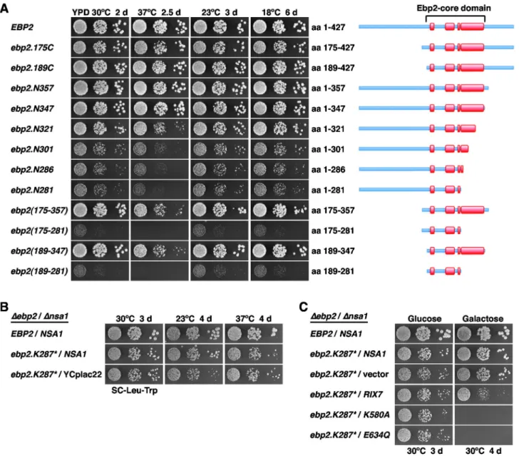
growth (Figure S14B). Notably, Ebp2 variants lacking most of or the complete C-terminal extension (ebp2.N357 and ebp2.N347; amino acids 1-357 and 1-347, respectively), showed wild-type growth at all tested temperatures (Figure 7A). To further delineate the C-terminal border, we tested the growth of additional deletion constructs (ebp2.N321, ebp2.N301, ebp2.N286 and ebp2.N281), remarkably revealing that even the construct lacking the complete last predicted α-helix of the central domain supported growth (Figure 7A). The slow-growth phenotype associated with this ebp2.N281 allele was almost identical to the one of the ebp2.N286 allele, which corresponds to a ‘clean’ variant of the original ebp2.K287* mutant. Next, we determined whether the central Ebp2 domain would be sufficient to mediate the function of Ebp2. The constructs lacking both the N- and C-terminal extensions, ebp2(175-357) and ebp2(189-347), conferred wild-type growth at all temperatures, except at 37°C where a growth defect was clearly visible (Figure 7A). Furthermore, the ebp2(175-281) and ebp2(189-281) constructs, having additionally the long predicted α-helix deleted from the C-terminus of the central domain, still sustained growth, albeit at much lower rate (Figures 7A and S14B). Altogether, we conclude that Ebp2 contains a conserved central domain, referred to as the Ebp2-core domain (amino acids 189-347), which forms the minimal unit ensuring almost complete functionality. Strikingly, deletion of the prominent C-terminal α-helix from the Ebp2-core domain still supports growth, indicating that the minimal structural and functional information for Ebp2 to at least partially fulfil its essential task resides in a short segment of ~90 amino acids between residues 189 and 281.
The ebp2.K287* allele suppresses the lethality of the
∆nsa1 null mutant
Next, we tested the capability of the ∆nop16 allele as well as the ebp2, rpl14a and rpf1 alleles, isolated via the mak5 sl-screens, to suppress the lethality of the ∆nsa1 null mutant. To this end, we generated double shuffle strains (for the essential EBP2 and RPF1 genes and the quasi-essential RPL14A gene) or an NSA1/∆nop16 shuffle strain. While expression of the ebp2.K287* mutant was promoting robust growth in the absence of Nsa1 (Figure S15A), the ∆nop16, rpl14a.L123*, rpf1.L199P and rpf1.C139R alleles did not restore growth of ∆nsa1 null mutant cells (Figure S15B-D). To better assess the extent of suppression, we compared the growth properties of ebp2.K287* cells in the presence and absence of NSA1 after plasmid shuffling on 5-FoA-containing plates. This analysis revealed that the ebp2.K287* allele very efficiently suppressed the lethality of the ∆nsa1 null mutant (Figure 7B). As observed before for the mak5.R728*, nop1.M232K and nop4.S460L alleles (Figure 1A-C), suppression was more efficient at 30°C than at 23°C (Figure 7B); moreover, growth of the ebp2.K287* mutant was even slightly better in the absence than in the presence of Nsa1 at 37°C (Figure 7B). Moreover, in validation of the above findings that dominant-negative alleles of RIX7 still exert their phenotypes in the mak5.R728* and nop4.S460L ∆nsa1 null ’suppressor’ strains (Figure 3A and 3B), galactose-induced over-expression of the dominant-negative RIX7.K580A and E634Q mutants also conferred lethality to
ebp2.K287*-Figure 7. Ebp2 contains an essential, central core domain and the ebp2.K287* allele suppresses ∆nsa1 lethality. (A) Growth phenotypes of viable ebp2 deletion mutants. Plasmid-borne wild-type EBP2 or the indicated ebp2 deletion mutants under the control of the authentic promoter were transformed into the EBP2 shuffle strain. After plasmid shuffling on plates containing 5-FoA, cells were restreaked on YPD plates and then spotted in 10-fold serial dilution steps onto YPD plates, which were incubated for 2 d at 30°C, 2.5 d at 37°C, 3 d at 23°C and 6 d at 18°C. The proteins encoded by the N- and/or C-terminally truncated ebp2 mutants are schematically depicted on the right, predicted α-helices within the central Ebp2-core domain are highlighted in red. (B) Bypass suppression of ∆nsa1 null lethality by the ebp2.K287* allele. The EBP2/NSA1 double shuffle strain was co-transformed with plasmids harbouring the EBP2 wild-type gene or the ebp2.K287* allele and a plasmid carrying NSA1 or the empty vector (YCplac22). After plasmid shuffling on plates containing 5-FoA, cells were restreaked on SC-Leu-Trp plates and then spotted in 10-fold serial dilution steps onto SC-Leu-Trp plates, which were incubated for 3 d at 30°C, 4 d at 23°C and 4 d at 37°C. (C) Dominant-negative RIX7 alleles confer lethality to ebp2.K287*/∆nsa1 cells. The EBP2/NSA1 double shuffle strain was co-transformed with plasmids harbouring the EBP2 wild-type gene or the ebp2.K287* allele and plasmids carrying the NSA1 wild-type gene or, expressed under the control of the GAL1-10 promoter, wild-type RIX7 and the dominant-negative RIX7.K580A or RIX7.E634Q alleles or empty vector. After plasmid shuffling on plates containing 5-FoA, cells were restreaked on SC-Leu-Trp plates and then spotted in 10-fold serial dilution steps onto SC-Leu-Trp (Glucose) and SGal-Leu-Trp (Galactose) plates, which were incubated for 3 d or 4 d at 30°C, respectively.
suppressed ∆nsa1 null mutant cells (Figure 7C). As observed for the mak5.R728* ∆nsa1 null ’suppressor’ strain (Figure 3A), over-expression of wild-type RIX7 already had a strong negative effect on the growth of ebp2.K287* cells lacking Nsa1 (Figure 7C).
Rix7 over-expression reduces growth and 60S formation in mak5 and ebp2 mutants
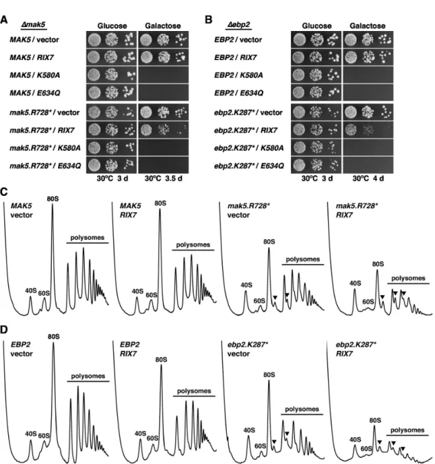
To expand on the observation that over-expression of wild-type RIX7 particularly affected the growth of the mak5.R728* and ebp2.K287* alleles in cells lacking Nsa1 (Figures 3A and 7C), we next determined the effects of galactose-induced over-expression of RIX7 from the GAL1-10 promoter on mak5.R728* and ebp2.K287* mutant cells in the presence of Nsa1. While over-expression of RIX7 had only a very minor effect on the growth of MAK5 and EBP2 wild-type cells, growth of the mak5.R728* and, even more dramatically, of the ebp2.K287* mutant was severely affected by RIX7 over-expression (Figure 8A and 8B). For control purposes, we also tested over-expression of the dominant-negative RIX7.K580A and E634Q mutants, conferring, as expected, lethality to wild-type as well as mak5.R728* and ebp2.K287* cells.
Next, we assessed the effects of RIX7 over-expression on 60S subunit biogenesis by polysome profile analysis. To this end, we pre-grew cells in synthetic liquid medium with raffinose as carbon source and then induced RIX7 expression by addition of galactose for ~16 h before preparing the cell extracts. In agreement with the mild effect on growth, RIX7 over-expression only resulted in very subtle changes of the polysome profile of MAK5 and EBP2 wild-type cells, as indicated by a slight decrease in free 60S subunits (Figure 8C and 8D). On the other hand, over-expression of RIX7 in mak5.R728* and ebp2.K287* cells aggravated their 60S subunit deficiency, as evidenced by a further decrease in free 60S subunits and a drastic reduction of polysome content (Figure 8C and 8D). We conclude that Rix7, when highly abundant, negatively acts on pre-60S subunits from mak5 and ebp2 mutant cells, which are likely structurally deranged and might therefore be prematurely channelled into a clearance pathway by excess Rix7 (see Discussion).
Discussion
A genetic network defines the ’Mak5 cluster’ as a novel 60S biogenesis module
In this study, we have identified, based on synthetic lethal interactions, the functional environment around the DEAD-box protein Mak5, which includes Ebp2, Nop16, Rpf1 and the r-protein Rpl14 (Figure 9). The intimate genetic interconnection between all four protein trans-acting factors indicates that Mak5, Ebp2, Nop16 and Rpf1 constitute a novel biogenesis cluster, which we propose to refer to as the ’Mak5 cluster’. Importantly, our genetic analysis represents the crucial first step towards the elucidation of the precise molecular function of Mak5, in conjunction with its genetically defined partner proteins, in one of the multiple assembly steps during the nucleolar phase of pre-60S maturation. Several lines of evidence support the genetic conclusion that Mak5, Ebp2,
Nop16 and Rpf1 form a functional unit acting together at a distinct step during the assembly of 60S subunits. First, Ebp2, Nop16 and Rpf1 are stoichiometric components of the late nucleolar pre-60S particle defined by the Nsa1-TAP bait [39], and we have shown here that Mak5 is also associated with the Nsa1 pre-60S particle (Figure 4). Notably, these four biogenesis factors are absent from the nucleoplasmic, Rix1-defined pre-60S particle (Figure 4 and [40]). In agreement, affinity purifications of Ebp2-TAP and Rpf1-HA reveal, as observed for the Nsa1 pre-60S particle, a predominant association with 27SB pre-rRNAs [39,73,85]. Moreover, genetic depletion or mutational perturbation of Mak5, Ebp2 and Rpf1 elicit similar pre-rRNA processing phenotypes, i.e. delayed conversion of 27SA2 into 27SB pre-rRNAs and reduced formation of mature 25S rRNA from the 27SB pre-rRNAs [58,83–85,89], altogether indicating an instability of 27SB pre-rRNA containing pre-60S ribosomes. Moreover, mutations in ebp2 and mak5 likely entail similar structural alterations within pre-60S particles since both the ebp2.K287* and the mak5.R728* allele suppress the lethality of ∆nsa1 null mutant cells (Figures 1A and 7B). Finally, in support of the specificity of the genetic interactions reported here, there have previously no synthetic lethal relations been unveiled for Nop16, Rpf1 and Rpl14. A sl-screen with the ebp2-14 allele, however, recently revealed a functional interaction between Ebp2 and Brx1 [73], which is, as Rpf1, a member of the σ70-like motif family of
RNA-binding ribosome biogenesis factors [85,90]. Yeast two-hybrid data further suggest a direct physical interaction between Ebp2 and Brx1 [73]. However, Brx1 clearly remains associated with pre-60S particles that already contain 7S pre-rRNA and mature 25S and 5.8S rRNAs [73,85], and its genetic depletion mainly leads to the accumulation of the 27SA2 pre-rRNA [90].
Altogether these observations suggest an additional or slightly different role for Brx1, therefore, future experiments are needed to address the interesting possibility that Brx1 might also be functionally connected with the other ’Mak5 cluster’ factors. The ’Mak5 cluster’ factors may orchestrate the structural arrangement of a eukaryote-specific 60S subunit surface composed of r-proteins Rpl6, Rpl14, and Rpl16 and rRNA expansion segments ES7L and ES39L
What could be the role(s) of the ’Mak5 cluster’ during pre-60S maturation? The functional connection between the ’Mak5 cluster’ factors and the r-protein Rpl14a may offer valuable insight into the potential role of these biogenesis factors. The almost exclusively eukaryote-specific Rpl14 (L14e) is located close to the P-stalk on the solvent-side of the 60S r-subunit (Figure S10B and [56]). Interestingly, Rpl14 is found in some, but not all, archaeal organisms; its two prominent C-terminal α-helices at the least, however, are strictly eukaryote specific (Figure S11A). The α-helical part of Rpl14, not including its C-terminal α-helix, is clamped between the long bent helix of the eukaryote-specific rRNA expansion segment ES7L and a short single-stranded region of ES39L on the surface of 60S subunits (Figure S10C and [56]). On the other hand, the β-stranded part of Rpl14 dives a little bit deeper into the 60S subunit where it makes both contact with the
Figure 8. Over-expression of RIX7 enhances the growth defect and 60S subunit deficiency of mak5.R728* and ebp2.K287* mutants. The MAK5 (A) and EBP2 (B) shuffle strains were co-transformed with plasmids harbouring either the MAK5 or EBP2 wild-type genes or the mak5.R728* or ebp2.K287* mutant alleles and empty vector or plasmids expressing wild-type RIX7 and the dominant-negative RIX7.K580A or RIX7.E634Q alleles under the control of the GAL1-10 promoter. After plasmid shuffling on plates containing 5-FoA, cells were restreaked on SC-Leu-Trp plates and then spotted in 10-fold serial dilution steps onto SC-Leu-Trp (Glucose) and SGal-Leu-Trp (Galactose) plates, which were incubated at 30°C for 3 d, 3.5 d or 4 d, respectively. (C and D) Over-expression of RIX7 affects 60S subunit biogenesis in mak5.R728* and ebp2.K287* mutant cells. The MAK5 (C) and EBP2 (D) shuffle strains were co-transformed with plasmids harbouring either the MAK5 or EBP2 wild-type genes or the mak5.R728* or ebp2.K287* mutant alleles and empty vector or a plasmid expressing wild-type RIX7 under the control of the GAL1-10 promoter. After plasmid shuffling on plates containing 5-FoA, cells were restreaked on SC-Leu-Trp plates. Transformed cells were first pre-grown in SC-Leu-Trp medium, then diluted into SC-Leu-Trp medium with raffinose as carbon source and, finally, expression of wild-type RIX7 was induced by addition of galactose to the medium. After ~16 h of galactose induction, cell extracts were prepared under polysome-conserving conditions and eight A260 units were resolved in 10-50% sucrose gradients. The absorption profiles were recorded by continuous monitoring at A254. Sedimentation is from left to right. The peaks of free 40S and 60S subunits, 80S free
couples/monosomes and polysomes are indicated. Half-mers are highlighted by arrowheads. doi: 10.1371/journal.pone.0082741.g008
eukaryote-specific r-protein Rpl20 (L20e) and the backbone of a short segment (nucleotides 1183-1185) of the 25S rRNA [56]. The N-terminal extension of Rpl14 contacts, by forming a short parallel β-strand, a three-stranded β-sheet of Rpl9 (L6) (Figure S10C and [56]), however, this interaction does not seem to take place in higher eukaryotic 60S subunits (Figure S10A and [57]). Most notably, the eukaryote-specific C-terminal α-helix of Rpl14, which is truncated in the rpl14a.L123* allele (Figures
S10A and S10C), is involved in a series of eukaryote-specific interactions, comprising contacts with (i) the long C-terminal, eukaryote-specific α-helix of Rpl16 (L13), (ii) Rpl6 (L6e) and (iii) ES39L (Figures S10C and S11B; [56]). The C-terminal α-helices of Rpl14 and Rpl16 are sandwiched between two distinct structural elements of ES39L, and Rpl6, which is located above the early-assembling Rpl33 (L33e) [91], connects the base of the long bent helix of ES7L with ES39L Figure 9. Model summarizing the genetic networks established in this study. Nsa1 (green) is a component of late nucleolar pre-60S particles (light bordeaux) whose release and recycling is mediated by the AAA-type ATPase Rix7 (turqoise) [39]. Mutant alleles of the genes encoding the 60S biogenesis factor Ebp2 (red), the DEAD-box RNA helicase Mak5 (light blue), the methyltransferase component of C/D-box snoRNPs Nop1 (chartreuse) and the RRM-containing RNA-binding protein Nop4 (pink) suppress the lethality of ∆nsa1 null mutant cells. These bypass suppressions are indicated by red double arrows, whose line thickness correlates with the observed strength of the suppression. Mak5 forms a genetic network with Ebp2, Nop16, Rpf1 and the r-protein Rpl14. Synthetic lethal and synthetic enhancement interactions amongst these are depicted by blue continuous or dashed ’negative’ arrows, respectively. We propose that Ebp2, Mak5, Nop16 and Rpf1, referred to as the ’Mak5 cluster’ factors, may orchestrate the structural arrangement of a eukaryote-specific 60S subunit surface composed of the r-proteins Rpl6, Rpl14 and Rpl16 and the rRNA expansion segments ES7L and ES39L (not depicted). Since dominant-negative alleles of RIX7 retain their phenotype in the absence of Nsa1, Rix7 may have additional nuclear substrates besides Nsa1. Finally, over-expression of Rix7 negatively affects growth of mak5 and ebp2 mutant cells both in the absence and presence of Nsa1, suggesting that Rix7, at least when excessively abundant, may act on structurally defective pre-60S subunits and may subject them to degradation (gray trashcan). We therefore propose that Rix7 may, besides specifically releasing and recycling Nsa1, sense the structural integrity of pre-60S particles and, if these are excessively damaged, channel them into a clearance pathway.