HAL Id: hal-02990072
https://hal.archives-ouvertes.fr/hal-02990072
Submitted on 14 Dec 2020
HAL is a multi-disciplinary open access
archive for the deposit and dissemination of sci-entific research documents, whether they are pub-lished or not. The documents may come from teaching and research institutions in France or abroad, or from public or private research centers.
L’archive ouverte pluridisciplinaire HAL, est destinée au dépôt et à la diffusion de documents scientifiques de niveau recherche, publiés ou non, émanant des établissements d’enseignement et de recherche français ou étrangers, des laboratoires publics ou privés.
Hybrid giant lipid vesicles incorporating a PMMA-based
copolymer
Ylenia Miele, Anne-Françoise Mingotaud, Enrico Caruso, Miryam Malacarne,
Lorella Izzo, Barbara Lonetti, Federico Rossi
To cite this version:
Ylenia Miele, Anne-Françoise Mingotaud, Enrico Caruso, Miryam Malacarne, Lorella Izzo, et al.. Hybrid giant lipid vesicles incorporating a PMMA-based copolymer. Biochimica et Biophysica Acta (BBA) - General Subjects, Elsevier, 2020, pp.129611. �10.1016/j.bbagen.2020.129611�. �hal-02990072�
Hybrid giant lipid vesicles incorporating a PMMA-based
1copolymer
2Ylenia Mielea, Anne-Françoise Mingotaudb, Enrico Carusoc, Miryam C. Malacarnec, Lorella 3
Izzo*c
, Barbara Lonetti*b, Federico Rossid 4
a Department of Chemistry and Biology, University of Salerno, Via Giovanni Paolo II, 132, 5
84084 Fisciano – Italy. 6
b Laboratoire des IMRCP, Université de Toulouse, CNRS UMR 5623, Université Toulouse III - 7
Paul Sabatier, 118 Rte de Narbonne, F-31062 Toulouse cedex 9 – France. 8
c Dipartimento di Biotecnologie e Scienze della Vita, Università degli Studi dell’Insubria, via J. 9
H. Dunant, 3, 21100 Varese – Italy. 10
d Department of Earth, Environmental and Physical Sciences – DEEP Sciences – Pian dei 11
Mantellini 44, 53100 Siena – Italy. 12
* Corresponding Author: L.I. lorella.izzo@uninsubria.it
13 B.L lonetti@chimie.ups-tlse.fr 14 15 16 17 18
STATISTICAL SUMMARY 1 Words: 6494 2 Figures/Tables: 5 3 HIGHLIGHTS 4
• Methacrylate based polymer is mixed with a lipid to produce giant vesicles 5
• Electroformation is not a suitable method to obtain such hybrid systems 6
• Phase transfer method is used to obtain hybrid giant vesicles 7
• Phase transfer method useful when the polymer has a relatively high Tg 8
ABSTRACT 1
Background 2
In recent years, there has been a growing interest in the formation of copolymer-lipid hybrid self-3
assemblies, which allow combining and improving the main features of pure lipid-based and 4
copolymer-based systems known for their potential applications in the biomedical field. As the 5
most common method used to obtain giant vesicles is electroformation, most systems so far used 6
low Tg polymers for their flexibility at room temperature. 7
Methods 8
Copolymers used in the hybrid vesicles have been synthesized by a modified version of the 9
ATRP, namely the Activators ReGenerated by Electron Transfer ATRP and characterized by 10
NMR and DSC. Giant hybrid vesicles have been obtained using electroformation and droplet 11
transfer method. Confocal fluorescence microscopy was used to image the vesicles. 12
Results 13
Electroformation enabled to obtain hybrid vesicles in a narrow range of compositions (15 mol % 14
was the maximum copolymer content). This range could be extended by the use of a droplet 15
transfer method, which enabled obtaining hybrid vesicles incorporating a methacrylate-based 16
polymer in a wide range of compositions. Proof of the hybrid composition was obtained by 17
fluorescence microscopy using labelled lipids and copolymers. 18
Conclusions 19
This work describes for the first time, to the best of our knowledge, the formation of giant hybrid 20
polymer/lipid vesicles formed with such a content of a polymethylmethacrylate copolymer, the 21
glass temperature of which is above room temperature. 22
General Significance 23
This work shows that polymer structures, more complex than the ones mostly employed, can be 1
possibly included in giant hybrid vesicles by using the droplet transfer method. This will give 2
easier access to functionalized and stimuli-responsive giant vesicles and to systems exhibiting a 3
tunable permeability, these systems being relevant for biological and technological applications. 4
KEYWORDS liposomes; polymersomes; hybrid GUVs; self-assembly; surfactant; polymer; 5
phase transfer method 6
1. INTRODUCTION 7
Liposomes and polymersomes are self-assembled vesicles formed by lipids and 8
copolymers respectively and characterized by the presence of a hydrophobic double layer 9
delimitating an internal water pool. Their structure allows the encapsulation of both hydrophobic 10
and hydrophilic components with high loading efficiency, which mainly boosted the research in 11
applications related to drug delivery [1–4]. Besides, the last 10 years of research witnessed a 12
growing interest in polymersomes used as reactors [5]. For this particular application, the low 13
bilayer permeability of the high molecular weight constituents represents a drawback. Block 14
copolymers forming intrinsically permeable polymersomes [6,7], polymersomes whose 15
permeability can be modulated by a stimulus, i.e. pH [8], light [9], CO2 [10], and self-adaptive 16
polymersomes [5] have been proposed. A further, less explored possibility is the use of hybrid 17
lipid/polymer vesicles, which could exploit the complementary properties of liposomes and 18
polymersomes, i.e. biocompatibility, biodegradability and permeability of the former and high 19
mechanical stability and chemical versatility of the latter. These hybrid vesicles have higher 20
permeability to small dyes like carboxyfluorescein or calcein with respect to polymersomes [11– 21
13] and it has been recently shown that the hybrid vesicles permeability could be increased by 22
adding phospholipase A2 that can selectively degrade lipids [14]. Examples of hybrid nanoscale 1
polymersomes have been proposed for applications in nanomedicine and seemed to improve the 2
targeting efficiency [15,16]. Interestingly, hybrid vesicles at the micron scale are valid 3
alternatives to giant liposomes as simplified cell models for a deeper understanding of 4
fundamental biological processes [17,18]. They have indeed the potential for mimicking the 5
membrane functionalities like the presence of raft-like domains [19,20], membrane asymmetry 6
[21] transport or recognition properties thanks to the insertion of proteins in the membrane [22– 7
24] and even chemical energy-driven ATP production [25] or cell compartmentalization [26]. To 8
date, the potential responsiveness of block copolymers has not yet been exploited for hybrid 9
vesicles. Indeed, only examples of temperature-responsive hybrid vesicles are reported in the 10
literature, exploiting the phase transition of dioleylphosphatidyl choline based lipids [27]. 11
One of the reasons of the lack of polymer/lipid hybrid systems is the limited choice of the 12
amphiphilic copolymers which were generally restricted to a small group based on the 13
combination of poly(ethylene oxide) or poly(oxazoline) as hydrophilic unit and either 14
poly(dimethyl siloxane) (PDMS) [28,29], poly(butadiene) (PBD) [30,31], polyisobutylene (PIB) 15
[32] and very recently butylacrilate [33] as hydrophobic moiety. 16
Different methodologies have been used to form polymer based giant unilamellar vesicles 17
(GUVs). They have been mainly inspired from those developed for lipids [34]. Electroformation 18
method is the most employed one. It is a solvent-free method where a dry amphiphilic thin film 19
is hydrated in the presence of an alternating current probably inducing fluctuations and interlayer 20
repulsions, which favours the swelling and release of giant vesicles [35]. It was applied to block-21
copolymers for the first time in the case of poly(ethylene oxide)-block-poly(ethyl ethylene) [36]. 22
Electroformation can be applied to a limited class of block-copolymers [33,37,38], as the choice 23
of the nature of the block copolymer is usually dictated by the hydrophobic block glass transition 1
temperature, since this should be as low as possible in order to guarantee a sufficient flexibility 2
and mobility of the polymer chains. Gentle hydration has been described even earlier than 3
electroformation for brain phospholipids [39] and is quite difficult to be applied to block-4
copolymers as witnessed by the very limited number of publications [40–42]. Gel assisted 5
hydration using polyvinyl alcohol (PVA) [43] or agarose [44] have been proposed for lipid 6
vesicles and PVA was also used for polymer-based vesicles [45]. In this case, the presence of 7
unwanted impurities in the bilayer or lumen cannot be excluded. 8
Emulsion phase (or droplet) transfer has been developed for lipids [46], improved in 9
microfluidic devices [47,48] or special centrifugation set-ups [49–51] (continuous droplet 10
interface crossing encapsulation, i.e. cDICE) and has been rarely used for block copolymers 11
[21,52]. This method is based on a water-in-oil emulsion where water droplets are coated with 12
the amphiphilic species. These droplets are allowed to cross the interface of an oil-on-water 13
biphasic system stabilized by an amphiphilic monolayer, thus forming the vesicles. A known 14
drawback of this method is the presence of residual oil between the lipid leaflets [53]. However, 15
membrane bending rigidity analysis revealed that mineral oil doesn’t affect the mechanical 16
properties of the membrane [54]. 17
Because hybrid GUVs are essential tools for developing synthetic reactors or biomimetic cells 18
and are so far very limited, in this work we propose to use block copolymers based on 19
methylmethacrylate (MMA) and N,N-dimethylaminoethyl methacrylate (DMAEMA) mixed 20
with palmitoyl oleyl phosphatidyl choline (POPC) in order to obtain giant hybrid vesicles. The 21
presence of sensitive DMAEMA unit could provide the access to systems with pH-22
responsive permeability. In the literature MMA/DMAEMA based block-copolymers have been 23
mainly used to develop nano-sized polymersomes. Polymeric nano-vesicles based on butyl-1
methacrylate (BMA) and DMAEMA were developed as reactors for the urea-urease system, for 2
example [55]. We recently showed that the copolymer poly(ethylene glycol monomethyl ether)-3
block- poly( methyl methacrylate –random- N,N-dimethyl amino ethyl methacrylate),
mPEG-4
block-P(MMA-ran-DMAEMA), was able to form nano-vesicles in a wide range of DMAEMA
5
chemical compositions and architectures (linear and branched) [56]. Furthermore, they exhibited 6
a pH-dependent swelling characterized by a strong increase in size, up to 10 times passing from 7
pH 7.4 to pH 4.4. This swelling was attributed to electrostatic repulsions at low pH, linked to the 8
concurrent increase of protonated DMAEMA units. 9
As for giant vesicles, which were not necessarily unilamellar ones, poly(methacrylic acid)-10
block-poly(methylmethacrylate-random-methacrylic acid), PMAA-block-P(MMA-ran-MAA)
11
could form giant vesicles in a photo-polymerization induced self-assembly process in an aqueous 12
methanol solution [57,58]. The authors investigated the pH-responsive behaviour of the vesicles 13
showing that they were disrupted in basic environment (pH 12) and they could be reversibly 14
reconstructed at neutral pH. Yoshida also characterized polymeric giant vesicles with 15
copolymers containing DMAEMA units inserted in the P(MMA-ran-MAA) block [59]. These 16
vesicles were unstable and had many holes on the surface. When quaternized DMAEMA was 17
used the vesicles were stable, but they had much smaller size (below 1 µm). 18
Hybrid GUVs made of poly(cholesteryl methacrylate)-block-poly(2-(dimethylaminoethyl 19
methacrylate) (pCMA-block-pDMAEMA) were characterized with confocal microscopy by W. 20
Zong et al. [16] to confirm the presence of both phospholipids and the block copolymer in the 21
same membrane, pCMA constitutes the hydrophobic block, while the pH-sensitive pDMAEMA 22
content was 70% w/w (corresponding to 3.6 mol % using Mn=47.5 kD as reported in Table 1 of 1
the cited work). Also, hybrid nano-vesicles were formed by this copolymer mixed with 2
phospholipids containing palmitoyl and oleyl as fatty acyl groups and different charged 3
headgroups (i.e. phosphatidylcholine, ethylphosphocholine and phosphatidylserine) showed a 4
prevalent cytosolic localization when incubated with mouse macrophages, indicating a good 5
potential for applications in drug delivery [16]. 6
In this paper, we want to show that a block copolymer with high molecular weight and high Tg 7
can be successfully incorporated into hybrid giant vesicles. We propose to use mPEG-block-8
P(MMA-grad-DMAEMA) in combination with 1-palmitoyl-2-oleoyl-sn-glycero-3-9
phosphocholine (POPC) to prepare vesicles by means of two formation methods, namely 10
electroformation and droplet transfer; it will be shown that the high Tg polymer diminishes the 11
efficiency of the electroformation method even at low (~ 15 mol %) polymer content, whilst the 12
droplet transfer method allows the formation of hybrid vesicles for a wide range of polymer/lipid 13
ratios. With this approach, the polymer content in the hybrid GUVs has been sensibly increased 14
with respect to that already reported in the literature (~ 4 mol %). 15
2. MATERIALS AND METHODS 16
Materials
17
Copper bromide (CuBr2), 2,2’–bipyridine (bpy), tin (II) 2-ethylhexanoate (Sn(EH)2), 2,4-18
dimethyl-3-ethylpyrrole, trifluoroacetic acid (TFA), dichlorodicyanoquinone (DDQ), 19
triethylamine (Et3N), boron trifluoride etherate (BF3.Et2O), butylated hydroxytoluene (BHT), 20
basic alumina, Sephadex LH-20, 1-palmitoyl-2-oleoyl-sn-glycero-3-phosphocholine, Mineral oil 21
M5904, toluene, methanol, dimethylformamide (DMF), n-hexane, poly(ethylene glycol) 22
monomethylether (mPEG, Mn = 2000 gmol-1, Mw/Mn = 1.16 and Mn = 5000 gmol-1, Mw/Mn = 1
1.02) and pyranine were purchased from Sigma-Aldrich and used without further purification. 2
18:1 Liss Rhodamine PE was used as received by Avanti Polar Lipids. Methylmethacrylate 3
(MMA) and N,N-(dimethylamino)ethylmethacrylate (DMAEMA) (Sigma-Aldrich) were purified 4
prior copolymerizations by means of a column filled with basic alumina to remove the inhibitors. 5
Dichloromethane (Sigma-Aldrich) used for BODIPY synthesis was distilled over CaCl2. 6
All manipulations involving air-sensitive compounds were carried out under nitrogen 7
atmosphere using Schlenk techniques. 8
Synthesis of the fluorescent-labeled DMAEMA monomer
2,6-diethyl-1,3,5,7-tetramethyl-8-[4-9
[8-(2-methacryloylethyl)dimethylammoniumbromideoctyloxy]phenyl]-4,4’-difluoroboradiaza
10
indacene (BODIPY-DMAEMA)
11
As a first step, the precursor 2,6-diethyl-1,3,5,7-tetramethyl-8-[4-(8-bromooctyloxy)phenyl]-4,4’-12
difluoroboradiaza indacene was synthesized. 4-(8-bromooctyloxy)benzaldehyde was prepared as 13
previously described [60]. 720 µL of 4-(8-bromooctyloxy)benzaldehyde (2.3 mmol) and 756 µL 14
of 2,4-dimethyl-3-ethylpyrrole (5.6 mmol) were then dissolved in 50 mL of absolute CH2Cl2 15
under N2 atmosphere, ten drops from a Pasteur pipette of TFA were added and the solution was 16
stirred at RT overnight or until TLC analysis showed complete consumption of the aldehyde. At 17
this time, 785 mg of DDQ (3.45 mmol) were added and stirring continued for 20 min. Then, 5 mL 18
of Et3N and 5 mL of BF3.OEt2 were added. The mixture was stirred for 12 h and the organic layer 19
containing the crude product was subsequently washed three times with water; the organic 20
solution was dried over Na2SO4, and evaporated to dryness. The raw material was purified by 21
chromatography (SiO2, petroleum ether - CH2Cl2, 1:1) to afford 246 mg of product (yield: 18.2%) 22
in the form of orange needles. C31H42BBrF2N2O, MM = 587.39 gmol-1, UV-vis (CH2Cl2): 528 nm 1
(ε = 92600 M-1 cm-1). Quantum efficiency of fluorescence Φfluo (in CH2Cl2): 0.45 (544 nm) was 2
calculated according to the following equation: 3
Φfluosample=Φfluostandard×(Ifluosample/Ifluostandard)×(Absstandard/Abssample) 4
where Ifluo is the fluorescence intensity at the specific excitation wavelength, Abs denotes the 5
absorbance at the excitation wavelength. 6 1H NMR (CDCl 3) : 1.00 (t, 6H, 2×CH3); 1.46 (s, 6H, 2×CH3); 1.45-1.55 (m, 8H, 4×CH2); 7 1.82-1.93 (m, 4H, 2×CH2); 2.32 (q, 4H, 2×CH2); 2.58 (s, 6H, 2×CH3); 3.45 (t, 2H, CH2Br); 4.03 8 (t, 2H, CH2O); 6.00 (s, 2H, 2×CH); 7.02 (d, 2H, 2×CH); 7.18 (d, 2H, 2×CH). 9
To obtain the fluorescent-labeled DMAEMA monomer, 10 mg (0.045 mmol) of 2,6-diterbutyl-10
4-methylphenol (BHT) were added to a mixture of 236 mg of 2,6-diethyl-1,3,5,7-tetramethyl-8-11
[4-(8-bromooctyloxy)phenyl]-4,4’-difluoroboradiaza indacene (0.402 mmol) in 3 mL of 12
DMAEMA (17.75 mmol), reaction scheme and chemical structures are reported in Figure S1 of 13
the supporting information. The mixture was kept at 70 °C for 24 h. During the reaction time, a 14
solid product was formed. After filtration, the product was washed 4 times with 20 mL of diethyl 15
ether affording 214 mg (71.4%) of product. C39H57BBrF2N3O3, MM = 744.60 gmol-1(ESI-MS 16
spectrum is reported in Figure S2 of the supporting information). 1H NMR (CDCl
3) : 0.97 (t, 17 6H, 2×CH3); 1.33 (s, 6H, 2×CH3); 1.40 (m, 8H, 4×CH2); 1.80 (m, 4H, 2×CH2); 1.95 (s, 3H, 18 CH3), 2.25-2.33 (q, 4H, 2×CH2); 2.51 (s, 6H, 2×CH3); 3.56 (s, 6H, 2×CH3); 3.61-3.65 (t, 2H, 19 CH2); 3.97-4.02 (t, 2H, CH2); 4.15 (t, 2H, CH2); 4.65 (t, 2H, CH2); 5.68 (s, 1H, CH); 6.15 (s, 1H, 20
CH); 6.96-6.99 (d, 2H, 2×CH); 7.13-7.15 (d, 2H, 2×CH). Details of 1H peak assignment and 21
HSQC-NMR spectra are reported in Figures S3 – S5 of the supporting information. 22
Synthesis of mPEG-block-P(MMA-grad-DMAEMA) copolymers.
Macroinitiator mPEG-Br was synthesized according to the procedure reported in literature. [61] 1
In a typical procedure, the copolymerization of MMA/DMAEMA was carried out in a 50 mL 2
glass flask charged, under nitrogen atmosphere, with 0.1 g of mPEG-Br macroinitiator in 5 mL 3
of toluene. Subsequently, 100 μL of CuBr2 (1 × 10⁻³ M in DMF, 10-4 mmol), 100 μL of bpy (1 × 4
10−2 M in DMF, 10-3 mmol), 100 μL of Sn(EH)
2 (1 × 10⁻² M in toluene, 10-3 mmol), 2-4 mL of 5
MMA (18.8-37.6- mmol) and 1.2 – 2.5 mL of DMAEMA (7.10 – 14.8 mmol) were added (the 6
volumes of MMA and DMAEMA were varied to change the final composition in the 7
copolymer). The mixture was thermostated at 60 °C and magnetically stirred. The reaction was 8
stopped after 4h by adding n-hexane. The copolymer was recovered by filtration, washed with
9
cold methanol (~ 4 °C) and dried in vacuum at 40 °C. 1H-NMR (400 MHz, CDCl3): 0.87-1.03 10
(-CH3 main chain), 1.83-1.91 (-CH2- main chain), 2.30 (-N(CH3)2)), 2.58 (–OCH2 CH2 N(CH3)2), 11
3.61 (-OCH3), 3.66 (-OCH2CH2-), 4.08 (-OCH2 CH2 N(CH3)2). 13C-NMR(400 MHz, CDCl3): 12
16.9-19.1 (-CH3 main chain), 44.9-45.3 (quaternary carbon in the main chain), 46.2 (-N(CH3)2), 13
52.2 (-OCH3, MMA), 54.6 (-CH2- main chain), 57.6 (-OCH2 CH2 N(CH3)2), 63.5 (-OCH2 CH2 14
N(CH3)2), 70.9 (-OCH2 CH2-), 176.3-178.2 (-C=O). Details of 1H peak assignment and 13C NMR 15
spectra are reported in Figure S6 of the supporting information. 16
Chemical composition and the molecular weight (Mn) were evaluated via 13C NMR. The molar 17
fractions of the components in the final copolymers were evaluated through the equations: 18
XmPEG = ImPEG
ImPEG+ 2IMMA+ IDMAEMA (1)
19
XMMA = 2IMMA
ImPEG + 2IMMA+ IDMAEMA (2)
XDMAEMA= IDMAEMA
ImPEG+ 2IMMA+ IDMAEMA (3)
1
where ImPEG is the integration of the signal relative to mPEG units: –OCH2CH2 –, IMMA is the 2
signal corresponding to the units –OCH3 of MMA and IDMAEMA integrates the two carbons of the 3
amine group of DMAEMA (-N(CH3)2). Mn was calculated from the following equation: 4
Mn= DPmPEGMM EO
( )
+ DPMMAMM MMA(
)
+ DPDMAEMAMM DMAEMA(
)
(4)5
where: 6
DPmPEG = MnPEG/44 7
DPMMA = DPmPEG (2IMMA/ImPEG) 8
DPDMAEMA = DPmPEG (IDMAEMA/ImPEG) 9
The synthesis of the fluorescent-labelled copolymer was carried out under the same experimental 10
conditions described above by adding 2 mL of MMA (18.8 mmol), 1.2 mL of DMAEMA (7.10 11
mmol), 2 mL of DMF and 5 mg of the fluorescent monomer BODIPY-DMAEMA (6.7 × 10⁻³ 12
mmol) to the reaction mixture. The applied procedure of purification [62,63]was based on the 13
use of lipophilic Sephadex LH-20 to separate molecules with different molecular weights. A 14
mini column (Pasteur pipette) was loaded with the lipophilic stationary phase (~1.5 g of 15
Sephadex previously swollen for one hour in 10 mL of methanol) and filled with methanol to 16
elute the samples. 1H-NMR (400 MHz, CDCl3): 0.87-1.04 (-CH3 main chain), 1.27 ppm (6 17
CH2 of the alkyl chain of the BODIPY-DMAEMA), 1.83-1.91 (-CH2- main chain), 2.30 (-18
N(CH3)2)), 2.59 (–OCH2 CH2 N(CH3)2), 3.61 (-OCH3), 3.65 (-OCH2CH2-), 4.08 (-OCH2 CH2 19
N(CH3)2). 13C-NMR (400 MHz, CDCl3): 16.5-18.7 (-CH3 main chain), 29.7 ppm (6 CH2 of the 20
alkyl chain of the BODIPYDMAEMA), 44.6-44.9 (quaternary carbon in the main chain), 45.8 (-1
N(CH3)2), 51.8 (-OCH3, MMA), 54.4 (-CH2- main chain), 57.2 (-OCH2 CH2 N(CH3)2), 63.1 (-2
OCH2CH2 N(CH3)2), 70.6 (-OCH2CH2-), 176.3-178.2 (-C=O). Details of 1H peak assignment, 3
13C NMR and HSQC-NMR spectra are reported in Figures S7 and S8 of the supporting 4
information. 5
Chemical composition and the molecular weight (Mn) were evaluated via 13C NMR through 6 the equations: 7 mPEG mPEG BODIPYDMAEMA mPEG 2 MMA DMAEMA
2 I X I I I I = + + + (5) 8 MMA MMA BODIPYDMAEMA
mPEG MMA DMAEMA
2 2 2 I X I I I I = + + + (6) 9 DMAEMA DMAEMA BODIPYDMAEMA mPEG 2 MMA DMAEMA
2 I X I I I I = + + + (7) 10 BODIPYDMAEMA BODIPYDMAEMA BODIPYDMAEMA
mPEG MMA DMAEMA
2 2 2 I X I I I I = + + + (8) 11
where ImPEG is the integration of the signal relative to mPEG units: -OCH2CH2-; IMMA is the 12
integration of the signal relative to MMA units: -OCH3; IDMAEMA is the integration of methyl 13
group of the signal relative to DMAEMA units; IBODIPYDMAEMA are the 4 CH2 of the fluorescent 14
mojety (signals labeled i1-4 in the NMR spectra, figures S7-S8 of the supporting info). The 15
degrees of polymerization and hence the molecular weights were calculated from the following 1
equation: 2
Mn=DPmPEG(MMEO) + DPMMA(MMMMA) +
+ DPDMAEMA(MMDMAEMA) + DPBODIPYDMAEMA(MMBODIPYDMAEMA) (9) 3 where: 4
(
)
(
)
(
)
mPEG n(mPEG)MMA mPEG MMA mPEG
DMAEMA mPEG DMAEMA mPEG
BODIPYDMAEMA mPEG BODIPYDMAEMA mPEG
DP = M /44 DP = DP 2I /I DP = DP I /I DP = DP I /2I 5 NMR Analysis. 6
1H and 13C NMR spectra were recorded at 25 °C in CDCl
3 using a Bruker Avance 400 MHz 7
spectrometer (D1 = 5 s for 13C NMR). Samples were prepared by introducing 20 mg of 8
copolymer and 0.5 mL of CDCl3 into a tube (5 mm outer diameter). Chemical shifts () are listed 9
as parts per million: 1H NMR spectra are referenced using the residual solvent peak at = 7.26 10
ppm, in 13C NMR spectra the residual solvent peak is at = 77.2 ppm. 11
12 13
DSC.
14
The glass transition temperatures were measured with Dynamic Scanning Calorimetry (Mettler
15
Toledo DSC1 STAR SYSTEM FRS5). About 10 mg of sample were inserted in a 100 L 16
aluminum pan and heating/cooling cycles were registered at 20 K/min. In the first run, the 17
sample was heated from 25 °C to 165 °C, followed by cooling to -60 °C. In the other two runs, 1
the sample was heated from -60 °C to 165 °C and cooled from 165 °C to -60 °C. The Tg was 2
measured on the second heating run where the inflection point is generally more evident. 3
Microscopy.
4
Images were acquired on an inverted optical microscope (Eurotek Orma INV100TFL) using a 5
20× objective and then analysed with the software ImageJ. The mixed membranes were 6
characterized with a confocal microscope (Leica TCS SP2 and TIRF OLYMPUS FV1000). The 7
fluorescent polymer and the fluorescent lipid were excited respectively at 488 nm, with an Ar 8
laser, and at 561 nm, with a DPSS 561 laser and the fluorescence was collected with 9
photomultipliers tubes PMTs in the wavelength ranges 498 – 530 nm and 571 – 630 nm, 10
respectively. 11
Preparation of Giant Vesicles.
12
Droplet Transfer Method. Stock solutions of [POPC] = 3 mM (2.28 mg mL-1) and [copolymer] 13
= 5 mg mL-1 (0.36 mM by using the copolymer average molecular weight Mn = 13850 g mol-1) 14
were prepared in mineral oil. The fluorescent probes, being less soluble in mineral oil, were 15
dissolved in CHCl3 at the concentration [fluo-polymer]CHCl3 = 10 mg mL-1 (0.027 mM using an 16
average molecular weight Mn = 370928 g mol-1) and [18:1 Liss Rhod PE]CHCl3 = 0.77 mM (1 mg 17
mL-1). Giant Unilamellar vesicles were prepared with the Droplet Transfer Method described by 18
Pautot et al. for lipid-based GUVs [46] using lipid:copolymer molar ratios 93:7, 85:15 and 60:40 19
(corresponding to lipid:copolymer weight ratios 0.42:0.58, 0.24:0.76, 0.08: 0.92 by using the 20
copolymer average molecular weight Mn = 13850 g mol-1). An Eppendorf tube was filled with 21
500 μL of an aqueous phase containing 200 mM of glucose (O-solution), then 300 μL of a 22
hydrophobic interfacial phase containing lipids and polymers at various ratios (obtained by 1
mixing the POPC and polymer stock solution) was poured on top of the O-solution. A second 2
Eppendorf tube was used to prepare a water/oil microemulsion: 20 μL of an aqueous solution 3
containing 200 mM of sucrose (inner solution, I-solution), were mixed by pipetting with 600 μL 4
of an oil phase containing the same amount of lipids and polymers as the interfacial phase and 5 5
μL of chloroform solutions of fluorescent probes (chloroform was firstly evaporated in argon 6
stream before and then oil containing other amphiphiles was added). The microemulsion was 7
then poured over the interfacial phase and vesicles were formed by centrifuging the tube at 6000 8
rpm for 10 minutes at room temperature. Vesicles were collected as a pellet at the bottom of the 9
aqueous phase and gently washed with 100 μL of O-solution and finally observed at the 10
microscope. In these experimental conditions, however, pure polymeric vesicles could not be 11
formed. When POPC was not present in the initial oil solution, the precipitation of a drop of 12
coloured polymer (i.e. containing the fluorescent polymeric probe or pyranine when employed) 13
was always detected at the bottom of the Eppendorf tube. Problems might be related with 14
difference in density between the I- and O-solution that could be overcame by employing a 15
different nonpolar solvent or by changing the concentrations of the sugars. 16
Electroformation. GUVs were prepared on a Vesicles prep pro instrument produced by Nanion
17
through the electroformation method described by Angelova et al. [64] for lipid-based giant 18
vesicles. Stock solution of [POPC] = 1.32 mM (1 mg mL-1) and [copolymer] = 1 mg mL-1 (0.072 19
mM if we consider a copolymer average weight of 13850 g/mol) and POPC where prepared at 1 20
mg mL-1 (both polymer and lipid) in chloroform and [copolymer] = 1 mg mL-1(0.072 mM by 21
using the copolymer average molecular weight Mn = 13850 g mol-1). For electroformation, they 22
were mixed in different ratios so that the total final concentration of the polymer plus POPC was 23
0.25 mg mL-1 and the total deposited amount was fixed in order to avoid the formation of a thick 1
film. The POPC concentration was 0.136 mM, 0.08 mM and 0.05 mM in the samples with 2
POPC:copolymer molar ratios equal to 93:7, 85:15 and 70:30 respectively (corresponding to 3
lipid:copolymer weight ratios 0.42:0.58, 0.24: 0.76, 0.11: 0.89 by using the copolymer average 4
molecular weight Mn = 13850 g mol-1). 5
5 μL of the obtained chloroform solutions of the polymer and lipid at different molar ratios 6
were spread onto ITO-coated glass slide and dried under vacuum for at least 3 hours. The dry 7
film was surrounded with a 1 mm O-ring in order to delimit the electroformation chamber which 8
was subsequently filled with 250 μL of a 240 mM sucrose solution. Another ITO-coated glass 9
slide was used to close the chamber, which was further connected to an alternate voltage 10
generator. A peak-to-peak voltage of 3 V and a frequency of 10 Hz were applied at 65 °C for at 11
least 2 hours to form the GUVs. The temperature and the voltage were then slowly decreased and 12
the vesicles were collected with a pipette. 13
3. RESULTS AND DISCUSSION 14
A mandatory condition that has to be satisfied to control the formation, the stability and the 15
characteristics of mixed polymer/lipid vesicles is the similarity of the solubility parameter (δ) 16
[65,66] of the phospholipids’ fatty acids chains with the hydrophobic part of the amphiphilic 17
synthetic copolymer. In this respect, the comparable values of PMMA (δ = 18.7 MPa1/2 [67]) 18
with that of the fatty acid chains (δ = 18.2 MPa1/2 [20]), allows us to consider as good the 19
compatibility between an amphiphilic block copolymer containing PMMA and the phospholipid 20
chosen for the formation of the hybrid vesicles (POPC). 21
However, a rational design in terms of molecular weight, composition and 1
hydrophilic/hydrophobic ratio is also needed since one of the main issue for the formation of 2
hybrid vesicles also includes the similitude or the discrepancy of the size of the hydrophobic 3
segments in the copolymers and phospholipids, other than a proper glass transition temperature 4
(Tg) of the synthetic copolymer [20]. 5
To this end, several MMA-based block copolymers having different properties (Mn, 6
Hydrophilic-Lipophilic Balance HLB, etc.) were synthesized by a modified version of the 7
ATRP, namely the Activators ReGenerated by Electron Transfer ATRP (ARGET-ATRP) 8
[68,69]. 9
The copolymers have a linear, A-block-(BC), architecture and consist of a hydrophilic block 10
(A) based on poly(ethylene glycol) monomethylether (mPEG), and a hydrophobic block (BC) 11
made of a copolymer chain containing methyl methacrylate (MMA, B) and N,N-12
(dimethylamino)ethyl methacrylate (DMAEMA, C) as represented in Figure 1. 13
The DMAEMA monomer was introduced both to modulate the PMMA Tg, and to give 14
potential pH-sensitivity for further applications to the hybrid vesicles [56,70–72]. However, 15
though the chain based on MMA/DMAEMA has an increasing hydrophilicity with decreasing 16
pH, at pHs ranging from neutrality to basic conditions, the hydrophobicity, and consequently the 17
δ value, remains compatible with the POPC fatty acid chain. 18
Copolymers were thus synthesized using mPEG-Br (Mn = 2000 g mol-1 and Mn = 5000 g mol -19
1) as macroinitiator in presence of CuBr
2/bpy and Sn(EH)2 as reducing agent in toluene at 60 °C 20
(Figure 1). The experimental conditions, meaning relative amount of MMA and DMAEMA in 21
the feed, were systematically changed in order to find the most suitable microstructure for the 1
formation of mixed vesicles with POPC. 2
3
Figure 1. Reaction scheme for the synthesis of mPEG-block-P(MMA-grad-DMAEMA) copolymers
4
The large difference in the reactivity ratios found between MMA and DMAEMA 5
suggested that the “BC” block is characterized by a gradient-like composition with the initial 6
part of the chain (i.e. the one closer to the mPEG block) richer in the monomer DMAEMA, that 7
smoothly changes towards a prevalent MMA composition [73]. This microstructure in principle 8
could favour the formation of blended vesicles allowing the insertion of the hydrophobic moiety 9
of the copolymer within the phospholipid carbon chains. The characteristics of all copolymers 10
synthesized are reported in Table 1. 11
Table 1. Chemical composition, molar masses, HLB and Tg of the copolymers synthesized
12 Sample n mPEG XmPEG (mol) XMMA (mol) XDMAEMA (mol) Mna (kg mol-1) HLBb Tgc (°C)
1 2000 0.24 0.76 --- 16.3 2.45 97
2 5000 0.14 0.51 0.35 62 1.61 85
3 5000 0.44 0.35 0.21 23 4.35 50
4 5000 0.50 0.27 0.23 19.4 5.15 55
5 2000 0.34 0.26 0.40 13.8 2.90 44
a Determined by 13C-NMR using equation (4)
1
b Calculated by Griffin method: HLB = 20(M
h/M) where Mh is the molar mass of the hydrophilic portion (mPEG)
2
and M is the molar mass of the copolymer 3
c heating rate 20 K/min
4 5
Considering that the Tg of the homopolymers is ~ - 65 °C for PEG, ~ 105 °C for PMMA 6
and ~ 20 °C for PDMAEMA, data reported in Table 1 show that the lower is the content of 7
MMA and the value of molecular weight (e.g. samples 3 – 5), the lower is the Tg of the 8
corresponding copolymer. Among the copolymers in Table 1, we selected the one with a molar 9
composition of 34% – 26% – 40% of mPEG – MMA – DMAEMA, respectively and Mn = 13.8 10
kg mol-1 (sample 5) to produce hybrid GUVs using the electroformation method. This copolymer 11
has the lowest Tg and average molecular weight together with a HLB (hydrophilic-lipophilic 12
balance) value similar to the one of POPC (3.98), it then appears the most suitable for the 13
preparation of hybrid vesicles. It is reasonable that the HLB value together with a low steric 14
mismatch between copolymers and POPC molecular weight should ensure a major compatibility 15
with the lipid. Besides, as it has the lowest Tg value is the best candidate for electroformation 16
where the higher the polymer chain mobility the higher the yield of vesicle formation. 17
Giant vesicles, containing different percentages of lipid and copolymer were produced by 18
using two different processes: an electroformation and a phase transfer method. 19
As copolymer 5 did not form pure GUVs by electroformation, we decided to explore 20
compositions with high molar amount of POPC. A large number of electroformed POPC giant 21
vesicles could be easily obtained within 30 minutes at room temperature, as reported in Figure 1
S9 a) of the Supporting Information. Even a small amount of copolymer (< 5 mol%) added to 2
POPC inhibited the vesicle formation at 25 °C and the temperature had to be increased till 65 °C 3
in order to be able to obtain a few vesicles after 2 hours, as shown in Figure S9 b) in the 4
Supporting Information. The maximum initial copolymer content clearly allowing vesicles 5
formation was 15 mol%, though the typical microscopy images showed few electroformed 6
aggregated vesicles (often containing solid residues of presumably precipitated polymers and/or 7
lipids), which hardly detached from the film. In the presence of higher initial percentage of 8
polymer, only scant irregular objects could be detected. 9
These results clearly show that the presence of the polymer, even at low percentage (< 15 10
mol%), with a high glass transition temperature hinders POPC vesicles electroformation. For this 11
reason we decided to use the phase transfer method, developed for giant liposomes and 12
previously employed for the synthesis of asymmetric hybrid polymer-lipid giant vesicles [21]. 13
With this method, the same amount of copolymer and lipid was dispersed in both the interfacial 14
phase and in the emulsion in order to favour a symmetric composition of the two leaflets of the 15
bilayer. In order to get insight into the nature of the formed vesicles, two fluorescent probes were 16
employed to highlight the possible partitioning of the two different components, lipid and 17
copolymer, within the hybrid GUVs. A fluorescent lipid, 18:1 Liss Rhod PE, known for its 18
affinity for the liquid disordered domains in phase separated model membranes [74] and also 19
used as a marker for the lipid-rich region in hybrid vesicles [75–78], was chosen. To stain the 20
polymer-rich domains, a modified DMAEMA with a pendant group consisting of a BODIPY-21
based fluorescent dye was synthesized and used to obtain a labeled mPEG-block-P(MMA-grad-22
DMAEMA) copolymer (composition:, mPEG = 4.4%, MMA = 51%, DMAEMA = 38%, 23
BODIPY = 6.2%, indicated as copolymer). The chemical structure of this BODIPY-1
copolymer should guarantee a preferential partition of the probe in the polymer-rich region. The 2
affinity between the BODIPY-copolymer probe and the copolymer 5 has been checked by DSC 3
measurements (Figure S10 in the Supporting Information). The presence of a single Tg transition, 4
different from the ones of neat copolymers, clearly indicates that they are molecularly miscible. 5
On the other hand, blends of the BODIPY-copolymer and POPC did not show changes in phase 6
transitions (Figure S11 in the Supporting Information). 7
Because of the difficulties related to the phase transfer method in determining the final 8
composition of vesicles [79–81], the percentages of lipids and polymers reported hereon refer to 9
the initial amount of the components in the solutions prior the centrifugation phase. In a first 10
step, lipid vesicles stained with the fluorescent dye were prepared at pH ~ 7 to characterize the 11
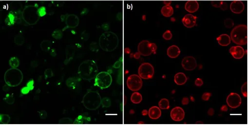
absorption and emission spectra useful for the observation at the confocal microscope. Figure 2 12
a) and b) show confocal images, respectively, at the characteristic emission wavelengths for the 13
fluorescence of BODIPY (498 < λ < 530 nm) and for 18:1 Liss Rhod PE (571 < λ < 630 nm), 14
when blended in the double layer of POPC vesicles. Figure 2 a) clearly shows that a very low 15
amount of the BODIPY-copolymer = 0.02 mol% with respect to the total amount of amphiphiles, 16
can be homogenously dispersed into the POPC phospholipid membranes and confirms that the 17
solubility parameter of the synthesized copolymer is close to that of pure PMMA. 18
1
Figure 2 POPC vesicles labelled with fluorescent probes. a) POPC and BODIPY-copolymer 99.98 %: 0.02 %;
2
b) POPC and 18:1 Liss Rhod PE 99 %: 0.8%. Small round spots in a) and b) are probably due to mineral oil
3
residues. White bar scales 10 μm.
4
Mixed vesicles were successfully prepared at pH = 7, where 50% of the copolymer is 5
protonated (pKa ~ 7, see Figure S12 in Supporting Information), by varying the unlabeled 6
copolymer mole percentage in the range 8 – 40 % (58 – 89 wt%), higher concentration of 7
copolymer hindered the self-assembly of the amphiphiles and the formation of a pellet at the end 8
of the phase transfer procedure was not obtained. Furthermore, we could not observe the 9
formation of pure polymer GUVs. 10
Vesicles with the compositions mol % POPC – mol % copolymer 5: 92% – 8%, 85% – 15%, 11
60% – 40% labeled with 18:1 Liss Rhod PE and BODIPY-copolymer are shown in Figure 3 a) – 12
c), respectively. In each figure, the green channel represents the fluorescence of the copolymer, 13
the red channel corresponds to the fluorescence of the lipid and the yellow channel represents 14
their overlap. When the concentration of the copolymer in the vesicles was below ~20 mol% the 15
mixed membranes were mainly heterogeneous, with small regions where copolymer blended into 16
the lipid layers. Interestingly, the presence of the copolymer in the membranes promoted the 17
C hapt er 4. P reparat ion of giant hybrid polymer/ lipid vesicles 103
The electroformed vesicles are difficult to detach from the plate, this can be a consequence of electrostatic interactions between the charged copolymer and the conductive glass slide (this effect was also noticed in mixed vesicles PEO:PBD where the detachment was favoured with the addition of PBD:PEO-COOH104). The selected experimental conditions made demanding the further
characterization of the produced vesicles (the main obstacles are the difficult detachment of thevesiclesfrom theplateand thenecessity to adopt microfluidic channels to load the enzymatic reaction in the sealed chamber slide), therefore the electroformation method was dismissed and the droplet transfer method chosen for t he vesicles preparat ion and charact erizat ion.
4.4.5 P reparat ion of hybrid polymeric vesicles t hrough
droplet t ransfer met hod
Vesicles made of POPC: P2kMD40% were marked with afluorescent lipid (18:1
Liss Rhod PE) andfluorescent polymeric markers (P5kMD53% andP2kMD20% )
to identify the presence of lipid and polymeric regions. Firstly, experiments of control (images in Figure 4.24) with just the lipid marker or just the polymeric marker were devised t o set t he condit ions for t he confocal acquisit ion.
F igur e 4.24: Pure POPC vesicles labelled withfluorescent probes. a) POPC and 18:1 Liss Rhod PE 99 %: 0.8% b) POPC and P5kMD53% 99.98 %: 0.02 %.
T he scale bar represent s 10µm.
Mixed vesicles were prepared by increasing P2kMD40% mole percentage from
7 % to 40 %. Vesicles with the compositions POPC-P14: 92% - 8%, 85% - 15%, 60% - 40% labelled with 18:1 Liss Rhod PE and P5kMD53% (long
chainfluorescent polymer) are shown respectively in Figures 4.25, 4.26 and 4.27. In eachfigure, the green channel represents the fluorescence of the polymer P5kMD53%, the red channel corresponds to thefluorescence of 18:1
fusion of single vesicles into budded structures, as shown in Figure 3 a), or large aggregates of 1
fused vesicles. The welding surface between vesicles was indeed characterized by a large 2
concentration of copolymer, highlighted by the intense yellow colour, which probably favours 3
electrostatic interaction between the outer leaflets of the membranes, as previously observed in 4
the presence of charged fatty acids incorporated in the double layer of POPC vesicles [82,83]. 5
6
Figure 3 Mixed POPC- copolymer vesicles having molar composition: a) 92% – 8%; b) 85% – 15%; c) 60% –
7
40%. All systems were stained with 18:1 Liss Rhod PE (0.8%) and BODIPY-copolymer (0.02%). More
8
pictures are reported in the Supporting Information (Figure S13).
9
When the percentage of copolymer increased, the length of the domains where it was present 10
seemed to increase accordingly (Figure 3 b), until a homogeneous distribution of lipids and 11
copolymers in the membrane was attained when the concentration of latter was around 40 mol%, 12
as shown in Figure 3 c). It is worth noting that mixed vesicles were obtained in presence of the 13
same amount of Liss Rhod PE (0.8%) and BODIPY-copolymer (0.02%) for all the investigated 14
compositions. At very low concentration of copolymer 5 (8%), image 3 a) shows the prevailing 15
presence of POPC as stained by Liss Rhod PE while, increasing the copolymer amount, the 16
images 3 b) and 3 c) show an increase in homogeneity of the membrane with consequent 17
overlapping of both the probes. The increasing spreading of BODIPY-copolymer in the 1
membrane, despite its fixed concentration, demonstrates both the presence of the copolymer into 2
the membrane and the prevailing affinity of the fluorescent copolymer for copolymer-rich 3
regions. Although the membrane composition became more homogeneous at higher copolymer 4
percentages, the total number of stable vesicles detected in the samples drastically diminished, 5
until amphiphilic structures could not be observed at copolymer amount > ~40 mol%. The 6
concentration dependence of both the lipid/copolymer distribution in the membrane and the 7
successful formation of vesicles might depend on the complex attractive interactions taking place 8
between the two molecules, as observed for a DPPC/PIB37-b-PEO48 system [32]. 9
It is interesting to compare our results with those previously reported in the literature 10
concerning POPC-based giant vesicles blended with PBD or PDMS. POPC is a lipid in the fluid 11
state at room temperature and in the case of PBD [13,31], homogeneous hybrid GUVs are 12
formed in a restricted range of composition (i.e. in the region of low and high mole polymer 13
content). In these homogeneous vesicles lipid molecules are dispersed in a “sea” of copolymers 14
and vice versa. This situation is the most energetically favoured. Indeed, apart from the chemical 15
affinity, also the structural compatibility has to be taken into account when dealing with 16
lipid/copolymer GUVs. The thickness of polymer bilayers varies between 5 and 50 nm, while the 17
lipid bilayer is 3-4 nm [20,84], i.e. there is a so called hydrophobic mismatch. As a consequence 18
of this hydrophobic mismatch, in phase separated systems, the copolymer has to distort in order 19
to adjust to the lipid bilayer and avoid as much as possible the contacts with water. This has a 20
high energy cost corresponding to high line tension at lipid-polymer boundary. Then, domain 21
formation is most of the time energetically disfavoured. Interestingly in the case of PDMS 22
triblock copolymers [29], hybrid GUVs were formed in all the weight concentration range and 23
were homogeneous in the polymer-rich region (till 85 wt % polymer content for 1.5k 1
copolymers, 50 wt% for 3k copolymers), while domain formation was observed at lower 2
polymer content. This phase separation was accompanied by budding and the phase-separated, 3
budded vesicles were not stable and budding degenerated in vesicle fission in the lipid rich 4
region. For molecular weight as high as 5k stable phase separated vesicles could not be observed 5
and pure lipid or pure polymer vesicles were present together with homogeneous structures. 6
Budding phenomena can evolve in fission when the line tension at boundary is higher than the 7
curvature energy associated to bending. The authors concluded that line tension at the lipid-8
polymer boundary were too high in the case of the 5k copolymers. Besides, the authors stressed 9
that block copolymers with the same molecular weight showed even higher instability towards 10
fission than the triblock structures. As observed for PDMS based copolymers, the copolymer 11
presented here forms homogeneous GUVs at high polymer content. On the contrary domain 12
formation is observed without fission between 24 w % and 42 w % POPC despite their higher 13
hydrophobic mismatch (13.8k compared to 5k in the literature). At these compositions the 14
bending costs associated with budding and fission are still higher than the boundary line energy, 15
so phase separation is still energetically favoured to fission. We speculate that our polymer has a 16
higher Tg than PDMS based ones, and consequently a higher curvature energy. 17
In a different set of experiments, we investigated the influence of the copolymer on the 18
distribution of the water-soluble fluorescent probe pyranine (structure reported in Figure S14 in 19
the Supporting Information) that was dissolved in the I-solution. This probe is membrane 20
impermeable and in pure POPC vesicles it is firmly confined into the aqueous lumen [85,86] as 21
highlighted in Figure 4 a). However, when the copolymer was present, the pyranine tended to 22
accumulate along the membrane (Figure 4 b). The intensity profiles shown in Figure 4 c) and 4 23
d) highlight the different distribution of pyranine. In pure POPC vesicles the fluorescence is 1
constant along the line and decreases at the border, in the mixed vesicle, in contrast, two peaks 2
appear in proximity of the membrane where pyranine accumulates. 3
1
Figure 4 Vesicles made of POPC - copolymer (93% - 7% mol/mol) and pyranine 50 M. a) Pure POPC
2
vesicles; b) Mixed vesicles; c) Intensity profile taken along a line for vesicles 1 and 2 of Panel a); d) Intensity
3
profile taken along line 1 and line 2 for the vesicle of Panel b).
4
This behaviour could be attributed to a tendency of pyranine to accumulate in copolymer-rich 5
domains, possibly due to the interaction of its OH moieties with the pH-sensitive units of the 6
DMAEMA group. This different behaviour observed for pyranine in the absence and in the 1
presence of the copolymer is another proof that the latter is indeed present in the POPC double 2
layer. 3
CONCLUSION 4
Hybrid lipid vesicles incorporating a copolymer with a Tg above room temperature have 5
been presented. Our study shows that electroformation cannot be used for these polymers to 6
efficiently obtain giant vesicles, probably linked to a low mobility in the electroformation 7
conditions. However, a method based on an emulsion phase transfer, so far used for lipids or 8
polymers such as PDMS or PBD has been successfully adapted. This can be preferably applied 9
to polymers compatible with lipids based on their solubility and affinity. For this, the solubility 10
parameter and the HLB value can be used to select appropriate candidates. The method presented 11
here will open up new possibilities to develop hybrid systems with a much larger chemical 12
structure variety, leading to new types of synthetic micro reactors or biomimetic compartments. 13
Possible future developments will therefore be adaptive micro-reactor with catalyst groups fixed 14
to the membrane, or pH-sensitive micro- or nano-reactors or synthetic cells with a controlled 15
permeability versus an external stimulus. 16
ACKNOWLEDGMENTS 1
The authors thank COST ACTION CM1304 and ERASMUS+ exchange program for funding 2
the mobility of Y.M. from the University of Salerno to the University of Toulouse “Paul 3
Sabatier” and the EU (FEDER-35477 “Nano-objets pour la biotechnologie”) for financial support. 4
Thanks are due to Dr.Patrizia Iannece (University of Salerno) for ESI-MS spectral measurements 5
and Dr. Patrizia Oliva (University of Salerno) for the bidimensional NMR spectral measurements 6
and Dr Stephanie Dauvillier (IPBS, University of Toulouse and CNRS) for assistance with 7
confocal microscope. L.I. thanks Università degli Studi dell’Insubria for the funding granted via 8
“Fondo dell’Ateneo per la Ricerca” (FAR 2018). 9
LIST OF ABBREVIATIONS 1
Monomethoxypoly(ethyleneglycol) mPEG
Poly(ethylene glycol) methyl ether 2-bromoisobutyrate
2,2′-Bipyridyl
mPEG-Br
bpy
1-palmitoyl-2-oleoyl-sn-glycero-3-phosphocholine POPC
1,2-dioleoyl-sn-glycero-3-phosphoethanolamine-N-(lissamine rhodamine B sulfonyl) (ammonium salt)
18:1 Liss Rhod PE
Methylmetacrylate MMA
N,N-(dimethylamino)ethyl methacrylate DMAEMA
2,6-diethyl-1,3,5,7-tetramethyl-8-[4-[8-(2- methacryloylethyl)dimethylammoniumbromideoctyloxy]phenyl]-4,4’-difluoroboradiaza indacene
BODIPY-DMAEMA
fluorescent mPEG-block-P(MMA-grad-DMAEMA) BODIPY-copolymer 2
ASSOCIATED CONTENT 3
Supplementary information is available. It describes the characterization of the products in more 4
details and report more pictures about hybrid GUVs. 5
REFERENCES 1
[1] F. Meng, Z. Zhong, J. Feijen, Stimuli-responsive polymersomes for programmed drug 2
delivery, Biomacromolecules, 10 (2009) 197–209. 3
[2] M. Huo, J. Yuan, L. Tao, Y. Wei, Redox-responsive polymers for drug delivery: from 4
molecular design to applications, Polym. Chem., 5 (2014) 1519–1528. 5
[3] L. Messager, J. Gaitzsch, L. Chierico, G. Battaglia, Novel aspects of encapsulation and 6
delivery using polymersomes, Curr. Opin. Pharmacol., 18 (2014) 104–111. 7
[4] M. Li, C. Du, N. Guo, Y. Teng, X. Meng, H. Sun, S. Li, P. Yu, H. Galons, Composition 8
design and medical application of liposomes, Eur. J. Med. Chem., (2019). 9
[5] H. Che, J.C.M. van Hest, Adaptive Polymersome Nanoreactors, ChemNanoMat, 5 (2019) 10
1092–1109. 11
[6] I. Louzao, J.C. van Hest, Permeability effects on the efficiency of antioxidant nanoreactors, 12
Biomacromolecules, 14 (2013) 2364–2372. 13
[7] K. Langowska, C.G. Palivan, W. Meier, Polymer nanoreactors shown to produce and 14
release antibiotics locally, Chem. Commun., 49 (2013) 128–130. 15
[8] X. Liu, D. Appelhans, B. Voit, Hollow Capsules with Multiresponsive Valves for 16
Controlled Enzymatic Reactions, J. Am. Chem. Soc., 140 (2018) 16106–16114. 17
[9] O. Rifaie-Graham, S. Ulrich, N.F. Galensowske, S. Balog, M. Chami, D. Rentsch, J.R. 18
Hemmer, J. Read de Alaniz, L.F. Boesel, N. Bruns, Wavelength-selective light-responsive 19
DASA-functionalized polymersome nanoreactors, J. Am. Chem. Soc., 140 (2018) 8027– 20
8036. 21
[10] Q. Yan, J. Wang, Y. Yin, J. Yuan, Breathing polymersomes: CO2-tuning membrane 1
permeability for size-selective release, separation, and reaction, Angew. Chem. Int. Ed., 52 2
(2013) 5070–5073. 3
[11] M. Mohammadi, S. Taghavi, K. Abnous, S.M. Taghdisi, M. Ramezani, M. Alibolandi, 4
Hybrid Vesicular Drug Delivery Systems for Cancer Therapeutics, Adv. Funct. Mater., 28 5
(2018) 1802136. 6
[12] W. Shen, J. Hu, X. Hu, Impact of amphiphilic triblock copolymers on stability and 7
permeability of phospholipid/polymer hybrid vesicles, Chem. Phys. Lett., 600 (2014) 56– 8
61. 9
[13] S.K. Lim, H.-P. de Hoog, A.N. Parikh, M. Nallani, B. Liedberg, Hybrid, Nanoscale 10
Phospholipid/Block Copolymer Vesicles, Polymers, 5 (2013) 1102–1114. 11
[14] M. Mumtaz Virk, E. Reimhult, Phospholipase A2-induced degradation and release from 12
lipid-containing polymersomes, Langmuir, 34 (2017) 395–405. 13
[15] Z. Cheng, D.R. Elias, N.P. Kamat, E.D. Johnston, A. Poloukhtine, V. Popik, D.A. Hammer, 14
A. Tsourkas, Improved Tumor Targeting of Polymer-Based Nanovesicles Using Polymer– 15
Lipid Blends, Bioconjug. Chem., 22 (2011) 2021–2029. 16
[16] W. Zong, B. Thingholm, F. Itel, P.S. Schattling, E. Brodszkij, D. Mayer, S. Stenger, K.N. 17
Goldie, X. Han, B. Städler, Phospholipid–Block Copolymer Hybrid Vesicles with 18
Lysosomal Escape Ability, Langmuir, 34 (2018) 6874–6886. 19
[17] E. Rideau, R. Dimova, P. Schwille, F.R. Wurm, K. Landfester, Liposomes and 20
polymersomes: a comparative review towards cell mimicking, Chem. Soc. Rev., 47 (2018) 21
8572–8610. 22
[18] C. G. Palivan, R. Goers, A. Najer, X. Zhang, A. Car, W. Meier, Bioinspired polymer 1
vesicles and membranes for biological and medical applications, Chem. Soc. Rev., 45 2
(2016) 377–411. 3
[19] M. Schulz, W.H. Binder, Mixed Hybrid Lipid/Polymer Vesicles as a Novel Membrane 4
Platform, Macromol. Rapid Commun., 36 (2015) 2031–2041. 5
[20] J.-F. Le Meins, C. Schatz, S. Lecommandoux, O. Sandre, Hybrid polymer/lipid vesicles: 6
state of the art and future perspectives, Mater. Today, 16 (2013) 397–402. 7
[21] A. Peyret, E. Ibarboure, J.-F. Le Meins, S. Lecommandoux, Asymmetric hybrid polymer– 8
lipid giant vesicles as cell membrane mimics, Adv. Sci., 5 (2018) 1700453. 9
[22] M. Bieligmeyer, F. Artukovic, S. Nussberger, T. Hirth, T. Schiestel, M. Müller, 10
Reconstitution of the membrane protein OmpF into biomimetic block copolymer– 11
phospholipid hybrid membranes, Beilstein J. Nanotechnol., 7 (2016) 881–892. 12
[23] M. Schulz, S. Werner, K. Bacia, W.H. Binder, Controlling molecular recognition with 13
lipid/polymer domains in vesicle membranes, Angew. Chem. Int. Ed., 52 (2013) 1829– 14
1833. 15
[24] J. Kowal, D. Wu, V. Mikhalevich, C.G. Palivan, W. Meier, Hybrid Polymer–Lipid Films as 16
Platforms for Directed Membrane Protein Insertion, Langmuir, 31 (2015) 4868–4877. 17
[25] L. Otrin, N. Marušič, C. Bednarz, T. Vidaković-Koch, I. Lieberwirth, K. Landfester, K. 18
Sundmacher, Toward artificial mitochondrion: mimicking oxidative phosphorylation in 19
polymer and hybrid membranes, Nano Lett., 17 (2017) 6816–6821. 20
[26] R.J.R.W. Peters, M. Marguet, S. Marais, M.W. Fraaije, J.C.M. van Hest, S. 21
Lecommandoux, Cascade Reactions in Multicompartmentalized Polymersomes, Angew. 22
Chem. Int. Ed., 53 (2014) 146–150. 23
[27] J. Nam, T. Kyle Vanderlick, P. A. Beales, Formation and dissolution of phospholipid 1
domains with varying textures in hybrid lipo-polymersomes, Soft Matter, 8 (2012) 7982– 2
7988. 3
[28] T.P.T. Dao, F. Fernandes, M. Fauquignon, E. Ibarboure, M. Prieto, J.F.L. Meins, The 4
combination of block copolymers and phospholipids to form giant hybrid unilamellar 5
vesicles (GHUVs) does not systematically lead to “intermediate” membrane properties, 6
Soft Matter, 14 (2018) 6476–6484. 7
[29] T.P.T. Dao, F. Fernandes, E. Ibarboure, K. Ferji, M. Prieto, O. Sandre, J.-F.L. Meins, 8
Modulation of phase separation at the micron scale and nanoscale in giant polymer/lipid 9
hybrid unilamellar vesicles (GHUVs), Soft Matter, 13 (2017) 627–637. 10
[30] C. Magnani, C. Montis, G. Mangiapia, A.-F. Mingotaud, C. Mingotaud, C. Roux, P. Joseph, 11
D. Berti, B. Lonetti, Hybrid vesicles from lipids and block copolymers: Phase behavior 12
from the micro-to the nano-scale, Colloids Surf. B Biointerfaces, (2018). 13
[31] J. Nam, P.A. Beales, T.K. Vanderlick, Giant Phospholipid/Block Copolymer Hybrid 14
Vesicles: Mixing Behavior and Domain Formation, Langmuir, 27 (2011) 1–6. 15
[32] M. Schulz, D. Glatte, A. Meister, P. Scholtysek, A. Kerth, A. Blume, K. Bacia, W.H. 16
Binder, Hybrid lipid/polymer giant unilamellar vesicles: effects of incorporated 17
biocompatible PIB–PEO block copolymers on vesicle properties, Soft Matter, 7 (2011) 18
8100–8110. 19
[33] A. Kubilis, A. Abdulkarim, A.M. Eissa, N.R. Cameron, Giant polymersome protocells dock 20
with virus particle mimics via multivalent glycan-lectin interactions, Sci. Rep., 6 (2016) 21
32414. 22
[34] E. Rideau, F.R. Wurm, K. Landfester, Self-Assembly of Giant Unilamellar Vesicles by 1
Film Hydration Methodologies, Adv. Biosyst., 3 (2019) 1800324. 2
[35] M.I. Angelova, D.S. Dimitrov, Liposome electroformation, Faraday Discuss. Chem. Soc., 3
81 (1986) 303–311. 4
[36] B.M. Discher, Y.-Y. Won, D.S. Ege, J.C.-M. Lee, F.S. Bates, D.E. Discher, D.A. Hammer, 5
Polymersomes: Tough Vesicles Made from Diblock Copolymers, Science, 284 (1999) 6
1143–1146. 7
[37] M. Dionzou, A. Morere, C. Roux, B. Lonetti, J.-D. Marty, C. Mingotaud, P. Joseph, D. 8
Goudouneche, B. Payre, M. Leonetti, A.-F. Mingotaud, Comparison of methods for the 9
fabrication and the characterization of polymer self-assemblies: what are the important 10
parameters, Soft Matter, 12 (2016) 2166–2176. 11
[38] A.M. Eissa, M.J. Smith, A. Kubilis, J.A. Mosely, N.R. Cameron, Polymersome-forming 12
amphiphilic glycosylated polymers: Synthesis and characterization, J. Polym. Sci. Part 13
Polym. Chem., 51 (2013) 5184–5193. 14
[39] J.P. Reeves, R.M. Dowben, Formation and properties of thin-walled phospholipid vesicles, 15
J. Cell. Physiol., 73 (1969) 49–60. 16
[40] R. Dimova, U. Seifert, B. Pouligny, S. Förster, H.-G. Dobereiner, Hyperviscous diblock 17
copolymer vesicles, Eur. Phys. J. E, 7 (2002) 241–250. 18
[41] Y. Zhou, D. Yan, Real-Time Membrane Fission of Giant Polymer Vesicles, Angew. Chem. 19
Int. Ed., 44 (2005) 3223–3226. 20
[42] E. Rideau, F.R. Wurm, K. Landfester, Giant polymersomes from non-assisted film 21
hydration of phosphate-based block copolymers, Polym. Chem., 9 (2018) 5385–5394. 22
[43] A. Weinberger, F.-C. Tsai, G.H. Koenderink, T.F. Schmidt, R. Itri, W. Meier, T. Schmatko, 1
A. Schröder, C. Marques, Gel-Assisted Formation of Giant Unilamellar Vesicles, Biophys. 2
J., 105 (2013) 154–164. 3
[44] K.S. Horger, D.J. Estes, R. Capone, M. Mayer, Films of Agarose Enable Rapid Formation 4
of Giant Liposomes in Solutions of Physiologic Ionic Strength, J. Am. Chem. Soc., 131 5
(2009) 1810–1819. 6
[45] T.P.T. Dao, M. Fauquignon, F. Fernandes, E. Ibarboure, A. Vax, M. Prieto, J.-F. Le Meins, 7
Membrane properties of giant polymer and lipid vesicles obtained by electroformation and 8
PVA gel-assisted hydration methods, Colloids Surf. Physicochem. Eng. Asp., 533 (2017) 9
347–353. 10
[46] S. Pautot, B.J. Frisken, D.A. Weitz, Production of Unilamellar Vesicles Using an Inverted 11
Emulsion, Langmuir, 19 (2003) 2870–2879. 12
[47] C. Martino, A.J. deMello, Droplet-based microfluidics for artificial cell generation: a brief 13
review, Interface Focus, 6 (2016) 20160011. 14
[48] S. Deshpande, C. Dekker, On-chip microfluidic production of cell-sized liposomes, Nat. 15
Protoc., 13 (2018) 856–874. 16
[49] M. Abkarian, E. Loiseau, G. Massiera, Continuous droplet interface crossing encapsulation 17
(cDICE) for high throughput monodisperse vesicle design, Soft Matter, 7 (2011) 4610– 18
4614. 19
[50] M.C. Blosser, B.G. Horst, S.L. Keller, cDICE method produces giant lipid vesicles under 20
physiological conditions of charged lipids and ionic solutions, Soft Matter, 12 (2016) 7364– 21
7371. 22
[51] K. Dürre, A.R. Bausch, Formation of phase separated vesicles by double layer cDICE, Soft 1
Matter, (2019). 2
[52] J. Petit, I. Polenz, J.-C. Baret, S. Herminghaus, O. Bäumchen, Vesicles-on-a-chip: A 3
universal microfluidic platform for the assembly of liposomes and polymersomes, Eur. 4
Phys. J. E, 39 (2016) 59. 5
[53] K. Kamiya, R. Kawano, T. Osaki, K. Akiyoshi, S. Takeuchi, Cell-sized asymmetric lipid 6
vesicles facilitate the investigation of asymmetric membranes, Nat. Chem., 8 (2016) 881– 7
889. 8
[54] A. Moga, N. Yandrapalli, R. Dimova, T. Robinson, Optimization of the Inverted Emulsion 9
Method for High-Yield Production of Biomimetic Giant Unilamellar Vesicles, 10
ChemBioChem, 20 (2019) 2674–2682. 11
[55] H. Che, S. Cao, J.C.M. van Hest, Feedback-Induced Temporal Control of “Breathing” 12
Polymersomes To Create Self-Adaptive Nanoreactors, J. Am. Chem. Soc., 140 (2018) 13
5356–5359. 14
[56] S. Villani, R. Adami, E. Reverchon, A.M. Ferretti, A. Ponti, M. Lepretti, I. Caputo, L. Izzo, 15
pH-sensitive polymersomes: controlling swelling via copolymer structure and chemical 16
composition, J. Drug Target., 25 (2017) 899–909. 17
[57] E. Yoshida, pH response behavior of giant vesicles comprised of amphiphilic poly 18
(methacrylic acid)-block-poly (methyl methacrylate-random-mathacrylic acid), Colloid 19
Polym. Sci., 293 (2015) 649–653. 20
[58] E. Yoshida, Giant vesicles comprised of mixed amphiphilic poly (methacrylic acid)-block-21
poly (methyl methacrylate-random-methacrylic acid) diblock copolymers, Colloid Polym. 22
Sci., 293 (2015) 3641–3648. 23