Cultural characteristics, pathogenicity and vegetative compatibility of
Fusarium udum isolates from pigeonpea (Cajanus cajan (L.) Millsp.) in
Kenya
E.K. Kiprop1, A.W. Mwang’ombe2, J.P. Baudoin3, P.M. Kimani4and G. Mergeai3,∗
1Department of Botany, Faculty of Science, Moi University, P.O. Box 1125, Eldoret, Kenya;2Department of Crop
Protection, Faculty of Agriculture, University of Nairobi, P.O. Box 30197, Nairobi, Kenya;3Phytotechnie
Tropicale et Horticulture, Facult´e universitaire des Sciences Agronomiques de Gembloux, Passage des D´eport´es, 2, B-5030, Gembloux, Belgium;4Department of Crop Science, Faculty of Agriculture, University of Nairobi,
P.O. Box 30197, Nairobi, Kenya;∗Author for correspondence (Fax: +32 81614544)
Accepted 15 November 2001
Key words:chlorate resistant, Fusarium wilt, heterokaryon, mutant, vegetative compatibility group, virulence
Abstract
Seventy-nine single-spore isolates of Fusarium udum, the causal agent of wilt disease of pigeonpea, from Kenya, India and Malawi were characterized according to their cultural characteristics, pathogenicity and vegetative com-patibility group (VCG). The isolates exhibited high variation in pathogenicity on a wilt-susceptible pigeonpea vari-ety, and in mycelial growth and sporulation on potato dextrose agar medium. The 79 isolates were categorized into two virulence groups, two groups of radial mycelial growth and four groups of sporulation. Radial mycelial growth showed a moderate negative correlation (r = −0.40; P = 0.01) with sporulation. However, mycelial growth and sporulation had no correlation with virulence. Pairings between complementary nitrate non-utilizing (nit) mutants of F. udum generated on chlorate containing minimal medium revealed that all the isolates belonged to a single VCG (VCG 1) with two subgroups, VCG 1 I and VCG 1 II. Vegetative compatibility was independent of cultural characteristics and pathogenicity. This is the first report of vegetative compatibility in F. udum.
Introduction
Pigeonpea (Cajanus cajan (L.) Millsp.) is one of the most widely-grown and eaten grain legumes in the semi-arid tropics of the world. Wilt caused by Fusarium
udum Butler is the most important disease of this crop (Kannaiyan et al., 1984). The disease is widely distributed in pigeonpea growing districts in Kenya. Comparisons of single-spore isolates of F. udum from the same or different geographical origin have shown that the fungus is highly variable in cultural char-acteristics (Reddy and Chaudhary, 1985; Gaur and Sharma, 1989) and pathogenicity (Shit and Sen Gupta, 1978; Gupta et al., 1988). However, studies on genetic diversity using isozyme markers revealed low varia-tion in F. udum (Shit and Sen Gupta, 1980; Okiror, 1986). Vegetative compatibility grouping (VCG) has
been used frequently as a means of identifying iso-lates of a fungus that are closely related (Leslie, 1993). This methodology has been applied to a long and growing list of Fusaria, but not to F. udum. In some cases, VCG has a correlation with pathogenic-ity, while in others no relationship was observed (Leslie, 1993; Clark et al., 1995). Thus, vegetative compatibility studies on F. udum help in understand-ing the genetic variability within this fungus result-ing in an improvement in wilt resistance pigeonpea breeding programmes. In view of the economic impor-tance of wilt and the fact that little has been reported on genetic variability of F. udum, the objectives of this study were to group the isolates from Kenya by VCGs, cultural characteristics and pathogenicity, and to determine if a relationship exists between these parameters.
Materials and methods Fungal isolates
Seventy-five isolates of F. udum were obtained from wilt-infected pigeonpea plants collected from 55 sites in 12 districts of Kenya in 1997. All the isolates were purified on tap water agar and established as single-spore isolates. Two single-single-spore isolates were obtained from each strain of F. udum from Malawi (IMI number 275452) and India (IMI number 205514), both strains received from the International Mycological Institute, UK. All these isolates were maintained on Spezieller Nahrastofarmer agar (Nirenberg, 1976) at −20◦C. The medium consisted of 1 g KH2PO4, 1 g KNO3, 0.5 g MgSO4.7H2O, 0.5 g KCl, 0.2 g glucose, 0.2 g sucrose and 20 g agar per litre.
Pathogenicity tests
The conidial suspensions of the 79 single-spore iso-lates of F. udum were prepared in 100 ml sterile dis-tilled water (SDW) from 7-day-old cultures on PDA. The conidial suspension was filtered through two layers of cheesecloth and the concentration was adjusted to 106 conidia ml−1. Seven-day-old pigeonpea seedlings of wilt susceptible variety KAT 60/8, grown on ster-ile riverbed sand, were uprooted carefully, the roots washed with SDW and trimmed to about 4 cm from the collar region using a sterile scalpel. The seedlings were dipped in inoculum suspension for 30 min and trans-planted in 20 cm diam plastic pots having a sterile mix-ture of red soil (Vertisol) and riverbed sand (3 : 1 v/v). Five inoculated seedlings were planted per pot with four replications for each isolate. Control seedlings were dipped in SDW. The plants were kept in a green-house having a maximum temperature between 23 and 30◦C with 12 h natural light per day at the University of Nairobi (Kenya). The plants were checked daily for the appearance of wilt symptoms. The records on percent-age of wilted seedlings were taken 6 weeks after inocu-lation (Reddy and Raju, 1997). The data were analysed by ANOVA using SAS system software (SAS Institute Inc., Cary, NC, USA). The experiment was repeated once.
Cultural characteristics
All the isolates were grown on PDA plates in tripli-cate for 8 days at 25◦
C in a 12 h light/dark cycle to
determine the radial mycelial growth (colony diameter) and sporulation (conidia ml−1). The colony diameter of each culture in the plate was measured using a vernier calliper by taking an average of four radial measure-ments at random. To estimate sporulation, conidial sus-pension was prepared from each culture plate using a modification of Calpouzos and Stallknecht (1965). Two discs of 6 mm diam each were cut from the oppo-site sides of the centre of the colony, but 1.5 cm from the middle point, and suspended in a universal bot-tle with 10 ml of SDW. This was put on a rotary shaker at 250 rpm for 20 min, the spore suspension then filtered through two layers of sterile cheesecloth and the conidial concentration determined by using a haemo-cytometer. A mean of four counts was obtained per colony. The data were analysed by ANOVA using SAS system software (SAS Institute Inc., Cary, NC, USA). The experiment was repeated once.
Vegetative compatibility
To generate nitrate non-utilizing mutants a 5 mm2 mycelial block of each isolate from 5-day-old cultures on PDA was transferred to the centre of a 9 cm diam Petri dish containing nitrate minimal medium with chlorate (MMC). This medium consisted of minimum medium (MM) (per litre: 30 g sucrose, 2 g NaNO3, 1 g KH2PO4, 0.5 g MgSO4·7H2O, 0.5 g KCl, 0.01 g FeSO4·7H2O, 0.2 ml trace element solution (5 g citric acid, 5 g ZnSO4·7H2O, 1 g Fe(NH4)2(SO4)2·6H2O, 0.25 g CuSO4·5H2O, 0.05 g MnSO4·H2O, 0.05 g H3BO4, 0.05 g NaMoO4·2H2O, and 95 ml distilled water) and 20 g agar) amended with 15 g KClO3 and 1.6 g l-asparagine. Ten MMC plates with a single mycelial block were used per isolate. The plates were incubated at 25◦C in a 12 h dark/light cycle and exam-ined periodically for the appearance of fast-growing sectors (chlorate-resistant) from the initial colony. Transfers were made from the leading margin of fast-growing sectors onto MM. Colonies having a thin expansive growth with no aerial mycelium on MM were considered nit mutants. The nit mutants were identi-fied as nit1, nit3 and NitM depending on their growth on nitrate, nitrite and hypoxanthine medium (Correll et al., 1987).
To test for complementation or heterokaryon forma-tion between nit mutants, a 5 m2
block of mycelium was transferred from MM to a fresh 9 cm Petri dish containing MM with a NitM mutant in the centre of a daisy configuration and four nit1and/or nit3 mutants
from different isolates on the outer circle at 3 cm apart. One to three nit1 and/or nit3 obtained from each isolate were used for complementation reaction. The plates were incubated as described above for 7 days but mutants that exhibited moderate to weak reactions were incubated for up to 20 days (Katan et al., 1991; Katan and Katan, 1999). NitM mutants obtained from isolates MS05, IND01a, MB01, KR03, TT04, MR06, MS07 and TK03 were used as testers in pairings with
nit1 and/or nit3 mutants of all isolates for vegetative compatibility tests. Four types of scoring were made: strong reaction (++) if wild-type growth developed within 4–7 days where the thin expansive growth of the nit mutants converge, moderate or weak reaction (+) if wild-type growth developed within 8–20 days, no reaction (−), and uncertain reaction (+−) (Katan et al., 1991; Katan and Katan, 1999). If a wild-type growth appeared at the intersection of colonies on pair-ing of complementary nit mutants (NitM and nit1 or
nit3) from different isolates on minimal medium, then the isolates were of the same VCG and if it remained thin, then the isolates were of different VCG. Comple-mentary and similar nit mutants from the same isolate were also paired. Complementation reactions between NitM and nit1 and/or nit3 mutants of different isolates were repeated at least two times.
Results
Pathogenicity tests
All 79 isolates of F. udum were pathogenic to the wilt-susceptible pigeonpea variety KAT 60/8, although they showed high significant (P = 0.01) variation in virulence (Table 1). Fusarium wilt symptoms on pigeonpea plants were observed from the eighth day of inoculation, and the fungus re-isolated from the stems of infected plants on the 14th day. The wilt incidence on infected plants ranged from 35% to 100%. The iso-lates were classified into two groups; those causing up to 60% wilt were moderately virulent and those causing more than 60% wilt were highly virulent. The number of isolates in the former group was 17 (21.5%) and in the latter group was 62 (78.5%). The highly virulent group was widely distributed in all the districts. The districts with a high proportion of the moderately vir-ulent group were Meru with 57% and Thika with 50% of isolates in this group. Some isolates from the same group collected from the same site showed significant variation in pathogenicity.
Cultural characteristics
The F. udum isolates exhibited high variation in radial mycelial growth and sporulation on PDA. There were significant differences (P = 0.01) among the isolates in terms of radial mycelial growth and sporulation (Table 1). Based on inter-quartile ranges and one-sample t-tests, the 79 single-spore isolates were classi-fied into different groups using radial mycelial growth and sporulation. There were 61 (77.2%) isolates with fast radial mycelial growth and 18 (22.8%) isolates with slow radial mycelial growth (over 79.0 mm and below 79.0 mm colony diameter, respectively). Eight (10.1%) isolates had very high sporulation, 23 (29.1%) isolates high sporulation, 28 (35.4%) isolates moderate sporulation and 20 (25.3%) isolates low sporulation, with over 15.9 × 10⁵, 8.0–15.9 × 10⁵, 5.0–7.9 × 10⁵ and below 5.0 × 10⁵ conidia ml−1, respectively. The radial mycelial growth of isolates was highly vari-able in most districts with the exception of Thika, Meru and Kirinyaga districts, and Malawi having only isolates with fast growth. The districts that had pre-dominantly high to very high sporulation groups were Machakos and Mbeere, while those with predomi-nantly low to moderate sporulation groups were Meru, Tharaka Nithi, Thika and Nairobi. Some isolates col-lected from the same site were significantly different (P = 0.01) with respect to radial mycelial growth and/or sporulation. For example, isolates that showed significant differences in radial mycelial growth and sporulation from the same site were TN01 and TN02, NY07 and NY08, MB02 and MB03, and TT04 and TT05.
Generally, isolates with fast radial mycelial growth had low to high sporulation while those with slow radial mycelial growth had moderate to very high sporulation. The correlation coefficient between radial mycelial growth and sporulation was negative and moderate (r = −0.40; P = 0.01). Pathogenicity was not cor-related with mycelial growth (r = −0.05, P = 0.67) and sporulation (r = 0.11; P = 0.33) and was statis-tically non-significant (P = 0.05).
Vegetative compatibility
1465 chlorate-resistant sectors were produced on MMC from 79 single-spore isolates of F. udum with a mean of 18.5 sectors per isolate (Table 1). The iso-lates differed considerably in their sectoring frequency with a range of 1.2 (IND01a) to 3.0 (TK06) sectors per colony. The majority of the sectors appeared in the
Table 1. Pathogenicity, radial mycelial growth and sporulation of Fusarium udum isolates from pigeonpea, and characterization of their chlorate-resistant mutants
Isolate Sitea Wiltb (%) Colony diameter (mm) Spores (conidia ml−1×10⁵) Chlorate-resistant sectors nit mutants
nitmutant phenotypes nit1 nit3 NitM
MK01 Makueni 1 75 81.3 9.4 18 7 7 0 0 MK02 Makueni 2 90 81.9 13.4 21 8 6 2 0 MK03 Makueni 3 75 81.2 9.6 18 7 7 0 0 MK04 Makueni 4 85 78.8 7.1 25 7 6 1 0 MK05 Makueni 5 80 80.7 11.0 24 10 6 4 0 MK06 Makueni 6 55 80.4 11.5 18 7 7 0 0 MK07 Makueni 7 75 80.2 5.8 14 7 4 3 0 MK08 Makueni 11 40 77.3 17.9 13 2 2 0 0 MK09 Makueni 11 60 77.8 11.2 18 7 6 1 0 MK10 Makueni 12 80 84.2 6.5 21 7 5 2 0 MS01 Machakos 13 90 80.8 11.2 20 5 5 0 0 MS02 Machakos 13 65 82.0 3.8 18 5 5 0 0 MS03 Machakos 14 75 80.8 7.3 26 11 8 3 0 MS04 Machakos 14 85 80.8 15.3 26 11 4 7 0 MS05 Machakos 15 85 77.3 5.7 14 5 4 0 1 MS06 Machakos 16 65 79.8 8.9 13 3 3 0 0 MS07 Machakos 17 65 77.5 17.7 20 6 3 2 1 MS08 Machakos 17 55 79.5 9.2 19 8 4 4 0 MS09 Machakos 17 55 79.5 2.7 14 2 2 0 0 MS10 Machakos 18 85 81.4 3.6 18 4 4 0 0 KT01 Kitui 19 95 80.2 6.4 27 18 12 6 0 KT02 Kitui 19 90 78.7 24.6 23 10 10 0 0 KT03 Kitui 20 65 82.9 6.2 18 6 6 0 0 KT04 Kitui 21 70 76.7 8.9 24 10 8 2 0 KT05 Kitui 23 80 83.3 3.8 14 3 2 1 0 MB01 Mbeere 24 55 82.6 5.0 21 6 3 0 3 MB02 Mbeere 25a 80 84.5 8.6 18 4 4 0 0 MB03 Mbeere 26b 90 78.9 5.6 19 5 3 2 0 MB04 Mbeere 27 85 78.3 11.1 17 4 4 0 0 MB05 Mbeere 27 95 72.7 12.8 19 4 4 0 0 MB06 Mbeere 28 70 81.9 11.0 19 13 9 4 0 MB07 Mbeere 28 85 80.9 11.1 17 7 7 0 0 MR01 Meru 33 100 84.1 7.1 21 7 4 3 0 MR02 Meru 34 55 82.6 6.7 14 3 3 0 0 MR03 Meru 35 70 83.9 4.8 27 6 6 0 0 MR04 Meru 37 65 79.5 6.9 21 9 8 1 0 MR05 Meru 38 55 83.0 5.2 13 2 2 0 0 MR06 Meru 38 55 84.3 6.5 17 8 4 2 2 MR07 Meru 39 55 84.9 9.8 18 5 5 0 0 NY01 Nyambene 40 85 84.2 4.0 13 5 5 0 0 NY02 Nyambene 41 95 84.3 4.8 14 3 0 3 0 NY03 Nyambene 41 75 85.7 6.3 14 2 2 0 0 NY04 Nyambene 42 70 82.7 11.9 23 5 3 2 0 NY05 Nyambene 43 70 79.6 6.1 17 6 6 0 0 NY06 Nyambene 43 70 81.5 7.8 18 9 7 2 0 NY07 Nyambene 44 100 78.4 21.1 13 7 5 2 0 NY08 Nyambene 44 100 81.6 5.8 14 2 2 0 0 TN01 T-Nithi 47 65 80.2 9.4 17 3 3 0 0 TN02 T-Nithi 47 70 75.1 5.8 13 3 3 0 0 TN03 T-Nithi 48 90 81.5 3.6 26 10 7 3 0 TN04 T-Nithi 50 95 85.4 4.0 25 9 7 2 0 TN05 T-Nithi 51 55 76.6 3.5 29 15 12 3 0
Table 1. Continued
Isolate Sitea Wiltb (%) Colony diameter (mm) Spores (conidia ml−1×10⁵) Chlorate-resistant sectors nit mutants
nitmutant phenotypes nit1 nit3 NitM
TT01 T-Taveta 55 100 78.6 16.9 17 6 5 1 0 TT02 T-Taveta 56 60 81.5 6.0 18 9 7 2 0 TT03 T-Taveta 57 75 86.3 9.0 18 4 4 0 0 TT04 T-Taveta 58 90 80.3 8.2 18 5 4 0 1 TT05 T-Taveta 58 100 81.8 4.4 20 10 7 3 0 TT06 T-Taveta 59 100 82.5 17.5 14 4 4 0 0 TT07 T-Taveta 62 85 81.2 6.2 17 5 5 0 0 TT08 T-Taveta 63 80 71.4 21.4 14 4 4 0 0 TT09 T-Taveta 64 85 82.0 4.0 19 6 6 0 0 ML01 Malindi 70 75 83.5 4.8 12 4 4 0 0 KR01 Kirinyaga 75 60 83.5 17.1 18 5 5 0 0 KR02 Kirinyaga 76 65 84.6 1.4 14 5 5 0 0 KR03 Kirinyaga 76 55 81.9 3.8 26 12 6 5 1 TK01 Thika 77 80 88.8 5.0 18 4 4 0 0 TK02 Thika 77 65 87.7 4.6 17 3 3 0 0 TK03 Thika 78 90 88.5 3.1 21 9 6 2 1 TK04 Thika 79 45 85.4 3.8 21 9 6 3 0 TK05 Thika 79 35 84.3 8.8 21 5 5 0 0 TK06 Thika 79 55 85.7 6.1 30 14 9 5 0 NB01 Nairobi 83 75 83.9 4.8 14 2 2 0 0 NB02 Nairobi 83 90 82.6 4.4 21 8 8 0 0 NB03 Nairobi 84 80 81.2 6.8 17 4 4 0 0 NB04 Nairobi 84 75 73.6 7.6 14 4 4 0 0 MAL01a Malawi 80 80.1 5.1 17 5 5 0 0 MAL01b Malawi 90 80.4 5.4 23 5 2 3 0 IND01a India 70 73.6 7.5 12 8 6 0 2 IND01b India 65 73.3 8.8 13 8 5 1 2 Total 1465 505 399 92 14 Mean 74.9 81.2 8.2 18.5 6.4 5.1 1.2 0.2 L.S.D. (P = 0.05) 16.4 1.0 0.3
aDistrict in Kenya or country with the field number. T-Nithi = Tharaka Nithi, T-Taveta = Taita. Strain from India was collected at Hyderabad, and Malawi strain from unknown site.
bPathogenicity on Fusarium wilt susceptible pigeonpea variety (KAT 60/8).
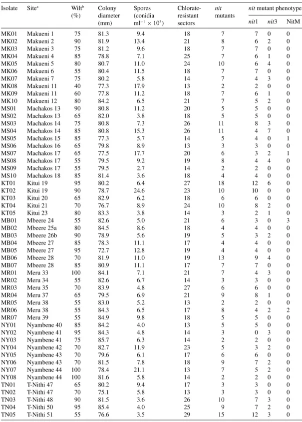
first and second week of incubation, and a few in the third week (Figure 1). The chlorate-resistant sectors that were unable to utilize nitrate as the sole source of nitrogen and consequently grew as thin expansive colonies with no aerial mycelium on MM were recov-ered at a mean frequency of between 0.14 and 0.68 sectors per colony. These sectors were designated nit mutants and were 34.5% of the total chlorate-resistant sectors. The nit mutants that did not utilize nitrate but utilized nitrite and hypoxanthine on the respective media as the sole source of nitrogen were designated
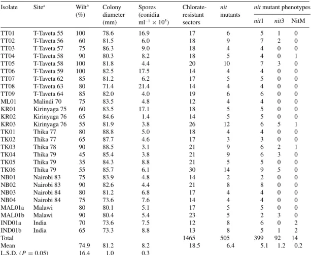
nit1, those that did not utilize nitrite designated nit3, and those that did not utilize hypoxanthine designated NitM. All nit mutants produced wild-type growth on PDA medium (Figure 2). Five hundred and five sectors
were nit mutants, of which 79.0%, 18.2% and 2.8% were nit1, nit3 and NitM, respectively.
Vegetative compatibility reaction was more robust in the wild-type with dense aerial mycelial growth between nit1 and NitM phenotypes of different isolates than between nit3 and NitM. Compatibility reaction between nit1 and nit3 mutants of different isolates was moderate to weak. A total of 1248 vegetative com-patibility reactions between eight NitM mutants and
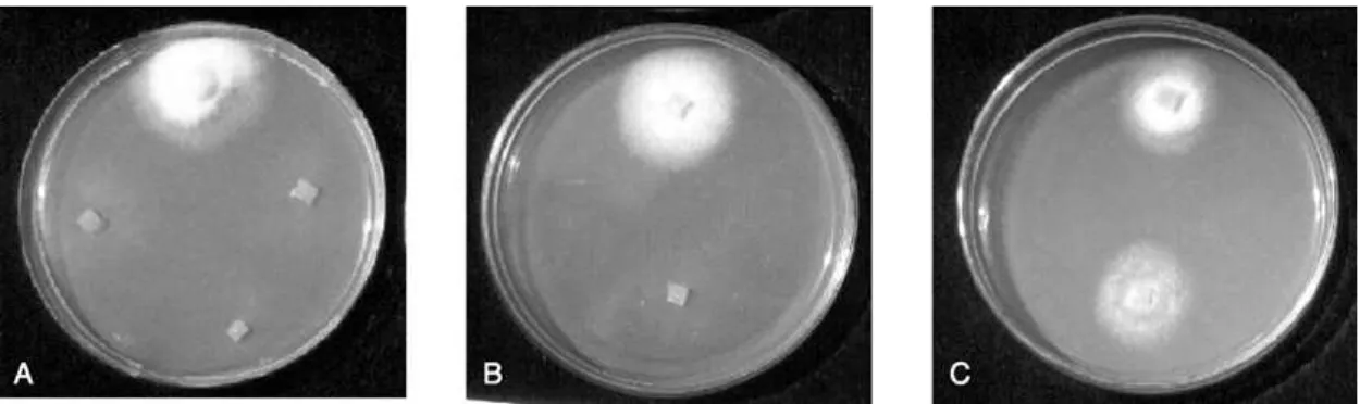
nit1 and/or nit3 mutants of different F. udum isolates were performed with varying degrees of heterokaryon formation. All the isolates formed strong or moder-ate/weak heterokaryons with at least one NitM and could be linked to other NitMs (Figure 3). Forty-nine (62%) isolates formed strong or moderate/weak
A B
Figure 1. Formation of chlorate-resistant sectors by isolates MK10 (plate A) and MK07 (plate B) of F. udum three weeks after incubation on nitrate minimal medium amended with chlorate.
Figure 2. Plate A shows wild-type growth of parental strain (top block), and thin growths of mutants NitM (right block), nit1 (bottom block) and nit3 (left block) of isolate TK03 on nitrate minimal medium; plate B shows wild-type growth of nit1 (top block) and thin growth of NitM (bottom block) of isolate MS05 on hypoxanthine medium; and plate C shows wild-type growths of nit1 (top block) and NitM (bottom block) of isolate MS05 5 days after incubation on nitrite medium.
A B
Figure 3. Formation of complementary heterokaryons in pairings between nit mutants of F. udum isolates 12 days after inoculation on nitrate minimal medium: Plate A shows strong reaction of NitM from isolate MS05 (centre block) with nit1 from isolate MB06 (upper block) and a moderate/weak reaction with nit1 from isolate TN01 (lower block), and plate B shows strong reactions of NitM from isolate IND01a (centre block) with nit1 from isolate KT02 (upper block) and nit1 from isolate MAL01b (lower block).
heterokaryons with all the eight NitM. Isolate MS04 did not develop heterokaryons with NitMs from iso-lates KR03, TT04 and TK03 but it formed wild-type mycelial growth with the remaining five NitM. Isolates MS05, MS10, MR02, TT05, MAL01a, for example, developed wild-type mycelial growth with the eight NitM. Therefore, isolate MS04 could be linked to NitM of KR03, TT04 and TK03 by the above isolates. Isolate MR05 formed moderate/weak heterokaryons with only NitM of IND01a and TK03 but could be linked to the other six NitM through isolates MR06 or NY07. Using a similar criterion, the 30 isolates that did not develop heterokaryons with all NitM were linked to 49 isolates that showed wild-type mycelial growth with all NitM. It was using this criterion that the F. udum single-spore isolates from Kenya, and one strain each from Malawi and India were grouped into one vegetative compatibil-ity group, VCG 1. However, 73 (92.4%) isolates formed strong reactions with at least one NitM while six (7.6%) isolates namely MR05, TN01, TN02, TN03, NB01 and NB02 formed either moderate/weak, uncertain or no reactions with NitMs. This enabled the subdivision of VCG 1 into two subgroups, namely VCG 1 I with 73 isolates and VCG 1 II with six isolates. The isolates of F. udum in subgroups VCG 1 I and II were highly variable with respect to cultural characteristics (radial mycelial growth and sporulation) and pathogenicity.
Discussion
Variations in pathogenicity of F. udum isolates enabled their classification into two virulence groups. Both virulence groups were found in eight districts while only the highly virulent group was found in five dis-tricts indicating that virulence may vary depending on the geographical origin of the isolates. Isolates with moderate virulence were dominant in Meru and Thika districts. Variability in pathogenicity of F. udum isolates from different geographical origins has been observed elsewhere (Shit and Sen Gupta, 1978; Okiror, 1986; Gupta et al., 1988; Gaur and Sharma, 1989).
Single-spore isolates of F. udum showed variation in their cultural characteristics. In the present study,
F. udum was classified into two groups by radial
mycelial growth and four groups by sporulation on PDA medium. Other workers have also indicated con-siderable variability in F. udum with respect to these characteristics (Shit and Sen Gupta, 1978; Okiror, 1986; Gupta et al., 1988; Gaur and Sharma, 1989). It
was found in this study that, on average, isolates with slow growth had higher sporulation while those with fast growth had lower sporulation agreeing with ear-lier findings (Gupta et al., 1988). Pathogenicity was not correlated with sporulation and radial mycelial growth, which also supports earlier observations (Okiror, 1986; Gaur and Sharma, 1989).
Isolates of F. udum differed considerably in their sec-toring frequency on chlorate medium with a mean of 0.14 and 0.68 sectors per colony. Earlier reports have also revealed a wide variation in sectoring frequency in isolates of other species of Fusarium (Correll et al., 1987; Clark et al., 1995; Sunder and Satyavir, 1998). These findings indicate that the sectoring of individual isolates of a particular fungal species differs and hence also the number of chlorate-resistant sectors recovered. Although the recovery frequency of chlorate-resistant sectors in the present study was lower than that recov-ered from other Fusarium species, the differences could be due to the type of fungal species, type of medium used for sectoring and culturing conditions. All the nit mutants recovered from F. udum could be divided into three distinct phenotypic classes: nit1, nit3 and NitM. The majority of the nit mutants recovered on MMC were nit1 mutants (79%), followed by nit3 mutants (18%), and NitM mutants (3%). Correll et al. (1987) recovered nit mutants of F. oxysporum at frequencies of 59–66% for nit1, 10–28% for nit3 and 10–25% for NitM on MMC. Clark et al. (1995) recovered nit mutants of F. lateritium at frequencies of 77.2% for
nit1, 7.4% for nit3 and 15.4% for NitM on KPS, and 77.7% for nit1, 9.7% for nit3 and 12.6% for NitM on KMM. These findings indicate that the majority of nit mutants recovered in fungal populations are usually
nit1 while nit3 and NitM are in the minority.
In the present study, isolates of F. udum from Kenya, Malawi and India comprised one vegeta-tive compatibility group, VCG 1. However, VCG 1 could be divided into two subgroups due to differ-ential reactions of mutants. Subgroup VCG 1 I con-stituted 73 isolates with at least one strong reaction with NitM while subgroup VCG 1 II constituted six isolates with either moderate/weak, uncertain or no reaction with NitM. Populations of other fusaria like
F. oxysporum f.sp. canariensis (Plyler et al., 2000),
F. oxysporum f.sp. vasinfectum (Katan and Katan,
1988), F. oxysporum f.sp. dianthi (Katan et al., 1989) have also been reported to be comprised single VCG, while those of F. oxysporum f.sp. radicis-lycopersici (Katan and Katan, 1999) and F. moniliforme (Sunder
and Satyavir, 1998) represented eight and 10 VCGs, respectively. More than one subgroup in VCG 0090, VCG 0091 and VCG 0094 of F. oxysporum f.sp.
radicis-lycopersici have been observed (Katan and
Katan, 1999). In the present study, the isolates of
F. udumin the two subgroups of VCG 1 were found
to have isolates with significant differences in cultural characteristics and pathogenicity. Sunder and Satyavir (1998) observed that isolates of F. moniliforme from different VCGs and also within the same VCG varied considerably in virulence and gibberrelic acid (GA3) production. Correll et al. (1986) have reported a cor-relation of VCG and colony size, with virulence of
F. oxysporumf.sp. apii isolates; however in the present study, vegetative compatibility showed no definite rela-tionship with cultural characteristics and pathogenic-ity. On the basis of vegetative compatibility as a genetic marker, the data presented in this study indicate that
F. udumcould be derived from a single lineage. How-ever, to draw more definite conclusions, it will be neces-sary to study more isolates from different geographical origins. Further investigations using molecular mark-ers and race typing are necessary to fully undmark-erstand the genetic variation within F. udum.
Acknowledgements
Financial support for this study was provided by the European Union through an INCO-DC project, contract number ERBIC18CT960130; the two fungal strains from Malawi and India were obtained from the International Mycological Institute, UK.
References
Calpouzos L and Stallknecht GF (1965) Sporulation of Cercospora beticolaaffected by an interaction between light and temperature. Phytopathology 55: 1370–1371
Clark CA, Hoy MW and Nelson PE (1995) Variation among iso-lates of Fusarium lateritium from sweetpotato for pathogenic-ity and vegetative compatibilpathogenic-ity. Phytopathology 85: 624–629 Correll JC, Klittich CJR and Leslie JF (1987) Nitrate non-utilizing mutants of Fusarium oxysporum and their use in vegetative compatibility tests. Phytopathology 77: 1640–1646
Correll JC, Puhalla JE and Schneider JF (1986) Identification of Fusarium oxysporum f.sp. apii on the basis of colony size,
virulence, and vegetative compatibility. Phytopathology 76: 396–400
Gaur VK and Sharma LC (1989) Variability in single spore iso-lates of Fusarium udum Butler. Mycopathologia 107: 9–15 Gupta O, Kotasthane SR and Khare MN (1988) Strain
varia-tion in Fusarium udum in Madhya Pradesh, India. Internavaria-tional Pigeonpea Newsletter 7: 22–25
Kannaiyan J, Nene YL, Reddy MV, Ryan JG and Raju TN (1984) Prevalence of pigeonpea diseases and associated crop losses in Asia, Africa and America. Tropical Pest Management 30: 62–71
Katan T and Katan J (1988) Vegetative-compatibility grouping of Fusarium oxysporum f.sp. vasinfectum from tissue and the rhizosphere of cotton plants. Phytopathology 78: 852–855 Katan T and Katan J (1999) Vegetative compatibility
group-ing in Fusarium oxysporum f.sp. radicis-lycopersici from UK, the Netherlands, Belgium and France. Plant Pathology 48: 541–549
Katan T, Hadar E and Katan J (1989) Vegetative compatibility of Fusarium oxysporum f.sp. dianthi from carnation in Israel. Plant Pathology 38: 376–381
Katan T, Zamir D, Sarfatti M and Katan J (1991) Vegetative com-patibility groups and subgroups in Fusarium oxysporum f.sp. radicis-lycopersici. Phytopathology 81: 255–262
Leslie JF (1993) Fungal vegetative compatibility. Annual Review Phytopathology 31: 127–151
Nirenberg H (1976) Untersuchungen uber die morphologis-che und bioligismorphologis-che Differenzierung in der Fusarium-section Liseola. Mitteilungen aus der Biologischen Bundesanstalt fur Land-und forstwirtschaft, Berlin-Bahlem 169: 1–117 Okiror MA (1986) Breeding for resistance to Fusarium wilt of
pigeonpea (Cajanus cajan (L.) Millsp.) in Kenya. PhD Thesis, University of Nairobi, 202 pp
Plyler TR, Simone GW, Fernandez D and Kistler HC (2000) Genetic diversity among isolates of Fusarium oxysporum f.sp. canariensis. Plant Pathology 49: 155–164
Reddy NPE and Chaudhary KCB (1985) Variation in Fusarium udum. Indian Phytopathology 38: 172–173
Reddy MV and Raju TN (1997) Evaluation of pigeonpea (Cajanus cajan) varieties for resistance to wilt caused by Fusarium udumand sterility mosaic disease in a perennial system. Indian Journal of Agricultural Sciences 67: 437–439 Shit SK and Sen Gupta PK (1978) Possible existence of
physio-logical races of Fusarium oxysporum f.sp. udum, the incitant of the wilt of pigeonpea. Indian Journal of Agricultural Sciences 48: 629–632
Shit SK and Sen Gupta PK (1980) Pathogenic and enzymatic variation in Fusarium oxysporum f.s.p. udum. Indian Journal of Microbiology 20: 46
Sunder S and Satyavir (1998) Vegetative compatibility, biosyn-thesis of GA3 and virulence of Fusarium moniliforme iso-lates from bakanae disease of rice. Plant Pathology 47: 767–772