Addressing delivery and synthesis challenges for
peptide-based cancer vaccines
By
Rebecca Lynn Holden
B.S. Chemistry
Baylor University, 2015
Submitted to the Department of Chemistry
in Partial Fulfillment of the Requirements for the Degree of
Doctor of Philosophy
in Chemistry
at the
Massachusetts Institute of Technology
September 2020
© 2020 Massachusetts Institute of Technology
All rights reserved
Signature of Author: ________________________________________________
Department of Chemistry
July 20, 2020
Certified by: ______________________________________________________
Bradley L. Pentelute
Associate Professor of Chemistry
Thesis Supervisor
Accepted by: _____________________________________________________
Adam Willard
Associate Professor
Graduate Officer
This doctoral thesis has been examined by a committee of the Department of Chemistry as follows:
Professor Elizabeth M. Nolan ………. Thesis Committee Chair Ivan R. Cottrell Professor of Immunology
Professor Bradley L. Pentelute……….. Thesis Supervisor Associate Professor of Chemistry
Catherine L. Drennan……… Thesis Committee Member Professor of Biology and Chemistry, HHMI Investigator
Addressing delivery and synthesis challenges for
peptide-based cancer vaccines
By
Rebecca Lynn Holden
Submitted to the Department of Chemistry on July 20, 2020 in Partial Fulfillment of the Requirements for the Degree of Doctor of Philosophy in Chemistry
Abstract
Therapeutic peptide vaccines have the potential to elicit and direct an anti-cancer T cell response, but their clinical efficacy has been limited in part by poor delivery to the lymphatic system, inefficient cell uptake, and scalable synthesis in the case of personalized vaccines. The work presented in this thesis explores several approaches to address these challenges.
First, we use our fast-flow automated synthesis technology to confront the synthesis bottleneck for patient-specific ‘neoantigen’ vaccines, which have shown early promise but are hindered by slow production. We synthesize a particularly challenging set of peptides from a previous clinical trial as a test case, demonstrating that our technology can produce the majority of sequences in sufficient quantities with comparable or higher purity than a commercial vendor in a fraction of the time.
Turning towards vaccine design, we explore several approaches to address the lymphatic and intracellular delivery of peptide antigens. We demonstrate the generality and anti-tumor efficacy of vaccines containing cell-penetrating peptides (CPPs), sequences shown to enhance the cell uptake of various cargo. We characterize their mechanism and identify several unanticipated contributors, namely trafficking to the lymph nodes, serum stability, and extended presentation in vivo. We then expand on an existing approach to mediate lymphatic trafficking via binding serum albumin by exploring additional albumin binding moieties. Next, we develop a straightforward and general approach to directly quantify antigen presentation and implement this technique to explore two strategies to design more effective vaccines, including CPPs.
Finally, we build on previous work using CPPs to deliver antisense oligonucleotides (ASOs), another application that we pursued in tandem with cancer vaccines. We combine amphipathic and cationic CPPs to create chimeric sequences that synergistically enhance activity of an ASO and access new routes of uptake not utilized by either parent CPP.
Drawing from our experience using CPPs to deliver ASOs as well as our expertise in peptide synthesis and design, we provide insight into the rapid production of personalized vaccines and efficient delivery of vaccine antigens. This thesis represents a new area of research for our lab, one in which we will hopefully continue to apply our unique skillset and perspective. Thesis Supervisor: Bradley L. Pentelute
Acknowledgements
The past five years have been some of the greatest years of my life but also the hardest, challenging me in ways that I could not predict. Earning a PhD is a difficult journey; and earning a PhD while coming out is a real trip. I have learned so much, grown stronger as a person, and become a better and more confident scientist. I owe a great deal of gratitude to everyone who has helped me make it through.
I want to start by thanking my thesis advisor, Prof. Brad Pentelute. His enthusiasm from the moment I joined the lab set the tone for my entire grad school career. I am grateful for his guidance and mentorship, which always seemed to be exactly what I needed. He encouraged me to propose my own ideas early on and his confidence in me allowed me to grow as an independent scientist. At the same time, he provided direction and encouragement whenever I needed it. His support extended beyond scientific guidance, helping me improve my communication and presentation skills and providing invaluable professional advice. Finally, of course, I would like to thank Brad for cultivating a supportive and collaborative lab culture that has significantly shaped both my research and my entire experience at MIT.
I could not have asked for a more invested and supportive thesis committee chair than Prof. Liz Nolan. I want to thank her for being an amazing mentor. It was always evident during our meetings that she genuinely cared about my research and my growth as a scientist. I appreciate her many insightful questions and thoughtful suggestions over the years. I am especially grateful more recently for her postdoc application and general career advice.
Than you to Prof. Cathy Drennan, who has been another incredible mentor. I appreciated her encouragement throughout grad school, particularly during my second- and third-year exams. Her advice for my postdoc search and her support with fellowship applications has been invaluable, and I am extremely grateful.
I am thankful to my collaborators in the Irvine lab, beginning with Prof. Darrell Irvine, who has provided a great deal of advice and scientific guidance for many of the projects described in this thesis. I am also grateful to Coralie, whose insight and advice over the last few years helped me to be a better scientist and whose sincerity, thoughtfulness, and humor made her a great colleague and friend. Thank you to Kelly, who was both a collaborator and a mentor during my first couple of years in grad school. I am grateful for her organization and positivity getting several of these projects up and running. I would also like to thank Dan, Josetta, and Naveen for their contributions to our work.
I would like to express my gratitude to Prof. Cathy Wu and Prof. Nir Hacohen for a productive and exciting collaboration on their neoantigen vaccines. I have learned a lot working with them and look forward to learning more in the future. I would also like to thank Keerthi, Sisi, Matt, Zhuting, and the whole Neovax subgroup.
I cannot imagine what my experience in grad school would have been without my amazing colleagues and friends in the Pentelute lab. Thank you to my best friend Azin, whose compassion, vivacity, and sarcasm always brightened my day. I am grateful to have worked with someone so supportive, who was always willing to listen and help with anything going on in my research or personal life. Thank you to Nina, whose cheerfulness and style brightened our space. I am grateful for her thoughtfulness, generosity, and mentorship. I know I can turn to her any time in the future. I would like to thank Colin for being my first person in the lab and teaching me so much. He was a delight to work with and helped me through some of the hardest moments of grad school. Thank you to Justin for being an easygoing, practical, and encouraging friend and collaborator. I am grateful to Alex M. for being a willing teacher, a caring friend, and a fun roommate. He truly is the definition of precious. I would also like to thank Ethan for his commiseration and for the scientific input he happily provided anytime I asked. Thank you to Charlotte for being dependably upbeat and always willing to help. I owe a great deal of
gratitude to Anthony Q. for his tireless efforts to keep the Orbitrap running, as well as his down-to-earth kindness and his many, many, many jokes. I am thankful to Carly for being amazing to work with and a brilliant sand volleyball team captain. Thank you to Alex L. and Tuang for being my comrades throughout the grad school process, especially second- and third-year exams. I would like to thank Nick for being an enthusiastic collaborator and for organizing our immunotherapy super-group meetings. And finally, I would like to thank all of my other friends and colleagues who have made the past five years so enlightening and enjoyable: Katie, Jessie, Rachael, Zak, Mette, Diómedes, Joe, Aaron, Deborah, Surin, and everyone else in the Pentelute lab. I am also grateful to Christine, Katherine, and Alex (and Eli) from the Kiessling lab for their scientific input and friendship.
I want to extend a huge thank you to all of the friends that I made outside of lab. In particular, I would like to thank Lindsey, who became an immediate friend and who has been a source of endless support, commiseration, and sangria. I also want to thank Marty, who was one of the first friends I made in Cambridge and whose kindness and originality have been welcome constants throughout grad school. I enjoyed our trips together almost as much as I enjoyed being roommates. Thank you to Sheena, the gentlest spirit I know; Dmitro, one of my favorites; Zach, who will be relieved to know that he is also one of my favorites; and Brendan, who is a treasure. Thank you to Alex, Aubrey, Ashley, and Chad—Candace’s amazing friends who not only gave me their stamp of approval but became my friends, too. I am thankful to Erin for their care and wisdom. I also want to thank my comrades from Boston Feminists for Liberation, who have taught me so much. It has been a pleasure to organize with and befriend such incredible people.
I would like to thank Regina Spektor, Noname, Chance the Rapper, and Freddie Mercury. Their work has provided joy, comfort, and inspiration and seen me through the last five years. I am also grateful for the craft brewing and abundant greenery in the lovely cities of Cambridge and Somerville for bringing indispensable moments of peace, stress relief, and creativity.
Thank you to all of my friends from before I moved to Cambridge who helped me get through the last few years: Caitlin, Stefan, ChicAnya, Rae, Vincent, Chelsea, Ian, Niki, and Martin. Even being apart, they were there for me when I needed it and I am deeply grateful for their love and support. I am especially thankful to Caitlin, who is the kind of friend who hops on a ten-hour overnight bus when you need her without a second thought. She is my person and I am glad to have her.
And, of course, I want to thank my family. Thank you to my sister, Kelly, who brought the party—and her cat—with her when she visited and who can practically read my mind. Her determination and compassion continue to inspire me. Thank you to my parents for helping me through grad school and my whole education in more ways than I can count. I am so grateful to them for always listening, for their constant support and encouragement, and for their generosity. I want to thank my Dad for looking out for me and my mom for always checking in to make sure I was all right—and for her patience when I took a day or two (or more) to respond. Finally and most of all, I want to thank my partner, Candace. She has been the best part of my experience at MIT and I have a hard time imaging grad school without her. I am grateful to have someone so supportive, brilliant, and effortlessly funny in my life; someone who challenges me to be the best version of myself.
Table of Contents
Abstract 5 Acknowledgements 6 Table of Contents 8 List of Figures 12 List of Tables 14Chapter 1: Background and Overview 15
1.1 Cancer Immunotherapy and T cells 16
1.2 T cell activation and vaccine components 18
1.3 Vaccine antigen classes 19
1.4 Vaccine formats 20
1.5 Delivery of peptide vaccines 21
1.6 Cell-penetrating peptides 23
1.7 Overview of thesis 24
1.8 References 27
Chapter 2: Automated Flow Synthesis of Tumor Neoantigen Peptides for Personalized Immunotherapy 36
2.1 Introduction 37
2.2 Results and Discussion 39
2.3 Materials and Methods 52
2.3.1 Materials 52
2.3.2 Resin loading 52
2.3.3 Automated Flow Peptide Synthesis 52
2.3.4 Microwave Peptide Synthesis 55
2.3.5 Resin Cleavage 56
2.3.6 RP-HPLC analysis of IMPs by the commercial peptide vendor 56
2.3.7 RP-HPLC and LC/MS analysis of unpurified and purified IMPs 56
2.3.8 RP-HPLC purification of IMPs 57
2.3.10 Generation and detection of patient neoantigen-specific T cells 57
2.3.11 IFN-γ ELISPOT assay 57
2.4 Conclusions 58
2.5 Acknowledgements and Conflict Statements 59
2.6 References 60
Chapter 3: Sequences that promote both lymphatic and intracellular delivery enhance the efficacy of peptide cancer vaccines 62
3.1 Introduction 63
3.2 Results and Discussion 65
3.3 Materials and Methods 73
3.3.1 Reagents 73
3.3.2 Peptide synthesis 74
3.3.3 Peptide purification 74
3.3.4 Antigen conjugation 75
3.3.5 LC-MS analysis 75
3.3.6 Serum stability LC-MS assay 76
3.3.7 Cells 76
3.3.8 In vitro activation assay 76
3.3.9 Serum protein pulldown 76
3.3.10 Activation-based serum stability assay 77
3.3.11 DC2.4 uptake assay 77
3.3.12 Mice 78
3.3.13 Prophylactic vaccination 78
3.3.14 Tumor inoculation and therapy 78
3.3.15 Pmel-1 in vivo activation assay 78
3.3.16 Confocal imaging 78
3.3.17 Lymph node trafficking 79
3.3.18 Flow cytometry 79
3.3.19 Statistical analysis 79
3.4 Conclusions 79
3.5 Acknowledgements and Conflict Statements 80
3.6 References 81
Chapter 4: Enhancement of peptide vaccine immunogenicity by increasing lymphatic drainage using diverse albumin binders 85
4.1 Introduction 86
4.2 Results 87
4.3 Materials and Methods 93
4.3.1 Peptide and peptide conjugate synthesis 93
4.3.2 Mice and immunizations 95
4.3.3 Evaluation of murine immune responses and flow cytometry 95
4.3.4 Statistical analysis 96
4.4 Discussion 96
4.5 Acknowledgements and Conflicts Statement 97
4.6 References 97
Chapter 5: A direct elution and targeted analysis approach to characterize antigen presentation and design peptide vaccines 99
5.1 Introduction 100
5.2 Results 102
5.3 Materials and Methods 112
5.3.1 Reagents 112
5.3.2 Synthesis 113
5.3.3 Cleavage and Deprotection 114
5.3.4 Purification 114
5.3.5 LC-MS Characterization 114
5.3.6 Mammalian tissue culture and CD11c+ isolation 114
5.3.7 MHC presentation assay 115
5.4 Discussion 118
5.5 References 119
5.6 Appendix 122
Chapter 6: Chimeras of Cell-Penetrating Peptides Demonstrate Synergistic Improvement in Antisense Efficacy 134
6.1 Introduction 135
6.2 Results and Discussion 136
6.3 Materials and Methods 149
6.3.1 Materials 149
6.3.2 Methods for LC-MS Analysis 150
6.3.3 Fast-flow Peptide Synthesis 150
6.3.5 Peptide Purification 151
6.3.6 PMO-Azide Synthesis 151
6.3.7 PMO-Peptide Conjugation with Cu(I)-Catalyzed Azide-Alkyne Cycloaddition 152
6.3.8 Fluorophore Conjugation 152
6.3.9 Flow Cytometry 152
6.3.10 LDH Assay 153
6.3.11 Inhibitor Experiments 155
6.3.12 Flow cytometry assay with fluorophore-labeled conjugates 157
6.3.13 Live-Cell Confocal Imaging 158
6.3.14 Melting Temperature Analysis 160
6.3.15 Structural Prediction 161
6.4 Acknowledgements and Conflict Statements 162
6.5 References 163
List of Figures
Figure 2.1 Peptide design and production for a personalized neoantigen vaccine. 38
Figure 2.2. Comparison of peptide synthesis methods. 41
Figure 2.3. Analytical RP-HPLC traces of IMP 10, IMP 14, IMP 16, and IMP 23. 43
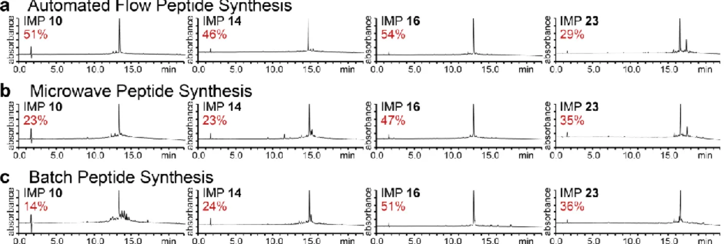
Figure 2.4. Analytical RP-HPLC traces of IMP 10, IMP 14, IMP 16, and IMP 23. 43
Figure 2.5. Summary of synthesis data for unpurified IMPs 10, 14, 16, and 23 synthesized by flow, microwave, and batch peptide synthesis. 44
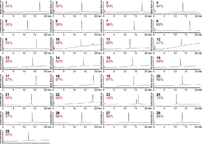
Figure 2.6. Unpurified neoantigen peptides produced by automated flow peptide synthesis. 45 Figure 2.7. Purified neoantigen peptides produced by automated flow peptide synthesis. 46
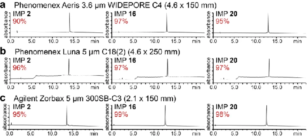
Figure 2.8. Analytical RP-HPLC traces of purified IMP 2, IMP 16, and IMP 20. 47
Figure 2.9. Characterization of IMPs produced by automated flow peptide synthesis. 47
Figure 2.10. Characterization of purified ASPs produced by automated flow peptide synthesis. 51
Figure 2.11. IFN-γ secretion by neoantigen-specific T cell lines against mutated ADAMTS7 (ASP41) peptide. 58
Figure 3.1 Conjugation to various CPPs increases T cell activation in vitro and improves immunogenicity of vaccine antigens. 65
Figure 3.2. Evaluating vaccine design and formulation. 67
Figure 3.3. CPPs are a general strategy to enhance vaccine immunogenicity and therapeutic efficacy. 68
Figure 3.4. CPPs increase intracellular delivery. 70
Figure 3.5. CPP conjugation promotes lymph node trafficking and enhances serum stability, likely due to association with serum proteins. 72
Figure 3.6. CPP conjugation extends antigen presentation in the draining lymph node. 73
Figure 4.1. Albumin-binding peptide conjugation enhances lymph node targeting of peptide antigens. 88
Figure 4.2. Albumin-binding peptides enhance vaccine immunogenicity. 89
Figure 4.3 Linker placement but not cyclization impacts immunogenicity of ABP-containing vaccine constructs. 91
Figure 4.4. Conjugation to -tocopherol enhances immunogenicity of a vaccine antigen.92 Figure 4.5. ABP vaccine construct characterization. 94
Figure 5.1. Direct elution followed by targeted LC-MS/MS analysis enables detection of MHC-presented antigens. 103
Figure 5.2. Our assay enables quantitation of MHC-presented antigens in a sensitive and
generalizable manner. 106
Figure 5.3. This technique enables characterization of various treatment parameters on antigen
presentation. 108
Figure 5.4. Single residue substitutions with a D-amino acid impacted the presentation of a murine tumor antigen in a position-dependent manner. 110 Figure 5.5. Conjugation to a cell-penetrating peptide increased presentation of a murine tumor antigen under certain treatment conditions. 111 Figure 5.6. Testing treatment and elution conditions. 117 Figure 6.1. PMO-peptide chimera conjugates enhance exon skipping. 138 Figure 6.2. The activity of PMO-CPP chimera conjugates is influenced by specific design
features. 140
Figure 6.3. PMO-peptide chimera conjugates exhibit dose dependent activity. 142 Figure 6.4. PMO-pVEC-Bpep conjugate undergoes energy-dependent uptake via a route
distinct from the PMO-CPPs. 143
Figure 6.5. PMO-pVEC-Bpep exhibits high internalization and high exon skipping activity.146 Figure 6.6. Peptide conjugation slightly alters PMO binding to a complementary nucleic
acid. 148
Figure 6.7. LDH Release from HeLa-654 Cells upon Treatment with 5 M PMO-peptide
conjugate. 154
Figure 6.8. LDH Release from HeLa-654 Cells upon Treatment with PMO-peptide conjugates
across a range of concentrations. 154
Figure 6.9. Effect of endocytosis inhibitors on PMO-Bpep, PMO-pVEC, and
PMO-pVEC-Bpep efficacy. 156
Figure 6.10. Comparison of PMO activity of unlabeled PMO-peptide conjugates with
SulfoCy5-labeled PMO-peptide conjugates. 158 Figure 6.11. PMO-SulfoCy5-pVEC-Bpep exhibits the most nuclear SulfoCy5
fluorescence. 160
Figure 6.12. Peptide conjugation has a minor impact on PMO binding to its target sequence.161 Figure 6.13. CPPs are predicted to contain significant helical character, with coiled and
List of Tables
Table 2.1. Sequences of IMPs 1–29 from a previous clinical trial. 40 Table 2.2 Summary of yield and purity data for unpurified and purified IMPs 1–29 synthesized
by automated flow peptide synthesis. 48
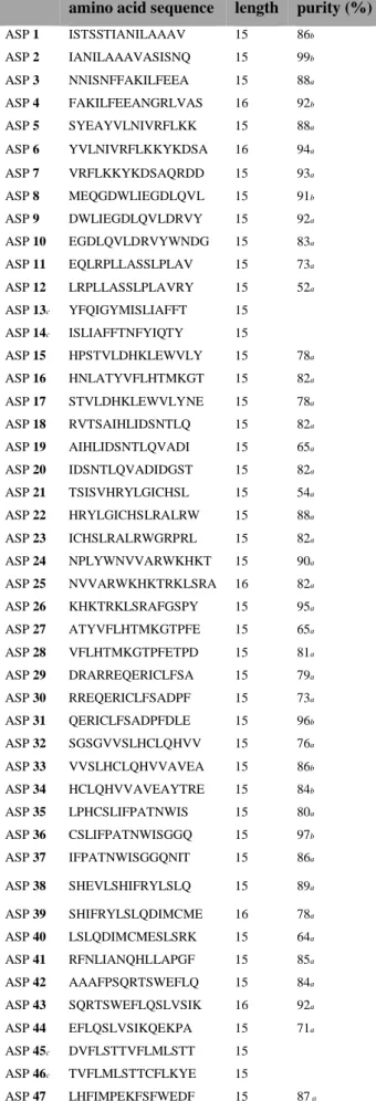
Table 2.3. Sequences from a set of ASPs for a personalized neoantigen vaccine. 50 Table 2.4. Summary of coupling and deprotection steps performed during automated flow
peptide synthesis. 54
Table 2.5. Summary of coupling and deprotection steps performed during microwave peptide
1.1 Cancer immunotherapy and T cells
Cancer immunotherapy has revolutionized the field of oncology in recent years, with several FDA-approved therapies becoming mainstays of treatment and an explosion of preclinical research providing promising new approaches.1–4 It incorporates a broad range of
therapies designed to act in some capacity on the patient’s immune system, rather than on the tumor itself, in order to elicit an anti-tumor immune response.5 In doing so, these therapies take
advantage of several key hallmarks of the immune system: precise targeting of distinct cell subsets; self-amplification of responses; and the formation immunological memory that provides a rapid response in the case of disease recurrence.5,6 While various approaches have
targeted different aspects of the immune system, most focus consistently on T cells.1–5
T cells are part of the adaptive immune response, meaning that they respond to specific antigens.6,7 Once naïve antigen-specific T cells are activated, they differentiate into effector
cells and exert their disease-controlling function. Eventually, this response diminishes and some of these effector cells differentiate into long-lived memory T cells which can be readily re-activated as needed.6 Two subtypes of T cells exist, distinguished by expression of two surface
markers: CD4+ and CD8+. Upon activation, CD8+ T cells become ‘cytotoxic T lymphocytes’
(CTLs), which can selectively kill target cells, while CD4+ T cells become ‘helper’ T cells,
which exert a range of functions that mediate and coordinate different elements of the immune response.6,7 While there is a growing interest in the CD4+ T cell response, CD8+ T cells have
been the focus of most immunotherapies to date due to their ability to directly kill tumor cells.7,8
Checkpoint blockade, which ‘takes the brakes off’ the CTL response, is the hallmark cancer immunotherapy.3,7 It comprises a series of monoclonal antibodies that block different ‘immune
checkpoints’ by binding inhibitory signaling receptors on T cells, antigen presenting cells, or tumor cells in order to block signals that either diminish CTL cell activation or dampen an ongoing CTL cell response. A handful of different checkpoint therapies have demonstrated remarkable clinical success, transforming the standard of care in melanoma and other highly mutagenic cancers.3–5 Ongoing work seeks to characterize additional immune checkpoints and
identify candidate antagonists for these inhibitory receptors.9 Checkpoint therapies are limited
by incomplete responses and off-target toxicity. While efficacy is often dramatic in patients who respond to checkpoint, a substantial fraction of patients exhibit little to no response.3,10
This discrepancy is attributed to an inconsistency in the level of pre-existing anti-tumor responses; when no response is present initially, there is little gain from a therapy that enhances the magnitude or quality of the T cell response.3,7 Additionally, significant autoimmune side
effects have been observed in some patients.3,7 There is a need for therapies that can stimulate
an anti-tumor T cell response and provide greater selectivity towards tumor cells.
Another key class of immunotherapy that has recently entered the clinic is chimeric antigen receptor T cells (CAR-Ts). In this approach, patient T cells are removed and the native T cell receptor is replaced by a chimeric antigen receptor ex vivo, which combines a binding domain against a previously-identified tumor antigen with native T cell receptor transmembrane and signaling domains.2,7 This approach has proved promising for several hematologic
malignancies, most notably B cell leukemias and lymphomas, but there are significant toxicity concerns associated with excessive activation and cytokine release.11,12 Additionally, the
process of identifying tumor-specific antigens and constructing and validating CARs is labor-intensive, further complicated by the heterogeneous nature of solid tumors which makes identification of broadly-applicable antigens highly challenging. Ongoing work is currently seeking to refine this approach and provide more nuanced control over the CAR-T response, but there is a need for immunotherapies that are more safe, cost-effective and scalable.2,13,14
A third class of cancer immunotherapy that has seen growing interest in the last decade is vaccination. Cancer vaccines supply tumor antigens to antigen-presenting cells in order to activate a tumor-specific T cell response.1,7,15–17 They differ from conventional vaccines in that
the approach is therapeutic, rather than prophylactic; i.e., designed to engage the immune system against an ongoing disease rather than simply guard against future encounters. Cancer vaccines have the potential to elicit anti-tumor immunity in patients without an existing response and to increase the magnitude of existing responses.18–22 However, these responses
can be limited in magnitude and their therapeutic efficacy is untested. They exhibit minimal side effects and indeed are designed to minimize off-target effects by guiding the immune response against tumor antigens. There is evidence that combining these vaccines with checkpoint blockade and possibly other immunotherapies can achieve greater anti-tumor efficacy.18,23–25 A range of different vaccine platforms and designs have been developed, which
will be explored in greater detail in section 1.4. While promising, cancer vaccine technologies require significant improvement to reach their full potential.
1.2 T cell activation and vaccine components
Cancer vaccines prime a T cell response against tumor antigens. Understanding their mechanism of action and designing improved vaccines requires insight into the process of T cell activation. Naïve T cells are activated upon interacting with antigen presenting cells (APCs), in particular dendritic cells (DCs), in the presence of several necessary stimuli: a cognate interaction between the T cell receptor, which varies between individual T cells, and an antigen peptide presented by the DC; and the interaction of co-stimulatory molecules at the surface of the T cell and DC.6,26,27 The DC co-stimulatory signaling molecules are upregulated
in the presence of inflammatory stimuli, e.g. upon innate immune activation.6,26,27 Successful
vaccines must therefore engage innate immunity in addition to providing relevant antigens. For vaccine which contain isolated antigen(s) rather than a whole pathogen, this is done by also including a component called an adjuvant designed to engage innate immune receptors and trigger an inflammatory response. 15,32 Optimal adjuvant design is a critical element of vaccine
development, and indeed most of the approved cancer vaccines consist only of an adjuvant designed to activate innate immunity. However, this work focuses on the TCR/cognate antigen signal and the antigen component of these vaccines.
Antigens presented to T cells are short, linear peptides that generally range in length from 8-11 amino acids for CD8+ T cells and ~13-20 amino acids for CD4+ T cells.6,26 They are bound
non-covalently to a surface protein called the major histocompatibility complex (MHC), also referred to as human leukocyte antigen (HLA) in humans. MHC class I, or MHC-I (HLA-I), binds CD8+ T cell antigens and facilitates interaction with the TCR in a process known as
antigen presentation, while MHC-II (HLA-II) presents antigen to CD4+ T cells.6,26 Vaccines
designed to elicit a T cell response must include one or more antigens, either within native proteins in whole pathogens in the case of conventional vaccines, or as isolated peptides, proteins, or antigen-encoding nucleic acids.6,15–17
Presentation of extracellular antigens by DCs, including vaccine antigens, involves uptake, proteolytic processing into short peptide epitopes, loading onto MHC, and display at the cell surface. This first requires trafficking to the lymph node, where these interactions occur, either as a result of passive antigen transport through the lymphatics or uptake by activated DCs which then home to the lymph nodes.6,26,28 The predominant pathway for processing CD8+ T cell
antigens occurs in the cytosol. In all other nucleated cells, healthy or tumor cells, peptide fragments resulting from proteasomal degradation of cytosolic peptides are transported to the endoplasmic reticulum for further proteolytic processing, loaded onto newly synthesized
MHC-I, and displayed at the cell surface in order to allow CD8+ T cell surveillance for infection or
transformation.6,26 In addition to this pathway, DCs present extracellular antigen in a process
known as ‘cross-presentation’ in which antigens are endocytosed, gain cytosolic access, and then follow the canonical MHC-I loading route.6,26,29 While other pathways exist, this is the
predominant pathway by which DCs present extracellular antigen to CD8+ T cells.29 It is worth
noting that endosomal escape into the cytosol is poorly characterized and may be a bottleneck in the presentation of vaccine antigens.
1.3 Vaccine antigen classes
The antigens included in cancer vaccines belong to two major classes, tumor associated antigens and tumor-specific antigens. Tumor associated antigens (TAAs) are present in healthy tissue but overexpressed or differentially expressed in tumor cells.30,31 TAAs for a given cancer
are present in many patients with little variability, simplifying their identification and production. However, vaccinating against TAAs can have predictable side effects, as stimulating an immune response against TAAs can damage healthy tissue as well.30,31 Over 70
clinical trials have used TAAs in therapeutic vaccines.17 The other general class of tumor
antigens used in cancer vaccines are tumor specific antigens (TSAs), which are present in tumor cells but absent in healthy tissue. This includes cancer-germline (also called cancer-testis) antigens, which are derived from proteins that are typically expressed in germline cells, but not presented due to a lack of MHC-I expression.30,31 TSAs also include antigens that arise from
mutations occurring within tumor cells that, if expressed, can yield antigen peptides that do not exist in healthy tissue, termed ‘neoantigens.’30,31
Neoantigens are attractive vaccine candidates that have elicited significant interest in recent years due to their high specificity, lower inherent risk, and their generally higher immunogenicity, as they represent new antigen sequences that have not been subject to immune tolerance.1,16 Several clinical trials have demonstrated that personalized vaccines are indeed
able to stimulate and strengthen T cell responses to neoantigens, although the magnitude and efficacy of these responses remains limited.18–21 As neoantigens arise from mutations, they are
tumor-specific and therefore patient-specific. While a handful of frequently-occurring mutations, often in cancer-driver genes, have been identified as ‘shared neoantigens,’ the majority of neoantigens will need to be addressed in personalized vaccines.1,7,16
Next-generation sequencing and recent computational tools to predict candidate antigen peptides have brought this process closer to a feasible reality.32 However, production of patient-specific
vaccines remains a significant challenge. The handful of clinical trials conducted to date, especially those using peptide vaccines, were hindered by technical challenges associated with vaccine synthesis and purification and generally limited to small patient cohorts.18–21 While a
range of challenges exist for these vaccines and for cancer vaccines broadly, addressing the production bottleneck for personalized neoantigen vaccines is a major concern. Developing a more rapid and practical approach to synthesize neoantigen vaccines could enable future clinical trials and, ultimately, make this potentially powerful therapy broadly accessible.
1.4 Vaccine formats
As described in section 1.1, vaccines consisting of defined tumor antigens have gained significant interest in the field of cancer immunotherapy. They offer control over antigen selection and design, have a generally high safety profile, and are more practical than many cell-based approaches, offering the possibility of off-the-shelf vaccines produced for shared tumor antigens.1,7 These antigens, which are ultimately presented to T cells as short, linear
peptides, are supplied in a number of different formats including whole DCs, viral or bacterial vectors, peptides, proteins, or by DNA or RNA encoding the appropriate antigen sequences.
Some approaches involve treating DCs or precursor cells with antigen ex vivo then administering these antigen-loaded cells to patients.33 Clinical trials using this approach saw
good safety profiles and some efficacy, with one system using tumor lysate to treat DCs ex vivo, Sipleucel-T, receiving FDA approval.34 Ongoing work seeks to more efficiently mature DCs
with a desired phenotype and introduce defined antigens.33,35 While promising, ex vivo
approaches can more be time-intensive and costly than other vaccination strategies. Approaches using bacterial or viral vectors have also been explored and while promising for their ability to self-amplify and generate high immune responses, associated safety concerns continue to cast doubt on their broad utility.36,37
Nucleic acid-based vaccines have been developed for shared tumor antigens as well as personalized neoantigens. Due to their biological role upstream of translation and, in some cases, their ability to self-amplify, they could be more efficient than peptide- or protein-based vaccines.38,39 Vaccines comprised of plasmid DNA are highly stable and have been shown to
elicit immune responses in animal models.49,51 However, their immunogenicity is limited by
poor intracellular and nuclear delivery.39–42 Moreover, they carry the potential risk of
integration into host DNA, which could cause transformation.43 RNA-based vaccines have
including a personalized neoantigen vaccine trial.20,38 Their decreased stability relative to DNA
is a concern and they face similar challenges regarding intracellular delivery. Substantial ongoing work seeks to address these challenges, for example: enhancing stability with non-natural bases or other chemical modifications, formulating RNA as liposomes or lipid nanoparticles to improve their stability and enhance trafficking to the spleen, and constructing self-replicating RNAs to amplify the level of antigen presentation.38,44–46
Peptide- and protein-based vaccines, which contain the actual peptide sequences presented to T cells, have been used extensively in preclinical and clinical research. Several clinical trials have indicated that proteins and peptides containing shared tumor antigens are safe and capable of eliciting a T cell response.15,47–50 While proteins offer a higher stability to
proteolysis than shorter peptides, peptide vaccines carry several practical advantages: they are straightforward to handle and benchtop stable and, similarly to DNA and RNA, they are readily accessible by chemical synthesis.51,52 They are also readily amenable to chemical modification
via incorporation of non-natural monomers. However, like other large biomolecules, they are hampered by poor delivery across biological membranes and cytosolic access is likely a bottleneck limiting their efficacy.15–17,51,53 In addition to dozens of clinical studies employing
shared tumor antigens, peptide vaccines have been used in a handful of clinical trials testing patient-specific neoantigen vaccines.15–19,22 While promising, further development is required
to enhance the magnitude and quality of vaccine-induced T cell responses and, in the case of neoantigen vaccines, expand peptide production capabilities. Peptide vaccines are of considerable interest in vaccine development and extensive, ongoing work seeks to address their associated limitations, namely: production (for neoantigen vaccines), intracellular and cytosolic delivery, proteolytic stability, and biodistribution.1,21–24 Taking these considerations
along with our expertise in peptide synthesis, we elected to focus on synthetic peptides as the antigen format in our efforts to improve cancer vaccines.
1.5 Delivery of peptide vaccines
Effective delivery to the appropriate tissues, cells, and subcellular compartments is a significant challenge hindering the efficacy of cancer vaccines, including peptide antigens. Substantial effort has been dedicated to addressing the various levels to this problem, including stability to proteolysis, trafficking to the lymph nodes or spleen, delivery to specific cell types, and intracellular—particularly cytosolic—delivery.7,15,51 A range of strategies have been
developed to address these challenges include various classes of nanoparticles, antibodies and other targeting moieties, albumin binding lipids, and cell-penetrating peptides.
Nanoparticle vaccine delivery systems potentially address several aspects of the peptide vaccine delivery challenge: increasing stability to proteolysis by encapsulating or occluding antigen peptides and allowing multivalent incorporation of targeting and/or membrane crossing moieties.53 They can offer several additional benefits to vaccine efficacy, including controlled
release of antigen or adjuvant molecules and co-delivery of antigen and adjuvant stimuli to the same antigen-presenting cells.54 However, biocompatibility remains a critical concern as the
immunogenicity of some nanoparticles, while providing intrinsic adjuvant activity, can cause severe or allergic reactions.55 The complex synthesis and assembly of many nanoparticles
further complicates their practical utility. Ultimately, reports of nanoparticle systems for cancer vaccine delivery focus largely on design and in vitro validation with some testing in animal models and clinical implementation is likely farther on the horizon than other approaches.53
Another approach aims to improve delivery of vaccine antigens to cell types of interest, namely various DC subsets, by covalent attachment to a targeting moiety. This has been primarily achieved by conjugating peptide or protein antigens to monoclonal antibodies (mAbs) targeting cell surface receptors such as DEC205 or Clec9A on cross-presenting DCs.56 Other
targeting moieties have also been explored, including a range of antibody derivatives such as scFvs and nanobodies, as well as other classes of ligands like synthetic polymers that bind DC receptors.57,58 In addition to directing vaccine antigens to the appropriate cell types, careful
selection of the target receptor might also promote favorable intracellular processing by guiding antigen to late v. early endocytic compartments.59 While promising, this work largely remains
limited to in vitro and animal studies.
Another recently-identified strategy to promote vaccine antigen trafficking to lymph nodes involves binding to serum albumin. It has been demonstrated that size is a major contributing factor in determining whether a molecule injected subcutaneously traffics to the blood stream or the lymphatics, with increasing size favoring partitioning to the lymphatics.60
This effect is likely a factor contributing to the success of nanoparticle and antibody-based approaches, as these alterations increase the overall size of the vaccine construct. Recently, Liu et al. determined that conjugation to lipid moieties that bind serum albumin could similarly facilitate lymph node trafficking, presumably by increasing the effective size of the vaccine molecule by associating with the 66 kDa protein.61 This strategy, deemed ‘albumin
hitchhiking,’ is similar to the dye-targeting to lymph nodes done during sentinel lymph node trafficking and demonstrated impressive increases in vaccine efficacy in animal models.61–63
Developing creative ways to enhance lymph node trafficking could yield more effective cancer vaccines.
Enhancing intracellular delivery is another key strategy to improve the efficacy of peptide vaccines.17 In particular, cytosolic delivery is an important goal for vaccines designed
to elicit a CD8+ T cell response. The most salient approach to address intracellular delivery uses
cell-penetrating peptides, which are discussed in detail in the following section (1.6).
1.6 Cell-penetrating peptides
Cell-penetrating peptides (CPPs) are a broad class of peptides demonstrated to enhance the intracellular and, in some cases, cytosolic delivery of a variety of cargo. Since the first CPPs were discovered thirty years ago, over 1,000 sequences have been identified including sequences derived from natural proteins, novel sequences that are rationally designed or randomly generated, and chimeras combining one or more known CPPs.64–66 They range in
length from approximately 5 to 40 amino acids and vary in their biophysical characteristics, with many sequences containing a large proportion of cationic residues while others are negatively charged or neutral. Most sequences are hydrophilic or amphiphilic, while some are more hydrophobic in nature.65
The mechanisms of CPP-mediated cell entry have been debated at length, and consist of two main classes: endocytosis and direct translocation across the cell membrane.64–67 Virtually
all CPPs appear to promote endocytosis to some extent, but the endocytic mechanism for different sequences as well as the efficiency of internalization vary.64–69 Early studies
supporting direct translocation as the primary route for some sequences were later called into question, but it appears that some degree of direct translocation across the cell and/or endosomal membrane occurs under some conditions by a variety of proposed mechanisms.70–75 CPPs have
been demonstrated to promote the intracellular delivery of a variety of cargo, including fluorophores and other small molecules, other peptide sequences, proteins, and nucleic acids as well as nucleic acid analogs.64,75–78 Notably, the efficiency and often the mechanism of cell
uptake depend in part on the cargo, as well as on the CPP sequence, cell type, and treatment concentration.64–66,79–82 All of these factors must be considered in selecting a CPP for a given
application.
CPPs have been applied to enhance vaccine efficacy, likely by boosting intracellular delivery.83,84 A handful of CPP sequences have been demonstrated to increase antigen delivery
nature.83–90 Characterization of the mechanism of uptake for two of these sequences indicated
that uptake is largely energy-dependent, that is, via endocytosis; further, analysis of one sequence demonstrated that antigen presentation proceeds primarily via the canonical MHC-I loading pathway, requiring cytosolic delivery.91,92 Several CPPs have been demonstrated to
enhance endogenous T cell priming against vaccine antigens in animal models, including non-human primates, and have shown efficacy in several murine tumor models.93–96 One sequence
designed by Derouazi and colleagues has entered a Phase I clinical trial.97 While highly
promising due to their ease of synthesis and versatility, only a handful of CPP sequences have been explored. Additionally, studies have been limited to a small number of model tumor antigens or, in many cases, an epitope from ovalbumin which is notoriously immunogenic relative to the majority of tumor antigens.85–87,93–96 Further, the mechanism by which CPPs
enhance peptide vaccine efficacy is incompletely characterized, especially in vivo with regards to the actual level of uptake into different antigen presenting cells, stability, and lymph node trafficking. Addressing these outstanding mechanistic questions, expanding the range of CPPs tested, and demonstrating their generalizability to different antigens could confirm CPPs as a viable strategy to enhance the therapeutic efficacy of peptide cancer vaccines.
1.7 Overview of thesis
This thesis will address challenges related to peptide cancer vaccines, with an aim to increase their effectiveness and generalizability by improving their synthesis and delivery. This work builds off our lab’s expertise in CPP optimization and design to aid in the delivery of synthetic nucleotide analogs for exon skipping as well as our automated fast-flow synthesis technology. Chapter 2 details the application of our synthesis technology to the production of a personalized neoantigen vaccine. Chapter 3 explores the generalizability and mechanism of CPPs for peptide vaccine delivery, implicating lymph node trafficking as well as intracellular delivery as critical factors in their success. Chapter 4 further expands on lymph node trafficking as a key strategy to enhance vaccine efficacy using two albumin binding moieties, including an albumin binding peptide. Chapter 5 details the development of a new technique to directly measure antigen presentation and applies this approach to evaluate two approaches to designing more effective peptide vaccines. The sixth and final chapter, which covers work towards our lab’s original CPP project, uses chimeric CPPs to deliver an antisense oligonucleotide that facilitates exon skipping.
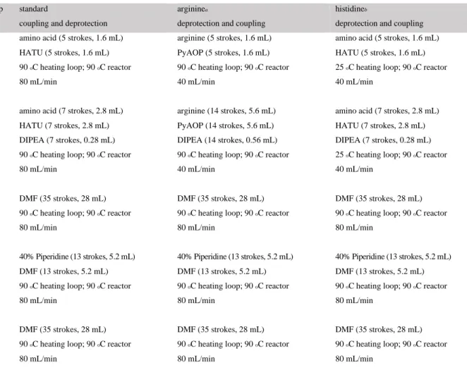
Chapter 2 addresses the production bottleneck in personalized cancer vaccines. As described above, high-throughput genome sequencing and computation have enabled rapid identification of mutation-derived, patient-specific neoantigens which can be synthesized and formulated as a vaccine, but generating the vaccine peptides for each patient in a rapid and affordable fashion remains difficult. High-throughput peptide synthesis technology is therefore urgently needed for personalized cancer vaccines to succeed in the clinic. Previously, we developed automated flow peptide synthesis technology that greatly accelerates the production of synthetic peptides. We show that this technology permits the synthesis of high-quality peptides for personalized medicine. Automated flow synthesis produces 30-mer peptides in less than 35 minutes and 15- to 16-mer peptides in less than 20 minutes. The purity of these peptides is comparable with or higher than the purity of peptides produced by other methods. This work illustrates how automated flow synthesis technology can enable personalized therapy by accelerating peptide synthesis and increasing purity. We envision that implementing this technology in clinical settings will greatly increase capacity to generate clinical-grade peptides on demand, which is a key step in reaching the full potential of personalized vaccines for the treatment of cancer and other diseases.
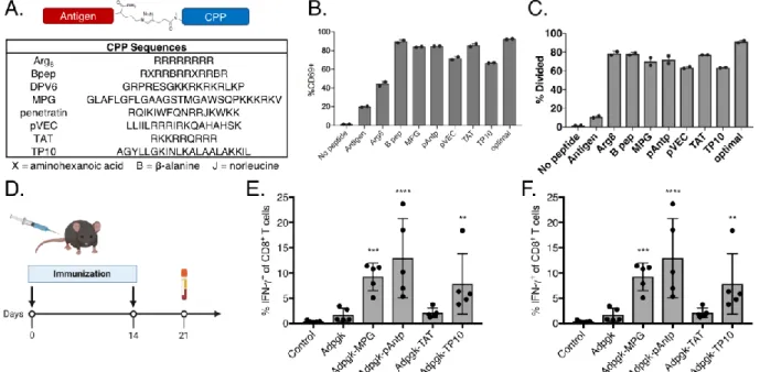
Chapter 3 confronts the delivery challenges associated with peptide vaccines using cell-penetrating peptides (CPPs). As highlighted previously, CPPs are a straightforward approach to enhance the anti-tumor efficacy of peptide vaccines by improving uptake by antigen presenting cells. However, their impact on antigen behavior in vivo remains largely uncharacterized and reports to date concern only a limited number of CPPs and model antigens. Here, we screen a set of CPPs to compare their impact on T cell priming as well their mechanism of action at the cellular and systems levels. All CPPs increased antigen uptake and T cell activation in vitro and the majority also enhanced endogenous T cell priming in vivo, by up to 20-fold. A top CPP, penetratin, similarly enhanced vaccine immunogenicity for other tumor antigens and provided significantly greater tumor protection compared to a vaccine with the unmodified antigen. After confirming that CPPs increase uptake by antigen presenting cells, according to their expected mechanism, we then explored their impact on antigen delivery and presentation in vivo. We determined that CPPs promote delivery to the lymph nodes and suggest passive trafficking mediated by serum protein binding as a likely mechanism. We demonstrate that CPPs prolong antigen serum stability and extend the duration of presentation in the draining lymph node, suggesting a temporal component to CPP-mediated vaccine immunogenicity. These findings support CPPs as a general strategy to boost the immunogenicity and efficacy of
therapeutic peptide vaccines and indicate several additional factors beyond intracellular delivery that contribute to their activity.
Chapter 4 further expands on the use strategies that promote lymphatic trafficking to address the delivery challenge for peptide vaccines. As described above, one previously reported strategy involves binding of endogenous albumin upon injection via a diacyl lipid, which allows peptide antigens to “hitchhike” to the draining lymph node in a similar manner to the mechanism of action of sentinel lymph node mapping dyes used clinically. Here, we explore the scope of potential albumin binding moieties by testing whether an albumin-binding peptide and a small molecule, a-tocopherol, can successfully boost vaccine immunogenicity in vivo. We demonstrate that multiple albumin-binding moieties conjugated to peptide antigens enhanced lymph node accumulation and subsequent T cell priming.
Chapter 5 describes the development of a new technique to measure presentation of an antigen of interest and its implementation screening two potential strategies for improving peptide vaccines. The ability to directly measure presentation of specific MHC-associated antigens could facilitate new avenues of investigation, including screening strategies to boost the presentation of vaccine antigens. We aimed to develop a new technique that is straightforward, quantitative, and higher throughput than existing techniques by combining direct elution of antigens from the surface of intact cells followed by targeted, high-sensitivity LC-MS/MS analysis. We validated the ability of this approach to quantify MHC-presented antigens in a generalizable manner, then applied it to characterize the dynamics of vaccine antigen presentation. With this technique in hand, we then began to explore the impact of D-amino acid substitution as well as the impact of CPP conjugation on the magnitude and kinetics of vaccine antigen presentation. While instrumentation limits its sensitivity to the level of other MS-based techniques, we anticipate that the simplicity of our approach could enable design of vaccine antigens with optimal presentation and characterization of this fundamental process.
Chapter 6 does not concern cancer vaccines and instead focuses on the design of chimeric CPPs for our lab’s original CPP application, improving the delivery of phosphorodiamidate morpholino oligonucleotides (PMOs). PMOs are a promising class of therapeutics for genetic disease. PMOs designed for “exon skipping” must be internalized into cells, reach the nucleus, and act on pre-mRNA to mediate their effects. One tactic for improving PMO delivery and exon skipping is to covalently conjugate PMOs to CPPs. Here we report the synthesis of PMOs conjugated to CPP chimeras, constructed by combining multiple CPPs into one sequence. The chimeric CPPs synergistically improve PMO activity up to 70-fold over the PMO alone, beyond the expected effects of each component peptide. By investigating the
design space of CPP chimeras, we demonstrate that all components must be covalently attached, that the order of the two sequences matters, and that peptide identity can tune activity. We identified one chimera (pVEC-Bpep) to investigate in more detail and found that it engages different mechanisms of endocytosis than its parent peptides. We also examined the extent to which the beneficial effect comes from improved cellular uptake as opposed to the downstream steps required for exon skipping. Given the complexity of intracellular delivery, we anticipate this work will lead researchers to consider combining molecules with different physicochemical properties in order to aid in the delivery of biologic cargoes. Earlier projects using CPPs to deliver PMOs informed both this project as well as the application of CPPs to cancer vaccines, which we explored in parallel.
This thesis tackles the synthesis bottleneck in producing personalized neoantigen vaccines using our in-house automated technology. It then applies our expertise in delivering large biomolecules gained with PMOs to the delivery of peptide vaccine antigens. After surveying a set of CPPs to demonstrate that they can deliver a range of vaccine antigens and enhance anti-tumor efficacy, we dissect their mechanism of action and determine that in addition to mediating intracellular delivery, they also promote lymph node trafficking via serum protein binding. We then assess several other albumin binding moieties for their ability to promote lymph node trafficking and vaccine efficacy. I also present a new technique to measure antigen presentation, facilitating vaccine optimization. The work described in this thesis represents a new direction for our lab that applies our existing synthesis and delivery capabilities to the challenge of therapeutic cancer vaccines.
1.8 References
(1) Sahin, U.; Türeci, Ö. Personalized Vaccines for Cancer Immunotherapy. Science 2018, 359 (6382), 1355–1360. https://doi.org/10.1126/science.aar7112.
(2) June, C. H.; O’Connor, R. S.; Kawalekar, O. U.; Ghassemi, S.; Milone, M. C. CAR T Cell Immunotherapy for Human Cancer. Science 2018, 359 (6382), 1361–1365. https://doi.org/10.1126/science.aar6711.
(3) Ribas, A.; Wolchok, J. D. Cancer Immunotherapy Using Checkpoint Blockade. Science 2018, 359 (6382), 1350–1355. https://doi.org/10.1126/science.aar4060.
(4) Jenkins, R. W.; Fisher, D. E. Treatment of Advanced Melanoma in 2020 and Beyond. Journal of Investigative Dermatology 2020. https://doi.org/10.1016/j.jid.2020.03.943. (5) Waldman, A. D.; Fritz, J. M.; Lenardo, M. J. A Guide to Cancer Immunotherapy: From
T Cell Basic Science to Clinical Practice. Nature Reviews Immunology 2020, 1–18. https://doi.org/10.1038/s41577-020-0306-5.
(6) Abbas, A.; Lichtman, A.; Pillai, S. Cellular and Molecular Immunology, 9th ed.; Elsevier, 2017.
(7) Waldman, A. D.; Fritz, J. M.; Lenardo, M. J. A Guide to Cancer Immunotherapy: From T Cell Basic Science to Clinical Practice. Nature Reviews Immunology 2020, 1–18. https://doi.org/10.1038/s41577-020-0306-5.
(8) Borst, J.; Ahrends, T.; Bąbała, N.; Melief, C. J. M.; Kastenmüller, W. CD4 + T Cell Help in Cancer Immunology and Immunotherapy. Nature Reviews Immunology 2018, 18 (10), 635–647. https://doi.org/10.1038/s41577-018-0044-0.
(9) Qin, S.; Xu, L.; Yi, M.; Yu, S.; Wu, K.; Luo, S. Novel Immune Checkpoint Targets: Moving beyond PD-1 and CTLA-4. Mol. Cancer 2019, 18 (1), 155.
https://doi.org/10.1186/s12943-019-1091-2.
(10) Hamid, O.; Robert, C.; Daud, A.; Hodi, F. S.; Hwu, W. J.; Kefford, R.; Wolchok, J. D.; Hersey, P.; Joseph, R.; Weber, J. S.; Dronca, R.; Mitchell, T. C.; Patnaik, A.; Zarour, H. M.; Joshua, A. M.; Zhao, Q.; Jensen, E.; Ahsan, S.; Ibrahim, N.; Ribas, A. Five-Year Survival Outcomes for Patients with Advanced Melanoma Treated with Pembrolizumab in KEYNOTE-001. Ann. Oncol. 2019, 30 (4), 582–588.
https://doi.org/10.1093/annonc/mdz011.
(11) Schuster, S. J.; Svoboda, J.; Chong, E. A.; Nasta, S. D.; Mato, A. R.; Anak, Ö.; Brogdon, J. L.; Pruteanu-Malinici, I.; Bhoj, V.; Landsburg, D.; Wasik, M.; Levine, B. L.; Lacey, S. F.; Melenhorst, J. J.; Porter, D. L.; June, C. H. Chimeric Antigen Receptor T Cells in Refractory B-Cell Lymphomas. N. Engl. J. Med. 2017, 377 (26), 2545–2554.
https://doi.org/10.1056/NEJMoa1708566.
(12) Laetsch, T. W.; Myers, G. D.; Baruchel, A.; Dietz, A. C.; Pulsipher, M. A.; Bittencourt, H.; Buechner, J.; De Moerloose, B.; Davis, K. L.; Nemecek, E.; Driscoll, T.; Mechinaud, F.; Boissel, N.; Rives, S.; Bader, P.; Peters, C.; Sabnis, H. S.; Grupp, S. A.; Yanik, G. A.; Hiramatsu, H.; Stefanski, H. E.; Rasouliyan, L.; Yi, L.; Shah, S.; Zhang, J.; Harris, A. C. Patient-Reported Quality of Life after Tisagenlecleucel Infusion in Children and Young Adults with Relapsed or Refractory B-Cell Acute Lymphoblastic Leukaemia: A Global, Single-Arm, Phase 2 Trial. The Lancet Oncology 2019, 20 (12), 1710–1718. https://doi.org/10.1016/S1470-2045(19)30493-0.
(13) Fiorenza, S.; Ritchie, D. S.; Ramsey, S. D.; Turtle, C. J.; Roth, J. A. Value and Affordability of CAR T-Cell Therapy in the United States. Bone Marrow Transplant. 2020. https://doi.org/10.1038/s41409-020-0956-8.
(14) Lim, F. L. W. I.; Ang, S. O. Emerging CAR Landscape for Cancer Immunotherapy. Biochem. Pharmacol. 2020, 114051. https://doi.org/10.1016/j.bcp.2020.114051. (15) Hollingsworth, R. E.; Jansen, K. Turning the Corner on Therapeutic Cancer Vaccines.
npj Vaccines 2019, 4 (1), 1–10. https://doi.org/10.1038/s41541-019-0103-y. (16) Hu, Z.; Ott, P. A.; Wu, C. J. Towards Personalized, Tumour-Specific, Therapeutic
Vaccines for Cancer. Nature Reviews Immunology 2018, 18 (3), 168–182. https://doi.org/10.1038/nri.2017.131.
(17) Ma, M.; Liu, J.; Jin, S.; Wang, L. Development of Tumour Peptide Vaccines: From Universalization to Personalization. Scandinavian Journal of Immunology n/a (n/a), e12875. https://doi.org/10.1111/sji.12875.
(18) Ott, P. A.; Hu, Z.; Keskin, D. B.; Shukla, S. A.; Sun, J.; Bozym, D. J.; Zhang, W.; Luoma, A.; Giobbie-Hurder, A.; Peter, L.; Chen, C.; Olive, O.; Carter, T. A.; Li, S.; Lieb, D. J.; Eisenhaure, T.; Gjini, E.; Stevens, J.; Lane, W. J.; Javeri, I.; Nellaiappan, K.; Salazar, A.; Daley, H.; Seaman, M.; Buchbinder, E. I.; Yoon, C. H.; Harden, M.;
Lennon, N.; Gabriel, S.; Rodig, S. J.; Barouch, D. H.; Aster, J. C.; Getz, G.;
Wucherpfennig, K.; Neuberg, D.; Ritz, J.; Lander, E. S.; Fritsch, E. F.; Hacohen, N.; Wu, C. J. An Immunogenic Personal Neoantigen Vaccine for Melanoma Patients. Nature 2017, 547 (7662), 217–221. https://doi.org/10.1038/nature22991.
(19) Keskin, D. B.; Anandappa, A. J.; Sun, J.; Tirosh, I.; Mathewson, N. D.; Li, S.; Oliveira, G.; Giobbie-Hurder, A.; Felt, K.; Gjini, E.; Shukla, S. A.; Hu, Z.; Li, L.; Le, P. M.; Allesøe, R. L.; Richman, A. R.; Kowalczyk, M. S.; Abdelrahman, S.; Geduldig, J. E.; Charbonneau, S.; Pelton, K.; Iorgulescu, J. B.; Elagina, L.; Zhang, W.; Olive, O.; McCluskey, C.; Olsen, L. R.; Stevens, J.; Lane, W. J.; Salazar, A. M.; Daley, H.; Wen, P. Y.; Chiocca, E. A.; Harden, M.; Lennon, N. J.; Gabriel, S.; Getz, G.; Lander, E. S.; Regev, A.; Ritz, J.; Neuberg, D.; Rodig, S. J.; Ligon, K. L.; Suvà, M. L.;
Wucherpfennig, K. W.; Hacohen, N.; Fritsch, E. F.; Livak, K. J.; Ott, P. A.; Wu, C. J.; Reardon, D. A. Neoantigen Vaccine Generates Intratumoral T Cell Responses in Phase Ib Glioblastoma Trial. Nature 2019, 565 (7738), 234–239.
https://doi.org/10.1038/s41586-018-0792-9.
(20) Sahin, U.; Derhovanessian, E.; Miller, M.; Kloke, B.-P.; Simon, P.; Löwer, M.; Bukur, V.; Tadmor, A. D.; Luxemburger, U.; Schrörs, B.; Omokoko, T.; Vormehr, M.;
Albrecht, C.; Paruzynski, A.; Kuhn, A. N.; Buck, J.; Heesch, S.; Schreeb, K. H.; Müller, F.; Ortseifer, I.; Vogler, I.; Godehardt, E.; Attig, S.; Rae, R.; Breitkreuz, A.; Tolliver, C.; Suchan, M.; Martic, G.; Hohberger, A.; Sorn, P.; Diekmann, J.; Ciesla, J.; Waksmann, O.; Brück, A.-K.; Witt, M.; Zillgen, M.; Rothermel, A.; Kasemann, B.; Langer, D.; Bolte, S.; Diken, M.; Kreiter, S.; Nemecek, R.; Gebhardt, C.; Grabbe, S.; Höller, C.; Utikal, J.; Huber, C.; Loquai, C.; Türeci, Ö. Personalized RNA Mutanome Vaccines Mobilize Poly-Specific Therapeutic Immunity against Cancer. Nature 2017, 547 (7662), 222–226. https://doi.org/10.1038/nature23003.
(21) Dillman, R. O.; Cornforth, A. N.; Nistor, G. I.; McClay, E. F.; Amatruda, T. T.; Depriest, C. Randomized Phase II Trial of Autologous Dendritic Cell Vaccines versus Autologous Tumor Cell Vaccines in Metastatic Melanoma: 5-Year Follow up and Additional Analyses. J Immunother Cancer 2018, 6 (1), 19.
https://doi.org/10.1186/s40425-018-0330-1.
(22) Chamani, R.; Ranji, P.; Hadji, M.; Nahvijou, A.; Esmati, E.; Alizadeh, A. M.
Application of E75 Peptide Vaccine in Breast Cancer Patients: A Systematic Review and Meta-Analysis. European Journal of Pharmacology 2018, 831, 87–93.
https://doi.org/10.1016/j.ejphar.2018.05.010.
(23) Moynihan, K. D.; Opel, C. F.; Szeto, G. L.; Tzeng, A.; Zhu, E. F.; Engreitz, J. M.; Williams, R. T.; Rakhra, K.; Zhang, M. H.; Rothschilds, A. M.; Kumari, S.; Kelly, R. L.; Kwan, B. H.; Abraham, W.; Hu, K.; Mehta, N. K.; Kauke, M. J.; Suh, H.; Cochran, J. R.; Lauffenburger, D. A.; Wittrup, K. D.; Irvine, D. J. Eradication of Large Established Tumors in Mice by Combination Immunotherapy That Engages Innate and Adaptive Immune Responses. Nat Med 2016, 22 (12), 1402–1410.
https://doi.org/10.1038/nm.4200.
(24) Collins, J. M.; Redman, J. M.; Gulley, J. L. Combining Vaccines and Immune
Checkpoint Inhibitors to Prime, Expand, and Facilitate Effective Tumor Immunotherapy. Expert Rev Vaccines 2018, 17 (8), 697–705.
https://doi.org/10.1080/14760584.2018.1506332.
(25) Grenier, J. M.; Yeung, S. T.; Khanna, K. M. Combination Immunotherapy: Taking Cancer Vaccines to the Next Level. Front Immunol 2018, 9, 610.
https://doi.org/10.3389/fimmu.2018.00610.
(26) Janeway, C. A.; Travers, P.; Walport, M.; Shlomchik, M. J. Immunobiology: The Immune System in Health and Disease, 5th ed.; Garland Science: New York, 2001. (27) Tai, Y.; Wang, Q.; Korner, H.; Zhang, L.; Wei, W. Molecular Mechanisms of T Cells
Activation by Dendritic Cells in Autoimmune Diseases. Front Pharmacol 2018, 9. https://doi.org/10.3389/fphar.2018.00642.
(28) Liu, H.; Moynihan, K. D.; Zheng, Y.; Szeto, G. L.; Li, A. V.; Huang, B.; Van Egeren, D. S.; Park, C.; Irvine, D. J. Structure-Based Programming of Lymph-Node Targeting in Molecular Vaccines. Nature 2014, 507 (7493), 519–522.
https://doi.org/10.1038/nature12978.
(29) Joffre, O. P.; Segura, E.; Savina, A.; Amigorena, S. Cross-Presentation by Dendritic Cells. Nat Rev Immunol 2012, 12 (8), 557–569. https://doi.org/10.1038/nri3254. (30) Vigneron, N. Human Tumor Antigens and Cancer Immunotherapy. Biomed Res Int
2015, 2015. https://doi.org/10.1155/2015/948501.
(31) Ilyas, S.; Yang, J. C. Landscape of Tumor Antigens in T-Cell Immunotherapy. J Immunol 2015, 195 (11), 5117–5122. https://doi.org/10.4049/jimmunol.1501657.
(32) Jurtz, V.; Paul, S.; Andreatta, M.; Marcatili, P.; Peters, B.; Nielsen, M. NetMHCpan-4.0: Improved Peptide-MHC Class I Interaction Predictions Integrating Eluted Ligand and Peptide Binding Affinity Data. J. Immunol. 2017, 199 (9), 3360–3368.
https://doi.org/10.4049/jimmunol.1700893.
(33) Wculek, S. K.; Cueto, F. J.; Mujal, A. M.; Melero, I.; Krummel, M. F.; Sancho, D. Dendritic Cells in Cancer Immunology and Immunotherapy. Nature Reviews Immunology 2020, 20 (1), 7–24. https://doi.org/10.1038/s41577-019-0210-z.
(34) Kantoff, P. W.; Higano, C. S.; Shore, N. D.; Berger, E. R.; Small, E. J.; Penson, D. F.; Redfern, C. H.; Ferrari, A. C.; Dreicer, R.; Sims, R. B.; Xu, Y.; Frohlich, M. W.; Schellhammer, P. F.; IMPACT Study Investigators. Sipuleucel-T Immunotherapy for Castration-Resistant Prostate Cancer. N. Engl. J. Med. 2010, 363 (5), 411–422. https://doi.org/10.1056/NEJMoa1001294.
(35) Tanyi, J. L.; Bobisse, S.; Ophir, E.; Tuyaerts, S.; Roberti, A.; Genolet, R.; Baumgartner, P.; Stevenson, B. J.; Iseli, C.; Dangaj, D.; Czerniecki, B.; Semilietof, A.; Racle, J.; Michel, A.; Xenarios, I.; Chiang, C.; Monos, D. S.; Torigian, D. A.; Nisenbaum, H. L.; Michielin, O.; June, C. H.; Levine, B. L.; Powell, D. J.; Gfeller, D.; Mick, R.; Dafni, U.; Zoete, V.; Harari, A.; Coukos, G.; Kandalaft, L. E. Personalized Cancer Vaccine
Effectively Mobilizes Antitumor T Cell Immunity in Ovarian Cancer. Sci Transl Med 2018, 10 (436). https://doi.org/10.1126/scitranslmed.aao5931.
(36) Sato-Dahlman, M.; LaRocca, C. J.; Yanagiba, C.; Yamamoto, M. Adenovirus and Immunotherapy: Advancing Cancer Treatment by Combination. Cancers (Basel) 2020, 12 (5). https://doi.org/10.3390/cancers12051295.
(37) Flickinger, J. C.; Rodeck, U.; Snook, A. E. Listeria Monocytogenes as a Vector for Cancer Immunotherapy: Current Understanding and Progress. Vaccines (Basel) 2018, 6 (3). https://doi.org/10.3390/vaccines6030048.
(38) Wadhwa, A.; Aljabbari, A.; Lokras, A.; Foged, C.; Thakur, A. Opportunities and Challenges in the Delivery of MRNA-Based Vaccines. Pharmaceutics 2020, 12 (2). https://doi.org/10.3390/pharmaceutics12020102.
(39) Hobernik, D.; Bros, M. DNA Vaccines-How Far From Clinical Use? Int J Mol Sci 2018, 19 (11). https://doi.org/10.3390/ijms19113605.
(40) Kutzler, M. A.; Weiner, D. B. DNA Vaccines: Ready for Prime Time? Nat. Rev. Genet. 2008, 9 (10), 776–788. https://doi.org/10.1038/nrg2432.
(41) Tejeda-Mansir, A.; García-Rendón, A.; Guerrero-Germán, P. Plasmid-DNA Lipid and Polymeric Nanovaccines: A New Strategic in Vaccines Development. Biotechnology and Genetic Engineering Reviews 2019, 35 (1), 46–68.
https://doi.org/10.1080/02648725.2018.1560552.
(42) Lim, M.; Badruddoza, A. Z. M.; Firdous, J.; Azad, M.; Mannan, A.; Al-Hilal, T. A.; Cho, C.-S.; Islam, M. A. Engineered Nanodelivery Systems to Improve DNA Vaccine Technologies. Pharmaceutics 2020, 12 (1), 30.