HAL Id: tel-02801967
https://hal.inrae.fr/tel-02801967
Submitted on 5 Jun 2020HAL is a multi-disciplinary open access archive for the deposit and dissemination of sci-entific research documents, whether they are pub-lished or not. The documents may come from teaching and research institutions in France or abroad, or from public or private research centers.
L’archive ouverte pluridisciplinaire HAL, est destinée au dépôt et à la diffusion de documents scientifiques de niveau recherche, publiés ou non, émanant des établissements d’enseignement et de recherche français ou étrangers, des laboratoires publics ou privés.
Fonctionnement des populations de virus multipartites
de plantes au cours des différentes étapes de leur cycle
de vie
Anne Sicard
To cite this version:
Anne Sicard. Fonctionnement des populations de virus multipartites de plantes au cours des différentes étapes de leur cycle de vie. Biologie végétale. 2014. Français. �tel-02801967�
Délivré par le
Centre international d’études supérieures
en sciences agronomiques
Montpellier
Préparée au sein de l’école doctorale SIBAGHE
Et de l’unité de recherche UMR - BGPI
Spécialité : Microbilogie/Parasitologie
Présentée par Anne SICARD
Soutenue le 12 décembre 2014 devant le jury composé de
Mme Isabelle JUPIN, Directrice de Recherche, CNRS, Paris Examinatrice Mme Eugénie HEBRARD, Chercheur, IRD, Montpellier Examinatrice M. Esteban DOMINGO, Professeur, Spanish Research Council Rapporteur M. Enrique MORIONES, Professeur, Consejo Superior de
Investigaciones Cientificas
Rapporteur M. Stéphane BLANC, Directeur de Recherche, INRA, Montpellier Dir. de thèse
Fonctionnement des populations de virus
multipartites de plantes au cours des
différentes étapes de leur cycle de vie
2 Résumé :
Les virus multipartites sont caractérisés par un génome divisé en au moins deux segments, chacun étant encapsidé individuellement. Bien que ces virus représentent près de 30% des genres de virus de plantes connus, les coûts et avantages engendrés pas cette organisation génomique restent mal connus. Basée sur l’étude d’un Nanovirus, le Faba bean necrotic stunt
virus (FBNSV), ma thèse vise à mieux comprendre les dynamiques des populations d’un virus
multipartite au cours de l’infection de son hôte et de sa transmission par pucerons.
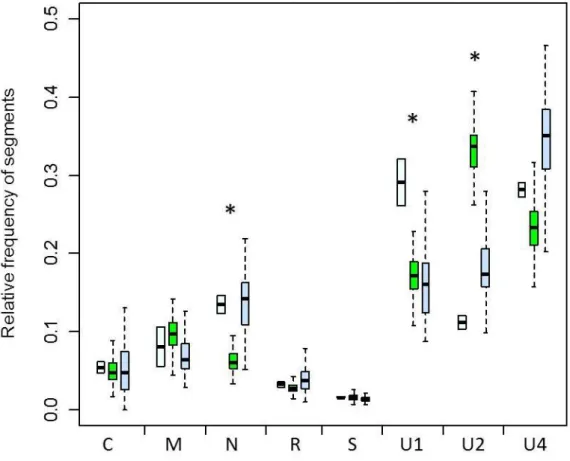
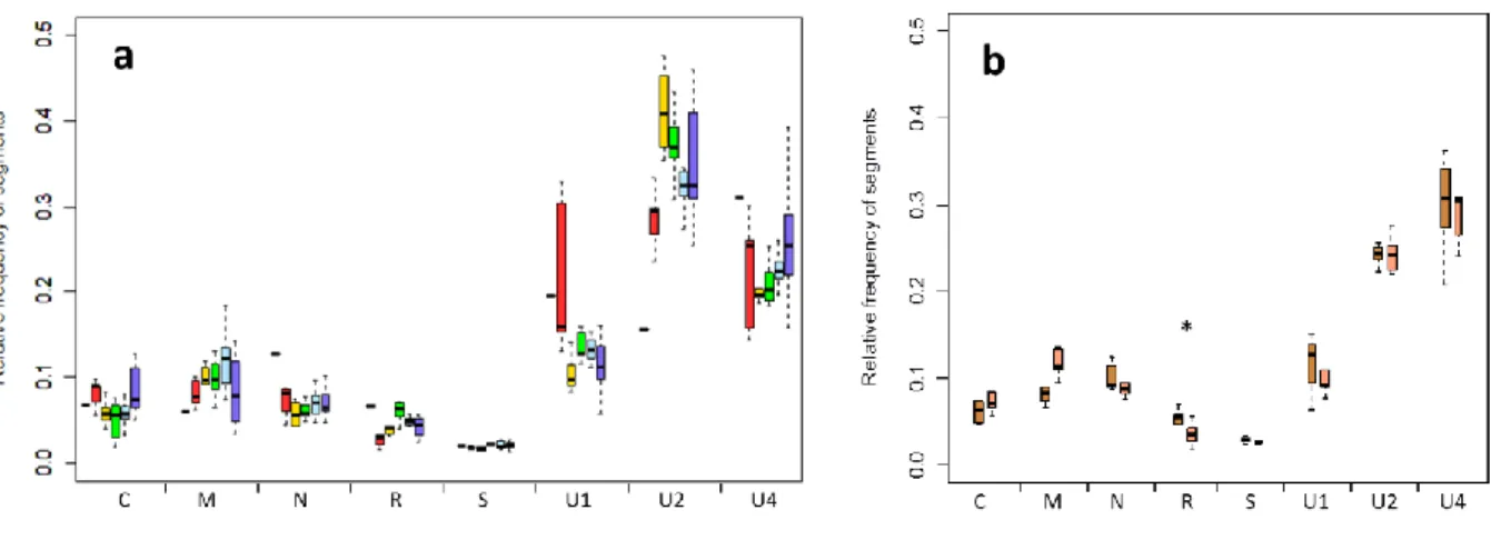
Dans un premier temps, nous avons démontré que chacun des huit segments composant le génome du FBNSV s’accumulait de manière reproductible à une fréquence relative spécifique, certains segments représentant près de 30% de la population virale au sein de la fève et d’autres n’excédant pas 2%. En changeant de plante hôte, nous avons pu montrer que les fréquences relatives des différents segments étaient hôte-spécifiques. Nous avons par ailleurs observé des changements de fréquences relatives au sein des pucerons vecteurs, changements révélant des interactions intimes entre le FBNSV –précédemment perçu comme traversant simplement les barrières cellulaires des pucerons- et son vecteur.
Cette plasticité et l’ajustement correspondant de la composition génétique de la population virale au sein de l’hôte pourrait permettre aux virus multipartites de s’adapter rapidement à un nouvel environnement grâce à un changement spécifique du nombre de copies de ses gènes. Cependant, ces différences de fréquences relatives pourraient également constituer un coût additionnel pour le virus si, comme il est généralement admis, tous les segments ont besoin de pénétrer au sein de la même cellule pour garantir l’infection. En tenant compte des segments rares au sein des populations virales, ce coût apparaît énorme et nous avons donc décidé de vérifier l’existence d’un tel coût expérimentalement.
Une étude de la distribution des différents segments génomiques au sein des cellules végétales est donc en cours et ce, dans le but de regarder si les huit segments sont toujours ensemble au sein des cellules ou s’ils peuvent être physiquement séparés. Cette question est d’importance majeure non seulement en ce qu’elle permet de réévaluer le coût potentiel des virus multipartites mais aussi en ce qu’elle permet de tester le concept du cycle de réplication « cellule-autonome » chez les virus.
Enfin, le maintien de l’intégrité génomique des virus multipartites reposant sur de relativement faibles goulots d’étranglement tout au long du cycle viral, des outils ont été mis en place pour quantifier ces goulots d’étranglement.
Mots clés : Nanoviridae, Faba bean necrotic stunt virus, Nombre de copies de gène,
3 Abstract :
Multipartite viruses are characterized by a genome divided into two or more segments, each encapsidated individually into separated virus particles. Although they represent more than 30% of the known plant virus genera, little is known on how such viral systems actually work and on the advantage(s) and cost(s) of their peculiar genome organization. Based on the study of a Nanovirus, the Faba bean necrotic stunt virus (FBNSV), my thesis aimed at better understanding the dynamics of a multipartite virus population during within host disease development and during aphid transmission.
We have first demonstrated that each of the eight segments composing the FBNSV genome reproducibly accumulates at a specific relative frequency, some segments representing up to 30 % of the total viral DNA within an infected plant and others not exceeding 2%. By changing the host plant species, we could further show that the relative frequency of the different segments is host-specific. Besides, we observed changes in segment frequencies within the vectors, thus revealing intimate interactions between the FBNSV –which was previously thought to simply traverse cellular barriers- and its vectors.
This plasticity and the corresponding adjustment of the genetic composition of the within host viral population could enable the multipartite virus to rapidly adjust to a novel environment, by specifically changing the copy number of its different genes. However, it could also constitute an additional cost for the virus if, as generally assumed, all segments need to enter the same cell to secure infection. When considering the rare segments in FBNSV populations, the cost appear enormous and we have decided to experimentally address this question.
We are currently looking at the distribution of the different genome segments within individual cells of host plants in order to empirically assess whether all 8 segments are always together within individual cells or whether they can be physically separated. This question is of major importance, not only for re-evaluating the possible costs in multipartite viral systems, but also for challenging the concept of a cell-autonomous replication cycle in viruses.
Finally, as the genomic integrity preservation of multipartite viruses is based on rather small bottlenecks during the whole viral life cycle, tools have been developed to quantify these bottlenecks.
Keywords : Nanoviridae, Faba bean necrotic stunt virus, Gene copy number , Circulative
4
Laboratoire d’accueil
Cette thèse a été réalisée au sein de l’équipe :
Interactions Virus Insecte Plante (VIP)
Sous la Direction de :
Dr Stéphane BLANC, Directeur de Recherche INRA, Montpellier - Directeur de thèse Dr Serafin Gutiérrez, Chercheur, CIRAD, Montpellier – Co-Directeur de thèse
UMR Biologie et Génétique des Interactions Plante-Parasite (BGPI)
INRA-CIRAD-SupAgro
Campus International de Baillarguet 34398 Montpellier cedex 5
5
Remerciements
Je souhaite tout d’abord remercier les membres du jury d’avoir accepté de juger mon travail : Mme Eugénie Hébrard, Mme Isabelle Jupin et Messieurs Esteban Domingo, et Enrique Moriones.
Je souhaite également remercier les membres de mon comité de thèse pour leurs conseils : Mme Sylvie Dinant, M. Bruno Gronenborn, M. Michel Lebrun et M. Philippe Roumagnac.
Merci à Philippe Rott et à Claire Neema pour leur accueil au sein de l’UMR BGPI et leurs investissements pour que cette UMR soit un lieu propice au travail et aux échanges. Je tiens tout particulièrement à remercier Stéphane Blanc d’avoir été un directeur de thèse très présent, à l’écoute et toujours très enthousiaste. Merci à toi Stéphane de m’avoir amenée à découvrir le monde fabuleux des virus multipartites et merci pour ton aide précieuse dans le rush final…
Merci également au Maître Jedi, Serafin Gutierrez, pour toutes les discussions que l’on a pu avoir et pour tous les conseils prodigués tout au long de cette thèse. Merci pour ta rigueur scientifique et tes idées judicieuses.
Je voudrais également remercier Dr T.L.German, mon professeur à l’université de Madison, WI de m’avoir initiée aux joies de la virologie et de m’y avoir fait prendre goût. Merci à Yannis Michalakis pour son implication dans ce projet et pour toutes ces réunions très fructueuses tout au long de ma thèse. Mais pourquoi diable avoir choisi un nanovirus ?...
Un grand merci à tous les membres de l’équipe VIP, cette grande famille au sein de laquelle il est bon de grandir…
Je souhaite plus particulièrement remercier Michel Yvon pour son aide dans ce projet et pour sa constante bonne humeur, travailler à tes côtés allège tellement la tâche.
Merci à Marie-Stéphanie Vernerey pour toute son aide en microscopie, son investissement dans le projet, pour les joggings matinaux et sa gentillesse.
Merci à Daniel Gargani pour toutes les « belles images » émises.
Manue, cette partie est pour toi… Merci pour ton humour et ta joie de vivre.
Merci à Aurélie Bak, Déborah Conflon et Zineb Belabess pour tous ces bons moments passés ensemble.
6
Merci à Jean-Louis Zeddam pour ses bonnes idées et à Maryline Uzest pour sa gentillesse et son aide.
Merci à « l’équipe gémini » pour tous les échanges techniques et conceptuels que nous avons pu avoir notamment au cours des réunions du lundi matin.
Je voudrais également remercier les membres de l’UMR BGPI pour tous les bons moments passés et tout particulièrement Katia Bonnemeyre.
Un grand merci à tous mes amis qui m’ont beaucoup soutenue tout au long de cette thèse et qui m’ont permis de passer des weekends formidables aux quatre coins de la France. Je vous attends tous en Californie !
Mes pensées se tournent évidemment vers ma famille qui m’a beaucoup encouragée et aidée à en arriver là. Je souhaite tout particulièrement remercier mes parents qui savent toujours trouver les mots justes et mes sœurs qui malgré la distance savent toujours se montrer présentes.
Enfin, merci à Marie-Steph et à Michel pour leur accueil chaleureux dans des endroits merveilleux lors de la rédaction de ce manuscrit.
Merci à Jérémie pour son aide dans l’édition de ce manuscrit et merci au comité de relecture, Hélène, David, Manuella et Déborah.
7
Table des matières
Laboratoire d’accueil ... 4
Remerciements ... 5
Table des matières ... 7
Liste des figures et tableaux ... 9
Liste des abréviations ... 10
Introduction générale ... 12
Introduction bibliographique ... 17
1. Review : The strange lifestyle of multipartite viruses ... 18
2. Le Faba bean necrotic stunt virus, un modèle pour l’étude des virus multipartites ... 49
Les nanovirus, des virus multipartites au génome segmenté « à l’extrême » ... 49
Répartition géographique et importance agronomique des Nanoviridae ... 49
Le Faba bean necrotic stunt virus comme modèle d’étude ... 50
Transmission du FBNSV ... 51
Information génétique du FBNSV ... 52
Fonction des 8 protéines du FBNSV et importance de celles-ci dans l’infection de la fève ... 54
Réplication du FBNSV ... 55
Evolution du FBNSV ... 56
Chapitre I : Le nombre de copies de gènes est régulé différentiellement chez un virus multipartite .. 59
1. Contexte et objectifs ... 59
2. Article 1 : Gene copy number is differentially regulated in a multipartite virus ... 62
3. Complément à l’article 1 ... 75
Essai de mise en place de l‘évolution expérimentale du FBNSV ... 75
Protocole ... 76
Résultats et discussion ... 79
Chapitre II : La transmission cirulante non-propagative des nanovirus : une vision simplificatrice ... 84
1. Contexte et objectif ... 84
2. Circulative non-propagative aphid-transmission of nanoviruses : an oversimplified view (Article 2) ... 88
3. Conclusion ... 110
Chapitre III : Les virus multipartites : un fonctionnement décentralisé ... 113
1. Introduction ... 113
2. Matériel et méthodes ... 116
3. Résultats ... 124
8
5. Perspectives à plus long terme ... 144
Chapitre IV : Insertion des marqueurs génétiques neutres dans le clone infectieux du FBNSV pour l’estimation des goulots d’étranglement au cours de son cycle de vie ... 147
1. Introduction ... 147
2. Matériel et méthodes ... 152
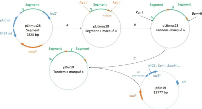
Construction des clones « segment-tige boucle » ... 152
Approche 2 : Construction de clones en tandem avec marqueurs génétiques ... 158
3. Résultats ... 163
4. Conclusion ... 165
Conclusion générale ... 168
Annexes ... 174
1. Annexe I : Purification du FBNSV ... 175
2. Annexe II : Calcul de la probabilité de perdre au moins un segment au cours de l’infection de la fève ... 176
3. Annexe III : Phloème ... 177
4. Annexe IV : Carte des plasmides utilisés au cours de cette thèse ... 178
9
Liste des figures et tableaux
Figure 1 : Schematic representation of plant virus families and genera ... 21
Figure 2: Genome formula of FBNSV in two different host plant species ... 27
Figure 3: Comparison of the genome or segment sizes in viruses ... 31
Figure 4: Relationships among putative 30K MP superfamily members determined by bootstrapped parsimony ... 34
Figure 5: Répartition géographique mondiale des nanovirus (jaune) et des babuvirus (rouge) ... 50
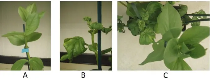
Figure 6: Symptômes causés par le Faba bean necrotic stunt virus sur la fève ... 51
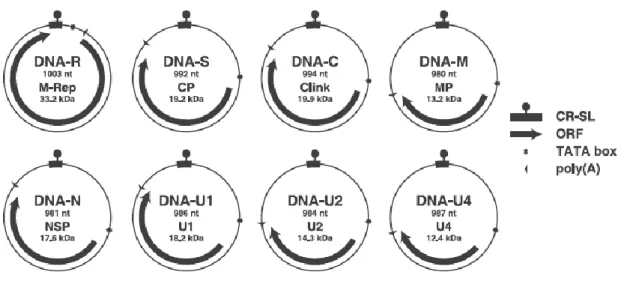
Figure 7: Organisation génomique du FBNSV ... 53
Figure 8: Particules virales de Faba bean necrotic stunt virus ... 53
Figure 9: Réplication des virus à ADN simple brin selon le mécanisme du cercle roulant ... 56
Figure 10 : Evolution expérimentale de la formule génomique du FBNSV ... 77
Figure 11 : Mesure de l’intensité de fluorescence de chaque segment dans une cellule coinfectée par les segments S (en rouge) et U4 (en vert), 29 jours post-infection avec le logiciel ZEN 2009 (ZEISS) 123 Figure 12 : Spécificité des sondes S-oligonuclétidique (ON-S, A) , S-random priming (RP-S, B), R-random priming (RP-R, C), U4-R-random priming (RP-U4, D), R1-R-random priming (RP-R1, E) et R2-random priming (RP-R2, F) ... 125
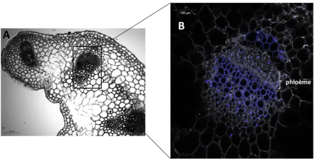
Figure 13: Hybridation fluorescente in situ (FISH) sur faisceaux conducteurs de pétioles de Vicia faba ... 128
Figure 14: Effets de l’infection du FBNSV sur les tissus phloémiens de pétiole de fève 29 jours post-inoculation ... 130
Figure 15 : Localisation des segments S et U4 du FBNSV dans les cellules de pétioles de fève. ... 132
Figure 16: Localisation des segments R et S du FBNSV dans les cellules de pétioles de fève ... 134
Figure 17: Localisation des segments R et S dans les cellules de pétioles de fève après traitement RNAse. ... 136
Figure 18 : Colocalisation des deux moitiés du segment R du FBNSV au sein du phloème des pétioles de fèves, 29 jours post-inoculation ... 138
Figure 19 : Localisation du segment R et de son produit d’expression dans des pétioles de fève infectée ... 140
Figure 20 : Etapes de construction des clones pCambia2300-segment-tige boucle... 153
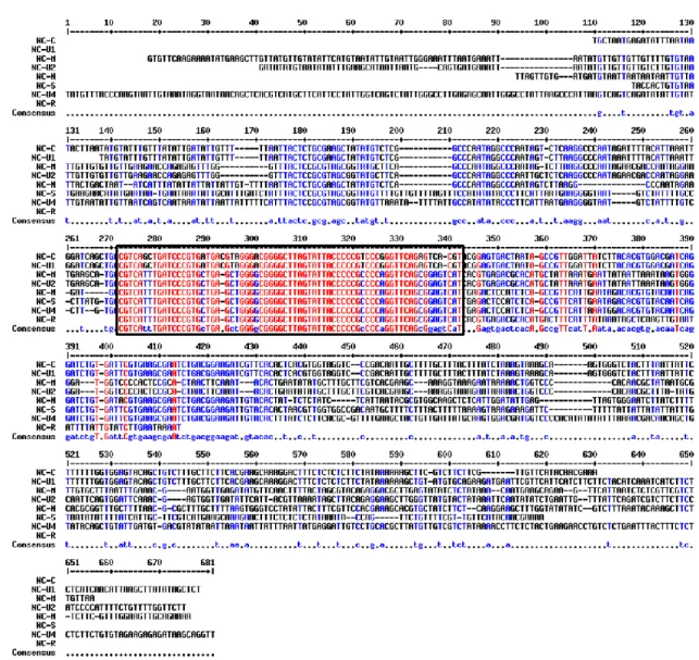
Figure 21 : Alignement des séquences non-codantes des huit segments du Faba bean necrotic stunt virus mettant en évidence la région commune de la tige-boucle (encadrée en noir) ... 155
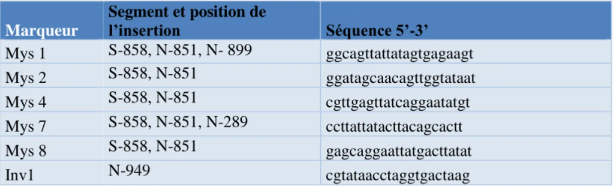
Figure 22 : Etapes de la construction de clones des segments tandem avec marqueurs génétiques 158 Figure 23 : Zones d’insertion des marqueurs au sein des segments S et N du FBNSV. ... 161
Figure 24 : Localisation du phloème au sein d’un pétiole de feuille de fève saine. ... 177
Figure 25 : Schéma représentatif des éléments du phloème ... 177
Figure 26 : Représentation schématique des étapes de différenciation des tubes criblés ... 177
Figure 27 : Carte du plasmide pLitmus 28 ... 178
Figure 28 : Carte du plasmide pCambia2300 ... 179
Figure 29 : Carte du plasmide pBin19 ... 180
Tableau 1 : Amorces utilisées en PCR pour amplifier les régions codantes de chaque segment ... 119
Tableau 2 : Séquences des amorces utilisées pour l’obtention des « megaprimers » ... 155
10
Liste des abréviations
Abréviations Virales
Acronyme Nom Genre Famille
BBTV Banana bunchy top virus Babuvirus Nanoviridae
BMV Brome mosaic virus Bromovirus Bromoviridae
CaLCuV Cabbage leaf curl virus Begomovirus Geminiviridae
CaMV Cauliflower mosaic virus Caulimovirus Caulimoviridae
CCMV Cowpea chlorotic mottle virus Bromovirus Bromoviridae
CMV Cucumber mosaic virus Cucumovirus Bromoviridae
CPMV Cowpea mosaic virus Comovirus Secoviridae
FBNSV Faba bean necrotic stunt virus Nanovirus Nanoviridae
FBNYV Faba bean necrotic yellows virus Nanovirus Nanoviridae
FLUV Influenza virus Influenzavirus A Orthomyxoviridae
FMDV Foot and mouse disease virus Apthovirus Picornaviridae
PVY Potato virus Y Potyvirus Potyviridae
RCNMV Red clover necrotic mosaic virus Dianthovirus Tombusviridae
SBWMV Soil-borne wheat mosaic virus Furovirus Virgaviridae
SSCSV Subterranean clover stunt virus Nanovirus Nanoviridae
TAV Tomato aspermy virus Cucumovirus Bromoviridae
TBSV Tomato bushy stunt virus Tombusvirus Tombusviridae
TRV Tobacco rattle virus Tobravirus Virgaviridae
TSWV Tomato spotted wilt virus Tospovirus Bunyaviridae
TUMV Turnip mosaic virus Potyvirus Potyviridae
TYLCV Tomato yellow leaf curl virus Begomovirus Geminiviridae
Abréviations courantes
CR-M Major common region
CR-SL Common Region-Stem Loop
FISH Fluorescence in situ hybridization
HC Helper component
GCN Gene copy number
MOI Multiplicity of infection qPCR quantitative PCR
11
12
Introduction générale
La nature, le fonctionnement et l’évolution des génomes viraux inspirent de nombreux biologistes depuis près de deux siècles (Zaitlin and Palukaitis, 2000). Assez tôt dans l’histoire de la découverte des virus, une architecture génomique particulièrement fascinante a été mise à jour ; il s’agit de celle qui caractérise les virus à composants multiples ou virus « multipartites ». Ces virus représentent un groupe polyphylétique dans lequel le génome est segmenté en deux à huit molécules d’acide nucléique, et dans lequel chacun des segments est encapsidé individuellement dans une particule virale. Les virus multipartites sont très fréquemment rencontrés chez les virus infectant les plantes –puisqu’ils constituent plus d’un tiers des genres et famille de virus de plante connus- et sont responsables de maladies dévastatrices sur des cultures d’importance majeure dans le monde entier. Les virus multipartites ont été aussi reportés chez les mycovirus mais, pour des raisons qui restent à ce jour totalement énigmatiques, ils n’ont jamais été décrits chez les bactériophages et pas non plus chez les animaux, exceptés chez de très rares espèces virales bipartites infectant les insectes. Vu les nombreux amalgames trouvés dans la littérature, il est important de souligner que, contrairement aux virus multipartites, les virus segmentés encapsident leurs différents segments génomiques au sein de la même particule virale. Les virus segmentés peuvent infecter à la fois les bactéries, les champignons, les plantes et les animaux.
L’origine des virus multipartites intrigue depuis des décennies les évolutionnistes et virologistes. Deux scénarios principaux ont ainsi été proposés pour expliquer l’évolution de cette architecture génomique. Dans le premier scénario, les virus multipartites seraient issus de la compétition entre un virus monopartite « parental » (ou ancestral) et des virus défectifs interférents produits au cours de leur réplication, ces derniers pouvant être fonctionnels par complémentation. Ils pourraient ainsi être sélectionnés comme un groupe de plusieurs segments interdépendants dans le contexte d’une forte multiplicité d’infection cellulaire (MOI) (Garcia-Arriaza et al., 2004; Iranzo and Manrubia, 2012; Nee, 1987; Simon-Loriere and Holmes, 2011). La production, le maintien et même la fixation dans la population virale de deux génomes défectifs complémentaires lors de passages en séries effectués à forte MOI en cultures cellulaires semblent corroborer ce scénario (Garcia-Arriaza et al., 2004; Ojosnegros et al., 2011). La co-infection d’une cellule par au moins deux virus distincts, qui auraient ensuite évolué pour fonctionner ensemble par complémentation, constitue le second scénario proposé
13
(Roossinck, 1997; Simon-Loriere and Holmes, 2011). L’exemple du Pea enation mosaic virus, virus représentant une symbiose obligatoire entre un composant apparenté aux luteovirus et un composant assigné au genre Umbravirus, pourrait étayer cette hypothèse. Cependant, l’indépendance de réplication de ces deux composants ainsi que l’absence d’homologie de séquence entre les régions terminales 3’ et 5’ des ARN-2 et ARN-1 -une condition obligatoire pour la reconnaissance de la matrice dans la réplication de tous les virus multipartites à ARN- ne permettent pas de classer ce virus parmi les virus multipartites (de Zoeten and Skaf, 2001). Si chacun des deux scénarios pourrait être à l’origine des virus multipartites constitués d’un petit nombre de segments, l’origine des virus composés d’un nombre beaucoup plus élevé de segments (>4) semble plus complexe. Une étude théorique récente menée par Iranzo et Manrubia (Iranzo and Manrubia, 2012) a montré que le nombre de particules virales qui doivent pénétrer et infecter efficacement chaque cellule (multiplicité d’infection cellulaire ou MOI) pour qu’un virus multipartite composé de plus de quatre segments puisse être fixé, était près de dix fois supérieur aux valeurs de MOI reportées jusqu’à présent. Les virus pour lesquels le nombre de segments génomiques est supérieur à 4 pourraient peut-être résulter de la conjonction des deux scénarios précédents : la segmentation d’un virus monopartite à laquelle se serait ajoutée ultérieurement une capture de gènes lors de co-infections par d’autres espèces virales (Iranzo and Manrubia, 2012).
Le premier scénario, celui de la coopération des génomes défectifs, repose sur l’existence d’avantages sélectifs associés à la segmentation du génome. De tels avantages potentiels ont été imaginés et étudiés quasi exclusivement par des approches de modélisation mathématique. A ce jour, les avantages proposés sont : (i) une fidélité de réplication accrue puisque les segments d’acide nucléique courts représentent des cibles plus petites pour la mutation (Pressing and Reanney, 1984) ; (ii) une durée de synthèse plus courte donc une réplication plus rapide pour les segment courts (Nee, 1987; Spiegelman et al., 1975) ; (iii) une plus grande stabilité des particules virales qui encapsident des segments plus courts (Ojosnegros et al., 2011) ; (iv) une facilitation des échanges génétiques intra génome par un échange de modules d’information que représente chaque segment, sans nécessité de crossing-over entre les acides nucléiques (Chao, 1988, 1991, 1992) et enfin (v) une réplication et transcription indépendantes permettant aux virus à génome segmenté de maintenir un cycle de réplication au cours duquel les segments peuvent être exprimés en quantités différentes (Wagner 2008). Quels que soient les avantages proposés jusqu’à ce jour, et quels que soient les scénarios envisagés pour expliquer l’origine des virus multipartites, tous reposent sur une nécessité commune : la
14
multiplicité d’infection cellulaire (MOI) doit être très élevée pour permettre une complémentation fonctionnelle entre les divers segments génomiques. Cette nécessité de complémentation fonctionnelle et la forte MOI requise sont unanimement perçues et modélisées comme le coût majeur, pour ne pas dire unique, que doivent supporter ou surmonter ces systèmes viraux. En effet, une vision communément partagée dans tous les articles traitant de l'évolution de ces virus, et dans la communauté des virologistes en général, est que tous les segments génomiques composant une espèce donnée doivent pénétrer au sein de la même cellule pour qu’il y ait complémentation fonctionnelle et donc succès de l’infection, et/ou pour éviter la perte d’une partie de l’information.
Il est très surprenant de noter que l’ensemble des études qui s’intéressent spécifiquement au fait que ces virus soient multipartites, s’y intéressent pour comprendre le « pourquoi » de leur existence mais jamais le « comment » ces systèmes fonctionnent réellement. Les études citées ci-dessus cherchent toutes à confronter le ratio coût/bénéfice, la plupart au travers d’approches purement théoriques, et très peu voire aucune démarche expérimentale simple et sans a priori ne cherche à décrire le fonctionnement de ces systèmes viraux (hormis des études purement moléculaires qui n’informent pas sur les propriétés du système). Cet état de fait est analysé dans un article de revue en cours de préparation, dont la version actuelle est proposée dans la partie I de cette introduction.
Vu cette carence en études expérimentales s’adressant au fonctionnement et aux propriétés des systèmes viraux multipartites, ma thèse avait un objectif simple qui était d’élargir le champ de connaissances sur la biologie de ces virus. Pour ce faire, le Faba bean necrotic stunt virus (FBNSV), appartenant à la famille des Nanoviridae, a été choisi comme modèle d’étude. Ce virus représente un des cas les plus extrêmes de virus multipartite puisque son génome est constitué de huit segments génomiques, le nombre le plus élevé décrit à ce jour. Les différentes caractéristiques du FBNSV sont présentées dans la partie II de l’introduction.
Dans un premier temps, nous avons démontré que chaque segment génomique du FBNSV s’accumule de manière très reproductible à une fréquence relative spécifique (et différente pour chaque segment) au sein des populations virales in planta, certains segments étant très fréquents et d’autres très rares. Nous avons aussi pu observer que le pattern des fréquences des segments était spécifique à l’hôte. Un nouvel avantage tenant compte de ces données a alors été proposé pour la segmentation et la multi-encapsidation des génomes viraux: la possibilité de réguler différentiellement le nombre de copies des gènes (gene copy number, GCN), un
15
paramètre régulant l’expression génique et phénotypique chez tous les organismes où il a été étudié (Hastings et al., 2009; Stranger et al., 2007). Le changement de fréquences relatives des segments en fonction de l’environnement dans lequel le virus se trouve pourrait être à l’origine d’une plasticité de ces systèmes viraux, leur conférant un niveau d’ajustement supplémentaire à un environnement changeant, sans impliquer la sélection immédiate de mutation adaptative. Cette partie importante de mon travail de thèse est décrite sous la forme d’un article publié, dans le Chapitre I.
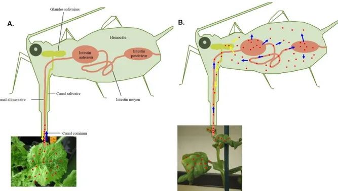
Par ailleurs, les différences de fréquences relatives de chacun des segments, et le fait que ces fréquences soient hôte-dépendantes, nous ont amenés à tester l’impact du passage à l’intérieur des pucerons vecteurs du FBNSV sur sa composition génétique. Nos résultats remettent en question une vision largement partagée quant au mode de transmission « circulant non-propagatif » des Nanoviridae (Chapitre II). En effet, ce mode de transmission invoque un simple passage de particules virales au travers des barrières cellulaires de leur insecte vecteur, sans décapsidation, réplication, ni même transcription. Les résultats obtenus montrent un changement des fréquences relatives des segments du FBNSV au sein des pucerons, qui ne peut que résulter d’une interaction intime plus complexe entre le Faba bean necrotic stunt virus et son insecte vecteur. Cette partie constitue le Chapitre II de ma thèse, et est présentée sous la forme d’un article qui est actuellement soumis.
Que ce soit dans les plantes hôtes ou dans les pucerons vecteurs, de telles différences de fréquences relatives entre les segments peuvent également augmenter le coût généralement associé à ces systèmes viraux. En effet, si toutes les particules virales se ressemblent quel que soit le segment qu’elles contiennent, l’entrée aléatoire des segments génomiques au sein des cellules pourrait être à l’origine de la perte des segments plus rares, sauf à imaginer une MOI qui devient totalement démesurée au cours de l’infection.
L’existence réelle de ce coût n’a, à notre connaissance, jamais été testée expérimentalement. Le chapitre III vise à pallier ce manque et ainsi, tout simplement, à vérifier si les différents segments ont toujours besoin de coexister au sein des cellules pour que le système viral puisse être fonctionnel.
Enfin, le maintien de l’intégrité génomique des virus multipartites est basé sur la nécessité que les goulots d’étranglement soient relativement faibles au cours de l’infection systémique de la plante, du passage de feuille à feuille, et aussi au moment de la transmission d’une plante à une autre par pucerons. Nous avons développé des outils destinés à quantifier ces goulots
16
d’étranglement, outils qui seront présentés au sein du chapitre IV bien que ce travail soit encore très préliminaire.
17
Introduction bibliographique
R
EVIEW
:
T
HE STRANGE LIFESTYLE
18
1. Review : The strange lifestyle of multipartite viruses
Préambule
Depuis la revue de Jaspars (1974) relatant la découverte des virus multipartites dans les années 1960, aucune autre synthèse n’a été publiée sur le sujet. Pourtant, de nombreux travaux ont été menés sur ces virus et peuvent être regroupés en trois grandes catégories (i) ceux sur les génomes, leurs caractéristiques, les fonctions des différents segments, et des différentes protéines dans le cycle viral, (ii) ceux traitant de l’évolution, et (iii) ceux plus théoriques discutant des avantages et coûts de ces systèmes viraux.
Nous avons donc décidé d’écrire une synthèse bibliographique (destinée à être publiée dans les mois à venir) confrontant ces différentes données. Cette synthèse vise à mettre en exergue les principales caractéristiques qui pourraient être spécifiques aux virus multipartites et, qui pourraient in fine représenter une base biologique pour expliquer les avantages et les inconvénients de cette architecture génomique particulière.
Cette synthèse regroupe et discute les résultats de différentes équipes dans le monde, y compris ceux obtenus lors de ma thèse, mais elle est aussi influencée par les nombreuses discussions, interprétations et questions que nous avons menées au laboratoire durant ces trois années. En conséquence, elle introduit d’inévitables redondances avec d’autres chapitres de ce mémoire. Nous avons néanmoins choisi de l’insérer en guise d’introduction bibliographique.
La version présentée peut être considérée comme une première version comportant les éléments qui nous paraissent importants ou pertinents. Elle sera in fine certainement modifiée et peut être formatée sous forme de revue plus succincte. Quoi qu’il en soit, elle reflète notre vision actuelle des virus multipartites et vise à dégager les principaux axes de recherche qui permettront de mieux appréhender leur fonctionnement.
19 Introduction
The general architecture, organization, and packaging of viral genetic information can be divided into three distinct categories, here distinguished under the terms “monopartite”, “segmented” and “multipartite” viruses. The monopartite type is a virus with all genetic information encoded by a single nucleic acid molecule protected in a shell made of proteins (and sometimes also lipids), forming the virus particle. The segmented type here refers to cases where the viral genome is divided into two or more nucleic acid segments that are all encapsidated together in a single virus particle. Finally, the multipartite viruses (the terms multicomponent viruses or coviruses are also used in the literature) are species where the genome is divided into two or more nucleic acid segments, just as the segmented type, but where each of these segments is individually packaged into its own virus particle. This latter peculiar organization is the only one resulting in virus particles that do not contain the entirety of the viral genetic information, and where the co-entry of several virus particles in a new cell or a new host is assumed as mandatory to maintain the integrity and functionality of the viral genome. As demonstrated below, the biology and evolution of multipartite viruses challenge some basic concepts of virology. In fact, at this point, it remains hard (if possible at all) to conceive how multipartite viruses have evolved and can actually be functional. This review is intended to briefly review the successive steps of the life cycle of a virus, trying to extract and highlight empirical or theoretical data specifically relevant for the biology of multipartite viruses. It also tries to identify major gaps and thus future research lines which would allow a better comprehension of the functioning of these peculiar biological systems.
The report of the called “multicomponent viruses” marked an important step in the history of the discovery of viruses. When analytical centrifugation techniques emerged, in particular the centrifugation through density gradient (Brakke, 1951), it was rapidly noted that some viral-like diseases appeared associated to two or more components of different density made of proteins and nucleic acid. The first reported cases were the purification of Tobacco rattle virus (Lister, 1968) and of Cowpea mosaic virus (Van Kammen, 1968), but at this time the multi-component nature of such disease-inducing agent could not be understood. Together with earlier dose-related infectivity studies (Bald, 1937, 1950; Fulton, 1962), further development of biochemistry, electron microscopy, and later molecular biology and sequencing, definitely evidenced that many viruses are composed of two or more physically separated particles, each containing a complementary portion of the genetic information (Jaspars, 1975; Hull, 2014; Zaitlin and Palukaitis, 2000). While monopartite and segmented viruses infect all possible
20
living organisms, multipartite viruses appear extremely successful in plants (and fungi), but not in animals where very rare cases have only been reported in insects (Hayakawa et al., 2000; Ribière et al., 2010; Wang et al., 2007). The success of multipartite viruses in plants, where 30 to 40 % of virus genera and families are multipartite (Hull, 2014), is a long-standing mystery for which a few tentative and questionable answers have been proposed. For example, it had early been speculated that the multipartite architecture of the genome could be related to its RNA nature, and logically proposed that multipartite viruses are so frequent in plants because most plant viruses are RNA viruses (Pressing and Reanney, 1984). However, it is now clear that multipartite viruses are also frequent among plant viruses with a DNA genome. In fact, multipartite viruses can be (+)ssRNA, (-)ssRNA, dsRNA, and ssDNA viruses, their genome size is highly variable, they can form icosahedral, rod-like, or filamentous virus particles, and none of these features demarcates them from monopartite and segmented viruses (Figure 1). Soon after their discovery, both virologists and evolutionary biologists begun to question and investigate the possible benefits and the counterpart costs in these complex biological systems. Putative benefits proposed and sometimes disputed are mostly related to the smaller size of the genome segments, as opposed to a larger single molecule, which may induce a better tolerance to high mutation rates (Pressing and Reanney, 1984), a faster replication (Nee, 1987, 1988), a facilitated genetic exchange between information modules born by the different genome segments (Chao, 1988, 1991, 1992), or a higher stability of viral particles (Ojosnegros et al., 2011). The counterpart cost opposed to these benefits is the reduced chance to infect new cells (and new hosts) with all components required for functionality and integrity of the viral genome (Chao, 1991; Iranzo and Manrubia, 2012; Nee, 1987; Pressing and Reanney, 1984). This cost is equally assumed in all studies striving to define conditions allowing multipartite viruses to evolve from monopartite ancestors. Contrary to the multiple proposed benefits, the reduced chance of success at infection steps appears so intuitively logical, that it is considered an evidence and has never been questioned. The quantitative importance of the cost has been suggested to be related directly to the number of genome segments, thus the number of virus particles, required to gather the entire genome information when entering a cell or host (Iranzo and Manrubia, 2012).
A main concern in this field of research is that most of the above cited studies are theoretical, and that empirical supports for the various benefits proposed and for the actual existence of the cost are rare, if available at all. When considering the common wisdom on viruses, not only in
21 Figure 1 : Schematic representation of plant virus families and genera
All drawings are approximately scaled, providing a comparative view of the sizes of the virus particles. Only genera most representative of the virus structure are indicated. Families and genera with a red asterisk are those whose member species are multipartite viruses. (N.B.: The genus Begomovirus is composed of both monopartite and bipartite viruses).
Reproduced from M. H. V. van Regenmortel et al., eds., Virus Taxonomy: 7th Report of the
22
a general public, but also in the wide majority of the scientific community, it appears clearly that a conceptual frame derived from the understanding of monopartite viruses largely dominates. As a consequence, it is quite possible that this frame biases the conception of theoretical models intended to explain the evolution of multipartite viral systems, and hampers the design of experiments that would relevantly address biological processes specifically adapted to the way of life of multipartite viruses.
Here below, we will illustrate step by step the fact that experimental investigations targeting the specifics of the biology of multipartite viruses are extremely scarce, and emphasize what we consider major gaps to be addressed in the near future.
1 - Replication
Multipartite viruses, just as monopartite and segmented viruses, are replicated through a diversity of mechanisms depending partly on their genome nature. Whatever the molecular details or the cellular location of viral replication, two specific benefits have been proposed and modeled to explain the evolution of genome segmentation, thus of segmented and multipartite viruses. The first putative replication-related benefit is that splitting a genome into several smaller segments should engender faster replication (Nee, 1987; Spiegelman et al., 1975). This is based on the trivial fact that, when the speed of the replicase is constant and when this replicase is not a limiting factor, the time required for the duplication of a genome of 10kb is twice longer than that for two segments of 5kb each. Experimental studies looking at the replication kinetics as a general function of genome length seem to corroborate this hypothesis (Mills et al., 1967; Sakai et al., 1999). However, the only study directly comparing a monopartite genome to its bipartite derivative obtained in infected cell cultures could not confirm the expected faster replication of the bipartite variant (Ojosnegros et al., 2011). This contrasting report calls for more studies comparing near-isogenic monopartite and multipartite viruses, in order to empirically confirm that faster replication can indeed benefit segmented genomes. In addition, while faster replication can be a competitive advantage in simple experimental designs or simple theoretical models, its effect in more realistic host and ecological context calls for possible nuances (see for example Goldhill and Turner, 2014). The second replication-benefit proposed for segmented genome, is that smaller segments are smaller targets for mutations, and thus result in a higher replication-fidelity (Nee, 1987; Pressing and Reanney, 1984). Initially invoked for RNA viruses replicated by error-prone
23
RNA-dependent RNA-polymerases (Drake, 1993), this argument could now be extended to ssDNA viruses of plants (Gemini- and nanoviruses) and animals (circo-, denso-, and parvoviruses), where mutation rates comparable to that of RNA viruses have been repeatedly documented (Duffy and Holmes, 2009; Grigoras et al., 2010; López-Bueno et al., 2006; Ren et al., 2013; Sarker et al., 2014; van der Walt et al., 2008; Yang et al., 2014). Different studies comparing the mutation rate in distinct families of RNA viruses have suggested a possible negative correlation between the mutation rate and the genome length (Drake, 1993; Sanjuan et al., 2010), suggesting that smaller genomes or segments can bear higher mutation frequencies. In contrast, very recently, a comprehensive study has compiled a large dataset of 118 mutation rates, from 91 genes of 28 viral species (Hicks and Duffy, 2014). By testing the relationship between mutation rates, several viral genome properties and ecological factors, the authors concluded that the nature of the target cells is the only good predictor of the viral mutation rates, whereas genome length and genome segmentation are not.
Altogether, it seems reasonable to conclude that the data currently available do not corroborate the hypotheses explaining the evolution of genome segmentation by a faster replication and/or an easier mutational escape of smaller genome segments. In addition, whatever the future outcome of this debate, it cannot specifically explain the separate encapsidation of segments in multipartite viruses.
As opposed to the above arguable benefits, one replication-associated constraint for segmented genomes is the need to bear similar origins of replication and regulatory elements on all segments, in order to be efficiently recognized and processed by the replication complex. The existence of conserved origins of replication among genome segments has been demonstrated for a number of segmented and multipartite viruses, as for example bromoviruses (Ahlquist et al., 1984; Sivakumaran et al., 2000), begomoviruses (Argüello-Astorga et al., 1994; Fontes et al., 1994; Gladfelter et al., 1997) and nanoviruses (Horser et al., 2001; Timchenko et al., 2000). A deletion/mutation in this region drastically affects the replication efficiency of the corresponding segments and thus, in order to coordinate the various functions distributed onto distinct segments, its concerted evolution is mandatory (Hughes, 2004; Savory and Ramakrishnan, 2014). While the conservation of these regulatory sequences may here appear has a burden for segmented and multipartite viral genomes, it might also promote genetic exchanges, as discussed in the next section.
24 2 - Genetic exchanges
The genetic exchanges in between viral genomes can depend directly on the replication mechanisms. However, it can also be replication-independent, through genome break and repair processes and, most of all, through reassortments in segmented and multipartite viruses. A reassortment can be defined as an exchange of homologous segments between two related virus isolate or species, thus a form of genetic exchange not involving intra-molecular cross-overs (Chao, 1991). Reassortment had been suggested to substitute for intramolecular recombination in segmented RNA genomes at a time when RNA cross-overs were believed to be rare, if not impossible (Chao, 1988, 1991; Pressing and Reanney, 1984; Van Vloten-Doting and Jaspars, 1977). Thus, for RNA viruses, reassortment was perceived as the major mean to promote genetic exchange and to both purge deleterious mutations and create new advantageous combinations (Chao, 1991).
When empirically evaluating whether reassortants are favored in the real world, the published results appear contradictory. Numerous studies describe the existence of reassortants from the analysis of sequence data sets, and postulate that they play an important role in the evolutionary history of the corresponding viruses. However, investigations explicitly demonstrating a selective advantage or cost in reassortants are few.
Specific reassortants were shown or suggested to have a higher fitness in segmented and multipartite viruses of animals and plants, such as for example Influenza virus, Tomato spotted
wilt virus, Cucumber mosaic virus and several nanoviruses (Grigoras et al., 2014; Ince et al.,
2013; Kuroda et al., 2005; Lin et al., 2004; Medina and García-Sastre, 2011; Neumann et al., 2009; Pierrugues et al., 2007; Qiu and Moyer, 1999; Roossinck, 2002). In contrast, in plant samples from fields where several related strains of Cucumber mosaic virus (CMV) co-circulate, two independent studies concluded that reassortants are much fewer than expected, and thus mainly counter selected (Bonnet et al., 2005; Nouri et al., 2014). Interestingly, even for Influenza virus or the plant nanoviruses, which are respectively segmented and multipartite viruses with an elevated number of segments (6 to 8), reassortments seem to be constrained to one or two segments only, suggesting that many other possible combinations do rarely emerge (Brown et al., 1998; Grigoras et al., 2014).
In 1986, Bujarski and colleagues (Bujarski and Kaesberg, 1986) demonstrated that Brome
mosaic virus (BMV), a monopartite (+)ssRNA plant virus could recombine by intramolecular
25
recombination rates as elevate as DNA viruses (Froissart et al., 2005; Grigoras et al., 2014; Tromas et al., 2014; Urbanowicz et al., 2005), and that intra-segment recombinants are as frequent as inter-segment reassortants in multipartite viruses (Bonnet et al., 2005; Grigoras et al., 2014; Nouri et al., 2014; Stainton et al., 2012; Urbanowicz et al., 2005). From this wealth of new data, it is now evident that both recombination and reassortment promote frequent genetic exchanges in multipartite viruses. How this is affecting the hypothesis that genome segmentation has evolved to allow sex through reassortment is unclear, but the demonstrated ease of intra-molecular recombination may alleviate some of its authority.
The mechanisms of intra-molecular recombination have a priori no reason to differ between monopartite, segmented and multipartite viruses, and are thus not detailed further. In contrast, as mentioned in section 1, the role of the conserved replication origin in distinct segments of the same viral genome deserves mention in this section. During the replication process, this conserved region promotes homology-driven inter-segment recombination.
This has been experimentally forced under high selection pressure for Brome mosaic virus (Bujarski and Kaesberg, 1986), and also observed in natural populations for Nanoviridae (Grigoras et al., 2014; Stainton et al., 2012). Because these replication origins are sometimes conserved in different species of the same family, homologous-recombination process can paradoxically occur in between heterologous segments originating from distinct species, as previously observed in nanovirus samples collected from the field (Sivakumaran et al., 2000; Timchenko et al., 2000). One could state that such recombination between segments encoding distinct functions is a non-sense. So, the only sound “raison d’être” of recombination events driven by the replication origin appears to be its facilitated exchange between segments of related genomes (Hughes, 2004; Savory and Ramakrishnan, 2014). This might help reassorting segments from distinct strains or species by rapidly matching their replication origin to the viral system into which they incorporate.
3 - Gene expression
Viruses have been extremely inventive in order to encode all required genes in very small genomes, and to regulate their coordinated expression. In particular, viruses have to deal with cell machinery, which generally limits the translation of mRNAs to only one ORF. Over ten distinct strategies have been described for gene expression of monopartite viruses, and most are also used by segmented and multipartite viruses (Hull, 2014). For example, viral genomes or
26
segments can encode a single or several genes, these genes can be expressed from a single mRNA or from subgenomic (or subsegment) mRNAs, as a single protein which can sometimes be subsequently cleaved in several products with specific functions. In other cases, the genomes or segments can have internal ribosome entry sites (IRES) allowing several ORF to be translated, leaky scanning sequences allowing polycistronic RNA to produce more than one protein, or strategies to promote reinitiation of translation of several ORFs encoded on a single mRNA.
In front of this complexity, the viral genome segmentation could provide an extreme simplification where each segment would encode a single gene. A long-foreseen advantage of segmented and multipartite viral systems would be that each segment could possess its own specific regulatory sequence (Godefroy-Colburn et al., 1985; Kwon and Chung, 2000; Shirasawa-Seo, 2005; Zagorski, 1978). Surprisingly enough, only two virus groups have evolved this ultimate simplification: the multipartite virus families Nanoviridae and
Partitiviridae. In all other segmented or multipartite virus species, the one-segment/one-gene
strategy is either not found or combined with other segments encoding multiple genes. In conclusion, that the genome segmentation could be a simple way to match the eukaryotic gene expression and RNA translation machinery does not seem totally satisfactory, because in most cases the control of gene expression in these viruses is observed to be as complex as in monopartite viruses.
One additional way to regulate gene expression, which is seemingly possible in segmented and multipartite, but not in monopartite viruses, is the differential regulation of gene copy numbers (Sicard et al., 2013). Despite, an important literature on the significant impact of gene copy number variations on gene expression in all cellular organisms (Hollox and Hoh, 2014; Katju and Bergthorsson, 2013; Mileyko et al., 2008), this idea has thus far hardly made its ways into our scientific community (Elde et al., 2012; Filée, 2013). This concept has been so ignored in virology that the relative frequency of the different genome segments had not been explicitly investigated until recently, neither in segmented nor in multipartite viruses. We directly addressed this question in the nanovirus Faba bean necrotic stunt virus (FBNSV) (Sicard et al., 2013), and found that each ssDNA segment (so each gene) accumulates reproducibly with a specific relative copy number in a given environment. We proposed that these copy numbers, each associated to a specific segment, define the “genome formula” (Figure 2A), which proved to be specific to the host plant species (Figure 2B). Because viral populations closer to the steady state or setpoint genome formula accumulated to higher levels, we hypothesized that the
27 Figure 2: Genome formula of FBNSV in two different host plant species
The genome formulae presented are in Vicia faba (A) and Medicago truncatula (B). The relative frequencies of the eight FBNSV segments have been calculated in within-host viral populations, and the median copy number of each segment is represented here as relative to the less abundant one. The core genome corresponds to the classical conception of the viral FBNSV genome (rectangle). Adapted from Sicard et al., 2013
28
differential regulation of gene copy number might be adaptive, and could stand as an unforeseen benefit for segmented and multipartite viruses. Several earlier hints indicating that multipartite viruses other than nanoviruses could also regulate their gene (or segment) copy number are discussed in (Sicard et al., 2013), and a direct demonstration has been published recently for the tripartite (+)ssRNA Alfalfa mosaic virus (Sanchez-Navarro et al., 2013). The genome formula has not been investigated upon infection of a host by a segmented virus, where it could play a role at the intracellular level. While multipartite viruses might control a differential copy number of their different genes (or segments) at cell or host entry, segmented viruses appear constrained at these specific steps by the fact that in most cases one copy of each segment is encapsidated in every single virus particles (McDonald and Patton, 2011; Poranen and Tuma, 2004; Rager et al., 2002; Reguera et al., 2013; Wichgers Schreur et al., 2014).
This discovery might represent a significant step forward in the understanding of the biology of multipartite viruses for several reasons: (i) it represents an unprecedented putative advantage for the regulation of gene expression in segmented viral genomes, (ii) this advantage applies best to multipartite viruses because GCN can be regulated at all viral step, whereas it could mainly be regulated at the level of individual cells for segmented virus, (iii) as discussed later, this advantage is the only one described thus far that imposes a constraint on the relative frequency of the segments, and that can explain that these viral systems have not evolved to the situation of the minimum cost where all segments would ideally accumulate at equal frequency.
4-Encapsidation
This step of the virus life cycle is really key in this review because it markedly distinguishes the segmented from the multipartite viruses. These are seemingly opposite strategies where in the former the virus obviously tries to have its whole genetic information travelling together within plant and from plant to plant, whereas in the latter it apparently tries not.
Segmented viruses have a very specific constraint at encapsidation, which is the sorting of distinct segments to ensure that at least one of each is present in every single particle. The molecular means by which this process is successfully accomplished are partly understood. Best-documented cases are those of Influenza virus or the phage Phi6. The segments bear different and complementary packaging signal sequences, which induce specific secondary RNA folding and a timely concerted interaction with the structural protein ensuring the sorting of one of each per virion (Chou et al., 2012; Mindich, 2004; Noda and Kawaoka, 2012).
29
A priori, multipartite viruses do not have to sort segments at encapsidation and they could encapsidate in ways similar to monopartite viruses, the frequency of encapsidated segments directly depending of the frequency of these segments within producing cells. As for monopartite viruses, the specific packaging of viral segments of multipartite genomes relies on the presence of assembly signal sequences (Basnayake et al., 2009; Choi and Rao, 2003). A noticeable difference with monopartite viruses, however, is that the same coat protein(s) have to accommodate the packaging of segments of different sizes, sequences, and secondary/tertiary structures. Multipartite virus particles can be either spherical (Partitiviridae, Nanoviridae,
Begomovirus, Secoviridae, Idaeovirus…), bacilliform (Ourmiavirus, Alfamovirus…),
rod-shaped (Virgaviridae, Varicosavirus, Benyvirus…) or filamentous (Closteroviridae,
Capiloviridae…). While rod-shaped, bacilliform and filamentous viruses can easily
accommodate segments of different sizes, by accordingly adjusting the length of the virus particles, physical constraints exist for viruses with an icosahedral structure. A nice illustration of this constraint are found in in vitro studies of particles made with the coat protein of Cowpea
chlorotic mottle virus (CCMV) with a symmetry T=3 (Comas-Garcia et al., 2012). The particles
can encapsidate segments from 100 to 12000 base-long RNAs, but they preferentially package one or more RNA segments with a total size around 3.2kb, consistently resulting in an optimal protein/RNA ratio of 6/1 that corresponds to the natural situation for this virus.
In some icosahedral viral species, all segments have a comparable size, indicating that this size may be optimal for particle stability. This is particularly striking for ssDNA multipartite nanoviruses, where the 8 genome segments vary between 920 and 1022 nt in all described species. The ssDNA bipartite geminiviruses of plants and bidensoviruses of insects also have very constrained genome segment sizes of around 2.7 kb and 6 kb, respectively. In other cases however the size of segments encapsidated in icosahedral particles can widely vary. In many species of the family Bromoviridae the RNAs, 1, 2, 3 and 4 are approximately of 3, 3, 2 and 1 kb, respectively. This is somehow intriguing because it could theoretically allow the encapsidation of two or more short segments in a single particle. A case study illustrating this is that of Brome mosaic virus (BMV) where some particles contain either one copy of a RNA-1 or one copy of RNA-2 (around 3kb each) whereas others contain one copy of RNA-3 and one of RNA-4, together also summing up to approximately 3kb (Annamalai and Rao, 2008; Choi and Rao, 2000). It was recently shown that the situation in BMV is even more complex, because some smaller virus particles have been found and might also accommodate a single copy of either RNA-3 or RNA-4 (Ni et al., 2014).
30
Once encapsidated, the different interactions occurring between the distinct segments and the capsid, resulting from distinct 3D structure of the packaged RNA, can engender different virion stability (Vaughan et al., 2014). This difference could actually constitute an advantage of multi-encapsidation since it may regulate a differential timing of RNA release and thus the kinetics of gene expression, as suggested for BMV (Ni et al., 2014; Vaughan et al., 2014). Likewise, in rod- or filamentous shape particles, the possible time shift associated with decapsidation of particles of different size could participate in the timely regulation of gene expression. Unfortunately, this possibility has been thus far proposed for BMV only and further work will be required to decide whether this might be a general feature in multipartite viral systems. Perhaps related to similar questions of encapsidation constraints is the very elegant work recently published on the experimental evolution of Foot and mouse disease virus (FMDV) (Ojosnegros et al., 2011). Through repeated passages in cell cultures at elevated multiplicity of infection, these authors observed that two defective molecules complementing each other could outcompete their ancestral monopartite FMDV. The selective advantage of this bipartite derivative was demonstrated to be associated to a higher stability and infectivity of the virus particles packaging smaller RNA segments, and not to a faster replication. The authors thus proposed that genome segmentation and more specifically the multi-encapsidation could in some cases result from a «trade-off between segment length and particle stability». While it has recently been shown that particle size was correlated to genome length (Zandi and van der Schoot, 2009), there is no clear general correlation between genome length and particle stability. For this reason, we believe that the case of segmented FMDV variant taking over the monopartite parental genome might be a specific example, related to experimental conditions, hardly expandable as a general rule to explain the evolution of multipartite viruses. Perhaps consistently, it should be noted that this FMDV experiment was conducted with a virus of vertebrates, where no multipartite natural systems have ever been described. Finally, and this might be a most definitive argument pleading against the genome multi-encapsidation as a way to preserve particle stability for oversized genomes, (i) multipartite viruses do not necessarily have longer genomes than monopartite viruses and (ii) they sometimes encapsidate segments which are longer than the whole genome of some monopartite viruses (Figure 3).
31 Figure 3: Comparison of the genome or segment sizes in viruses
The listed families and genera are those with significant differences in genome architectures. The size-range of whole genomes and that of individual segments are illustrated by black and grey lines, respectively. All size data come from Viralzone.
*All genera of the family Geminiviridae are composed of monopartite virus species, except for the genus Begomovirus
**The genus Begomovirus is composed of both monopartite and bipartite virus species. *** All genera of the family Virgaviridae except the genus Tobamovirus are composed of multipartite virus species.
32 5 - Within host movement
When viruses move from cell to cell or long distance to colonize systemically their host, the question of transferring all genome information becomes a real issue distinguishing multipartite viruses from both segmented and monopartite. Indeed, for the two latter, the whole genetic information may move as a whole, packaged as complete information units within individual virions, whereas multipartite viruses scatter away fragmentary genetic information in distinct virus particles, which must later reunite to resume infection. The question in this section is thus to see whether some striking specific features emerge in the known mechanisms of within host spread for multipartite viruses.
The discussion on this point is confused by the fact that molecular and cellular data available are showing that viral forms actually trafficking within their host plant is multifarious (reviewed in Hipper et al., 2013; Scholthof, 2005). Some plant virus species move both cell-to-cell and long distance as mature virus particles. Others can move cell-to-cell as nucleoprotein complexes not assembled into mature virus particles, which are only required for long distance movement. Finally, in rare cases, some viral species can spread both cell-to-cell and in the vasculature as nucleoprotein complexes that do not even contain the coat protein. All three cases have been suggested in both monopartite and multipartite viruses (Scholthof, 2005). For example monopartite Cauliflower mosaic virus (Carluccio et al., 2014) and multipartite Cowpea mosaic
virus (Pouwels, 2003) move both cell to cell and long distance as mature virions. On the
opposite, both the monopartite Tomato bushy stunt virus (Scholthof et al., 1995) and the bipartite Cabbage leaf curl virus (Pooma et al., 1996) move cell-to-cell and long distance within their host in the absence of the coat protein.
Whatever the viral form that is actually transported, with regard to limitations linked to the size exclusion limit of plasmodesmata, viruses have developed specific different mechanisms to insure their passage to the adjacent cells and into the vasculature, all depending on one or more movement proteins. Through intricate interactions with multiples host factors, some of these movement proteins simply enlarge the size of plasmodesmata, allowing passage of infectious material, whereas others polymerize into a tubular structure, which serves as a syringe to inject the virus into the next cells (Ritzenthaler, 2011; Tilsner and Oparka, 2012). These different mechanisms to pass through plasmodesmata are also shared by multipartite, segmented and monopartite viruses (Hipper et al., 2013; Scholthof, 2005), and thus the present data do not distinguish multipartite virus movement mechanisms from that of other viruses. One further illustrative example of this conclusion is the phylogeny of the viral movement proteins of the
33
30K super family, established by Melcher in 2000 (Melcher, 2000). In Figure 4, it is apparent that the relatedness between these 30K movement proteins does not reflect any commonalities within multi- or monopartite viruses.
If no distinct molecular or cellular features appear in the mechanisms of within host movement of multipartite viruses, one may try to look at it with a different angle, that of the number of virus particles or genomes that actually enter and infect individual cells (MOI, Multiplicity Of cellular Infection). For monopartite viruses the “independent action hypothesis” (IAH) stipulates that infection of a cell and/or host can be initiated by a single infectious unit, thus a single virus particle, and that each infectious unit can act independently (Druett, 1952). Theoretical models predict a deviation from IAH when one infecting segment depends on the presence of one or more others. This could be empirically verified in several instances (Sanchez-Navarro et al., 2013; Zwart et al., 2011, 2013) and indicate that the number of viral particles efficiently entering a cell (here simplified as MOI) must be much higher in multipartite viruses than it is in monopartite or segmented ones. The MOI is even predicted to be directly related to the number of genome segments and should reach very high values when multipartite viruses have more than 3 or 4 segments. In this case, Iranzo and Manrubia (Iranzo and Manrubia, 2012) have estimated that this number should reach hundreds in order to possibly explain the evolution of such viruses.
MOI values have been experimentally estimated in a number of monopartite viruses infecting bacteria (Turner et al., 1999), insects (Bull et al., 2001), vertebrates (Josefsson et al., 2011; Jung et al., 2002) and plants (Bergua et al., 2014; Gonzalez-Jara et al., 2009; Gutiérrez et al., 2010), and repeatedly found to be relatively small (from 1-13) most often below 5 (reviewed in Gutiérrez et al., 2012). Unfortunately, the only estimate of the MOI of a multipartite virus is that of the bipartite Soil-borne wheat mosaic virus, where solely RNA-2 segment was analyzed (Miyashita and Kishino, 2010). The estimated value is similar to that in monopartite viruses, in the order of 5, not clearly supportive of the prediction that multipartite viruses infect cells with higher numbers of virus particles or genome segments.
Due to this paucity of data, and considering the importance of this question in the biology of multipartite viruses, more investigation are wanted and should focus on multipartite viruses with a high number of segments, such as for example member species of the family
34 Figure 4: Relationships among putative 30K MP superfamily members determined by bootstrapped parsimony
Branches with less than 80% support (100 bootstrap replicates) were collapsed. 0, RT, N, A, I, II and III represent the type of polymerase encoded by the viruses: none, RNA-dependent DNA polymerase, negative-strand virus, ambisens-strand virus and positive-strand virus, supergroups I, II, III RNA-dependent DNA polymerases, respectively. The thin-lined polygon encloses those MPs known to form virion-bearing tubules. Genera with a red asterisk are those whose member species are multipartite viruses (N.B.: The genus Begomovirus is composed of both monopartite and bipartite viruses).
Reproduced from U. Melcher, The ‘30K’ superfamily of viral movement proteins. Journal of