Accelerator-Based Boron Neutron Capture Therapy
byWilliam Bruce Howard
B.S., Engineering Physics
United States Military Academy, 1987
Submitted to the Department of Physics
in Partial Fulfillment of the Requirements for the Degree of
Doctor of Philosophy in Physics
at the
Massachusetts Institute of Technology
February 1997
C 1997 Massachusetts Institute of Technology. All rights reserved
Signature of Author:
Department of Physics
Certified by:
,/ ryesso r-cquelyn \anch
Professor Lee Grodzins
Thesis
Con-,,imnpr;u,-Accepted by:Froressor ueorge Koster Chairman, Committee on Graduate Students Department of Physics OF TE e P, 9nc
FEB 1 21997
LIBRARIESAccelerator-Based Boron Neutron Capture Therapy
by
William Bruce Howard
Submitted to the Department of Physics on January 21, 1997 in Partial Fulfillment of the Requirements for the Degree of Doctor of Philosophy in
Physics
ABSTRACT
Boron Neutron Capture Therapy (BNCT) is a promising therapy modality for cancer. Clinical trials using BNCT are underway in the US. BNCT works by a selective loading of tumor cells with 1oB and subsequent irradiation of the tumor with thermal neutrons. The reaction 'lB(n,ac) is induced and releases approximately 2.3 MeV of energy, most of which is deposited locally. In Accelerator-Based BNCT (AB-BNCT), neutrons for this therapy are produced using ion induced reactions. Three reactions, 7Li(p,n) Ep=2.5 MeV, 9Be(p,n) Ep=3.0-4.0 MeV, and 9Be(d,n) Ed=2.6 MeV are investigated here. Complete
data for the 9Be(p,n) reaction were not previously available. Therefore, 28 thick target neutron spectra were measured, on an absolute basis, using time-of-flight techniques. Proton energies 3.0, 3.4. 3.7. and 4.0 MeV, and laboratory angles 0-145 degrees were used. The absolute accuracy of the data (better than 25% for most neutron energies) was confirmed by measuring a different reaction with a known spectrum. Using the three reactions, Monte Carlo techniques were used to design therapy beams for AB-BNCT. The reaction 7Li(p,n) yielded the highest dose rates. However, given lithium's poor thermal properties, lithium targets will be difficult to develop. Dose rates using 9Be(p,n)
were also high. Beryllium is known to be an excellent target material. Using 7Li(p,n) and
9Be(p,n), therapy times of 12-25. and 27-60 minutes, respectively, were predicted (tumor
depth 2-6 cm, 15 RBE-Gy total tumor dose, 10 kW accelerator power, 30 ppm boron tumor concentration). Using 9Be(p,n) and a 1 mA beam current, nearly equivalent
therapy beam parameters were predicted with 4.0 and 3.7 MeV protons. For equivalent accelerator power, 3.7 MeV protons would produce higher dose rates. Using 9Be(d,n) resulted in lower dose rates because the reaction's high energy neutrons must be extensively moderated. AB-BNCT therapies would result in an intense neutron and photon radiation field requiring significant facility shielding. Most of the occupational dose comes from (n,y) reactions in the facility walls. Effective facility shielding has been designed. Effective shielding for the patient can be made from lithium carbonate and polyethylene.
Thesis Supervisor: Jacquelyn C. Yanch
Acknowledgments
I would like to thank my thesis advisor, Professor Jacquelyn Yanch for the opportunity to
work on this project, and for her patience, support, and guidance. i would also like to thank her for the many times she has been a devoted and caring friend.
I also want to express my appreciation to the members of my thesis committee, especially Dr. Stephen Steadman and Professor Lee Grodzins, for their advice and support.
Much of the funding for this reseach was provided by a fellowship from the Hertz
Foundation. I am very thankful for their support. This work was also supported by a US Department of Energy grant (Grant No. DE-FG02-89ER60874).
This work would not have been possible without the assistance of the other members of the Laboratory for Accelerator Beam Applications (LABA). In particular, I would like to thank Haijun Song for his help.
Our collaborators at Newton Scientific have contributed immeasurably to the progress at LABA. I am especially grateful for their patience in answering many technical questions, and for their advice.
The members of The Ohio University Accelerator Laboratory have been very supportive, accommodating, and helpful during the past two years.
I am indebted to the MIT Reactor BNCT group for their contributions. They have been helpful in many ways, particularly in regards to therapy beam design and dosimetry. My thanks especially to Professor Otto Harling, Dr. Ron Rogus, Stead Kiger, and Kent Riley.
Finally, I want to express my deepest appreciation to my wife, Nancy, for all of her help and patience.
Sincerely,
William B. Howard 21 January, 1997
TABLE OF CONTENTS Page Number Abstract Acknowledgments 3 Table of Contents 4 List of Figures 10 List of Tables 14 CHAPTER ONE 17 Introduction I.A BNCT 17
I.A. 1 Medical Theory 17
I.A.2 BNCT dose components 22
I.A.3 Clinical use of BNCT 25
I.A.3.a Glioblastoma Multiforme 26
I.A.3.b Malignant melanoma 27
I.A.4 History of BNCT 28
I.A.4.a Early results 28
I.A.4.b Treatment of GBM in Japan 31
I.A.4.c Treatment of melanoma 31
I.A.5 Recent Progress 32
I.A.6 Non-reactor neutron sources 34
I.B Introduction to Accelerator-Based BNCT (AB-BNCT) 36
I.B.1 Target system 39
I.C The LABA Accelerator 42
I.C.1 Description of the accelerator 42
I.C.2 Accelerator performance to date 42
I.D Overview of New Research 46
CHAPTER TWO 53
Neutron Producing Reactions
II.A 7Li(p,n) 54
II.A. 1 Neutron yield and spectra 55
II.A.2 Gamma production 55
II.B 9Be(d,n) 58
II.C 9Be(p,n) 60
II.C. 1 Introduction 60
II.C. l.a Neutron production channels 64
II.C.1.b Previous measurements 65
II.C. .c Gamma production 68
II.C. L.d Introduction to recent measurements 69
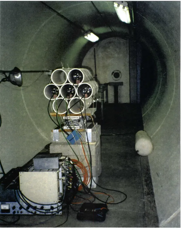
II.C.2 Experimental methods and equipment 71
II.C.2.a Introduction to time-of-flight 71
II.C.2.b The Ohio University TOF facility 74
II.C.2.c Neutron detectors 79
II.C.2.d Data processing 84
II.C.2.e Statistical and systematic error 87
II.C.3 Results and discussion 93
II.C.4 Total yield calculations 104
II.C.5 Conclusions and suggestions for future measurements 105
CHAPTER THREE 110
Therapy Beam Design and Verification
III.A Objectives and design criteria 112
III.A. 1 Objectives 112
III.A.2 Design parameters 114
1II.B The MCNP model 115
III.C MSR materials 120
III.C.1 Moderator materials 121
III.C.2 Reflector materials 124
III.C.3 Thermal neutron filter 124
III.D The optimization methodology 125
III.E Therapy beam design results and discussion 130
III.E. 1 Results using the 9Be(p,n) reaction 133
III.E. 1.a Initial results using 4.0 MeV protons 133
III.E. .b Results using 3.7 MeV protons 138
III.E. 1.c Results using 3.4 and 3.0 MeV protons 138 III.E.l.d Combined results for proton energies 4.0, 3.7, 3.4, and 3.0 MeV 143
III.E.2 Results using the 7Li(p,n) reaction 147
III.E.4 Gamma production at the target 151
III.E.5 Effect of changing target location 154
III.E.6 Results using 9Be(d.n) reaction 157
III.E.7 Effect of reflector diameter 161
III.E.8 Conclusions regarding therapy beam design 162
III.F Experimental verification 173
III.F. 1 The dual ionization chamber technique 173
III.F. l.a Equipment used 175
III.F. l.a.(i) Ionization chambers, flushing gas, and high voltage supply 175 III.F. 1.a.(ii) Water-filled brain phantom, and automated dosimetry 176 III.F. 1.a.(iii) Charge collection and electrometer 180 III.F. 1.b Calibration of the ionization chambers 180
III.F. 1.b.(i) Determining the A values 181
III.F. 1 .b.(ii) Determining the (B/A)TE valuE 185 III.F. 1 .b.(iii) Determining the (B/A)CG value 187 III.F. 1 .c Corrections to the ionization chamber signals 189 III.F. 1.d Thermal neutron response of the ionization chambers 190 III.F. .e Dual ionization chamber measurement techniques 191 III.F.2 The activation foil -cadmium difference method 192 III.F.2.a Calculation of the loB and 14N dose rates 194
III.F.2.b Experimental techniques -activation foils 194
III.F.3 Results and discussion 195
CHAPTER FOUR 207 Facility and Patient Shielding Evaluations
IV.A Facility shielding evaluation and dose assessment 208
IV.A.1 Purpose 208
IV.A.2 Description of the building before renovation 209
IV.A.3 Geometrical model for MCNP 213
IV.A.4 Radiation sources 218
IV.A.5 Dose rate prediction 221
IV.A.6 Shielding evaluation and improvement 221
IV.A.7 Dose estimations 224
IV.A.8 Discussion 228
IV.A.9 Initial measurements of the Bremsstrahlung radiation 229 IV.A. 10 Concluding remarks regarding facility shielding 230
IV.B Patient shielding evaluation 231
IV.B. 1 Methodology 232
IV.B.2 Results 236
IV.B.2.a Effect of increasing 6Li enrichment 238
IV.B.2.b Gamma Capture Reactions 240
IV.B.3 Concluding remarks regarding patient shielding 240
CHAPTER FIVE 244
Summary and Conclusions
V.A Summary of Research 244
APPENDIX A 251 Operation of the LABA Accelerator
A.1 Ion source 254
A.2 Injector 257
A.3 High voltage generation 258
A.4 Accelerating column 264
A.5 Stripping foils 265
A.6 Prototype high current beryllium target 267
A.7 Accelerator performance to date 269
APPENDIX B 271
Neutron Spectra from the 9Be(p,n) Reaction
APPENDIX C 331
LIST OF FIGURES
Page Number 17 CHAPTER ONE
Introduction
Figure I-A-1: Illustration of BNCT principles Figure I-B-1: Model of an AB-BNCT system Figure I-C-1: The accelerator at LABA
Figure I-C-2: Cut-away drawing of the LABA accelerator
CHAPTER TWO
Neutron Producing Reactions
Figure II-A-1: Figure II-A-2: Figure II-B-1: Figure II-B-2: Figure II-B-3: Figure II-C-1: Figure II-C-2: Figure II-C-3: Figure II-C-4: Figure II-C-5: Figure II-C-6: Figure II-C-7: Figure II-C-8: Figure II-C-9:
Neutron yield versus angle for 'Li(p,n) reaction Neutron energy spectrum for 7Li(p,n) reaction
Neutron yield versus deuteron energy for the reaction 9Be(d,n)
Average neutron energy for the reaction 9Be(d,n)
Neutron spectrum for the reaction 9Be(d,n), Ed=2.6 MeV
Excitation function for the reaction 9Be(pn)
Accelerator at OUAL Target chamber at OUAL
TOF tunnel and detectors at OUAL OUAL facility
Neutron spectrum for the reaction 27Al(d,n) Lithium glass efficiency
TOF electronics circuit
Comparison of two measurements of 9Be(d,n) CHAPTER TWO
Figure II-C-10: 9Be(p,n) neutron spectra at three angles, Ep=4.0 MeV Figure II-C-11: 9Be(p,n) neutron spectra at 0 degrees, three proton energies Figure II-C-12: Comparison of two measurements of 9Be(p,n)
Figure II-C- 13: Illustration of possible source of neutrons for 9Be(p,n)
Figure II-C-14: "Subtracted" spectra for 9Be(p,n)
CHAPTER THREE
Therapy Beam Design and Verification
Figure III-B-1: Therapy beam design model
Figure III-B-2: Dimenstions of therapy beam design model Figure III-B-3: Model of head for therapy beam design Figure III-C-1: Neutron cross section data for 27A1
Figure III-D-1: Neutron importance weighting for therapy beam design Figure III-D-2: Example of dose components for therapy beam desing Figure III-E-1: Relationship between tumor dose at 2 cm and at 6 cm Figure III-E-2: Therapy beam simulation results, 9Be(p,n), Ep=4.0 MeV Figure III-E-3: Results of the configuration: 4.0-27x10g-d
Figure III-E-4: Therapy beam simulation results, 9Be(p,n), E,=3.7 MeV Figure III-E-5: Results of the configuration: 3.7-25x10g-d
Figure III-E-6: Therapy beam simulation results, 9Be(p,n), E,=3.4 MeV
Figure III-E-7: Therapy beam simulation results, 9Be(p,n), Ep=3.0 MeV
Figure III-E-8: Comparison using same currents, 9Be(p,n), all energies
Figure III-E-9: Comparison using same power, 9Be(p,n), all energies Figure III-E-10: Therapy beam simulation results, 7Li(p,n), Ep=2.5 MeV
Figure III-E- 11: Results of the configuration: li-22x 10g-d
Figure III-E-12: Comparison using same power, 9Be(p.n) and 7Li(p,n)
Figure III-E-13: Target gamma ray production, 4.0-27x10g-d Figure III-E-14: Target gamma ray production, 4.0-34x10g-a Figure III-E-15: Target gamma ray production, li-22x10g-d
94 94 97 101 103 110 116 117 118 123 129 131 134 135 137 139 140 141 142 144 145 148 149 150 152 153 155
Figure III-E-16 Figure III-E- 17 Figure III-E-18 Figure III-E-19 Figure III-E-20 Figure III-E-21 Figure III-E-22 Figure III-E-23 Figure III-E-24 Figure III-E-25 Figure Figure Figure III-F-1: III-F-2: III-F-3:
Varying target location with equal distance to therapy port Varying target location with equal moderator length
Results of the configuration: 4.0-27x10 Og-d, target at A= +5 cm Therapy beam simulation results, 9Be(d,n), Ed=2.6 MeV
Results using 7Li(p,n) reaction and RBE set A
Results using 9Be(p,n) reaction, E,=4.0 MeV, RBE set A
Results using 9Be(p,n) reaction, Ep=3.7 MeV, RBE set A Results using 7Li(p,n) reaction and RBE set B
Results using 9Be(p,n) reaction, E,=4.0 MeV, RBE set B Results using 9Be(p,n) reaction, E,=3.7 MeV. RBE set B Picture of Far West ionization chamber
Picture of dosimetry phantom and stand
Expected neutron spectra in dosimetry phantom
CHAPTER FOUR
Facility and Patient Shielding Evaluations
Drawing of MIT building NW 13 and surrounding area Basement of MIT building NW 13
First floor of MIT building NW 13
MCNP model, basement of MIT building NW 13 MCNP model, first floor of MIT building NW 13 MSR designed for the reaction 7Li(p,n)
Shielding improvements to MIT building NW 13 MCNP model for patient shielding evaluations Patient model in MCPN shielding simulations Photon dose rate in phantom torso
Neutron dose rate in phantom torso
Sources of photon dose rate in phantom torso
156 158 159 160 163 164 165 166 167 168 177 179 182 207 Figure Figure Figure Figure Figure Figure Figure Figure Figure Figure Figure Figure IV-A- 1: IV-A-2: IV-A-3: IV-A-4: IV-A-5: IV-A-6: IV-A-7: IV-B-1: IV-B-2: IV-B-3: IV-B-4: IV-B-5: 210 211 212 214 215 217 223 234 235 237 239 241
APPENDIX A 251 Operation of the LABA Accelerator
Figure A-1: The accelerator at LABA 252
Figure A-2: Cut-away drawing of the accelerator 253
Figure A-3: The LABA accelerator ion source 255
Figure A-4: Components of the HV system 259
Figure A-5: Schematic drawing showing the first few stages of the CMC. 261 Figure A-6: First half cycle of the CMC operation 262 Figure A-7: Second half cycle of the CMC operation 263 Figure A-8: Accelerating column removed from the pressure vessel. 266 Figure A-9: Prototype high current beryllium target. 268
LIST OF TABLES
CHAPTER ONE Introduction
Table I-A-1i: Important thermal neutron capture reactions in human tissue
CHAPTER TWO
Neutron Producing Reactions
Page Number 17 24 53 Table II-A- 1: Table II-C-1: Table II-C-2: Table II-C-3: Table II-C-4:
Gamma production from proton bombardment of lithium Neutron producing reations for 9Be(p,n)
Previous measurements of the reaction 9Be(p,n)
Gamma production from proton bombardment of beryllium List of energies and angles used in measurement of 9Be(p,n)
CHAPTER THREE
Therapy Beam Design and Verification
Table Table Table Table Table Table Table Table III-C-1: III-E- 1: III-E-2: III-E-3: III-E-4: III-E-5: III-F- 1: III-F-2:
Possible therapy beam filter materials for BNCT MSR designations for Section III.E
Summary of 9Be(p,n) beam design results, using equal power RBE values for sets A and B
Summary of promising results using RBE weighting factors Time required to deliver 15 RBE-Gy to a tumor
Summary of previous (B/A)cG values
Measured and simulated dose components in phantom
55 64 66 68 93 110 122 132 146 162 169 170 188 196 CHAPTER THREE
CHAPTER FOUR 207 Facility and Patient Shielding Evaluations
Table IV-A-1: Predicted photon dose rate from Bremsstrahlung radiation 224 Table IV-A-2: Dose rates predicted using shielded MSR 225 Table IV-A-3: Dose rates predicted using simple moderator and reflector 226 Table IV-A-4: Dose rates predicted using 9Be(d,n) reaction 227
Table IV-B-l: The effect of using enriched 6Li in patient shielding 238
CHAPTER FIVE 244
Summary and Conclusions
Table V-A- 1: Time required to deliver 15 RBE-Gy to a tumor 248
APPENDIX B 271
Neutron Spectra from the 9Be(p,n) Reaction
Table B-1: Measured data, November 1995, 9Be(p,n) E,=4.0 MeV 273
Table B-2: Measured data, November 1995, 9Be(p,n) E,=3.7 MeV 277 Table B-3: Measured data, November 1995, 9Be(p,n) Ep=3.4 MeV 280
Table B-4: Measured data, November 1995, 9Be(p,n) Ep=3.0 MeV 281 Table B-5: Measured data, August 1996, 9Be(p,n) Ep=4.0 MeV 282
Table B-6: Measured data, August 1996, 9Be(p,n) E,=3.7 MeV 287
Table B-7: Measured data, August 1996, 9Be(p,n) Ep=3.4 MeV 292
APPENDIX C
Simulations Used in Therapy Beam Design
design configurations, design configurations, design configurations, design configurations, design configurations, design configurations, design configurations, 7Li(p,n)
9Be(p,n), Ep=3.0 MeV 9Be(p,n), Ep=3.4 MeV 9Be(p,n), E,=3.7 MeV 9Be(p,n), Ep=4.0 MeV
Varying target location Varying reflector radius
331 Table Table Table Table Table Table Table C-l: C-2: C-3: C-4: C-5: C-6: C-7: Therapy Therapy Therapy Therapy Therapy Therapy Therapy beam beam beam beam beam beam beam 332 341 349 354 364 389 404
CHAPTER ONE
Introduction
This chapter provides an introduction to BNCT, a promising modality for the treatment of cancer. The first section outlines the basic medical theory of BNCT and its proposed uses. A history of the therapy is provided, along with a presentation of recent progress. One approach to producing neutrons for BNCT is called Accelerator-Based BNCT (AB-BNCT). The unique features of AB-BNCT are described. The BNCT research program and accelerator at the Laboratory for Accelerator Beam Applications (LABA) at MIT are discussed. In the concluding section of this chapter, the new contributions to the field of AB-BNCT which are presented in this thesis will be outlined. These contributions are in the areas of neutron source reaction measurement, therapy
beam design and radiation shielding.
I.A BNCT
I.A. 1 Medical Theory
Boron Neutron Capture Therapy (BNCT) is a binary radiation therapy for cancer based on the nuclear reaction 'lB(n, c)7 Li [1]. Although this modality is currently being used clinically in Japan and is undergoing clinical trials in the US, several aspects of the therapy are still in the developmental stage. The therapy is binary in the sense that it
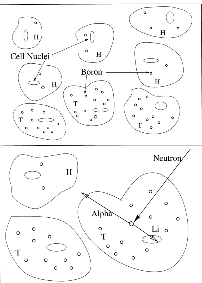
relies on two components combining at the tumor site to achieve the desired effect. The first component is a non-toxic, boron-containing pharmaceutical which is delivered to the tumor cells, and to the greatest extent possible, kept out of normal tissue. The second component is a low-energy neutron flux at the location of the tumor. When these two components combine, the fission reaction 'OB(n,ot) is induced. The thermal neutron cross section of this reaction is 3840 barns. The reaction cross section follows a 1/v behavior up to approximately 500 keV. The reaction proceeds to either the ground state or first excited state of 7Li releasing 2.79 MeV or 2.31 MeV, respectively, in the form of kinetic energy of the 7Li and 4He nuclei. The first excited state emits a single 0.48 MeV gamma photon as it decays to the ground state [2]. Since the alpha and 7Li are heavy charged particles, they are densely ionizing and have high linear energy transfer (LET) values (200 -300 keV/micrometer) and very short ranges in tissue (7 ýtm for alpha, and 4 ýpm for 7Li) [3]. Thus, their total combined energy is deposited in a volume approximately the size of a single cell. If high concentrations of 10B can be sufficiently localized in tumor cells, it is theoretically possible to destroy or inhibit these malignant cells without similar negative effects to nearby healthy cells. These concepts are depicted in Figure I-A-1.
BNCT, so illustrated, is deceptively simple. In this section, the medical theory of BNCT will be examined more closely and the various factors influencing its efficacy will be explained. It is important to note that BNCT is, by necessity, an interdisciplinary endeavor. The goal of selectively loading tumor cells with 1oB has been addressed primarily by pharmacologists and chemists. The design of neutron therapy beams
O
H
Li
o
cD
0 0 0 0 0Figure I-A-1: Boron delivered selectively to tumor cells (T), with less absorption by healthy cells (H). Tissue volume is irradiated by low energy neutrons which induce the reaction 10B(n,alpha) The fission products deposit most of their energy in the host cell.
suitable for BNCT has been accomplished mainly by nuclear engineers and physicists.
The radiobiologist has added expertise in many areas including microdosimetry and the
study of the biological effects of the various radiation dose components. Among other
tasks, the physician diagnoses the malignancy, refers the patient to a BNCT facility and
designs the treatment plan. Each of these groups must work together to ensure the safe
and effective treatment of the patient.
The approach used in radiation oncology for tumor control is to simultaneously
maximize radiation absorbed dose to the tumor and minimize the dose to normal tissue.
Complete eradication of malignant cells (tumor cure) is desired as even one cell could
seed the regrowth of the tumor. However, controlling the growth and spread of the tumor
(local control) without complete tumor cell kill could also lead to palliation of pain,
elimination of debilitating symptoms, and possibly increased survival times. Controlling
or eliminating the tumor requires high radiation doses to tumor cells. In practice, the dose
to the tumor is limited by the maximum allowable dose to healthy tissue, or tolerance
dose. This holds true for the BNCT modality.
The viability of BNCT, or for any other radiation therapy, depends greatly on the
selectivity of the treatment. In BNCT, this selectivity is achieved by preferentially
loading the tumor cells with '0B. This differentiation is accomplished by exploiting differences (physiological, chemical or metabolic) between healthy and tumor cells. One
controls the flow of chemicals in and out of the brain. In regions of rapid tumor growth,
this barrier has been shown to be less effective [4]. If a boron-containing chemical can be
found which would not penetrate the normal BBB, this same chemical might pass through
the "leaky" BBB found in tumor regions and be absorbed into the tumor cells. A second
approach uses antibodies which have been tagged with boron [1]. These antibodies seek
out antigens on the surface of tumor cells. Other approaches exploit the heightened
metabolic rate of tumor cells. These tumor cells might absorb and process more of
certain chemicals which can be loaded with boron. A similar approach seeks to tag boron
to nucleosides which then would be incorporated into the nuclei of tumor cells. This
approach has the advantage of placing the boron where the 'lB(n, ct) reaction can have its
greatest effect [1].
Two compounds in current clinical use for the delivery of loB to tumor cells are
sodium borocaptate and p-boronophenylalanine. Sodium borocaptate (Na12B12HilSH) is
commonly called BSH. BSH has been used clinically for the BNCT treatment of brain
tumors in Japan [5]. P-boronophenylalanine is commonly called BPA. BPA is an amino acid analog and is postulated to be taken up by various tumor cells, including melanomas,
due to their more active metabolism [6]. BPA has been used in clinical treatment of
melanoma patients in Japan and is being used in two clinical trials currently underway in
Unfortunately, it is not likely that boron will ever be delivered solely to tumor
cells. There will always be some residual boron in healthy tissue. A more realistic goal
is to maximize the concentration of boron in tumor cells relative to healthy cells. This
goal is quantified by such values as the uptake ratio, which is the ratio of the
concentration of boron in tumor cells (ppm) to the concentration in healthy cells. Some
examples of these ratios taken from clinical use and research will be presented later in
this chapter.
I.A.2 BNCT dose components
The radiation dose received during a BNCT procedure results from the following
reactions:
(1) 'lB(n.a)
(2) neutron capture reactions primarily from the reactions 14N(n,p) and 'H(n,y)
(3) neutron scattering resulting in energetic recoil nuclei
(4) incident photon irradiation which contaminates the therapeutic neutron beam
Of the reactions listed, only (1) delivers radiation dose selectively to the tissue
volume containing o1B. The boron concentration in tumor must be sufficiently large to offset the contaminating dose components which accompany the 'lB(n,a) dose and which
are not tumor specific (2,3, and 4). A 0.48 MeV photon, which contributes a non-specific dose component, accompanies 94% of the 'OB(n,ca) reactions. Previous research indicates
spherical tissue-equivalent model of the head with a uniform distribution of 10B [7]. This
0.48 MeV photon dose component will not be considered further. One advantage of
BNCT is that the 'lB(n,a) reaction produces high LET radiation, and the resultant dose
component is weighted by a large relative biological effectiveness (RBE) factor. The
RBE weighting factor is used to account for the fact that, for equivalent absorbed doses,
different types of radiation (gamma, alpha, heavy charged particles, etc.) result in
differing degrees of tissue damage. The RBE is referenced to the damage caused by a
standard radiation type (usually 250 kVp x-rays). The RBE is always defined for a
specific biological endpoint, such as cell death. The exact value of all weighting factors
for BNCT is the topic of continuing research. One set of typical values is: 1.0 for
photons, 4.0 for neutrons, and 4.1 for the 10B(n,a) reaction [8]. For this reason, physical dose (i.e. no RBE factors) will be used primarily in this thesis. When appropriate, RBE
weighted doses will be presented and the RBE values used will be stated.
Neutron capture reactions can occur in many nuclides commonly found in human
tissue. These nuclides, and the microscopic cross section for thermal neutron capture are
Nuclide Cross Section (barns) Boron- 10 3840 Oxygen-16 0.0002 Carbon-12 0.0037 Hydrogen-1 0.332 Calcium-40 0.44 Sodium-22 0.536 Nitrogen-14 1.75 Potassium-40 2.07
Table I-A-1. Important thermal neutron radiative capture reactions in human tissue [1].
Although the microscopic cross section for the reaction '0B(n,a) is much higher than those listed in Table I-A-1, the value of the 10B(n,ca) macroscopic cross section is a
direct function of the concentration of '0B in the tissue. It is the macroscopic cross section (microscopic cross section multiplied by number density) which determines the
relative probabilities of the various reactions in tissue. Since the number densities in
tissue of some isotopes, particularly 'H and 14N, are very large, the respective
macroscopic cross sections can also be significant. The 'H(n,y) reaction produces 2.2
MeV gamma rays which transfer energy to tissue primarily through Compton scattering.
The 14N(n,p) reaction releases a total kinetic energy of 0.62 MeV which is shared by the
proton (0.58 MeV) and 14C nucleus (0.04 MeV). The range of these heavy charged
particles is small, and the total kinetic energy is deposited within 1 mm of original '4N
nucleus [9].
As neutrons scatter from nuclei in tissue, energy is transferred to these nuclei,
which in turn directly ionize atoms in the cell. The amount of energy transfer is
the case of 'H, the neutron transfers an average of 50% of its energy in an elastic collision. This dose mechanism is tumor non-specific and must be minimized for
effective BNCT therapy. The common approach to this problem is to design neutron
therapy beams which contain few energetic (fast) neutrons.
Finally, the neutron therapy beam is always contaminated by some photons which
are produced concurrently with the neutrons, or later when neutrons interact with material
surrounding the therapy port. This photon dose can also be limited during therapy beam
design and optimization.
I.A.3 Clinical use of BNCT
BNCT is currently being proposed for treating several forms of malignant tumors,
including cervical [10], breast, head and neck [11], and metastatic cancer to the liver [12].
Current clinical research, however, is directed at two forms of cancer: brain (primarily
glioblastoma multiforme) and metastatic melanoma (a form of skin cancer). This thesis
primarily addresses the use of BNCT to treat tumors located in the brain. These include
glioblastoma multiforme and melanoma which has metastasized to the brain. In this
section, some clinical information on glioblastoma multiforme and melanoma is
I.A.3.a Glioblastoma Multiforme
The brain contains interstitial cells call glia which perform supportive functions.
There are three types of glia cells: (1) astroglia (astrocytes) (2) oligodendroglia and (3)
microglia. In the US. gliomas (tumor cells which arise from glia cells) account for
approximately 31-49% of all intracranial tumors, and of these, 72-89% are astrocytomas
[7]. Astrocytomas are further subdivided into four grades based on malignancy and
prognosis, with grade IV the most malignant. The malignancy of brain tumors is judged
based on the aggressiveness of the tumor cells and the location of the tumor relative to
sensitive structures in the brain. Forty to sixty percent of all gliomas are grade III and IV
astrocytomas. Grade IV astrocytomas are known as glioblastoma multiforme (GBM)
[13].
GBM is often characterized by a central tumor which is not well vascularized, and
the presence of extensions or "fingers" which branch off the main tumor and which are
the most active and malignant parts of the tumor. In contrast to the central tumor, the
extensions are well vascularized. Metastasis to other locations is rare [7] . The prognosis of all astrocytomas is poor and any cells that escape treatment almost always lead to the
regrowth of tumor. After diagnosis, median survival for the most favorable patient
populations (based on several prognostic factors) is around 12 months, even with surgery
and photon therapy. For elderly patients (age > 60), the median survival drops to 6 months [14]. Despite advances in treatment techniques, the prognosis of these patients is
not improving [15], and as a result, treatment is generally palliative and not curative [13]
[1].
I.A.3.b Malignant melanoma
The skin consists of three layers, the epidermis (outermost layer), the dermis
(middle layer), and the hypodermis or subcutaneous tissue (deepest layer). Melanin is a
pigment which is synthesized from tyrosine in melanocytes. This pigment is found
primarily in the epidermis of the skin. Melanin provides the pigmentation of the skin and
hair and also serves as a light absorbing material for the skin. Inability to produce
melanin leads to an increased susceptibility to sunburn and skin cancer [16] [13] [1].
Malignant melanoma is a cancer of the melanin-producing cell. The incidence of
melanoma is rising sharply in the US, with approximately 30,000 new cases each year,
resulting in over 6.000 deaths annually. Typically, melanoma is indicated by an enlarging
black-brown nodule on the skin, surrounded by erythema. This form of cancer is usually
easy to diagnose, but its progression is difficult to predict. Melanoma can take one of
several forms. The most dangerous form progresses by growing vertically into the deeper
layers of the skin. Tumors which extend into the deeper skin layers are also more likely
to metastasize, usually to the regional lymph nodes. Discovery of lymph node metastasis
severely degrades the prognosis. The median survival for patients with distant metastases
prognosis is very bad, and the tumor is considered untreatable [17]. These tumors are currently being targeted by BNCT.
I.A.4 History of BNCT I.A.4.a Early results
Most historical accounts of BNCT [18] [1] [19] begin in 1936 with a proposal for such a therapy by Locher [20]. Some initial radiobiological studies, including some with mice. were conducted before World War II [18]. In August 1950 the Brookhaven Graphite Research Reactor (BGRR) was completed, and the use of this reactor for slow neutron therapy was already being considered by the medical department at Brookhaven National Laboratory (BNL). Clinical experiments of boron uptake in brain tumor patients were conducted at the Massachusetts General Hospital (MGH), and in 1951, patients were referred to BNL for BNCT treatments. The first series of patient studies was conducted at BNL and MIT. The following is a summary of the conditions and results
[18]:
The first patient at BNL was irradiated on 15 February 1951, as part of a ten patient study. 1oB was delivered intravenously in the form of an aqueous solution of borax. The irradiations lasted 17-40 minutes at 40 MW reactor power. The thermal neutron flux was 0.44-1.93 x 10¹² neutrons/cm2-sec. This group had a median survival of 97 days post BNCT.
* A second group of nine patients was treated at BNL using sodium pentaborate with D-glucose. The incident neutron flux was 2.34-3.84 x 10¹² neutrons/cm2-sec. This second group suffered from radiation induced dermatitis of the scalp and scalp ulceration. The median survival of this group was 147 days post irradiation.
* A third group of nine patients was irradiated with a neutron flux of 0.39-1.5 x 10¹² neutrons/cm2-sec. The boron compound was delivered directly to the internal carotid artery of the affected hemisphere, and no severe dermatitis was experienced. The median survival was 96 days.
* In 1959, the Brookhaven Medical Research Reactor (BMRR) became operational, and between 1959 and 1961, an additional 17 patients were treated using BNCT. The median survival was 87 days, and was considered disappointing.
* Between 1959 and 1961, 18 patients were treated for brain tumors at the MIT reactor. For 16 patients, the 10B carrier used was the p-carboxy derivative of phenylboronic acid. The average survival was six months post BNCT. At MIT the scalp and skull were reflected to prevent the dermatitis seen in the second BNL group [19].
A total of 45 patients were irradiated at BNL, with an additional 18 at MIT. None of these early trials was considered successful. The failure of the early clinical trials has been blamed on two main factors. First, the boron delivery agents were not successful in
sustaining a high concentration of '0B in the tumor cells. This resulted in low values of the tumor-to-healthy-tissue loB ratios during the therapy. Upon autopsy, it was
discovered that radiation damage had occurred in the capillaries of the brain due to the
high concentrations of ' 0B in the patients' blood [7]. Second, the thermal neutrons did not penetrate deep enough to treat deep-seated parts of the tumor [1] . After these
disappointing results, clinical trials were halted in the US.
In the 1960s, solutions were sought for both of these deficiencies. Fairchild, at
BNL, promoted the use of epithermal neutron beams as a way of treating the deeper
portions of the brain [21]. Epithermal neutrons provide greater penetration than thermal
neutrons into the organ containing the tumor. The epithermal neutrons are moderated in
the tissue volume and arrive at the tumor site with a thermal distribution. There is no
exact energy range which defines an ideal epithermal beam for BNCT; however, the
lower limit is approximately 0.5-1.0 eV [22] [19] and the upper limit is approximately 20
- 100 keV [22] [19] [23]. The use of epithermal beams for BNCT has been generally accepted in the BNCT community. During the early 1960s, researchers in the US sought
a new drug that would enter GBM tumor cells easily, yet not cross the blood brain barrier
[4] into healthy tissue. One such compound, BSH, was investigated at MGH and later
I.A.4.b Treatment of GBM in Japan
The most extensive use of BNCT clinically was by Dr. Hiroshi Hatanaka in Japan.
Between 1968 and 1992, 119 patients were treated for intracranial tumors. All of these
patients were injected with BSH and subsequently irradiated with thermal neutrons from
a reactor. To increase neutron penetration, the scalp and skull were resected. Dr.
Hatanaka's results are encouraging and have significantly helped spark interest in the
BNCT field. For grade III and IV gliomas, with BNCT treatment only, the five and ten
year survival rates are reported as 19.3% and 9.6% [5]. The respective numbers for
conventional surgery followed by radiotherapy or chemotherapy are 4.6% and 0% [24].
For shallow tumors, the five and ten year survival rates were 58.3% and 29.2%,
respectively. The median survival of untreated GBM patients in the US is eight months
[16] . The most effective conventional treatment to date, which combines surgery and photon irradiation, results in a 13% survival after three years [7]. Dr. Hatanaka reported
tumor-to-blood ratios of boron as high as 8.95, with an average around 1.4, and average
boron concentrations in tumor around 15 ppm [5].
I.A.4.c Treatment of melanoma
In the early 1970s Dr. Yutaka Mishima developed a program to treat melanoma
by BNCT using a thermal neutron beam [25]. In 1976, he developed a boron containing
analog of dopa, BPA. Melanin is formed from dopa, and actively absorbed by tumor cells
due to their increased metabolic rate [26]. The first patient treated under this program
metastasis made surgery impossible. Administration of BPA was via perilesional injection, and the patient was treated with 1013 thermal neutrons/cm2. After 9 months the tumor had regressed with no regrowth [27].
A second patient was treated for a primary melanoma of the foot. This case resulted in regression of the tumor, with no regrowth after 18 months. To date, 16 patients have been treated successfully with thermal BNCT [25]. Specific statistical data
on these patients are difficult to obtain.
I.A.5 Recent Progress
By September 1994, two epithermal beams were in operation and ready for clinical trials in the US. The first was at the MIT Research Reactor (MIT-R), and the second was at the Brookhaven Medical Research Reactor (BMRR). On September 6th, 1994 the first BNCT treatment with an epithermal beam was conducted at MIT-R using the boron delivery agent BPA as part of a phase-I clinical study for metastatic melanoma of the extremities [28]. In one year, four subjects, each with subcutaneous metastatic melanoma nodules in the lower extremities, were irradiated. The subjects were first given 400 mg/(kg-body weight) of BPA orally for the purpose of evaluating the pharmacokinetic absorption and excretion of the BPA. After this evaluation, the subjects were given repeat administrations of BPA, prior to each of four neutron irradiation fractions. The maximum total normal tissue dose received by the subjects was between 1000 RBE-cGy and 1250 RBE-cGy. The dose to the tumor was 25-35% higher due to the
preferential absorption of BPA. No negative effects were observed, with the exception of
an adverse skin reaction with one of the subjects, which was resolved in two weeks. Two
of the four subjects manifested clear tumor regression, which has not reversed. Each of
the subjects is still alive [29].
Also in September 1994 (just a few days after the first MIT-R irradiation), a single
patient was treated for GBM using BPA as the delivery agent at the BMRR [30]. BNL
began a multi-patient trial in February 1995. This trial was designed to evaluate: (1) the
performance of BPA as a delivery agent to GBM cells, (2) the safety of BNCT using BPA
for GBM treatment, and (3) the efficacy of this treatment for GBM patients[31]. In the
period up to February 1996, ten patients were treated with this protocol. As in the MIT-R
study, a biodistribution evaluation was conducted. The boron concentrations in tumor
were found to be 24.7+12.6 ýpg 1 0B/g-tissue (mean + standard deviation) for a 250 mg/(kg-body weight) administration of BPA. The tumor-to-blood ratio was
approximately 2.0, and the non-necrotic tumor-to-blood ratio was greater than 3.5. The
patients were treated to a normal brain endothelium dose of 10.5 Gy-equivalent (peak
dose volume of 1 cm3). No serious complications were experienced [31]. Of these initial ten patients, at least eight experienced recurrence of the tumor. Following BNCT, the
median survival time was 13.5 months. Two of the patients are still alive [31]. In the
second of two protocols now underway, the tumor dose has been increased over that
A recent update indicates that 23 patients have been treated at BNL to date. The life extension was similar to that using conventional radiotherapy, and so the results are encouraging [32]. Conventional radiotherapy is very time consuming for the patient who may not have a long life expectancy. Typical radiotherapy treatment protocols may last six weeks. BNCT procedures may require as little as a few days of the patient's time. Initial analysis of the results indicates that tumor control will require a dose to tumor of approximately 30-50 RBE-Gy [32].
I.A.6 Non-reactor neutron sources
If BNCT proves to be a viable cancer therapy modality, additional sources of neutrons (i.e. other than reactors) would be needed. The death rate from GBM in the US alone is approximately 3200/year [7]. In addition, there are approximately 6,000 deaths from melanoma each year. The total death rate from these two cancers alone is over 25/day. New reactor construction, particularly at a hospital, seems unlikely due primarily to the prohibitive cost of construction and operation. At lease three other reactors in the US have been identified for possible conversion to BNCT facilities [33] although no construction progress has been made to date.
In recent years, alternative sources of neutrons have been investigated. These neutron sources include: 252Cf [23], spallation sources [34], photoneutron sources [35] and accelerator-based (p,n) or (d,n) sources [8][36] [37] [38].
The possibility of a 252Cf source has been investigated, and deemed unlikely primarily because the quantity of radioactive material needed exceeds the total production capacity of the Western world [23]. The other alternative neutron sources listed above are accelerator-based neutron sources. An evaluation of one spallation source, using 72 MeV protons on a tungsten target, found that the predicted dose distributions in a head phantom were comparable to those achievable with existing reactor facilities [34]. The proton currents required are approximately 100 pA. A moderator made of iron and graphite is proposed [39]. A photoneutron source has been proposed which uses 6 MeV electrons striking a material such as tungsten. The resulting Bremsstrahlung photons then create neutrons through interaction with D20 which also serves as the moderator [40]. Of
the accelerator-based neutron sources, spallation and photoneutron sources have received less attention than the charged particle reactions, particularly 7Li(p,n) and 9Be(p,n).
For the purpose of this thesis, the term Accelerator-Based BNCT (AB-BNCT) will be used to describe the production of neutrons through charged particle induced reactions such as (d,n) or (p,n), as opposed to spallation or photoneutron sources. Section I.B introduces the concepts and terminology used in AB-BNCT. Section I.C will describe the operation of an accelerator built specifically for use in AB-BNCT research. The original research contained in this thesis will be introduced in Section I.D.
I.B Introduction to Accelerator-Based BNCT (AB-BNCT)
The new contributions to BNCT which are contained in this thesis are in the field
of AB-BNCT. These contributions are contained in Chapters II, III, and IV. In this
section. an introduction to AB-BNCT is provided which will form the foundation for the
work presented in the later chapters.
AB-BNCT differs from reactor-based BNCT in the method of generating neutrons
and modifying the energy spectrum of these neutrons to create a useful epithermal therapy
beam. A typical configuration consists of a neutron producing target, such as beryllium
or lithium, which is bombarded by energetic protons or deuterons. This target is
surrounded by an object known as a Moderator-Shielding-Reflector-Assembly (MSR).
The patient would be placed in front of the MSR and exposed to neutron radiation which
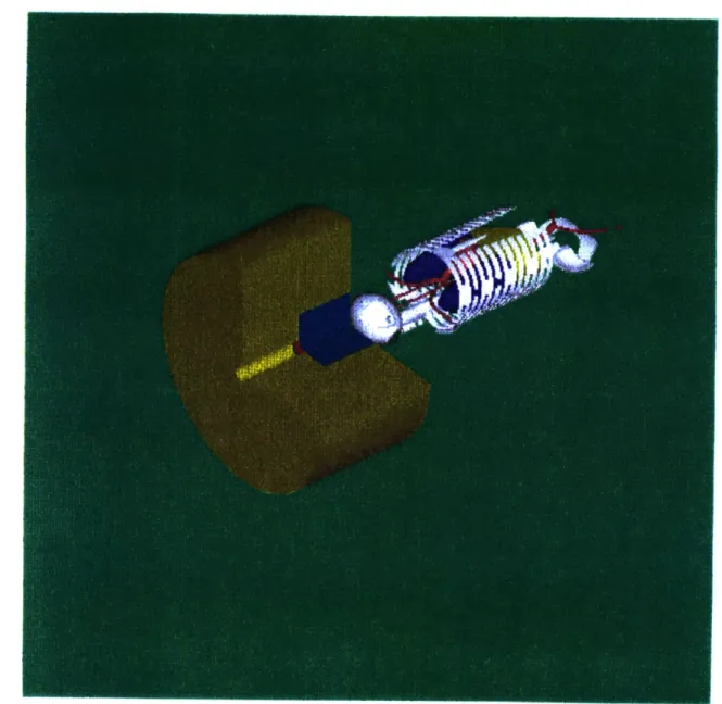
is emitted from the therapy port. A diagram representing such a configuration is shown in
Figure I-B-1.
Three basic components must work together to create a viable AB-BNCT therapy
beam: accelerator, neutron producing target, and MSR. The accelerator produces a
charged ion beam containing protons or deuterons at a current, I, and energy E. The ions
strike the target inducing a (p,n) or (d,n) reaction. Therefore, the target must be designed
to withstand a heat load equal to W=IE. For example, a 1 mA beam of 4 MeV protons
delivers 4 kW of power to the target. Neutrons are produced at the target with an energy
Figure I-B-1: Model of an AB-BNCT system. The reflector is shown in gold. The moderator is shown in blue. The neutron producing target is shown in red.
.... ... ... . . ...
This spectrum depends on the target material, ion species and ion energy. The total
neutron yield of a reaction, YTOT(Eion), is the neutron energy spectrum integrated over all
angles, On, and all neutron energies, En. The ideal reaction for AB-BNCT would produce
a copious and focused amount of purely epithermal neutrons, with no penetrating
contaminant radiation. The neutrons from this reaction could then be used, with very
little moderation, as a therapy beam. The kinematics of reactions capable of producing
epithermal beams at the target dictate that the ion energy must be kept only slightly above
the threshold energy of the reaction, ETH. Unfortunately, this approach generates
relatively small neutron yields, since the production cross section is low at ETH. The
alternative approach is to use an ion energy significantly above ETH, and generate
neutrons with an average energy well above the epithermal range. This neutron beam
cannot be used directly on a patient. The primary purpose of the MSR is to decrease the
average energy of the neutrons before they reach the patient position. This alternative
approach is the one used at LABA, depicted in Figure I-B-1. and described in Chapters
II-IV of this thesis.
In the most general sense, the design of an "optimized" AB-BNCT therapy beam
is the process of choosing all of the parameters of the combined system (accelerator,
target, and MSR) which will provide the most useful therapy beam, in a reasonable
amount of time, and with the least amount of other resources (money, space, shielding,
etc.). Each design parameter of the basic components depends on the others in a complex
reaction and ion energy. Increasing the ion energy increases the neutron production rate
at the target, but may result in a less optimal neutron spectrum at the patient position.
Also, if the ion energy is increased, the heat load on the target increases if the same beam
current is used.
I.B.1 Target system
Charged particle reactions can be divided into two classes: exothermic and
endothermic. Exothermic reactions, such as 9Be(d,n) release energy so that the resultant
neutrons have a maximum energy in excess of the bombarding charged particle energy.
Endothermic reactions, such as 7Li(p,n) absorb energy so that the maximum neutron energy is less than the charged particle energy. Both types are potentially useful for
AB-BNCT. The ideal neutron producing reaction for BNCT would have several
characteristics. First, the yield of the reaction would be high at low charged particle
energies and currents so the accelerator system requirements would be simplified and the
BNCT treatment could take place in a reasonable amount of time. Second, the neutron
energy spectrum would be as close to epithermal as possible to reduce the need for
extensive moderation/filtration. Third, the reaction would not produce penetrating
contaminant radiation (gamma-rays). Finally, the target material would be inexpensive,
safe to handle, capable of withstanding operating conditions such as high temperature and
vacuum, and easily made into a practical target. In all cases, a thick target would be used
Unfortunately, no target reaction meets these four requirements simultaneously.
Previous studies [41] [42] [22] [8] have concluded that three source reactions are currently plausible for AB-BNCT. These reactions are 7Li(p,n), 9Be(p,n) and 9Be(d,n). Chapter II of this thesis examines each of these reactions and describes the neutron
energy spectra for each. In Chapter III, the design of MSRs for each reaction is discussed.
I.B.2 Accelerator
An accelerator designed for use in AB-BNCT would be characterized by three
main performance parameters: ion current, ion energy and total available power. As a
minimum, such an accelerator must be capable of producing ion energies above the
threshold for the neutron producing reactions of interest. The highest threshold of the
reactions considered here is 2.059 MeV (9Be(p,n) ). Beyond this requirement, the method by which the performance parameters are chosen can be illustrated by an example. If the
7Li(p,n) reaction is to be used, the energy spectrum might be calculated or measured at
several proton bombarding energies. The resulting spectra could then be used, one at a
time, to design MSRs to be used with each proton energy. The combination of target
reaction, ion energy, and MSR which resulted in the most optimal beam would then be
implemented. These MSR designs would be based, in part, on assumptions concerning
issues such as: depth of tumor, total dose to tumor prescribed by a physician, tolerance
dose of healthy tissue, and the maximum time allowed for a therapy procedure. At the
end of the MSR design process, the therapy beam performance (based, for example on
One result of this evaluation would be the determination of the proton current required,
for each proton energy, which then sets the performance requirement for the accelerator.
As this example shows, the performance requirements for an accelerator are
dependent on the assumptions used in the therapy beam design. Although there is not yet
a consensus on these assumptions, the current clinical trials should help resolve some of
these issues. It is, however, possible to give an order-of-magnitude approximation to
some of the accelerator design specifications. Another example is helpful. As will be
shown in Chapter III, it is possible to design an AB-BNCT therapy beam with a tumor
dose rate, at a depth in tissue of six cm, of 2.5 cGy/(min-mA of accelerator beam current),
using a 4.0 MeV proton beam. Depending on the assumed RBE weighting factors, the
RBE-weighted dose rate could range from 6-10 RBE-cGy/min-mA. If one were to
assume that a physician prescribed a dose of 15 RBE-Gy, to be delivered in no more than
60 minutes, then a beam current of approximately 2.5 mA would be required, resulting in
an accelerator power of 10 kW. These results will, of course, change with changing
assumptions. Therapy beams are currently being designed within the AB-BNCT
community using ion bombarding energies and ion currents of approximately 2.5-4 MeV
and 4-30 mA respectively [43] [44] [45] [46] [47]. The accelerator power requirements,
In addition to adequate energy and current production, a viable accelerator for
BNCT would have the following characteristics. First, the production of charged
particles must be safe and reliable. Second, the accelerator (including auxiliary systems)
must be practical in terms of system cost, ease of use, size and weight. Several types of
accelerators have been examined to produce proton beams for AB-BNCT (Ep= 2-4 MeV,
I, = 4-30 mA). These include: radio frequency quadrupole (RFQ), electrostatic
quadrupole (ESQ) and electrostatic accelerators. Electrostatic and ESQ accelerators have
several properties which make them useful for the AB-BNCT application. First, beam
current and energy are continuously tunable over a wide range. Second, continuous
current is delivered to the target resulting in lower peak thermal load. Third, higher
accelerating gradients are possible resulting in a more compact system. Finally,
electrostatic accelerators have high electrical power efficiency which reduces cooling
requirements and system operating cost [48]. The accelerator which has been built at
LABA for AB-BNCT research is an electrostatic accelerator.
I.C The LABA Accelerator
I.C. Description of the accelerator
A detailed description of the LABA accelerator is given in Appendix A. A brief
description is given here. When examples are needed to highlight an aspect of the
accelerator operation, a proton beam will be assumed. The accelerator at LABA is a
tandem electrostatic linear accelerator capable of accelerating protons and deuterons. The
energy of 4.1 MeV) and a maximum power of 10 kW. The accelerator was designed by Newton Scientific Incorporated. Cambridge, MA. A picture of the accelerator is shown
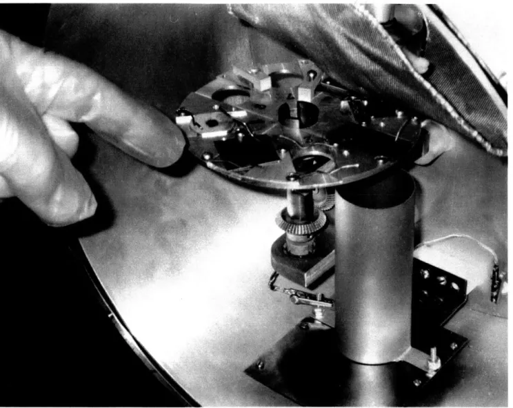
in Figure I-C-1. The complete system weighs approximately 1000 kg and measures 3.9 meters from the ion source to the high energy end of the pressure vessel. The largest diameter is 0.94 m. The entire outside surface of the accelerator is at ground potential.
A cut-away drawing of the accelerator, with the major components labeled, is shown in Figure I-C-2. Negative ions, such as H-, are produced in the ion source and injected into the low energy stage of the accelerating structure. The ions experience a constant acceleration provided by the electrodes of the low energy stage, and gain energy as they travel in vacuum towards the positively charged high voltage (HV) terminal. Inside the HV terminal, each ion passes through a stripping foil, where two electrons are stripped, leaving a positive ion, such as H+. These positive ions then experience a repulsive force from the terminal as they enter the high energy stage. The ions exit the accelerating structure, and enter the beam line extension, which is electrically grounded. In its present configuration, the beam line extension passes through a steering magnet, and quadrupole focusing magnet and terminates at the target housing. The steering magnet, quadrupole magnet and target are not shown in Figure I-C-2.
I.C.2 Accelerator performance to date
The accelerator has been operated, for limited duration, at a maximum continuous proton beam current of 0.8 mA at an energy of 2 MeV. The accelerator has been run
Fr -,
A
sC t rcri · *I c· ' cCiI
ill ar nr 1 tr a,~I
Figure I-C-1: The accelerator at LABA.
~ 1111
N
°TI
JoIPaaLaDP V9Vi aq4 0o 6ULMePJ RpMp 4n3 LeULmaW AH ,oAcacu I [assaA aJnssaAd aucnoS uoI "Z-3-I a@n6.[
routinely with deuteron beam currents up to 50 [LA at a beam energy of 2.6 MeV. The ion source current has been as high as 5 mA. The current research program aims to increase both the terminal voltage and the current in a systematic way, and to evaluate the performance of all accelerator systems.
I.D Overview of New Research
For AB-BNCT to become a viable and accepted cancer treatment modality several issues must be resolved. The purpose of this thesis is to address some of these issues, and report on the progress towards a solution for each. During the past few years, significant advances have been made in the field of AB-BNCT, both at LABA, and other facilities. The work presented in the remainder of this thesis will add to these advances. This section introduces the research presented as original contributions to the AB-BNCT field.
The thick target yield and spectra of the reaction 9Be(p,n) have been accurately
measured using time-of-flight techniques, using proton bombarding energies which are currently considered useful for AB-BNCT. This research will be presented in Chapter II. An important characteristic of these measurements is that they were accomplished on an absolute basis, without any normalization. The experimental procedures were confirmed indirectly by measuring a second reaction, 9Be(d,n). These measurements are important
not only to the BNCT community, but also to the scientific community at large, since this reaction can be used as a neutron source for a variety of applications.
A comparative study of possible therapy beams has been accomplished using
three likely neutron source reactions for AB-BNCT (9Be(p,n), 9Be(d,n), 7Li(p,n) ), and is
presented in Chapter III. Seventy different MSR configurations were studied using
Monte Carlo methods. In addition to examining the neutron production at the target, the
gamma contamination resulting from (p,y) reactions was addressed. The designs were
compared on the basis of equivalent accelerator current and power. Recommendations
are made concerning future beam design. Beam designs based on the reaction 9Be(p,n)
were studied for four different proton bombarding energies.
The BNCT dose components were measured experimentally using dual ionization
chambers and activation foil analysis. These measurements were accomplished on an
absolute basis and were compared with the values predicted by Monte Carlo simulation.
Recommendations for further measurements are presented.
The radiation dose rates resulting from operation of the LABA accelerator at a
shielded facility were investigated for several neutron producing reactions and MSR
configurations. This work is presented in Chapter IV. A system of electron suppression
magnets inside the accelerating column of the LABA accelerator appears to have
effectively reduced the bremsstrahlung radiation levels which were thought to be
problematic. Methods to shield the patient against non-therapeutic radiation dose rates
REFERENCES
1. Barth, R.F., A.H. Soloway, and R.G. Fairchild, Boron neutron capture therapy for
cancer. Scientific American, 1990. 263(4): p. 100-107.
2. Knoll, G.F., Radiation detection and measurement. Second ed. 1989, New York: John Wiley and Sons, Inc. 754.
3. Kiger, W.S.I., Neutronic design ofa fission converter-based epithermal beam for
neutron capture therapy, in MS Thesis, Nuclear Engineering. 1996, Massachusetts
Institute of Technology: Cambridge, MA. p. 471.
4. Goldstein, G.W. and A.L. Betz, The blood-brain barrier. Scientific American, 1986. 250((5)): p. 74-83.
5. Hatanaka, H. and Y. Nakagawa, Clinical results of long-surviving brain tumor
patients who underwent boron neutron capture therapy. International Journal of
Radiation Oncology Biology Physics, 1994. 28(5): p. 1061-1066.
6. Mallesch, J.L., et al., The pharmacokinetics ofp-boronophenylalanine fructose in
human patients with glioma and metastatic melanoma. International Journal of Radiation
Oncology Biology Physics, 1994. 28(5): p. 1183-1188.
7. Zamenhof, R.G., et al., Boron neutron capture therapy for the treatment of
cerebral gliomas. I: Theoretical evaluation of the efficacy of various neutron beams.
Medical Physics, 1975. 2(2): p. 47-60.
8. Yanch, J.C., et al., Accelerator-based epithermal neutron beam design for
neutron capture therapy. Medical Physics, 1992. 19(3): p. 709-722.
9. Attix, F.H., Introduction to radiological physics and radiation dosimetry. First ed.
1986, New York: John Wiley and Sons.
10. Lorvidhaya, V., et al. Status report on BNCT for cervical carcinoma in Thailand. in Seventh international symposium on neutron capture therapy for cancer. 1996. Zurich Switzerland: Elsevier Science -To be published.
11. Coderre, J.A. Application of BNCT to other types of tumors. in Seventh
international symposium on neutron capture therapy for cancer. 1996. Zurich,
![Table I-A-1. Important thermal neutron radiative capture reactions in human tissue [1].](https://thumb-eu.123doks.com/thumbv2/123doknet/13820771.442584/24.918.276.680.125.301/table-important-thermal-neutron-radiative-capture-reactions-tissue.webp)