HAL Id: hal-01096899
https://hal.univ-brest.fr/hal-01096899
Submitted on 18 Dec 2014
HAL is a multi-disciplinary open access
archive for the deposit and dissemination of
sci-entific research documents, whether they are
pub-lished or not. The documents may come from
teaching and research institutions in France or
abroad, or from public or private research centers.
L’archive ouverte pluridisciplinaire HAL, est
destinée au dépôt et à la diffusion de documents
scientifiques de niveau recherche, publiés ou non,
émanant des établissements d’enseignement et de
recherche français ou étrangers, des laboratoires
publics ou privés.
To cite this version:
Stéphanie Madec, Vianney Pichereau, Annick Jacq, Mathieu Paillard, Claire Boisset, et al..
Charac-terization of the Secretomes of Two Vibrios Pathogenic to Mollusks. PLoS ONE, Public Library of
Science, 2014, 9 (11), pp.0113097. �10.1371/journal.pone.0113097�. �hal-01096899�
Sud, 91405, Orsay, France,4 Centre de Recherche sur les macromole´cules ve´ge´tales, CERMAV-CNRS, BP53, 38041 Grenoble, France
Abstract
Vibrio tapetis causes the brown ring disease in the Japanese clam Ruditapes philippinarum while Vibrio aestuarianus is associated with massive oyster mortalities. As extracellular proteins are often associated with the virulence of pathogenic bacteria, we undertook a proteomic approach to characterize the secretomes of both vibrios. The extracellular proteins (ECPs) of both species were fractionated by SEC-FPLC and in vitro assays were performed to measure the effects of each fraction on hemocyte cellular parameters (phagocytosis and adhesion). Fractions showing a significant effect were subjected to SDS-PAGE, and proteins were identified by nano LC-MS/MS. 45 proteins were identified for V. aestuarianus and 87 for V. tapetis. Most of them belonged to outer membrane or were periplasmic, including porins or adhesins that were already described as virulence factors in other bacterial species. Others were transporter components, flagella proteins, or proteins of unknown function (14 and 15 respectively). Interestingly, for V. aestuarianus, we noted the secretion of 3 extracellular enzymes including the Vam metalloprotease and two other enzymes (one putative lipase and one protease). For V. tapetis, we identified five extracellular enymes, i.e. two different endochitinases, one protease, one lipase and an adhesin. A comparison of both secretomes also showed that only the putative extracellular lipase was common to both secretomes, underscoring the difference in pathogenicity mechanisms between these two species. Overall, these results characterize for the first time the secretomes of these two marine pathogenic vibrios and constitute a useful working basis to further analyze the contribution of specific proteins in the virulence mechanisms of these species.
Citation: Madec S, Pichereau V, Jacq A, Paillard M, Boisset C, et al. (2014) Characterization of the Secretomes of Two Vibrios Pathogenic to Mollusks. PLoS ONE 9(11): e113097. doi:10.1371/journal.pone.0113097
Editor: Yung-Fu Chang, Cornell University, United States of America
Received July 21, 2014; Accepted October 19, 2014; Published November 17, 2014
Copyright: ß 2014 Madec et al. This is an open-access article distributed under the terms of the Creative Commons Attribution License, which permits unrestricted use, distribution, and reproduction in any medium, provided the original author and source are credited.
Data Availability: The authors confirm that all data underlying the findings are fully available without restriction. All relevant data are within the paper and its Supporting Information files. The sequences of all the proteins have been published in Genbank and all the accession numbers are available in the manuscript. Funding: The work was funded by the Bivalife FP7 KBBE program (ref 266157). The funders had no role in study design, data collection and analysis, decision to publish, or preparation of the manuscript.
Competing Interests: The authors have declared that no competing interests exist. * Email: stephanie.madec@univ-brest.fr
.These authors contributed equally to this work.
Introduction
Vibrios have frequently been associated with bivalve mortalities, essentially at the larval stage but also in adults [1–4]. Since 1987, several mortality events have been reported in clams (Ruditapes philippinarum) in different sites of the French coastline. Before death, clams go back to the sediment surface and display a brown deposit on the inner surface of the valves, between the pallial line and the edge of the shell [5]. This disease, named the Brown Ring Disease (BRD), was also described in Spain and Portugal, and affects both reared and wild clams. Bacteriological studies led to the identification of a new bacterial species,Vibrio tapetis, capable of reproducing the BRD in healthy animals [6].
In France, shellfish production is a well-established industry mainly relying on the commercial farming of the Pacific oyster (Crassostrea gigas). Annual mass summer mortalities of C. gigas have been reported since 1980 on the French coast. Several studies have demonstrated that these mortality outbreaks resulted from
complex interactions between the physiological and/or genetic status of the oysters, environmental factors, and one or more infectious agents, among which the herpes virus, OsHV1 [7], and Vibrio sp. [8]. Analyses of both moribund and healthy oyster hemolymph revealed that Vibrio aestuarianus was the most frequently disease-associated species [2] until 2008. Since then, a more virulent pathogenic herpes virus OsHV1, genotype micro-var, emerged, reducing the occurrence ofV. aestuarianus while V. splendidus strains are still frequently isolated [9].
The observed variable virulence of the isolates could be linked to the varying toxicity of the bacterial extracellular products (ECPs), allowing bacteria to escape the host immune defenses. In a previous study, the ECPs of the pathogenic strainV. aestuarianus 01/32 were shown to cause lethality in C. gigas, as well as morphological changes and immunosuppression in oyster hemo-cytes [10]. Further biochemical and genetic approaches evidenced the major role of the Vam extracellular metalloprotease in the
the clam hemocytes and mantle cells [14], and its cytotoxic effects after phagocytosis resulted in cell rounding with loss of filipods [12].
It is recognized that the success of each step of the bacterial virulence process depends on the orchestrated activity of several specialized bacterial factors. In vibrios, such virulence factors have been more identified in human pathogens such asV. cholerae, V. parahaemolyticus and V. vulnificus [15–17] but also in V. anguillarum, V. harveyi and other fish, crustacean and mollusk pathogens [18]. Currently, the only virulence factor characterized inV. aestuarianus is the secreted zinc metalloprotease, Vam, a member of the thermolysin family [11]. No similar virulence factor has been described to date in V. tapetis, but a metalloprotease (Vsm), a homolog of Vam, was also identified as a major determinant of the toxicity of V. splendidus ECPs [19]. All this reinforced our objective to search for other secreted proteins potentially involved in the virulence of these two marine vibrios. So far only two vibrio secretomes have been described [20,21] and the importance of the extracellular compartment on host pathogen interaction led us to analyze more precisely the proteins of this compartment in both vibrios.
Materials and Methods
1. Bacterial strains, growth and culture conditions
V. aestuarianus 07/115 was isolated from the hemolymph of an oyster collected at Aber Benoıˆt (Brittany, France) in September 2007. It was identified by the sequencing of the 16S rRNA and gyrB genes and was found to be highly virulent when injected in adult oysters (Jean-Louis Nicolas, unpublished results). The V. tapetis CECT4600 strain was isolated in Aber Benoıˆt (France) in Landeda (France) in 1995 from BRD diseased Manila clam (Ruditapes philippinarum) [22]. These strains were respectively grown in Difco marine broth 2216 (BD, Franklin Lakes, USA) and Zobell broth (HiMedia Laboratories, Mumbai, India), or on Difco marine agar and Zobell agar at 18uC.
2. Preparation of extracellular products (ECPs) and fractionation by Size Exclusion Chromatography in Fast Purification Liquid Chromatography (SEC-FPLC) mode
ECPs were produced by the cellophane overlay method as previously described [10]. Total ECPs ofV. aestuarianus and V. tapetis culture supernatants were filtered through 0.22mm filter membranes and concentrated on an Amicon Ultra-4 membrane with a 10,000 molecular weight cut-off (MWCO) (Millipore, Billerica, MA, USA). The total protein content was quantified using a DC protein assay (Bio-Rad, Hercules, CA, USA) with 96-well micro-plates (Nunc) in a micro-plate reader (Bio-Tek Synergy HT) and the KC4 v3 software comparing the results with a calibration curve using standard proteins (Bovine Serum Albumin) provided with the DC protein assay kit. Then, ECPs were separated on an A¨ KTAFPLC system (GE Healthcare, Piscataway, NJ, USA) directed by the Unicorn 5.1 software. Aliquots containing 1.4 mg of total proteins dissolved in mobile phase (isocratic elution mode in PBS: 10 mM Phosphate Buffer pH 7.4, 137 mM NaCl, and 2.7 mM KCl) and filtered on a 0.22mm
3. In vitro assays : hemocyte cellular parameters
The effects of the obtained fractions were measured on oyster or clam hemocytes to assess the action of the ECPs on hemocyte adherence and phagocytosis capacities. Fractions showing inhib-itory or stimulatory effects were compared to the negative control (FSSW: Filtered Sterile Sea Water). For both tests, ECPs of each bacterial species were tested at 32mg.mL21of proteins, following previously described procedures [10]. Briefly, for phagocytosis tests, a sub-sample (150ml) of each hemolymph pool was distributed into a 5 ml polystyrene tube (Falcon, B-D Biosciences, San Jose, CA, USA), then underwent a two fold dilution with FSSW and was maintained on ice. Each sub-sample was subsequently combined with 30ml of a fluorescent bead (2.00mm in diameter, Fluoresbrite calibration grade, Polysciences, USA) working suspension (2% of the commercial suspension in FSSW, final concentration 16107beads.mL21), and incubated at 18uC for 120 min. The cells were then analyzed on a flow cytometer (FACSCalibur, BD San Diego, USA). The results were expressed as the percentage of hemocytes containing three beads or more [10].
To estimate hemocyte adhesive capacity, the sub-samples (100ml) of each hemolymph pool were distributed into 24-well microplates maintained on ice, as already described by Choquetet al. [12]. 100ml of FSSW or ECPs was added in triplicate to each sub-sample. After three hours of incubation at 18uC, the cells were fixed by addition of 200ml of a 6% formalin solution in FSSW. The supernatants were then transferred to cytometry tubes. The hemocyte number present in each supernatant was determined by flow cytometry. The results are expressed as average of non-adherent cells per ml i.e. an increase of the value compared to that of the negative control shows a cytotoxic effect of the tested ECPs.
4. Proteins electrophoresis (SDS-PAGE)
The fractions showing a significant effectin vitro on hemocyte phagocytosis or adherence were concentrated with Corning Spin-X UF Concentrators (Corning, Lowell, MA, USA) with a 10 kDa MWCO and applied on a Criterion precast acrylamide gradient gel 8–16% in Tris-HCl (Biorad, Hercules, CA, USA). After staining by Coomassie blue (Biosafe Coomassie, Biorad), the gel bands were cut out manually and conserved at –20uC before trypsin digestion.
5. Protein identification
5.1. In-gel digestions and peptides recovery. Excised gel plugs were washed 3 times with water, 100 mM ammonium bicarbonate and 100% acetonitrile successively. Cysteins were reduced by a treatment with a 65 mM DTT solution for 15 minutes at 37uC followed by alkylation with 135 mM iodoacetamide at room temperature in the dark. Gel plugs were washed again with 100 mM ammonium bicarbonate/acetonitrile (1:1), 100% acetonitrile, 100 mM ammonium bicarbonate and 100% acetonitrile successively before being dried. Gel pieces were then re-swollen in a solution of trypsin (12.5 ng/mL in 50 mM ammonium bicarbonate; Promega), and digestion was performed overnight at 37uC. The resulting peptides were then extracted
from the gel by sequential incubation in the following solutions: acetonitrile (ACN)/H2O/trifluoroacetic acid (TFA), 70:30:0.1 (v/
v/v), 100% ACN and ACN/H2O/TFA, 70:30:0.1 (v/v/v), and
extracts were eventually concentrated by evaporation to a final volume of 30mL.
5.2. Mass spectrometry (MS) analysis. Peptide mixtures were separated on a nano-HPLC system (Ultimate 3000, Dionex, Jouy-en-Josas, France), with an injection volume of 22mL: first, they were concentrated into a reversed-phase C18-PepMap trapping column (5mm, 300 A˚ /300mm i.d. x 5 mm, Dionex), and were then eluted with a 75-min gradient of ACN (from 2 to 90%) in aqueous 0.05% formic acid, at a flow rate of 250 nL/min. The nano-LC apparatus was coupled on-line with an Esquire HCT Ultra PTM Discovery mass spectrometer (Bruker Daltonik, GmbH, Bremen, Germany), equipped with a nanoflow ESI source and an ion trap analyser (ITMS). The mass spectrometer was operated in the positive ionization mode. The EsquireControl software (Bruker Daltonik, GmbH) automatically alternated MS and CID MS-MS acquisitions with the following criteria: up to seven ions per MS scan with an intensity threshold of 30,000 and a dynamic exclusion of 15 sec.
5.3. Protein identification. The DataAnalysis 3.4 software (Bruker Daltonik, GmbH) was used to create the peak lists from raw data. For each acquisition, a maximum of 2,000 MS/MS spectra were detected with an intensity threshold of 100,000 and the charge state of precursor ions was automatically determined by resolved-isotope deconvolution. The proteinScape 2.0 software (Bruker Daltonik, GmbH) was used to submit the MS/MS data to
the genomic V. aestuarianus 02/041 database (3693 CDS sequences; 1125373 residues, unpublished results), the only V. aestuarianus sequences available at that time. Peptide sequences were found to be 100% identical to the identified proteins in the database. Similarly, the MS/MS data for V. tapetis were submitted to theV. tapetis CECT4600 database (5498 sequences; 1633991 residues, unpublished). Submission to randomized versions of these databases (decoy) was used to determine the false positive rate (FPR), defined as the number of validated decoy hits/(number of validated target hits + number of decoy hits)*100, using the Mascot algorithm (Mascot server v2.2.07; http://www. matrixscience.com). Trypsin was selected as the cleaving enzyme with one allowed missed cleavage. In addition, carbamidomethy-lation of cysteins was set as fixed modifications and methionine oxidation were considered as variable modifications. The mass tolerance for parent and fragment ions was set to 0.6 and 0.5 Da, respectively. Peptide identifications were accepted if the individual ion Mascot scores were above 25 or the identity threshold (the ion score is 210*log(P), where P is the probability that the observed match is a random event, p-value,0.05). In case of ambiguous assignments (one compound fitting more than one peptide), the peptide sequence with the highest score was retained. The compilation of peptides identified to proteins was performed with the ProteinExtractor algorithm [23], so that every protein reported was identified by at least one peptide with a significant ion Mascot score (above the identity threshold) that could not be mapped to a higher-ranking protein already in the result list. This means that the final protein lists contain only those proteins and protein Figure 1. UV spectrum of total ECPs ofV. aestuarianusandV. tapetison a Superdex S200 10/30 column. Eluted fractions were collected with a flow of 1 mL/min. Fractions are numbered according to their elution time (top and bottom X-axes, respectively). Gel filtration profile was expressed in milliabsorbance units (mAU).
variants that could be distinguished directly by MS/MS. For every protein reported in the identification lists, a combined protein score (metascore) was calculated from the peptide scores with the ProteinExtractor algorithm. Finally, protein identifications were accepted if the False Positive Rate of the search was lower than 1%.
5.4. Bioinformatics. For each result of proteomic identifi-cation, we used various softwares and algorithms to determine i/a score of identification; this score was given by the MASCOT software, ii/the presence or not of a signal peptide and the predicted position of the cleavage site; the algorithm SignalP 3.0 (probability.0.93) was used except in the case of TolC for which SignalP 4.01 was used instead (http://www.cbs.dtu.dk/services/ SignalP/) and iii/the subcellular localization using PsortB and
Psort Gram negative bacteria (http://psort.hgc.jp/form.html); in case of ambiguity (score above threshold for two locations), the highest score was chosen. Lipoproteins and their localisation (outer membrane associated versus inner membrane associated) were predicted using LipoP1.0 (http://www.cbs.dtu.dk/services/ LipoP/). In general, lipoproteins are periplasmic but anchored to one or the other membrane by their acyl moiety (indicated by P/OM for instance). In most cases, they were associated with the OM. In some cases, they could be associated with the OM and facing outward.
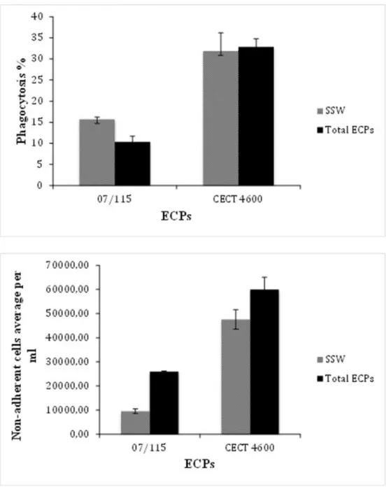
Figure 2. Effect ofV. aestuarianus07/115 (right bars) andV. tapetisCECT 4600 (left bars) total ECPs on oyster and clam, respectively, hemocyte phagocytosis capability (top panel) and hemocyte adhesion (bottom panel). Tests were carried out in triplicates as described in M&M and the error bars correspond to SD. Incubation of hemocytes with sterile sea water (SSW) was used as a negative control.
Results and Discussion
1. Preparation and fractionation of V. aestuarianus 07/115 and V. tapetis CECT4600 ECPs
The proteins from the extracellular compartment are of particular interest for functional investigation of bacterial patho-gen virulence, because they come into direct contact with host tissues and are often effectors of pathogenicity. Several lines of evidence highlight an important role of ECPs in the virulence of pathogenic vibrios. For example, a previous study on V. aestuarianus 01/032 showed that its ECPs displayed immuno-suppressive activities on oyster hemocyte functions [10]. Similar effects were described inV. tapetis, in which ECPs were shown to significantly decrease adhesive- [12] and phagocytic- [13] activities of clam hemocytes. However, although the biological activity ofV. aestuarianus ECPs has been associated with the secretion of the zinc metalloprotease, Vam [11], few studies have been carried out to date in V. tapetis and nothing is known about the molecular components responsible for the biological activity of theV. tapetis ECPs.
The extraction of secreted proteins was performed under conditions known to induce virulence [11,12]. ECPs were fractionated, their biological activity against hemocytes was assayed, and their protein contents were analyzed, as described in Materials and Methods.
In the case ofV. aestuarianus, fractionation of total ECPs gave four major peaks (Fig. 1). A first symmetrical peak eluted in the void volume of the column, suggesting that it was composed of a mixture of protein aggregates or complexes larger than 600 kDa.
Three poorly resolved additional peaks eluted at 16, 18 and 22 minutes, respectively. The elution diagram obtained with V. tapetis ECPs comprised a first peak also eluting in the void volume, and a second broad peak, lower in absorbance than the three peaks ofV. aestuarianus, but exactly superimposed. The fractions were recovered every minute and numbered according to the elution time. Determination of fraction protein contents allowed us to select a set of fractions (8, 9, 16 to 23 forV. aestuarianus ECPs and 8, 9, 14 to 21 for V. tapetis ECPs) showing a minimal concentration of 0,3 mg/ml of protein, to carry out further analyses.
2. Effects of V. aestuarianus and V. tapetis ECPs on phagocytosis and adherence activities of oyster and clam hemocytes respectively
We first assayed the activities of unfractionated ECPs. In both cases, the biological parameters assayed were hemocyte adhesion and phagocytosis. We found thatV. aestuarianus ECPs induced a decrease of phagocytosis and adherence properties of oyster hemocytes as shown in Fig. 2. This result is in keeping with previous results obtained by Labreuche et al. [10]. Similarly,V. tapetis ECPs triggered a decrease of hemocytes adherence as previously described [12]. However,V. tapetis total ECPs did not impact the phagocytic ability of clam hemocytes, contrary to what was found by Allam and Ford [13] who previously described a decrease in phagocytosis after treatment by bacterial supernatants obtained from liquid cultures. This discrepancy may be due to the different conditions used to prepare the ECPs (liquid culture versus cellophane overlay on plate).
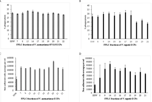
Figure 3. Effects ofV. aestuarianus(panels A and C) andV. tapetis(panels B and D) fractionated ECPs on oyster (A and C) and clam (B and D) hemocyte phagocytosis capacity (A and B) and adhesion properties (C and D). Tests were carried out in triplicates as described in Materials and Methods and the error bars correspond to SD.
The different FPLC fractions of ECPs previously obtained were then similarly tested for biological activity towards oysters (V. aestuarianus)- and clam (V. tapetis)- hemocytes. The results presented in Fig. 3 showed that all the assayed fractions obtained fromV. aestuarianus decreased the adhesive capacities of oyster hemocytes, with an increase of non-adherent hemocytes ranging from a factor 1.8 (fraction 16) to 2.5 (fraction 20). The only extracellular virulence factor described to date forV. aestuarianus is the Vam metalloprotease, which causes aggregation and the loss of pseudopods of oyster hemocytes [11]. Only the fractions 16 and 17 contained an azocaseinase activity (data not shown), suggesting that Vam is not responsible for this loss of adhesion and thatV. aestuarianus 07/115 extracellular products, in particular in fraction 20, contain additional factors playing a role in adherence decreasing.
However, although totalV. aestuarianus ECPs decreased the phagocytic activity of hemocytes (Fig. 2), none of the V. aestuarianus ECP fractions affected the oyster hemocyte phago-cytic activity (Fig. 3A). This result suggests that phagocytosis inhibition by ECPs may involve the joined activity of several factors that have been eluted in separate fractions.
In the case ofV. tapetis ECPs (Fig. 3B), a reduction in clam hemocyte phagocytosis capacity was recovered in fractions 19–20– 21, in accordance with previously published results [13]. This suggests that an inhibitor of this activity is present in the totalV. tapetis ECPs, which was separated during fractionation. As in the case of V. aestuarianus, all the recovered fractions displayed an effect on hemocyte adhesion, but with more variations amongst fractions. For example, fraction 14 triggered in excess of a 4-fold increase in non-adherent hemocytes whereas fraction 8 had only a 2-fold effect (Fig. 3D).
In summary, our results indicate that both V. tapetis and V. aestuarianus ECPs have major effects on hemocyte properties including loss of adherence and inhibition of phagocytosis, especially in the case ofV. tapetis. In the case of V. aestuarianus, inhibition of adhesion is independent of Vam, and is maximal in fraction 20. In the case of V. tapetis, we could partly separate adhesion inhibition activity (maximal in fraction 14) and
phagocytosis inhibition activity (maximal in fractions 19–21). In contrast, the observed phagocytosis inhibition inV. aestuarianus was lost upon fractionation, suggesting that it requires several factors acting in a complementary way while withV. tapetis, it was detected only after fractionation, suggesting the presence of an inhibitor in total ECPs.
3. Proteomic analysis of the two secretomes
Fractions combining both a significant effect in vitro on hemocytes, and sufficient protein amounts were further charac-terized by proteomic analysis. Accordingly, fractions 8 and 16 to 23 forV. aestuarianus and 9, 14 to 17 and 19 to 21 for V. tapetis were subjected to SDS-PAGE for further protein identification by nano LC-MS/MS. Several fractions (18 to 22 forV. aestuarianus and 19, 20, 21 for V. tapetis) did not show any band after Coomassie Blue staining, but were directly trypsinolyzed and analyzed by liquid chromatography tandem mass spectrometry (LC-MS/MS) starting from a total protein content of 5mg. As shown in Fig. 4, active fractions from both bacteria contained multiple proteins. In order to identify them, 16 and 43 distinct bands were excised forV. aestuarianus and V. tapetis, respectively, and analyzed by mass spectrometry, as described in Materials and Methods. Using the known proteome from both species, our proteomic analysis ofV. aestuarianus and V. tapetis secretomes led to the unambiguous identification of 45 and 87 proteins, respectively (Tables 1 and 2). Only five proteins in the ECPs of V. tapetis and none in the V. aestuarianus secretome were predicted to be cytoplasmic, emphasizing the quality of our protocol and the absence of cell lysis.
98% and 70% of the proteins in theV. aestuarianus and V. tapetis secretomes, respectively, were predicted to have a signal peptide (see Materials and Methods for the algorithms used), indicating that they are periplasmic or outer membrane compo-nents (see Tables 1 & 2). Most of the proteins appeared to be normal components of the outer membrane and the periplasmic space, suggesting that they were released in the medium most probably as membrane vesicles, as was previously described for other bacteria [24]. In accordance with this hypothesis, 98% of the Figure 4. SDS-PAGE of different FPLC fractions showing a significative effect on hemocyte adherence and/or phagocytosis properties: fractions 8A, 15A, 16A forV. aestuarianus(A) and 9T, 14T, 15T, 16T, 17T forV. tapetis(B).
Table 1. Proteins identified in the V. aestuarianus secretome. Protein function Genbank accession N r M W (kDa) Score* Nr of peptides coverage FPLC fraction Signal peptide cleavage site
** Predicted localization*** Structural component, envelope biogenesis and quality control, stress response OmpU: Outer membrane p rotein U KM588597 37 1682 20 68 8 21–22 OM Lpp: Major outer membrane lipoprotein KM588630 9 3 35 4 6 2 8 22–23 P/OM OmpA: Outer membrane p rotein A KM588598 35 327 5 22 8 21–22 OM BamA/YaeT: Outer membrane p rotein assembly factor KM588599 91 897 1 7 2 3 8 20–21 OM Tsp: carboxy-terminal protease for penicillin-binding protein 3 KM588600 75 198 5 8 8 23–24 P Peptidoglycan-associate d outer membrane lipoprotein Pal KM588601 19 100 2 15 8 25–26 P/OM Periplasmic component o f the Tol-Pal system, YbgF-like KM588602 29 453 6 25 8 23–24 P Thiol-disulfide isomerase DsbA KM588603 22 146 4 20 8 19–20 P LptD: LPS-assembly p rotein KM588604 52 52 2 3 8 24–25 OM Putative lipoprotein LpoA, activator o f penicillin binding protein 1A KM588605 67 226 5 9 8 26–27 P/OM Putative penicillin-binding protein activator LpoB-like, outer-membrane lipoprotein KM588606 22 894 1 2 7 1 8 25–26 P/OM NlpI lipoprotein KM588607 37 334 7 19 8 37–38 P/OM Energetic metabolism TorA: trimethylamine-N-oxide reductase (TMAO reductase) KM588608 92 119 3 4 8 33–34 P NapA: nitrate reductase, p eriplasmic, large subunit KM588631 93 1012 19 24 8 29–30 P Transporter components LamB: Maltoporin KM588632 43 800 1 2 4 3 8 22–23 OM Putative outer membrane porin; locus of qsr prophage KM588610 35 117 3 12 8 19–20 OM HisJ : ABC type h istidine transporter periplasmic histidine b inding protein KM588611 28 1169 19 72 8 21–22 P AapJ : ABC type L Aminoacid transporter subunit; periplasmic-binding KM588611 37 720 1 3 4 9 8 25–26 P PtsS1: ABC type p hosphate transporter, periplasmic phosphate binding protein KM588633 29 613 1 1 4 2 8 22–23 P TRAP-type u ncharacterized transport system, periplasmic component KM588612 34 524 1 2 3 2 8 24–25 P MglB: methyl-galactoside ABC transporter periplasmic-binding protein KM588613 35 460 7 21 8 22–23 P
Table 1. Cont. Protein function Genbank accession N r M W (kDa) Score* Nr of peptides coverage FPLC fraction Signal peptide cleavage site
** Predicted localization*** Putative ABC type d ipeptide transporter, periplasmic peptide-binding protein KM588637 57 266 6 11 8 23–24 P Putative ABC type tungstate transport system, substrate binding protein KM588614 30 78 2 8 8 23–24 P Pts2: ABC type phosphate transport system, periplasmic component KM588635 30 68 2 7 8 21–22 P TolC: o uter membrane efflux channel o f type I secretion system KM588615 48 64 2 5 8 22–23 OM FadL2: L ong-chain fatty acid outer membrane porin; bacteriophage T2 receptor KM588616 46 225 4 15 8 25–26 OM Putative YceI-like p rotein: lipid/polyisoprenoid-binding protein KM588636 20 89 2 8 8 22–23 P Motility/Flagell a FliD: F lagellar hook-associated p rotein 2 KM588617 71 123 3 6 8 -E Extracellular proteins Vam: secreted Zinc Metalloprotease Vam KM588637 66 1011 14 27 16, 17 25–26 E Putative extracellular triacylglycerol lipase KM588638 83 631 1 2 1 6 8 20–21 E Vpp: Vam p rotease processing e nzyme KM588639 101 4 22 6 8 8 23–24 E Unknown function Lipoproteins Putative outer membrane lipoprotein KM588618 21 469 6 39 8 17–18 P/OM Maltose operon p eriplasmic protein (MalM), outer membrane associated lipoprotein KM588640 71 139 4 12 8 24–25 P/OM Conserved lipoprotein of unknown function KM588619 14 193 4 33 8 21–22 P/OM Putative outer membrane lipoprotein KM588620 20 51 1 7 8 27–28 P/OM Conserved lipoprotein of unknown function KM588621 12 155 3 27 8 22–23 P/OM Conserved lipoprotein of unknown function KM588622 14 78 1 1 5 8 21–22 P/OM Putative outer membrane lipoprotein KM588623 28 190 3 18 8 20–21 P/OM Conserved o uter membrane lipoprotein of unknown function KM588641 21 362 9 62 8 17–18 P/OM Others Putative outer membrane p rotein KM588624 17 110 2 13 8 19–20 OM Outer membrane p roteins KM588625 84 110 4 5 8 26–27 OM Conserved o uter membrane o f unknown function KM588626 25 83 1 6 8 23–24 OM
biological functions (Tables 1 and 2) 1/Structural components, envelope biosynthesis and quality control, stress response, 2/ energetic metabolism, 3/transporter components, 4/iron acquisi-tion (except in V. aestuarianus), 5/catabolism, including chitin utilization, 6/motility, flagellar genes, 5/extracellular proteins, 6/ unknown function.
4. Identification of known and potential virulence factors in the vibrio secretomes
The only extracellular virulence factor characterized to date in V. aestuarianus is the secreted zinc metalloprotease, Vam, which was shown to cause lethality ofC. gigas oysters [11]. This protein was clearly identified and found to be quantitatively dominant in two active soluble fractions (16 and 17) in our study. More interestingly, we also identified a second extracellular protease in theV. aestuarianus secretome which we named Vpp (for Vam processing protease). Vpp is a homologue of Epp, a secreted protease which processes the secreted metalloprotease EmpA in Vibrio anguillarum [25], EmpA being a homologue of Vam. Hence, Vpp might be the Vam processing enzyme. Vpp is also a homologue of PrtV ofV. cholerae. In V. cholerae, PrtV was found to play a role in resistance to grazing by natural predator, outside the host, rather than in pathogenicity to humans [26]. Further studies should clarify the role of Vpp inV. aestuarianus, especially as fraction 8 that contains Vpp was found to decrease oysters hemocyte adherence.
Up to now, no secreted virulence factors have been described in V. tapetis. The only virulence factor characterized to date is the inner membrane protein DjlA, which was shown to be required for cytotoxicity towards clam hemocytes [27]. In contrast to V. aestuarianus, no metalloprotease was found in the V. tapetis secretome, suggesting different virulence mechanisms between the two species. However, two serine proteases (i.e. KM596588 and KM596661) carrying a signal peptide (Table 2) have been identified in two different fractions. As secreted serine protease was already shown to be involved in the virulence of several pathogenic bacteria [28], these two proteins could also play a role in the pathogenesis ofV. tapetis.
The secretomes of both vibrio species contained an extracellular triacylglycerol lipase (Table 1). This protein belongs to the same family as the phospholipase Pla1, a secreted virulence factor of Aeromonas hydrophila [29] and Cef, a toxin with cell elongation activity produced by Vibrio hollisae, which causes diarrhea in humans [30]. Phospholipases can act as potent membrane destructors and can manipulate host signalling pathways [31].
Another protein of interest is KM596634, identified in fraction 16 of theV. tapetis secretome, which contains the signatures of RTX toxins and autotransporters. Autotransporters are bacterial virulence factors that contain an N-terminal extracellular ("pas-senger") domain and a C-terminal b barrel ("b") domain that anchors the protein to the outer membrane. Upon autocleavage, the passenger domain is secreted. RTX (Repeat in toxins) toxins are virulence factors containing glycine- and aspartate-rich repeats binding Ca(2+) ions [32]. Such proteins were shown as virulence factors in other vibrio species [33,34].
Table 1. Cont. Protein function Genbank accession N r M W (kDa) Score* Nr of peptides coverage FPLC fraction Signal peptide cleavage site
** Predicted localization*** Conserved exported protein of unknown function KM588627 29 356 7 41 8 18–19 P Conserved o uter membrane p rotein of unknown function KM588628 35 137 2 7 8 20–21 OM Conserved o uter membrane p rotein of unknown function KM588629 20 63 2 1 2 8 19–20 OM * score given by the MASCOT software, ** as given by the algorithm SignalP 3.0 a nd some lipoproteins where putative SignalPeptidaseII cleavage sites w ere d etected b y LipoP 1.0, *** algorithm used PsortB and Psort Gram negative bacteria. Lipoproteins a nd their localisation were p redicted using LipoP1.0 (see details in materials and m ethods, b ioinformatics). (OM: outer membrane, IM: inner extracellular). doi:10.1371/journal.p one.0113097.t001
Table 2. Proteins identified in the V. tapetis secretome. Protein function Genbank accession N r MW (kDa) Score* Nr of peptides coverage FPLC fraction Signal peptide cleavage
site** Predicted localization*** Structural components, envelope biogenesis and quality control, stress response OmpH: Outer membrane porin H KM596581 35 2838 33 71 16 19–20 O M OmpU: Outer membrane p rotein U KM596582 37 2318 24 77 16 21–22 O M OmpV: Outer m embrane p rotein V KM596645 28 303 6 29 16 23–24 O M BamA/YaeT: Outer membrane p rotein assembly factor KM596583 90 204 3 4 9 20–21 O M OmpA: Outer m embrane p rotein A KM596646 35 665 9 27 9 2 0–21 OM Lpp : major outer membrane lipoprotein KM596647 10 54 1 1 2 9 22–23 O M Outer membrane p rotein KM596648 27 93 2 7 9 1 8–19 OM TolB : p eriplasmic component of the T ol-Pal system KM596584 49 787 1 7 3 4 9 -P Periplasmic component of the T ol-Pal system, YbgF-like KM596585 29 104 1 11 9 2 3–24 P Tsp: carboxy-terminal protease for penicillin-bindi ng protein 3 KM596586 75 397 9 16 9 2 2–23 OM SurA: p eptidyl-prolyl cis-trans isomerase KM596587 48 261 5 15 9 2 2–23 P DegP serine e ndoprotease KM596588 48 194 5 9 9 27–28 P Putative lipoprotein LpoA, activator o f p enicillin binding protein 1A KM596589 68 529 1 1 1 7 9 44–45 P /OM MltC : m embrane-bound lytic murein transglycosylase C KM596590 45 76 2 6 9 -IM/P Putative MltA-interacting M ipA KM596591 50 141 3 10 9 3 3–34 OM NlpI lipoprotein KM596592 35 199 3 17 9 2 2–23 P/OM SodB : iron superoxide d ismutase KM596593 21 303 5 32 15 -C SodM : superoxyde dismutase Mn/Fe KM596649 23 161 4 17 15 -C LptD : LPS-assembly lipoprotein KM596594 87 126 3 4 9 24–25 O M HslJ: Heat-shock lipoprotein KM596595 16 78 2 2 0 9 23–24 P /OM Energetic metabolism TorA: trimethylamine N -oxide (TMAO) reductase I catalytic subunit KM596596 92 754 1 2 1 4 1 4 3 3–34 P TrxA : thioredoxin 1 KM596597 12 213 3 26 9 -C Transporter components Putative outer membrane efflux channel of type I secretion system KM596598 48 136 3 8 9 19–20 O M Putative outer membrane efflux channel of type I secretion system KM596599 51 55 2 4 9 2 2–23 OM
Table 2. Cont. Protein function Genbank accession N r MW (kDa) Score* Nr of peptides coverage FPLC fraction Signal peptide cleavage
site** Predicted localization*** TolC: outer membrane efflux channel o f type I secretion system KM596600 47 1434 21 54 9 2 2–23 OM LamB: maltoporin KM596650 46 249 5 16 9 2 4–25 OM Outer membrane porin KM596601 37 143 2 7 9 19–20 O M Putative outer membrane porin KM596602 40 436 1 1 3 4 9 22–23 O M AapJ : ABC type L Aminoacid transporter subunit; periplasmic-bind ing KM596603 36 1004 17 47 9 2 5–26 P TRAP-type uncharacterized transport system, periplasmic component KM596604 35 996 1 7 4 4 9 24–25 U BtuB: Vitamin B12/cobalamin outer membrane transporter, TonB dependent KM596605 68 747 1 3 2 3 9 22–23 O M Putative chitoporin KM596606 50 1046 14 39 9 2 3–24 OM Putative (GlcNAc)2 periplasmic substrate-binding protein of ABC transporter KM596607 63 667 1 3 2 3 9 27–28 P Putative (GlcNAc)2 ABC transporter, periplasmic substrate-bindin g p rotein of ABC transporter KM596608 63 152 4 7 9 23–24 P Putative substrate binding protein component of oligopetpide/dip eptide ABC transporter KM596609 66 643 1 4 2 7 9 20–21 P Putative ABC-type o ligopeptide/dip eptide transport system, substrate-binding p eriplasmic component KM596651 69 151 4 7 9 20–21 P Fad L1: L ong-chain fatty acid outer membrane porin KM596652 44 164 4 16 9 2 1–22 OM FadL2: L ong-chain fatty acid outer membrane porin KM596610 43 441 6 18 9 2 5–26 OM ABC-type sugar transport system, p eriplasmic component KM596611 45 326 7 24 9 2 1–22 P Putative outer membrane p rotein of unknown function KM596653 40 326 6 15 9 2 1–22 OM Putative ABC-type transport system, p eriplasmic substrate-bindin g component KM596612 33 270 5 25 9 2 5–26 P TRAP-type C4-dicarboxylate transport system, periplasmic component KM596613 37 228 4 18 9 2 2–23 P Putative TonB-dependent receptor p rotein KM596654 77 91 2 3 9 2 2–23 OM Spermidine/putr escine ABC transporter: substrate binding periplasmic protein PotD2 KM596614 39 61 1 4 9 2 4–25 P Putative oligogalacturonate-sp ecific porin KM596615 26 522 1 0 2 6 9 20–21 O M Rutative Rhs family protein, m ay bind carbohydrates KM596616 259 5 4 1 1 1 4 -IM Iron a cquisition Putative outer membrane siderophore receptor KM596655 65 65 2 3 9 2 5–26 OM
Table 2. Cont. Protein function Genbank accession N r MW (kDa) Score* Nr of peptides coverage FPLC fraction Signal peptide cleavage
site** Predicted localization*** FbpA : P eriplasmic ferric iron-binding protein o f ABC transporter KM596617 37 302 6 18 9 2 1–22 P Putative TonB-dependent vibriobactin receptor KM596656 71 75 2 4 9 2 9–30 OM Putative iron-regulated p rotein with peptidase M75 domain KM596618 46 235 4 9 9 24–25 O M Catabolism Chitin utilization GbpA: N-acetyl glucosamine (GlcNAc) b inding protein A KM596619 54 358 7 14 17 24–25 E Chitinase KM596657 53 472 8 20 15 19–20 U ChiA: endochitinase A KM596620 89 367 6 6 1 4 2 1–22 E Endochitinase KM596621 88 935 1 1 1 2 1 6 2 2–23 E Putative chitinase KM596658 70 1876 26 35 16 28–29 O M Putative chitinase KM596659 48 192 4 9 9 20–21 P Others Aryl/Alkyl sulfatase KM596660 73 251 6 12 9 1 9–20 P Ggt : G amma-glutamyltransp eptidase KM596622 63 212 6 11 16 24–25 P PepD : a minoacyl-histidine d ipeptidase (peptidase D) KM596623 53 162 5 10 14 -C CpdB: 2 9:3 9-cyclic-nucleotide 2 9-phosphodiesterase KM596624 72 67 2 2 9 2 5–26 P UshA: bifunctional UDP-sugar h ydrolase and 5 9-nucleotidase, outer membrane associated lipoprotein KM596625 61 1949 32 50 9 2 5–26 P/OM Motility/Flag e lla Putative flagellar hook-length control p rotein FliK KM596626 71 1359 16 31 9 -IM Flagellin KM596627 40 300 6 17 9 -E FlgJ: peptidoglycan hydrolase FlgJ KM596628 34 650 1 3 4 4 9 -P FlgF: Flagellar basal body rod p rotein KM596629 27 147 3 19 9 -P FlgE: Flagellar hook protein KM596630 47 1313 20 45 9 -OM FlgD: Flagellar basal body rod modification p rotein KM596631 25 781 1 0 5 5 9 -P FlgC: Flagellar component of cell-proximal portion o f basal-body rod KM596632 15 656 7 72 9 -P FlgB: Flagellar basal body rod p rotein KM596633 14 500 7 63 9 -P Extracellular proteins Putative extracellular protease KM596661 38 387 7 29 9 2 4–25 E Putative extracellular lipase KM596662 90 1006 16 23 9 2 2–23 E/OM Putative autotransporter adhesin/RTX toxin KM596634 136 9 36 12 11 16 27–28 E /OM
Table 2. Cont. Protein function Genbank accession N r MW (kDa) Score* Nr of peptides coverage FPLC fraction Signal peptide cleavage
site** Predicted localization*** Unknown function Lipoproteins Conserved outer membrane lipoprotein of unknown function KM596635 12 474 8 56 9 2 4–25 P/OM Conserved outer membrane lipoprotein of unknown function KM596667 15 312 4 32 9 2 7–28 P/OM Putative outer membrane associated lipoprotein KM596636 51 1626 29 58 15 -P /OM Putative outer membrane associated lipoprotein KM596637 16 121 2 18 9 2 4–25 P/OM Conserved outer membrane lipoprotein of unknown function KM596638 14 240 4 35 9 1 8–19 P/OM Conserved outer membrane lipoprotein of unknown function KM596663 24 104 1 8 9 22–23 P /OM Conserved outer membrane lipoprotein of unknown function KM596639 24 66 1 5 9 2 4–25 OM Others Conserved outer membrane p rotein of unknown function KM596640 25 284 3 14 9 2 2–23 OM Conserved outer membrane p rotein of unknown function KM596641 36 86 2 1 2 9 23–24 O M Conserved outer membrane p rotein of unknown function KM596642 84 352 6 11 9 2 9–30 OM Conserved outer membrane p rotein of unknown function KM596664 19 564 6 29 17 23–24 O M Conserved putative inner membrane p rotein of unknown function KM596643 67 504 1 0 1 6 1 5 2 3–24 IM Conserved outer membrane p rotein of unknown function KM596644 46 407 7 16 9 1 8–19 OM Conserved p rotein of unknown function KM596665 19 68 1 6 9 -C Conserved p rotein of unknown function KM596666 56 417 6 19 16 21–22 O M * score given by the MASCOT software, ** as given by the algorithm SignalP 3.0, *** algorithm u sed for subcellular localization: PsortB or Psort G ram n e gative bacteria. Lipoproteins localisation w ere details in materials and m ethods, b ioinformatics). (OM: outer membrane, IM: inner m embrane, P: periplasmic, E: extracellular, C: cytosolic, U: unk nown) doi:10.1371/journal .pone.0113097.t002
Finally, it should be noted that, contrary toV. aestuarianus, the V. tapetis secretome contains one receptor (GbpA) and several chitinases, underscoring the role of chitin as a carbon source in the environment. Besides, chitinases have already be shown to be bacterial virulence factors, eg inListeria monocytogenes [35], and Legionella pneumophila [36]. Chitin is also a component of the shell organic matrix, andV. tapetis as a pathogen forms biofilms on the inner surface of the shell, typically at the level of the pallial line at the growing edge of the shell [37]. Hence, chitin use may be especially relevant toV. tapetis pathogenicity.
5. Proteins common to the V. aestuarianus and V. tapetis secretomes
Finally, the sequence of each secretome protein of a given species was comparedin silico (using blastP) to the full proteome of the other species, allowing us to identify 21 common proteins. The results are presented in Table 3. The only potential virulence factor is the putative extracellular lipase (Pla1) already mentioned above. The other proteins corresponded to normal components of the envelope in gamma proteobacteria, and/or in the Vibrio genus.
Conclusion
Extracellular products, especially secreted proteins, enter in direct contact with the host cells, and play a major role in the virulence of pathogenic bacteria. As a consequence, secretomic approaches are of particular relevance to identify the proteins involved in the infection process, and several studies have been carried out for different pathogens in recent years [38–41]. It should be noted that to date, only two secretomes of vibrios have been reported in the literature, i.e. those ofV. coralliilyticus [20] andV. cholerae [42].
In this paper, we characterized the extracellular proteome ofV. aestuarianus and V. tapetis, two vibrio species pathogenic to mollusks, as a first step towards the identification of new potential virulence factors. Although the extracellular products from both species were shown to be involved in bacterial virulence, only one extracellular virulence factor has been characterized to date, in the case ofV. aestuarianus, the Vam zinc metalloprotease [11].
This protein appeared as a major component of the V. aestuarianus secretome. However, we showed that a metallopro-tease-free fraction (fraction 8) also displayed biological activity to hemocytes, thus suggesting the occurrence of other potential virulence factors in this species.
KM596594 LptD KM588604
KM596600 TolC KM588615
KM596589 Putative lipoprotein LpoA, activator
of penicillin binding protein 1A
KM588605
KM596596 TorA KM588608
KM596586 Tsp KM588600
KM596603 AapJ KM588611
KM596585 Periplasmic component of the
Tol-Pal system, YbgF-like
KM588602
KM596635 Conserved lipoprotein of unknown function KM588621
KM596592 NlpI KM588607
KM596582 OmpU KM588597
KM596638 Conserved lipoprotein of unknown function KM588619
KM596583 BamA (YaeT) KM588599
KM596640 Conserved outer membrane
protein of unknown function
KM588626
KM596642 Putative outer membrane
protein of unknown function
KM588625
KM596650 LamB KM588632
KM596647 Lpp KM588630
KM596662 Putative extracellular lipase KM588638
KM596652 Long-chain fatty acid outer
membrane porin FadL
KM588616
KM596646 OmpA KM588598
KM596641 Conserved outer membrane protein
of unknown function
KM588628
active fractions of theV. aestuarianus and V. tapetis secretomes. Our data constitute the first valuable resource to further investigate the virulence factors of these two marine pathogen vibrios. Future works will aim at assessing the actual role of specific secreted proteins in the virulence.
the experiments: SM VP MP. Analyzed the data: SM VP AJ FG CP. Contributed reagents/materials/analysis tools: VP CP JLN. Contributed to the writing of the manuscript: SM VP AJ CP JLN.
References
1. Paillard C, Le Roux F, Borrego JJ (2004) Bacterial disease in marine bivalves, a review of recent studies: Trends and evolution. Aquat Living Resour 17: 477– 498.
2. Garnier M, Labreuche Y, Garcia C, Robert M, Nicolas JL (2007) Evidence for the involvement of pathogenic bacteria in summer mortalities of the Pacific
oysterCrassostrea gigas. Microb Ecol 53: 187–196.
3. Gay M, Renault T, Pons A-M, Le Roux F (2004) Two vibrio splendidus related
strains collaborate to killCrassostrea gigas: taxonomy and host alterations. Dis
Aquat Organ 62: 65–74.
4. Go´mez-Leo´n J, Villamil L, Lemos ML, Novoa B, Figueras A (2005) Isolation of Vibrio alginolyticus and Vibrio splendidus from aquacultured carpet shell clam (Ruditapes decussatus) larvae associated with mass mortalities. Appl Environ Microbiol 71: 98–104.
5. Paillard C (2004) A short-review of brown ring disease, a vibriosis affecting
clams,Ruditapes philippinarum and Ruditapes decussatus. Aquat Living Resour
17: 467–475.
6. Paillard C, Maes P (1995) The Brown Ring Disease in the Manila Clam, Ruditapes philippinarum. J Invertebr Pathol 65: 101–110.
7. Segarra A, Pe´pin JF, Arzul I, Morga B, Faury N, et al. (2010) Detection and description of a particular Ostreid herpesvirus 1 genotype associated with
massive mortality outbreaks of Pacific oysters,Crassostrea gigas, in France in
2008. Virus Res 153: 92–99.
8. Samain J, McCombie H (2008) Summer mortality of pacific oysterCrassostrea
gigas. The MOREST project. Versailles.
9. Romalde JL, Dieguez AL, Lasa A, Balboa S (2014) NewVibrio species associated
to molluscan microbiota: a review. Front Microbiol 4: 413.
10. Labreuche Y, Soudant P, Gonc¸alves M, Lambert C, Nicolas J-L (2006) Effects of
extracellular products from the pathogenicVibrio aestuarianus strain 01/32 on
lethality and cellular immune responses of the oysterCrassostrea gigas. Dev
Comp Immunol 30: 367–379.
11. Labreuche Y, Le Roux F, Henry J, Zatylny C, Huvet A, et al. (2010)Vibrio
aestuarianus zinc metalloprotease causes lethality in the Pacific oyster Crassostrea gigas and impairs the host cellular immune defenses. Fish Shellfish Immunol 29: 753–758.
12. Choquet G, Soudant P, Lambert C, Nicolas J-L, Paillard C (2003) Reduction of
adhesion properties of Ruditapes philippinarum hemocytes exposed toVibrio
tapetis. Dis Aquat Organ 57: 109–116.
13. Allam B, Ford SE (2006) Effects of the pathogenicVibrio tapetis on defence
factors of susceptible and non-susceptible bivalve species: I. Haemocyte changes following in vitro challenge. Fish Shellfish Immunol 20: 374–383.
14. Lopez-Cortes L, Castro D, Navas JI, Borrego JJ (1999) Phagocytic and
chemotactic responses of manila and carpet shell clam haemocytes againstVibrio
tapetis, the causative agent of brown ring disease. Fish Shellfish Immunol 9: 543– 555.
15. Matson JS, Withey JH, DiRita VJ (2007) Regulatory networks controllingVibrio
cholerae virulence gene expression. Infect Immun 75: 5542–5549.
16. Makino K, Oshima K, Kurokawa K, Yokoyama K, Uda T, et al. (2003)
Genome sequence ofVibrio parahaemolyticus: a pathogenic mechanism distinct
from that ofV cholerae. Lancet 361: 743–749.
17. Chen C-Y, Wu K-M, Chang Y-C, Chang C-H, Tsai H-C, et al. (2003)
Comparative genome analysis ofVibrio vulnificus, a marine pathogen. Genome
Res 13: 2577–2587.
18. Austin B (2010) Vibrios as causal agents of zoonoses. Vet Microbiol 140: 310– 317.
19. Le Roux F, Binesse J, Saulnier D, Mazel D (2007) Construction of aVibrio
splendidus mutant lacking the metalloprotease gene vsm by use of a novel counterselectable suicide vector. Appl Environ Microbiol 73: 777–784. 20. Santos E de O, Alves N, Dias GM, Mazotto AM, Vermelho A, et al. (2011)
Genomic and proteomic analyses of the coral pathogenVibrio coralliilyticus
reveal a diverse virulence repertoire. ISME J 5: 1471–1483.
21. Sikora AE, Zielke RA, Lawrence DA, Andrews PC, Sandkvist M (2011)
Proteomic analysis of theVibrio cholerae type II secretome reveals new proteins,
including three related serine proteases. J Biol Chem 286: 16555–16566.
22. Paillard C, Maes P (1990) Etiologie de la maladie de l’anneau brun chezTapes
philippinarum: pathoge´nicite´ d’un Vibrio sp. C R Acad Sci Se´rie 3 310: 15–20. 23. Thiele H, Glandorf J, Hufnagel P (2010) Bioinformatics strategies in life sciences: from data processing and data warehousing to biological knowledge extraction. J Integr Bioinform 7: 141.
24. Unal CM, Schaar V, Riesbeck K (2011) Bacterial outer membrane vesicles in disease and preventive medicine. Semin Immunopathol 33: 395–408. 25. Varina M, Denkin SM, Staroscik AM, Nelson DR (2008) Identification and
characterization of Epp, the secreted processing protease for the Vibrio
anguillarum EmpA metalloprotease. J Bacteriol 190: 6589–6597.
26. Vaitkevicius K, Lindmark B, Ou G, Song T, Toma C, et al. (2006) AVibrio
cholerae protease needed for killing of Caenorhabditis elegans has a role in protection from natural predator grazing. Proc Natl Acad Sci U S A 103: 9280– 9285.
27. Lakhal F, Bury-Mone´ S, Nomane Y, Le Goı¨c N, Paillard C, et al. (2008) DjlA, a membrane-anchored DnaJ-like protein, is required for cytotoxicity of clam
pathogenVibrio tapetis to hemocytes. Appl Environ Microbiol 74: 5750–5758.
28. Ruiz-Perez F, Nataro JP (2014) Bacterial serine proteases secreted by the autotransporter pathway: classification, specificity, and role in virulence. Cell Mol Life Sci 71: 745–770.
29. Merino S, Aguilar A, Nogueras M, Regue M, Swift S, et al. (1999) Cloning, sequencing, and role in virulence of two phospholipases (A1 and C) from
mesophilicAeromonas sp. serogroup O: 34. Infect Immun 67: 4008–4013.
30. Kothary M, Claverie E, Miliotis M, Madden J, Richardson S (1995) Purification
and characterization of a chinese hamster ovary cell elongation factor ofVibrio
hollisae. Infect Immun 63: 2418–2423.
31. Lang C, Flieger A (2011) Characterisation of Legionella pneumophila
phospholipases and their impact on host cells. Eur J Cell Biol 90: 903–912. 32. Linhartova´ I, Bumba L, Masˇı´n J, Basler M, Osicˇka R, et al. (2010) RTX
proteins: a highly diverse family secreted by a common mechanism. FEMS Microbiol Rev 34: 1076–1112.
33. Olivier V, Haines GK, Tan Y, Satchell KJF (2007) Hemolysin and the multifunctional autoprocessing RTX toxin are virulence factors during intestinal
infection of mice with Vibrio cholerae El Tor O1 strains. Infect Immun 75:
5035–5042.
34. Ziolo KJ, Jeong H-G, Kwak JS, Yang S, Lavker RM, et al. (2014)Vibrio
vulnificus biotype 3 multifunctional autoprocessing RTX toxin is an adenylate cyclase toxin essential for virulence in mice. Infect Immun 82: 2148–2157. 35. Chaudhuri S, Gantner BN, Ye RD, Cianciotto NP, Freitag NE (2013) The
Listeria monocytogenes ChiA chitinase enhances virulence through suppression of host innate immunity. MBio 4: e00617–12.
36. DebRoy S, Dao J, So¨derberg M, Rossier O, Cianciotto NP (2006)Legionella
pneumophila type II secretome reveals unique exoproteins and a chitinase that promotes bacterial persistence in the lung. Proc Natl Acad Sci U S A 103: 19146–19151.
37. Paillard C, Allam B, Oubella R (2004) Effect of temperature on defense
parameters in manila clam Ruditapes philippinarum challenged with Vibrio
tapetis. Dis Aquat Organ 59: 249–262.
38. Pocsfalvi G, Cacace G, Cuccurullo M, Serluca G, Sorrentino A, et al. (2008)
Proteomic analysis of exoproteins expressed by enterotoxigenicStaphylococcus
aureus strains. Proteomics 8: 2462–2476.
39. Barbey C, Budin-Verneuil A, Cauchard S, Hartke A, Laugier C, et al. (2009)
Proteomic analysis and immunogenicity of secreted proteins fromRhodococcus
equi ATCC 33701. Vet Microbiol 135: 334–345.
40. Benachour A, Morin T, He´bert L, Budin-Verneuil A, Le Jeune A, et al. (2009)
Identification of secreted and surface proteins from Enterococcus faecalis.
Can J Microbiol 55: 967–974.
41. Mendez JA, Soares NC, Mateos J, Gayoso C, Rumbo C, et al. (2012) Extracellular proteome of a highly invasive multidrug-resistant clinical strain of Acinetobacter baumannii. J Proteome Res 11: 5678–5694.
42. Barh D, Barve N, Gupta K, Chandra S, Jain N, et al. (2013) Exoproteome and
secretome derived broad spectrum novel drug and vaccine candidates inVibrio