HAL Id: hal-02379536
https://hal.archives-ouvertes.fr/hal-02379536
Submitted on 21 Sep 2020
HAL is a multi-disciplinary open access
archive for the deposit and dissemination of
sci-entific research documents, whether they are
pub-lished or not. The documents may come from
teaching and research institutions in France or
abroad, or from public or private research centers.
L’archive ouverte pluridisciplinaire HAL, est
destinée au dépôt et à la diffusion de documents
scientifiques de niveau recherche, publiés ou non,
émanant des établissements d’enseignement et de
recherche français ou étrangers, des laboratoires
publics ou privés.
Distributed under a Creative Commons Attribution - NonCommercial - NoDerivatives| 4.0
International License
Rodent malaria in Gabon: Diversity and host range
Larson Boundenga, Barthélémy Ngoubangoye, Stephan Ntie, Nancy-Diamella
Moukodoum, Francois Renaud, Virginie Rougeron, Franck Prugnolle
To cite this version:
Larson Boundenga, Barthélémy Ngoubangoye, Stephan Ntie, Nancy-Diamella Moukodoum, Francois
Renaud, et al.. Rodent malaria in Gabon: Diversity and host range. International Journal for
Parasitology: Parasites and Wildlife, Elsevier, 2019, 10, pp.117-124. �10.1016/j.ijppaw.2019.07.010�.
�hal-02379536�
Contents lists available atScienceDirect
IJP: Parasites and Wildlife
journal homepage:www.elsevier.com/locate/ijppaw
Invited article
Rodent malaria in Gabon: Diversity and host range
Larson Boundenga
a,*, Barthélemy Ngoubangoye
a, Stephan Ntie
b, Nancy-Diamella Moukodoum
a,
François Renaud
c, Virginie Rougeron
c,1, Franck Prugnolle
c,1aCentre International de Recherches Médicales de Franceville (CIRMF), BP. 769, Franceville, Gabon
bLaboratoire de Biologie Moléculaire et Cellulaire (LABMC), Département de Biologie, Université de Sciences et Techniques de Masuku (USTM), BP 941, Franceville,
Gabon
cLaboratory MIVEGEC, UMR 224-5290, IRD, CNRS, University of Montpellier, Montpellier, France
A R T I C L E I N F O Keywords: Rodent Malaria Plasmodium Central Africa Mus musculus Host range A B S T R A C T
Malaria parasites infect a wide range of vertebrate hosts, such as reptiles, birds and mammals (i.e., primates, ungulates, bats, and rodents). Four Plasmodium species and their subspecies infect African Muridae. Since their discoveries in the 1940s, these rodent Plasmodium species have served as biological models to explore many aspects of the biology of malaria agents and their interactions with their hosts. Despite that, surprisingly, little is known about their ecology, natural history and evolution. Most field studies on these parasites, performed from the 1940s to the early 1980s, showed that all rodent Plasmodium species infect only one main host species, the thicket rat. In the present study, we re-explored the diversity of Plasmodium parasites infecting rodent species living in peridomestic habitats in Gabon, Central Africa. Using molecular approaches, we found that at least two
Plasmodium species (Plasmodium vinckei and Plasmodium yoelii) circulated among five rodent species (including
the invasive species Mus musculus). This suggests that the host range of these parasites might be larger than previously considered. Our results also showed that the diversity of these parasites could be higher than cur-rently recognized, with the discovery of a new phylogenetic lineage that could represent a new species of rodent
Plasmodium.
1. Introduction
Malaria is a mosquito-borne disease caused by protozoan parasites of the genus Plasmodium (Déchamps et al., 2010). This genus comprises many species that infect a large range of vertebrate groups including birds, reptiles and mammals (Boundenga et al., 2016,2017;Yotoko and Elisei, 2006). In mammals, besides the parasites infecting humans, those infecting rodents and more specifically Muridae (Plasmodium
berghei, Plasmodium chabaudi, Plasmodium vinckei, and Plasmodium yoelii) have been the most studied and have served, since their
dis-coveries in the 1940s, as biological models to explore many aspects of the biology of malaria agents and their interactions with their hosts (vertebrate and arthropod vectors) (LaCrue et al., 2011;Landau and Chaubaud, 1994;Stephens et al., 2012;Vanderberg, 1991;Wargo et al., 2007). Due to their importance as model systems, rodent malaria agents were among the first eukaryotes (and among the first protozoans) to have their genome entirely sequenced (in 2002 for P. yoelii yoelii
17XNL, in 2005 for P. chabaudi chabaudi AS) (Carlton et al., 2002;Hall et al., 2005).
Despite the huge amount of knowledge acquired on the biology of
Plasmodium species infecting rodents during the last 80 years,
para-doxically little is known about their ecology, natural history, and evo-lution (Landau et al., 1970; Ramakrishnan et al., 2013; Vanderberg, 1991). Indeed, most field studies on these parasites were performed from the 1940s to the early 1980s (Landau and Chaubaud, 1994), and to our knowledge, no study has been carried out in their natural habitat since then.
Previous studies have shown that the distribution of rodent malaria agents is limited to the Congo basin: Cameroon, Central African Republic, Congo, Democratic Republic of the Congo, and Nigeria (Culleton, 2005;Killick-Kendrick, 1978;Landau and Chaubaud, 1994). It is thought that most of these agents (except P. berghei) infect one main host species, the thicket rat (Grammomys poensis – previously called
Thamomnys rutilans) that inhabits tropical lowland forests ( Killick-Kendrick, 1978;Landau and Chaubaud, 1994). A recent study carried out in Uganda, Kenya, Zambia and Mozambique did not detect any infection in sylvatic rodents, confirming the absence or the rarity of these rodent
Plasmodium species outside the Congo basin (Lutz et al., 2016).
https://doi.org/10.1016/j.ijppaw.2019.07.010
Received 11 March 2019; Received in revised form 26 July 2019; Accepted 27 July 2019
*Corresponding author.
E-mail address:[email protected](L. Boundenga).
1Co-managed this work.
IJP: Parasites and Wildlife 10 (2019) 117–124
2213-2244/ © 2019 The Authors. Published by Elsevier Ltd on behalf of Australian Society for Parasitology. This is an open access article under the CC BY-NC-ND license (http://creativecommons.org/licenses/BY-NC-ND/4.0/).
These parasites were first characterized and defined using mor-phological features, leading to the definition of four species (P. yoelii, P.
chabaudi, P. berghei and P. vinckei) and several subspecies (e.g., P. yoelii yoelii and P. yoelii nigeriensis) (Landau and Chaubaud, 1994). Isoenzyme analysis first and later multiple gene sequencing of several isolates collected in the 1970s (Carter, 1978) and stored at the European Ma-laria Reagent Repository (www.malariaresearch.eu) largely confirmed this diversity, and suggested that most subspecies should be considered as species. This is particularly true for the P. vinckei subspecies because they show high genetic divergence and do not seem to recombine when present in sympatry (Carter, 1978;Perkins et al., 2007;Ramiro et al., 2012)).
The only known vector of murine malaria parasites was, up to re-cently, Anopheles dureni millecampsi, the invertebrate host of P. berghei and P. v. vinckei in Katanga (Democratic Republic of the Congo) (
Killick-Kendrick, 1978;Stephens et al., 2012). However, a recent study in the forests of Gabon identified five other anopheles species infected by rodent Plasmodium parasites (Anopheles moucheti, Anopheles nili,
Ano-pheles gabonensis, AnoAno-pheles vinckei, and AnoAno-pheles marshalii), suggesting
that a far larger number of anopheles species could play the role of vector within their geographic range (Makanga et al., 2016; Rahola et al., 2014).
Recent studies on Plasmodium parasites of different groups of ver-tebrates demonstrated the utility of re-evaluating their diversity in the molecular era, by showing that their diversity has been, most of the time, historically underestimated (Boundenga et al., 2016; Prugnolle et al., 2010;Valkiūnas et al., 2008). For instance, the use of molecular tools has led to revising the diversity of the Plasmodium parasites in-fecting apes and ungulates (Boundenga et al., 2016; Liu et al., 2010;
Perkins and Schaer, 2016;Prugnolle et al., 2010).
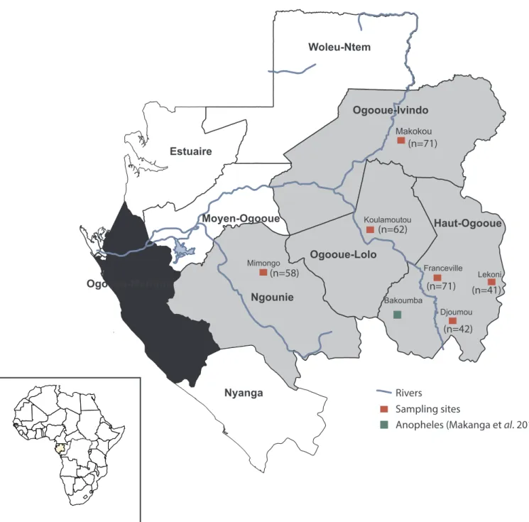
Fig. 1. Location of the provinces of Gabon where rodent samples were collected. The map shows the sites where rodents were captured (in red) and the site where
some Anopheles infected with rodent malaria parasites were found in a previous study (in green) (Makanga et al., 2016). The number of individuals collected in each province is indicated between brackets. (For interpretation of the references to colour in this figure legend, the reader is referred to the Web version of this article.)
L. Boundenga, et al. IJP: Parasites and Wildlife 10 (2019) 117–124
In the present study, we examined the genetic diversity and the host range of malaria parasites infecting Muridae in Gabon (Central Africa) by analysing the mitochondrial cytochrome b gene (CYTB). We focused on rodent parasites because information regarding their distribution and the ecology in these hosts is currently non-existent in Gabon. The present study is the first molecular overview of protozoan blood para-sites of the order Haemosporida recorded in rodents from the forested regions of Gabon.
2. Materials and methods 2.1. Study area and sample collection
Gabon (Fig. 1) is a Central African country located in the west part of the Congo basin. Most of its surface (85%) is covered by forest that is amongst the richest African forests in terms of diversity and endemism (Lee et al., 2006). Gabon has a humid and hot climate, typical of equa-torial regions. There are four seasons: two dry (from June to September and from December to February) and two rainy (from mid-September to mid-December and from mid-February to mid-June).
The Plasmodium diversity circulating among wild rodents in Gabon was analysed by screening rodent samples collected by different teams of the Centre International de Recherches Médicales de Franceville (CIRMF) in four provinces (Fig. 1) as part of a research programme on rodent patho-gens between 2009 and 2016 (SeeTable 1andTable S1for more details on sampling). Rodents were captured using Tomahawk and Shermann traps, as described in (Duplantier, 1989), in peridomestic habitats (up to 250m from the houses). Each individual was identified using morphological features described in previous studies (Duplantier et al., 1993). For this study, analyses were performed on 345 rodent samples that belonged to seven rodent species [Lemniscomys striatus (n = 50), Lophuromys nudicaudus (n = 40), Mastomys natalensis (n = 35), Praomys sp. (n = 30), Grammomys
poensis (n = 10), Rattus rattus (n = 100), and Mus musculus (n = 80)]. For
each animal, whole blood was preferentially used when available, other-wise a mix of liver/spleen was chosen (seeSupplementary Table S1for details on sampling). Biological samples (whole blood or liver/spleen) were collected and kept in liquid nitrogen until their arrival at the CIRMF where they were stored at −80 °C for molecular analyses.
2.2. Ethical approval
The study was performed outside protected areas in Gabon. Rodent trapping and sampling were conducted after authorization by the Wildlife and Hunting Department of the Gabonese Ministry of Water and Forestry (N°003/MEFE-PA/SG/DGEF/DCF and N°0021/MEFE-PA/ SG/DGEF/DCF). Animal capture, handling, euthanasia and sample transfer across country borders were performed in accordance with the guidelines of the American Society of Mammalogists (http://www. mammalsociety.org/committees/animal-care-and-use) and in strict ac-cordance with the recommendations of the Gabonese National Ethics Committee (Authorization N°PROT/0020/2013I/S G/CNE).
2.3. Molecular analyses
2.3.1. Molecular characterization of malaria parasites
For each sample, total DNA was extracted using the DNeasy Blood and Tissue Kit (Qiagen, Courtaboeuf, France) from approximately 200 μl of blood or 100 mg of liver/spleen according to the manufac-turer's procedures. Total DNA was then used as template for the de-tection of malaria parasites according to a previously described proto-cols based on the amplification by nested PCR of a portion of the
Plasmodium CYTB gene (Boundenga et al., 2017;Prugnolle et al., 2010). All amplified products (10 μl) were run on 1.5% agarose gels in Tris-acetate-EDTA buffer. Amplicons (700bp) were then sequenced by Eurofins MWG (France). Moreover, in all positive samples, the nature of the host species was confirmed by amplifying and sequencing a portion
Table 1 Description of the rodent species analysed in this study. The table shows the number of samples collected per species and the infection rate by Plasmodium agents. Year of samples collection Province Sites 2009 2010 2011 2012 2013 2014 2015 2016 Total of samples Blood (n/N) Organ (n/N) Blood (n/N) Organ (n/N) Blood (n/N) Organ (n/N) Blood (n/N) Organ (n/N) Blood (n/N) Organ (n/N) Blood (n/N) Organ (n/N) Blood (n/N) Organ (n/N) Blood (n/N) Organ (n/N) (n/N) Haut-Ogooue Franceville – 16 – 15 – – – – – 1/10 – – 3 10 2/8 9 3/71 Djoumou – – – – – – – – – – 9 2/20 – – 10 3 2/42 Lekoni – 5 – 1/17 – – – 2/19 – – – – – – 3/41 Ogooue-Lolo Koulamoutou – – – – – – 20 – 12 – 1/30 1/62 Ngounie Mimongo – – – 15 – – – – – 1/14 10 9 10 – – 1/58 Ogooue-Ivindo Makokou – – – – 6 15 – – 10 9 2/10 14 – 1/7 – – 3/71 Total of samples 21 30 6 32 10 10 48 24 36 21 47 18 42 13/345
of the cytochrome B gene, as previously described in (Steppan et al., 1999;Steppan and Schenk, 2017). All sequences identified in our study were deposited in GenBank under the following accession numbers: MK395253 to MK395265 (Plasmodium species), and MK519268 to MK519280 (host species). Due to the nature of the used biological material (frozen blood or organs), microscopic analyses could not be performed.
2.3.2. Phylogenetic tree analyses and estimation of divergence
The phylogenetic analyses were done after multiple alignments of the obtained partial CYTB sequences (700 nucleotides) and of the Genbank reference sequences using ClustalW (v 1.8.1 in BioEdit
v.7.0.9.0. Software) (Hall, 1999). It was previously shown that enough phylogenetic data can be obtained from CYTB sequences to study the phylogenetic relationships between malaria parasites and to recover major clades (Pacheco et al., 2011; Perkins, 2008). Maximum Like-lihood (ML) methods were used for tree construction (Boundenga et al., 2017). Sequence evolution was modelled with General Time Reversible (GTR) + Gamma, as determined using ModelTest (Posada and Crandall, 1998). The highest-likelihood DNA tree and the corre-sponding bootstrap support values were obtained with PhyML (Guindon et al., 2010) (freely available at the ATGC bioinformatics facility:http://www.atgc-montpellier.fr/) using Nearest Neighbour In-terchange (NNI) + Subtree Pruning Regrafting (SPR) branch swapping
Fig. 2. Phylogenetic relationships between the CYTB sequences of Plasmodium parasites obtained in our study (in colour) and the sequences obtained from existing
databases (in black). The tree was built using partial CYTB sequences (700 bp-long). The names of our isolates (for instance, n14GB-Ron48_Mus musculus-DJM) include: 1) the year and country of collection (n14GB: n14: 2014 and GB: Gabon); 2) the sample number (Ron48: Rodent number 48); 3) the rodent species and 4) the abbreviation of the sample site (FCV: Franceville; MIM: Mimongo, LEK: Lekoni, DJM: Djoumou; MKK: Makokou; KLM: Koulamoutou). The name of isolates clustering with Plasmodium yoelii is in green, and of isolates clustering with Plasmodium vinckei is in blue. CAM: Cameroon and CAR: Central African Republic. In our study we called P. sp. GAB the new Plasmodium lineage found in some Gabonese rodents. (For interpretation of the references to colour in this figure legend, the reader is referred to the Web version of this article.)
L. Boundenga, et al. IJP: Parasites and Wildlife 10 (2019) 117–124
and 100 bootstrap replicates. In our study, ML trees were rooted using
Plasmodium praefalciparum and Plasmodium reichenowi. The pairwise
genetic distances (p-distances) between species were estimated using MEGA 6 (Tamura et al., 2013).
3. Results
This study investigated the diversity of species belonging to the
Plasmodium genus and infecting rodents in four regions of Gabon
(Fig. 1). Plasmodium parasites were detected in five of the seven studied rodent species (for more details seeTable 1). The global infection rate was 3.71% (n/N = 13/345) and varied among rodent species: 11.43% for M. natalensis (n/N = 4/35), 3.33% for L. striatus (n/N = 2/50), 10% for Praomys sp. (n/N = 3/30), 3.75% for M. musculus (n/N = 3/80), and 10% for G. poensis (n/N = 1/10). No infection was found in the captured R. rattus and L. nudicaudus samples (for more details, see
Table 1).
Phylogenetic analyses revealed that the 13 parasite sequences could be grouped with those of two Plasmodium species (P. vinckei and P.
yoelii) that had been previously identified in central African rodents
(Fig. 2). Specifically, eight sequences clustered with the subspecies P.
vinckei lentum. All the five positive rodent host species were found to be
infected by this subspecies: M. natalensis (n = 4); G. poensis (n = 1), L.
striatus (n = 1), Praomys sp. (n = 1), and M. musculus (n = 1) (Figs. 2 and 3). These eight sequences clustered with other sequences found in
An. gabonensis (KU318034; KU318035 and KU318033) from Gabon
(Makanga et al., 2016) and the sequences found among rodents (DQ414653 and DQ414654) of the Democratic Republic of the Congo
(Carter and Walliker, 1976) (Fig. 2). Moreover, P. vinckei lentum was found in different regions of Gabon during our study (seeFig. 3).
The other five Plasmodium samples were related to P. yoelii, but formed two distinct phylogenetic groups (Fig. 2). The first group con-tained three sequences obcon-tained from L. striatus and Praomys sp. (Ron23_L. striatus; Ron301_Praomys sp. and n14GB-Ron152_Praomys sp.), and clearly clustered with the clade including both P. yoelii nigeriensis and P. yoelii yoelii (Fig. 2). These subspecies were not genetically different based on the CYTB sequence portion used in our study, and therefore could not be distinguished. The second group contained two sequences isolated from M. musculus, in associa-tion with two sequences previously obtained from infected forest
Ano-pheles of Gabon (KU318084 and KU318083) (Makanga et al., 2016). Although this clade (Plasmodium sp GAB) is related to P. yoelii, it was genetically quite different from all subspecies identified so far. It was well supported (high bootstrap values), and the average divergence (p-distance) measured between the P. yoelii subspecies and our two se-quences (n14GB-Ron11-Mus musculus and n14GB-Ron63-Mus musculus) was d = 0.03. This value was higher than the one observed between the different P. yoelii subspecies (d = 0.01) and similar to the divergence currently observed between different species of rodent Plasmodium (e.g., P. berghei – P. yoelii: d = 0.03). Like for P. vinckei, these infections were discovered in different regions of Gabon (Fig. 3).
4. Discussion
The present study is the first molecular overview of malaria para-sites recorded in rodents from Gabon and the first on the natural history
Fig. 3. Map of malaria parasite distribution for each host species and infection rate for each Plasmodium species or lineage (Plasmodium yoelii spp, Plasmodium vinckei lentum, and Plasmodium sp GAB). a) for Mus musculus; b) for Lemniscomys striatus; c) for Mastomys natalensis; d) for Praomys sp. and e) for Grammomys poensis. Plasmodium sp GAB (P. sp. GAB) corresponds to the new phylogenetic lineage of rodent Plasmodium described in our study.
of rodent malaria agents since almost 40 years. The analyses were performed on a set of 345 samples (whole blood, liver, or spleen) ob-tained from seven rodent species (Muridae family), all captured in proximity or within villages or cities of Gabon. All parasites detected using molecular methods belonged to the Plasmodium genus and were identified in five of the seven rodent species captured (L. striatus,
Praomys sp., M. natalensis, G. poensis, and M. musculus). Among the four Plasmodium species that are currently recognized to infect African
Muridae (P. chabaudi, P. berghei, P. vinckei, and P. yoelii) (Bafort, 1971;
Landau et al., 1970;Perkins et al., 2007;Stephens et al., 2012), two were identified in our study: P. yoelii and P. vinckei (with sequences that clustered specifically within the P. vinckei lentum subspecies). The dis-covery of these two parasite species in this region was not a surprise because they had been previously identified in neighbouring countries (Cameroun, Congo, Democratic Republic of the Congo, and the Central African Republic) (Landau and Chabaud, 1966;Landau and Chaubaud, 1994;Perkins et al., 2007;Stephens et al., 2012) (Fig. 4). The circu-lation of these two Plasmodium species was also reported recently in Gabonese forests in a study on Anopheles vectors showing that ap-proximately 11% of the infected mosquitoes captured carried rodent
Plasmodium parasites of these two species (Makanga et al., 2016,2017).
4.1. Host range
On the other hand, the diversity of rodent host species infected by these parasites was more a surprise. Indeed, murine malaria agents are considered to naturally infect one main host in the Congo basin, as reviewed byKillick-Kendrick (1978): “With the exception of one of the
parasites of the Cameroun and P. berghei of Katanga, the principal and probably sole host of murine malaria parasites in Africa south of the Sahara in natura is the thicket rat, T. rutilans” (now known as G. poensis). Our
study confirmed that G. poensis (T. rutilans) was infected, but it was not the only host or the host with the highest recorded prevalence (10%; n/ N = 1/10,Table 1). Rodent malaria agents were detected with similar prevalence rates in two other native species of Gabon (11.42% in M.
natalensis and 10% in Praomys sp.;Table 1) and with lower rates in another native species (L. striatus: 4%) and in one invasive species (M.
musculus: 3.75%). Although these results need to be confirmed in other
areas, they suggest that rodent malaria agents might be less specific than previously thought, and they are more in agreement with previous data on Plasmodium infectivity towards different murid hosts ( Killick-Kendrick, 1978). Indeed, it has been experimentally shown that P. yoelii and P. vinckei can infect a wide range of murid rodents (Bafort, 1971;
Fig. 4. Map showing the distribution of rodent malaria parasite species in Central Africa. This map is based on the data provided inLandau and Chabau (1994)(blue) and the data obtained in our study (red). P. v.: Plasmodium vinckei; P. y.: Plasmodium yoelii; P. c: Plasmodium chabaudi; P. v: Plasmodium vinckei; P. berghei: Plasmodium
berghei and P. sp. GAB: Plasmodium sp GAB. (For interpretation of the references to colour in this figure legend, the reader is referred to the Web version of this article.)
L. Boundenga, et al. IJP: Parasites and Wildlife 10 (2019) 117–124
Culleton, 2005;Fagbenro-Beyioku and Oyerinde, 1989). Our findings are also coherent with the fact that different Anopheles species can play the role of vector (Makanga et al., 2016). This diversity of vectors could favour the infection of different rodent species due to different trophic behaviours.
This difference compared with all previous studies could be ex-plained by the fact that at that time, malaria infection diagnosis was only based on microscopy analysis (Garnham, 1966; Landau and Chaubaud, 1994). This might not have provided enough power (com-pared with molecular methods) to detect chronic infections or low parasitaemia infections in other species than G. poensis. Another ex-planation could also be that the previously studied ecosystems and consequently the communities of rodents were different from those of the present study. We think that a more systematic analysis of rodent communities from different habitats (peridomestic and wild) and their
Plasmodium infections should be performed in the Congo basin.
Finally, the presence of Plasmodium species in M. musculus is an interesting observation. M. musculus is an invasive species that might have been introduced in Africa following the arrival of European set-tlers and has expanded across the continent (Terashima et al., 2006), capturing new local pathogens along the way. It would be interesting to understand how this parasite adapted to this new host species and to determine the physiological responses of this host species to these
Plasmodium infections. 4.2. Parasite diversity
Our study also shows that the rodent Plasmodium species diversity has historically been underestimated. Indeed, against all expectations, two of our sequences (Ron11_Mus musculus and n14GB-Ron63_Mus musculus), in addition to two sequences previously obtained from infected Anopheles, form a distinct and divergent clade, although close to P. yoelii (P. sp. GAB,Fig. 2). We think that this lineage may represent a new species of rodent Plasmodium on the basis of the di-vergence measured between this clade and P. yoelii (p-distance = 0.03), the closest known species. This observation needs to be confirmed using whole genome data (our analysis was based only on CYTB sequencing data) to ensure that recombination does not occur between this and other rodent Plasmodium clades (e.g., P. yoelii).
5. Conclusion
In conclusion, this study contributes to the knowledge and under-standing of the diversity of Plasmodium species that circulate among rodents of central Africa, particularly in Gabon. Our results show that at least two Plasmodium species circulate in Gabon and infect several ro-dent species. Our molecular analysis reveals the existence of a poten-tially new species of Plasmodium (Plasmodium sp. GAB), closely related to P. yoelii. Additional information must be obtained to assign this new lineage to a new species. Better characterizing the diversity of naturally circulating rodent Plasmodium species is a prerequisite to understand their evolution and to determine the forces that drove lineage diversi-fication (geographic isolation, host shift, co-speciation with host, vec-tors …). Additional studies on wild and commensal rodents over the entire range are necessary to re-define this diversity in the molecular era.
Acknowledgements
Authors thank the two reviewers for their very helpful comments. The authors declare that they have no competing interests. The study was funded by Centre International de Recherches Médicales de Franceville (CIRMF, Gabon) and Centre National de Recherches Scientifiques (CNRS, Franceville). We thank all people who were in-volved in sampling.
Appendix A. Supplementary data
Supplementary data to this article can be found online athttps:// doi.org/10.1016/j.ijppaw.2019.07.010.
References
Bafort, J., 1971. The biology of rodent malaria with particular reference to Plasmodium vinckei vinckei Rodhain 1952, Annales de la Société Belge de Médecine Tropicale. Societe Belge de Medecine Tropicale, pp. 3–203.
Boundenga, L., Makanga, B., Ollomo, B., Gilabert, A., Rougeron, V., Mve-Ondo, B., Arnathau, C., Durand, P., Moukodoum, N.D., Okouga, A.P., Delicat-Loembet, L., Yacka-Mouele, L., Rahola, N., Leroy, E., Ba, C.T., Renaud, F., Prugnolle, F., Paupy, C., 2016. Haemosporidian parasites of antelopes and other vertebrates from Gabon, central Africa. PLoS One 11, e0148958.
Boundenga, L., Perkins, S.L., Ollomo, B., Rougeron, V., Leroy, E.M., Renaud, F., Prugnolle, F., 2017. Haemosporidian parasites of reptiles and birds from Gabon, central Africa. J. Parasitol. 103, 330–337.
Carlton, J.M., Angiuoli, S.V., Suh, B.B., Kooij, T.W., Pertea, M., Silva, J.C., Ermolaeva, M.D., Allen, J.E., Selengut, J.D., Koo, H.L., 2002. Genome sequence and comparative analysis of the model rodent malaria parasite Plasmodium yoelii yoelii. Nature 419, 512.
Carter, R., 1978. Studies on enzyme variation in the murine malaria parasites Plasmodium berghei, P. yoelii, P. vinckei and P. chabaudi by starch gel electro-phoresis. Parasitology 76, 241–267.
Carter, R., Walliker, D., 1976. Malaria parasites of rodents of the Congo (Brazzaville): Plasmodium chabaudi adami subsp. nov. and Plasmodium vinckei lentum Landau, Michel, Adam and Boulard, 1970. Ann. Parasitol. Hum. Comp. 51, 637–646.
Culleton, R., 2005. A Pictorial Guide to Rodent Malaria Parasites. Edinburgh. pp. 11–17.
Déchamps, S., Maynadier, M., Wein, S., Gannoun-Zaki, L., Maréchal, E., Vial, H.J., 2010. Rodent and nonrodent malaria parasites differ in their phospholipid metabolic pathways. J. Lipid Res. 51, 81–96.
Duplantier, J.-M., 1989. Les rongeurs myomorphes forestiers du nord-est du Gabon: structure du peuplement, démographie, domaines vitaux.
Duplantier, J.-M., Granjon, L., Vincent, J., 1993. Les rongeurs du Sénégal: clé de détermination et critères d'identification.
Fagbenro-Beyioku, A., Oyerinde, J., 1989. Experimental infections of Plasmodium yoelii nigeriensis in mice and rats, and hosts' reactions. Afr. J. Med. Med. Sci. 18, 25–32.
Garnham, P., 1966. Immunity against the different stages of malaria parasites. Bulletin de la Societe de pathologie exotique et de ses filiales 59, 549–557.
Guindon, S., Dufayard, J.-F., Lefort, V., Anisimova, M., Hordijk, W., Gascuel, O., 2010. New algorithms and methods to estimate maximum-likelihood phylogenies: assessing the performance of PhyML 3.0. Syst. Biol. 59, 307–321.
Hall, N., Karras, M., Raine, J.D., Carlton, J.M., Kooij, T.W., Berriman, M., Florens, L., Janssen, C.S., Pain, A., Christophides, G.K., James, K., Rutherford, K., Harris, B., Harris, D., Churcher, C., Quail, M.A., Ormond, D., Doggett, J., Trueman, H.E., Mendoza, J., Bidwell, S.L., Rajandream, M.A., Carucci, D.J., Yates 3rd, J.R., Kafatos, F.C., Janse, C.J., Barrell, B., Turner, C.M., Waters, A.P., Sinden, R.E., 2005. A com-prehensive survey of the Plasmodium life cycle by genomic, transcriptomic, and proteomic analyses. Science (New York, N.Y.) 307, 82–86.
Hall, T.A., 1999. BioEdit: a User-Friendly Biological Sequence Alignment Editor and Analysis Program for Windows 95/98/NT, Nucleic Acids Symposium Series c1979-c2000. Information Retrieval Ltd., London, pp. 95–98.
Killick-Kendrick, R., 1978. Taxonomy, zoogeography and evolution. In: Killick-Kendrick, R., Peters, W. (Eds.), Rodent Malaria. Academic Press, London.
LaCrue, A.N., Scheel, M., Kennedy, K., Kumar, N., Kyle, D.E., 2011. Effects of artesunate on parasite recrudescence and dormancy in the rodent malaria model Plasmodium vinckei. PLoS One 6, e26689.
Landau, I., Chabaud, A., 1966. Decouverte dans la region de Brazzaville de Rongeurs infectes par des Plasmodium. Compte Hindu Hebdomadaire des Seances d VAcademie des Sciences, pp. 140–141.
Landau, I., Chaubaud, A., 1994. Plasmodium Species Infecting Thamnomys Rutilans: a Zoological Study.
Landau, I., Michel, J., Adam, J.-P., Boulard, Y., 1970. The life cycle of Plasmodium vinckei lentum subsp. nov. in the laboratory; comments on the nomenclature of the murine malaria parasites. Ann. Trop. Med. Parasitol. 64, 315–323.
Lee, M.E., Alonso, A., Dallmeier, F., Campbell, P., Pauwels, O.S., 2006. The Gamba complex of protected areas: an illustration of Gabon's biodiversity. Bull. Biol. Soc. Wash. 12, 229–241.
Liu, W., Li, Y., Learn, G.H., Rudicell, R.S., Robertson, J.D., Keele, B.F., Ndjango, J.-B.N., Sanz, C.M., Morgan, D.B., Locatelli, S., 2010. Origin of the human malaria parasite Plasmodium falciparum in gorillas. Nature 467, 420.
Lutz, H.L., Patterson, B.D., Peterhans, J.C.K., Stanley, W.T., Webala, P.W., Gnoske, T.P., Hackett, S.J., Stanhope, M.J., 2016. Diverse sampling of East African haemospor-idians reveals chiropteran origin of malaria parasites in primates and rodents. Mol. Phylogenetics Evol. 99, 7–15.
Makanga, B., Costantini, C., Rahola, N., Yangari, P., Rougeron, V., Ayala, D., Prugnolle, F., Paupy, C., 2017. “Show me which parasites you carry and I will tell you what you eat”, or how to infer the trophic behavior of hematophagous arthropods feeding on wildlife. Ecology and evolution 7, 7578–7584.
Makanga, B., Yangari, P., Rahola, N., Rougeron, V., Elguero, E., Boundenga, L., Moukodoum, N.D., Okouga, A.P., Arnathau, C., Durand, P., Willaume, E., Ayala, D., Fontenille, D., Ayala, F.J., Renaud, F., Ollomo, B., Prugnolle, F., Paupy, C., 2016. Ape malaria transmission and potential for ape-to-human transfers in Africa. In:
Proceedings of the National Academy of Sciences of the United States of America, vol. 113. pp. 5329–5334.
Pacheco, M.A., Battistuzzi, F.U., Junge, R.E., Cornejo, O.E., Williams, C.V., Landau, I., Rabetafika, L., Snounou, G., Jones-Engel, L., Escalante, A.A., 2011. Timing the origin of human malarias: the lemur puzzle. BMC Evol. Biol. 11, 299.
Perkins, S.L., 2008. Molecular systematics of the three mitochondrial protein-coding genes of malaria parasites: corroborative and new evidence for the origins of human malaria: full-length research article. DNA Sequence 19, 471–478.
Perkins, S.L., Sarkar, I.N., Carter, R., 2007. The phylogeny of rodent malaria parasites: simultaneous analysis across three genomes. Infect. Genet. Evol. 7, 74–83.
Perkins, S.L., Schaer, J., 2016. A modern menagerie of mammalian malaria. Trends Parasitol. 32, 772–782.
Posada, D., Crandall, K.A., 1998. Modeltest: testing the model of DNA substitution. Bioinformatics 14, 817–818.
Prugnolle, F., Durand, P., Neel, C., Ollomo, B., Ayala, F.J., Arnathau, C., Etienne, L., Mpoudi-Ngole, E., Nkoghe, D., Leroy, E., Delaporte, E., Peeters, M., Renaud, F., 2010. African great apes are natural hosts of multiple related malaria species, including Plasmodium falciparum. In: Proceedings of the National Academy of Sciences of the United States of America, vol. 107. pp. 1458–1463.
Rahola, N., Makanga, B., Yangari, P., Jiolle, D., Fontenille, D., Renaud, F., Ollomo, B., Ayala, D., Prugnolle, F., Paupy, C., 2014. Description of Anopheles gabonensis, a new species potentially involved in rodent malaria transmission in Gabon, Central Africa. Infect. Genet. Evol. : journal of molecular epidemiology and evolutionary genetics in infectious diseases 28, 628–634.
Ramakrishnan, C., Delves, M.J., Lal, K., Blagborough, A.M., Butcher, G., Baker, K.W., Sinden, R.E., 2013. Laboratory maintenance of rodent malaria parasites. Methods Mol. Biol. 923, 51–72.
Ramiro, R.S., Reece, S.E., Obbard, D.J., 2012. Molecular evolution and phylogenetics of rodent malaria parasites. BMC Evol. Biol. 12, 219.
Stephens, R., Culleton, R.L., Lamb, T.J., 2012. The contribution of Plasmodium chabaudi to our understanding of malaria. Trends Parasitol. 28, 73–82.
Steppan, S.J., Akhverdyan, M.R., Lyapunova, E.A., Fraser, D.G., Vorontsov, N.N., Hoffmann, R.S., Braun, M.J., 1999. Molecular phylogeny of the marmots (Rodentia: sciuridae): tests of evolutionary and biogeographic hypotheses. Syst. Biol. 48, 715–734.
Steppan, S.J., Schenk, J.J., 2017. Muroid rodent phylogenetics: 900-species tree reveals increasing diversification rates. PLoS One 12, e0183070.
Tamura, K., Stecher, G., Peterson, D., Filipski, A., Kumar, S., 2013. MEGA6: molecular evolutionary genetics analysis version 6.0. Mol. Biol. Evol. 30, 2725–2729.
Terashima, M., Furusawa, S., Hanzawa, N., Tsuchiya, K., Suyanto, A., Moriwaki, K., Yonekawa, H., Suzuki, H., 2006. Phylogeographic origin of Hokkaido house mice (Mus musculus) as indicated by genetic markers with maternal, paternal and bipar-ental inheritance. Heredity 96, 128.
Valkiūnas, G., Iezhova, T.A., Križanauskienė, A., Palinauskas, V., Sehgal, R.N., Bensch, S., 2008. A comparative analysis of microscopy and PCR-based detection methods for blood parasites. J. Parasitol. 94, 1395–1401.
Vanderberg, J.P., 1991. Rodent malaria models. Parasitol. Today 7, 340.
Wargo, A.R., Huijben, S., De Roode, J.C., Shepherd, J., Read, A.F., 2007. Competitive release and facilitation of drug-resistant parasites after therapeutic chemotherapy in a rodent malaria model. Proc. Natl. Acad. Sci. 104, 19914–19919.
Yotoko, K., Elisei, C., 2006. Malaria parasites (Apicomplexa, Haematozoea) and their relationships with their hosts: is there an evolutionary cost for the specialization? J. Zool. Syst. Evol. Res. 44, 265–273.
L. Boundenga, et al. IJP: Parasites and Wildlife 10 (2019) 117–124