HAL Id: hal-01543002
https://hal.uca.fr/hal-01543002
Submitted on 22 Jun 2017
HAL is a multi-disciplinary open access archive for the deposit and dissemination of sci-entific research documents, whether they are pub-lished or not. The documents may come from teaching and research institutions in France or abroad, or from public or private research centers.
L’archive ouverte pluridisciplinaire HAL, est destinée au dépôt et à la diffusion de documents scientifiques de niveau recherche, publiés ou non, émanant des établissements d’enseignement et de recherche français ou étrangers, des laboratoires publics ou privés.
Intracellular Oxygen Tension
Jerome Coste, Jean-Claude Vial, Gilles Faury, Alain Deronzier, Yves Usson,
Michel Robert-Nicoud, Jean Verdetti
To cite this version:
Jerome Coste, Jean-Claude Vial, Gilles Faury, Alain Deronzier, Yves Usson, et al.. NO Synthesis, Unlike Respiration, Influences Intracellular Oxygen Tension. Biochemical and Biophysical Research Communications, Elsevier, 2002, 290 (1), pp.97-104. �10.1006/bbrc.2001.6221�. �hal-01543002�
intracellular oxygen tension.
JEROME COSTE#, JEAN-CLAUDE VIAL*, GILLES FAURY# ALAIN DERONZIER+, YVES USSON@,
MICHEL ROBERT-NICOUD&and JEAN VERDETTI#
#
Groupe d'Electrophysiologie Moléculaire, +LEOPR-UMR CNRS 5630, *LSP-UMR CNRS 5588, Université J. Fourier, BP 53X, 38041 Grenoble, FRANCE.
@
Laboratoire TIMC-UMR CNRS 5525, &Laboratoire DYOGEN-INSERM U309, Institut Albert Bonniot, Université J. Fourier. Domaine de la Merci, 38706 La Tronche cedex, FRANCE.
Corresponding author: Jean Verdetti
Groupe d'Electrophysiologie Moléculaire, Université J. Fourier, BP 53X, 38041 Grenoble, FRANCE.
Tel: (33)476635820 Fax: (33)476514218
ABSTRACT
We have developed a new phosphorescent probe, PdTCPPNa4 , whose luminescence properties are affected by local variations of intracellular oxygen tension (PO2). Spectrofluorimetric measurements on living human umbilical venous endothelial cells loaded with this molecule show that a decrease in extracellular oxygen tension induces a fall of PO2, illustrating the phenomenon of oxygen diffusion and validating the use of this probe in living cells. Moreover, KCN- or 2.4-dinitrophenol-induced modifications of respiration do not lead to detectable PO2 variations, probably because O2 diffusion is sufficient to allow oxygen supply. On the contrary, activation by acetylcholine of endothelial nitric oxide synthase (eNOS), that produces NO while consuming oxygen, induces a significant decrease in PO2 whose amplitude is dependent on the acetylcholine dose, i.e. the eNOS activity level. Hence, activated cytosolic enzymes could consume high levels of oxygen which cannot be supplied by diffusion, leading to PO2 decrease. Other cell physiology mechanisms leading to PO2 variations can now be studied in living cells with this probe.
INTRODUCTION
In the cellular respiratory function, part of the energetic metabolism is accompanied by an oxygen intake and a CO2 discharge in the environment (1). In addition, non-mitochondrial enzymes, not involved in the respiratory function, can also use oxygen: nitric oxide synthase (NOS) is a cytoplasmic enzyme which produces a vasomodulator and neuromodulator gas, nitric oxide (NO), from the precursor L-Arginine, while consuming oxygen (2). In animal cells, extracellular oxygen influx occurs by diffusion through the cell membrane and the intracellular medium, according to the gradient of concentration existing because O2 is consumed into cells. Using data from the literature (3,4), the maximum oxygen flow entering a cell (diameter: 10µm) by simple diffusion can be calculated in the range of 310-15 mol O
2.sec-1.cell-1.
In biological tissues, various means can be used to measure oxygen, such as spectrophotometry (5), oxygen microelectrodes (6,7) and oxygraphy, the latter technique having allowed calculation of basal respiratory O2 consumption in human umbilical vein endothelial cells (HUVEC): 410-17 mol O2.sec-1.cell-1 (8). Also, non-invasive molecular probes, whose luminescence properties are affected by oxygen, have also been developed to study intracellular (PO2) or extracellular oxygen tension. For instance, the phosphorescent albumin-bound-Pd meso-tetra (4-carboxyphenyl) porphyrin, has been intravenously injected for in vivo tissue PO2 measurements in Hamster Skinfold (9) and in rat skeletal muscle (10). Recently, other investigators have measured intracellular PO2 modifications in response to variations of extracellular PO2 in isolated dead muscle skeletal fibers (poisoned with 2 mM NaCN) from Xenopus laevis by microinjection of albumin-bound Pd meso-tetra (4-carboxyphenyl) porphyrin (11).
In the present work, we have improved the solubility of the phosphorescent oxygen probe Pd-meso-tetra (4-carboxyphenyl) porphyrin (12) by using a chemically-prepared and purified tetra sodium salt derivative (PdTCPPNa4). In contrast to the porphyrin microinjection technique used by Lo
et al. in their studies on poisoned cells (12), our probe presents the capability of being loaded
non-invasively in living cells. On living human umbilical venous endothelial cells (HUVEC), we have studied the relative impacts on PO2 of two physiological intracellular mechanisms consuming O2, i. e. mitochondrial respiration and extramitochondrial NO synthesis, together with the effect of hypoxia on
the intracellular PO2, induced by a decrease of the extracellular PO2.
The spectrofluorimetry measurements indicate that both extracellular PO2 decrease and endogenous endothelial nitric oxide synthase (eNOS) activation induce a transient decrease in intracellular PO2 , whereas modulation of the mitochondrial respiration does not result in a significant variation of PO2. These findings lead to the hypothesis that maximal O2 diffusion across the cell membrane is sufficient for continuous oxygen supply in mitochondrial respiration, whereas O2 diffusion is insufficient to continuously provide with O2 the activated cytoplasmic enzymes, such as eNOS, that consume high levels of O2.
MATERIALS AND METHODS
Pd-meso-tetra (4-carboxyphenyl)porphyrin tetra sodium salt derivative.
0.185g of Pd-meso-tetra (4-carboxyphenyl)porphyrin (Porphyrin Products) was neutralized by addition of 13ml NaOH (0.065N). The resulting red solution was stirred for 2 hours at room temperature and filtered, before being evaporated near to dryness (0.5ml). The precipitate, Pd-meso-tetra (4-carboxyphenyl)porphyrin tetra sodium salt (PdTCPPNa4), was filtered off, washed with ethanol and dried under vacuum. Yield: 90 %, 169mg.
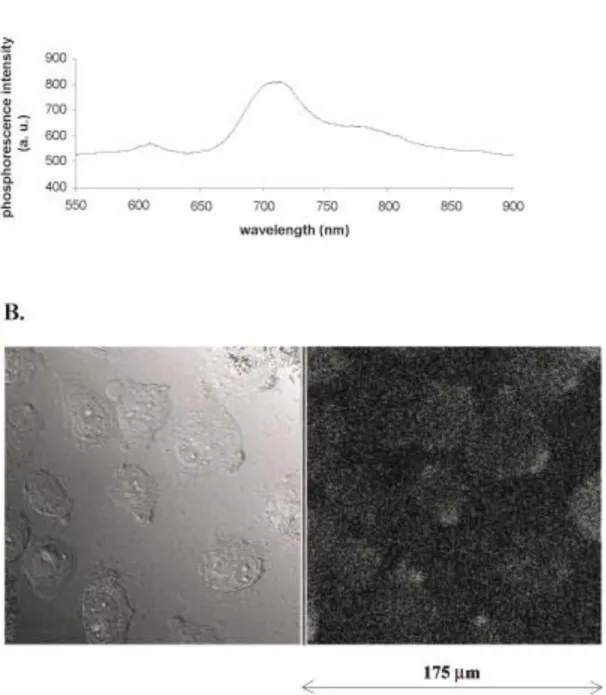
Intrinsic luminescent properties of the initial molecular phosphorescent probe are not affected by this treatment: emission spectra of PdTCPPNa4 in water solution, obtained with a 514nm laser beam, also shows a maximum emission peak at 710nm (Fig. 1A).
Similar to previously published investigations (13,14), the O2-dependence of the probe phosphorescence is described by the Sterm-Volmer relationship: 0/1+kq 0 PO2 where 0 and are the phosphorescence lifetimes (decay) in the absence of O2 and at a given PO2, respectively, and kq (the quenching constant) is a second order rate constant that is related to the frequency of collisions between O2 and the excited triplet state of PdTCPPNa4. An intracellular oxygen tension (PO2) decrease in the environment increases the phosphorescence intensity (I) and lengthens the phosphorescence decay () of PdTCPPNa4, and inversely. Here, I and were followed, as markers of the PO2 variations, in acellular solutions or in PdTCPPNa4–loaded endothelial cells.
Human Umbilical Venous Endothelial Cells (HUVEC).
HUVEC were used for several reasons. The endothelial cells have a glycolytic potential enabling them to stand hypoxia or short duration anoxia (15). Also, endothelial cells allow the investigation of three different mechanisms for inducing potential changes in PO2: i) decrease in extracellular oxygen (by argon bubbling), ii) use of uncouplers and inhibitors of the mitochondrial respiration, and iii) stimulation or blockade of eNOS.
HUVEC were obtained according to a protocole derived from the Jaffe's method and, for investigations of PO2 by fluorescence confocal microscopy, HUVEC were cultured on dishes whose bottom was a glass slide coatedwith 0.25mg.ml-1 fibronectin (16,17).
Determination of optimal active concentrations of KCN and DNP using oxygraphy technique. Respiration rate of HUVEC suspended in PSS (2ml, 5106 cells.mL-1) was measured polarographically in a stirred oxygraph vessel (37°C) including a Clark oxygen electrode. Different concentrations of the respiration inhibitor potassium cyanide (KCN) or of the uncoupler of mitochondrial phosphorylative oxidation 2.4-dinitrophenol (DNP) were then added and the effect on cellular O2 consumption was recorded.
Control of probe penetration in the cells.
Fluorescence images of adherent HUVEC were acquired by using a Zeiss LSM 410 confocal laser scanning microscope (Zeiss). Excitation: 514nm, dichroic 560nm, emission collection: 665nm long pass. Each image (optical sections passedthrough the nuclei) is the average of 4 successive image acquisition.
Intracellular Oxygen measurement in suspended HUVEC.
Sub-confluent adhering HUVEC were washed twice with PSS and trypsinized. Trypsinization was stopped by addition of PSS containing50% human serum. Cells were then centrifuged 5min at 200×g and resuspended in 6ml PSS with 0.45mg.ml-1 pluronic F-127, 5mg.ml-1 BSA added. Sixty microliters of a 10mM PdTCPPNa4 solution were added to a 6ml cell suspension (final concentrations: 100µM PdTCPPNa4) before a 60min incubation at 37°C, 5% CO2, humid atmosphere, in the dark. The cells werethen washed of the extracellular fluorescent probe by four centrifugation cycles (5min, 200×g) and pellet resuspension in 6ml PSS containing 4mM octanoate (106 cells·ml-1).
eNOS is activated using 0.1, 1 and 10µM acetylcholine (Ach) (18) or inhibited using 10µM N--nitro-L-arginine methyl ester (L-NAME), an analogue of the precursor L-Arginine which cannot be used by eNOS (19). L-NAME was added to the HUVEC suspension during PdTCPPNa4 incubation (60min). The cell suspension was then washed out of the probe and L-NAME by centrifugation and resuspension in PSS before measurement.
In addition, to evaluate the interaction between PdTCPPNa4 and the chemicals used in this study, the chemicals were added to a 10µM PdTCPPNa4 solution (without cells) during the phosphorescence
emission acquisition.
Two different parameters were measured, depending on the experimental condition: phosphorescence intensity (I) or phosphorescence decay (). and I were obtained using a pulsed water cooled continuous wave ArGa laser excitation (514nm) and a continuous laser beam (514nm), respectively, and the 90° phosphorescence emissionwas detected at 710nm with a AsGa photomultiplier.
Probe and Chemicals. Chemicals were obtained from Sigma except potassium cyanide (KCN) and 2,4 dinitrophenol (DNP) provided by Merck. PdTCPPNa4 is prepared as reported above.
Computer Analysis. Exponential decay curve analysis was performed by using the Origin 5.0 Microcal Origin Software. For suspended HUVEC intracellular origin measurements, phosphorescence acquisitionwas performed by using software writtenby the authors. The averaging of 100 successive exponential decays was used to measure .
RESULTS
Optimization of experimental conditions
Validation of the loading protocol
Figure 1B illustrates that PdTCPPNa4 enters human umbilical venous endothelial cells (HUVEC), as seen from the fluorescence confocal microscopy signal (optical slices passing through the nuclei). The weakness of the intracellular fluorescence is expected because of the low quantum yield of this probe. The strong photobleaching due to the repeated laser scanning of the whole microscopic field did not allow further sequential acquisition of images using the confocal microscopy technique. Consequently, all further experiments were carried out on HUVEC suspensions.
Optimal concentrations of KCN and 2.4-dinitrophenol for modulation of mitochondrial respiration in endothelial cells
On HUVEC suspension, using the oxygraphy technique, uncoupling by 100µM DNP stimulates the respiratory rate up to 2-fold within a few seconds, and 1mM KCN is an optimal concentration which induces almost an immediate 10-fold inhibition of respiration. However, 1mM KCN also induces a rapid morphological deterioration of cells observed in confocal microscopy. Since 100µM KCN does not induce cell morphological changes while still inhibiting the respiratory rate by 2-fold (data not shown), 100µM KCN and 100µM DNP have been used as respiration modulators in the following experiments.
Lack of interaction between the PdTCPPNa4 and the chemicals added
It has been verified that acetylcholine and L-NAME do not directly affect the optical properties of PdTCPPNa4 by direct molecular interaction in an acellular physiological solution (Fig. 2A and 2B). Besides, we have observed that the average values of the measured probe phosphorescence decays are different in physiological solution and in cells (about 6µsec and 90µsec, respectively). This can result from the different molecular environment around the probe. In accordance with the literature (12), we have observed, using bovine serum albumin (BSA) addition to PdTCPPNa4 solutions, that the presence of protein induces an increase in the probe luminescence. The same phenomenon likely
occurs when the probe is loaded in the protein-rich cytoplasm of the cell.
Experimental results
Intracellular PO2 variations during modification of extracellular oxygen tension
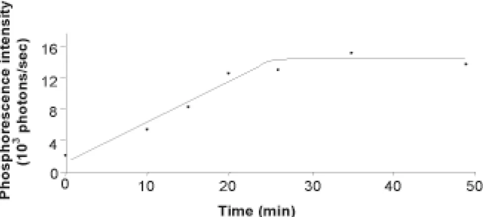
Argon bubbling (50 min) of a suspended HUVEC loaded with PdTCPPNa4 was used to deplete the extracellular medium of solubilized O2, and phosphorescence intensity was recorded. PdTCPPNa4 phosphorescence intensity increased by 7 fold, plateauing after 25min (Fig. 3). This indicates that PdTCPPNa4 strongly responds to the intracellular PO2 decrease induced by extracellular decrease in oxygen tension.
Mitochondrial respiration and PO2 variations
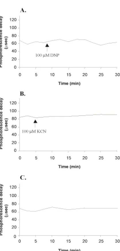
Under these conditions, the measurement of phosphorescence decay () avoids the problem of working with the very weak signal of PdTCPPNa4. As shown in figure 4, the addition of 100µM DNP or 100µM KCN does not modify the initial value of the phosphorescence decay () over 30min in PdTCPPNa4-loaded HUVEC, suggesting that mitochondrial respiration does not significantly influence PO2.
Non-respiratory oxygen consumption by eNOS and variations of PO2
Addition of 0.1, 1 and 10µM acetylcholine to HUVEC induce transient and dose-dependent increases
in (Fig. 5A, Fig. 6A, 6B, 6C), generally occurring after 2-5 minutes (up to 10min in a few
experiments), whereas no increase in is observed in the corresponding control experiments (Fig. 5B). These results suggest a significant reduction of the intracellular PO2 under acetylcholine stimulation. The average percentages of increase in amplitude of the phosphorescence decay peaks, as compared to baseline, are in the range of 4.2% (0.1µM Ach), 23% (1µM Ach) and 81.6% (10µM Ach) (Fig. 6D). A statistical analysis using a one-way ANOVA showed that the average percentages of increase in the amplitude of the phosphorescence decay peaks are significantly dependent upon the acetylcholine dose (P<0.05).
Figure 5C shows that there is no increase in in response to 1µM acetylcholine immediately after incubation with L-NAME. This tracing is comparable to the control (Fig. 5B). Nevertheless, 2 hours after the end of L-NAME incubation, after eNOS activity resumed, the acetylcholine-induced elevation –therefore PO2 decrease- is restored (Fig. 5D). The fact that no detectable modification of luminescence, following Ach addition on L-NAME-treated HUVEC, indicates a causal relationship between O2 consumption related to eNOS activation and the transient increase in . Moreover, the literature mentioned that a few porphyrins are sensitive to NO (20) or can be used as NO probe (21). Consequently, 10 and 100nmol NO (final concentrations: 3µM and 30µM, respectively) have successively been added to a PdTCPPNa4 solution (10 µM) in PSS, in order to verify that NO does not modify the value of corresponding to our probe (Fig. 2C). Therefore, these results depict actual PO2 modifications rather than NO level detection.
DISCUSSION
The present in vitro study validate a novel technique for measuring intracellular PO2 variations in acellular solutions as well as in living cells. Although, values of basal phosphorescent decay in acellular solutions (Fig. 2) and in living cells (Figs. 4 and 5) are different because the local environment surrounding the probe in the cell is likely to affect its response to PO2 variations. PdTCPPNa4 loaded into human umbilical venous endothelial cells (HUVEC) exhibits spectral properties similar to the well characterized phosphorescent oxygen probe Pd meso-tetra (4-carboxyphenyl) porphyrin (12). Moreover PdTCPPNa4 could be used for the measurement of intracellular oxygen tension (PO2) variations in living HUVEC, as shown by the intracellular response to extracellular PO2 decrease induced by argon bubbling (Fig. 3). The original loading protocol used in this study, in contrast with microinjection techniques which allows measurements in a few cells at a given time (11), enables the study of intracellular PO2 in a population of millions of cells. Furthermore, microscopy studies do not show any morphological cell damage due to PdTCPPNa4 loaded in HUVEC, even under focused light (Fig. 1B). In addition, the fact that low intracellular concentrations of PdTCPPNa4 were used is likely to keep the effect of any reactive O2 species produced, due to energy transfer from the porphyrin probe in its excited state to molecular oxygen, at a very low level.
Extracellular oxygen transport towards the inside of the cell occurs by diffusion according to the gradient of concentration and tends to maintain a constant PO2. Using data from the literature (3,4), theoretical maximum oxygen flow entering the cell by diffusion can be estimated in the range of 310-15 mol O2. sec-1.cell-1 and oxygen uptake measured by oxygraphy in HUVEC (37°C) has been found at a basal level, about 75 times lower: 410-17 mol O
2.sec-1.cell-1(8). Therefore, physiological modulations of mitochondrial respiration, which usually vary by a factor 2-4 times (8), should have no impact on intracellular PO2 , because O2 diffusion flow allows a O2 influx 20-50 fold higher. Thus, it is not surprising that, in these conditions, modulation of mitochondrial respiration using 100µM KCN or 100µM DNP, which respectively inhibits and stimulates respiratory function by 2-fold, does not measurably modify PO2 (Fig. 4).
Besides, oxygen consumption independent from respiration and resulting from activation of endothelial nitric oxide synthase (eNOS) has been evaluated. The use of a NO sensitive
microelectrode previously allowed to calculate NO concentration, at a distance of 50µm from the surface of rat aorta endothelial cell, in the range of 6.210-16 mol NO.sec-1.cell-1 (22). Therefore, since NO synthesis has a stoichiometry of 1/1 with regard to consumed O2, the basal production of NO by eNOS in rat aorta single endothelial cell requires the consumption of about 6.210-16 mol O2.sec-1.cell-1. Moreover, eNOS has been shown to be activated by endothelial cell stimulation with acetylcholine (Ach), leading to activation of NO release (23). In the endothelium from rabbit aorta, Ach increases NO extracellular level -therefore production- by 10 times (23) and probably more if intracellular NO degradation is taken into account. Therefore, the corresponding quantity of O2 transiently consumed by Ach-stimulated eNOS can be evaluated in the range of 10 times the basal consumption, i.e. 6.210-15 mol O2.sec-1.cell-1. This value is about 2 fold higher than the maximum rate of O2 diffusion through the cell membrane. Consequently, eNOS activation is a physiological mechanism which can induce a much stronger decrease in intracellular PO2 than modulation of respiration. Our experiments corroborate this calculation, since 0.1-10µM acetylcholine induce transient and dose-dependent increases in PdTCPPNa4-loaded cell phosphorescence decay, indicating a corresponding PO2 reduction (Fig. 5A and Fig. 6). On the contrary, no change in porphyrin phosphorescence was observed when cell respiration was increased or decreased by the use of DNP or KCN (Fig. 4). Also, as expected, no PO2 variation is detected in response to acetylcholine when eNOS is blocked by L-NAME (Fig. 5 C and D).
The present results suggest that other oxygen-related mechanisms of the cell physiology can now be further investigated using cell loading with PdTCPPNa4.
REFERENCES
1. Meyer, P. (1983). Physiologie humaine, pp. 1088-1099, Flammarion Medecine-Sciences, Paris. 2. Nathan, C., and Xie, Q. W. (1994). Nitric oxide synthases: roles, tolls, and controls. Cell 78, 915-8. 3. Clark, A., Jr., Clark, P. A., Connett, R. J., Gayeski, T. E., and C. R. Honig, C. R. (1987). How large is the drop in PO2 between cytosol and mitochondrion? Am. J. Physiol. 252, C583-7.
4. Hogan, M. C., Mathieu-Costello, O., Poole, D.C., and Wagner, P. D. (1994). Oxygen transport to tissue XVI. New York: Plemium Press, p. 22-23.
5. Kekonen, E. M., Jauhonen, V. P., and Hassinen, I. E. (1987). Oxygen and substrate dependence of hepatic cellular respiration: sinusoidal oxygen gradient and effects of ethanol in isolated perfused liver and hepatocytes. J. Cell Physiol. 133, 119-26.
6. Whalen, W. J. (1971). Intracellular PO2 in heart and skeletal muscle. Physiologist 14, 69-82.
7. Whalen, W. J., Riley, J., and Nair, P. (1967). A microelectrode for measuring intracellular PO2. J.
Appl. Physiol. 23, 798-801.
8. Steinlechner-Maran, R., Eberl, T., Kunc, M., Margreiter, R., and Gnaiger, E. (1996). Oxygen dependence of respiration in coupled and uncoupled endothelial cells. Am. J. Physiol. 271, C2053-61. 9. Buerk, D. G., Tsai, A. G., Intaglietta, M., and Johnson, P. C. (1998). Comparing tissue PO2 measurements by recessed microelectrode and phosphorescence quenching. Adv. Exp. Med. Biol. 454, 367-74.
10. Richmond, K. N., Shonat, R. D., Lynch, R. M., and Johnson, P. C. (1999). Critical PO(2) of skeletal muscle in vivo. Am. J. Physiol. 277, H1831-40.
11. Hogan, M. C. (1999). Phosphorescence quenching method for measurement of intracellular PO2 in isolated skeletal muscle fibers. J. Appl. Physiol. 86, 720-4.
12. Lo, L. W., Koch, C. J., and Wilson, D. F. (1996). Calibration of oxygen-dependent quenching of the phosphorescence of Pd-meso-tetra (4-carboxyphenyl) porphyrin: a phosphor with general application for measuring oxygen concentration in biological systems. Anal. Biochem. 236, 153-60. 13. Vanderkooi, J. M., Maniara, G., Green, T. J., and Wilson, D. F. (1987). An optical method for measurement of dioxygen concentration based upon quenching of phosphorescence. J. Biol. Chem. 262, 5476-82.
phosphorescence quenching. Adv. Exp. Med. Biol. 361, 61-6.
15. Buderus, S., Siegmund, B., Spahr, R., Krutzfeldt, A., and Piper, H. M. (1989). Resistance of endothelial cells to anoxia-reoxygenation in isolated guinea pig hearts. Am. J. Physiol. 257, H488-93. 16. Jaffe, E. A., Nachman, R. L., Becker, C. G., and Minick, C. R. (1973). Culture of human endothelial cells derived from umbilical veins. Identification by morphologic and immunologic criteria. J. Clin. Invest. 52, 2745-56.
17. Faury, G., Usson, Y., Robert-Nicoud, M., Robert, L. and Verdetti, J. (1998). Nuclear and cytoplasmic free calcium level changes induced by elastin peptides in human endothelial cells. Proc.
Natl. Acad. Sci. USA. 95, 2967-2972.
18. Lüscher, T.F., and Vanhoutte, P. M. (1990). The endothelium: modulator of cardiovascular function. Boca Raton: CRC Press.
19. Rees, D. D., Palmer, R. M. J., Schulz, R., Hodson, H. F., and Moncada, S. (1990). Characterization of three inhibitors of endothelial nitric oxide synthase in vitro and in vivo. Br. J. Pharmacol. 101, 746-752.
20. Pawlowski, M., and Wilson, D. F. (1994). Imaging oxygen pressure in tissue in vivo by phosphorescence decay. Adv. Exp. Med. Biol. 361, 83-91.
21. Clark, H. A., Barker, S. L. R., Brasuel, M., Miller, M. T., Monson, E., Parus, S., Shi, Z. Y., Song, A., Thorsrud, B., Kopelman, R., Ade, A., Meixner, W., Athey, B., Hoyer, M., Hill, D., Lightle, R., and Phylbert, M.A. (1998). Subcellular optochemical nanobiosensors: probbes encapsulated by biologically localised embedding (PEBBLEs). Sensors and Actuators. B 51, 12-16.
22. Brovkovych, V., Dobrucki, L. W., Brovkovych, S., Dobrucki, I., Do Nascimento, C.A., Burewicz, A., and Malinski., T. (1999). Nitric oxide release from normal and dysfunctional endothelium. J.
Physiol. Pharmacol. 50, 575-86.
23. Christie, M. I., Griffith, T. M., and Lewis, M. J. (1989). A comparison of basal and agonist-stimulated release of endothelium-derived relaxing factor from different arteries. Br. J.
FIGURE LEGENDS
Figure 1. Emission spectrum of the water soluble PdTCPPNa4 illuminated by a 514 nm laser beam (A.). Adherent HUVEC loaded with PdTCPPNa4 observed by confocal laser scanning microscopy in differential interferential contrast (left) and fluorescence (right) (B.). The images presented are representative of two experiments.
Figure 2. Absence of interaction of NO, Ach and L-NAME on the luminescence properties of PdTCPPNa4 (10µM) in PSS. (A.) Effects of 1µM acetylcholine or 10µM L-NAME on the 10µM porphyrin solution. (B.) Effects of 1µM acetylcholine and 10µM L-NAME on the 10µM porphyrin solution and corresponding control tracing. (C.) Sensitivity of the porphyrin probe (10µM) to successive additions of 10 and 100nmol NO. Arrows: product addition. A representative tracing is shown (n=2 in each case).
Figure 3. Effect of argon bubbling on the phosphorescence intensity of PdTCPPNa4-loaded HUVEC suspension. The argon bubbling starts at time 0min.
Figure 4. Effect of 100µM DNP (A.) and 100µM KCN (B.) on the phosphorescence decay of 100µM PdTCPPNa4-loaded HUVEC suspension. A control tracing is shown in C. A representative tracing is shown (n=2 in each case).
Figure 5. 1µM acetylcholine (Ach) induces a transient lengthening of phosphorescence decay () on 100µM PdTCPPNa4-loaded HUVEC (A.). Control tracing (B.). Effect, on 100µM PdTCPPNa4-loaded HUVEC, of 1µM Ach immediately after incubation with 10µM L-NAME (C.) and 2 hours after the end of L-NAME incubation (D.). A representative tracing is shown in each case (A: n=3 ; B,C,D: n=2).
Figure 6. Dose-effect of acetylcholine (Ach) on the phosphorescence decay () of 100µM PdTCPPNa4-loaded HUVEC. The amplitude of the phosphorescence decay peak induced by Ach addition increases with Ach concentration: 0.1 µM (A), 1 µM (B) and 10 µM (C). A representative tracing is shown in each case (A: n=5 ; B and C: n=4). The arrows indicate the time of Ach addition (A, B and C). The average percentages of increase in amplitude of the phosphorescence decay peaks induced by Ach, as compared to baseline, were calculated for each Ach dose using the results from all experiments, and represented in D.