HAL Id: tel-01426033
https://tel.archives-ouvertes.fr/tel-01426033
Submitted on 4 Jan 2017HAL is a multi-disciplinary open access
archive for the deposit and dissemination of sci-entific research documents, whether they are pub-lished or not. The documents may come from teaching and research institutions in France or abroad, or from public or private research centers.
L’archive ouverte pluridisciplinaire HAL, est destinée au dépôt et à la diffusion de documents scientifiques de niveau recherche, publiés ou non, émanant des établissements d’enseignement et de recherche français ou étrangers, des laboratoires publics ou privés.
Study of Exon Junction Complex in mouse neural stem
cells
Rahul Kumar Mishra
To cite this version:
Rahul Kumar Mishra. Study of Exon Junction Complex in mouse neural stem cells. Neurons and Cog-nition [q-bio.NC]. Université Pierre et Marie Curie - Paris VI, 2016. English. �NNT : 2016PA066201�. �tel-01426033�
! !
Université Pierre et Marie Curie
Ecole doctorale 515 - Complexité du vivant
Institut de Biologie de l’ENS – CNRS UMR8197, INSERM U1024 Equipe “Expression of eukaryotic mRNAs”
STUDY OF EXON JUNCTION COMPLEX
IN MOUSE NEURAL STEM CELLS.
Par M. RAHUL KUMAR MISHRA
Thèse de doctorat de :
BIOCHIMIE ET BIOLOGIE MOLECULAIRE DE LA CELLULE
Dirigée par Dr. Hervé Le Hir
Présentée et soutenue publiquement le 9 septembre 2016 Devant un jury composé de :
Dr. Alain Trembleau Président du Jury Dr. Florence Rage Rapporteur Dr. Alexandre Benmerah Rapporteur Dr. Hervé Moine Examinateur Dr. Nathalie Spassky Membre invité Dr. Hervé Le Hir Directeur de thèse
Resume
The Exon Junction Complex (EJC) plays a central role in coupling post‐ transcriptional processes in metazoans. This multi‐protein complex is assembled onto messengers RNAs (mRNAs) by the splicing machinery. Organized around a core complex serving as a platform for numerous factors, EJCs accompany mRNAs to the cytoplasm and is involved in mRNA transport, translation and stability. The physiological importance of the EJC is supported by observations associating defects in EJC component expression to developmental defects and human genetic disorders. Transcriptomic studies revealing the non‐ubiquitous deposition of EJCs strengthened the hypothesis that EJCs could participate to gene expression regulation. However, despite a precise picture of the structure of the EJC, functional links between EJC assembly and regulation of specific transcripts under physiological conditions is yet to be established.
During this thesis, I studied the expression of eIF4A3, Y14 and MLN51 three core proteins of the EJC in primary cultures of mouse neural stem cells (NSCs). NSCs can be differentiated into multiciliated ependymal cells that line all brain ventricles and have important physiological functions in brain development. We observed by immunofluorescence that in quiescent NSCs, all three proteins are concentrated in the vicinity of the centrosome at the base of the primary cilia. This localization reflects the presence of fully assembled EJCs as proved by the study of Y14 mutant that prevent EJC core mounting. Remarkably, the intense signals of EJC core proteins in the centrosomal region totally disappeared when quiescent cells are committed toward proliferation or differentiation. The presence of EJCs around centrosome is not restricted to NSCs as identical cellular localizations were observed in quiescent mouse embryonic fibroblasts (MEF) and IMCD3 cell lines. Immunopurification of EJC‐bound transcripts residing in the cytoplasm of NSCs coupled to large‐scale sequencing revealed that EJCs are enriched onto several transcripts encoding proteins linked to proliferation and differentiation.
Given that cytoplasmic EJCs mark mRNAs that have not yet experienced translation, our data plead for the presence of numerous untranslated transcripts around the centrosome in quiescent cells. The complete disappearance of centrosomal EJCs when cells enter the cell‐cycle or into differentiation most likely reflects the translation of EJC‐bound transcripts. Together, our results reveal a massive EJC‐associated spatio‐temporal program of post‐transcriptional gene regulation around the division apparatus of quiescent cells concomitant with cellular decisions for proliferation or differentiation.
2
TABLE OF CONTENTS
INTRODUCTION 1.0: mRNPs ...4 2.0: EJC ...5 2.1: The EJC core components ...7 2.1.1: eIF4A3 ...7 2.1.2: MAGOH‐Y14 …7 2.1.3: MLN51 ...8 2.2: The tetrameric EJC core …9 2.3: EJC assembly …11 2.3.1: The pre‐EJC core …12 2.3.2: The recruitment of eIF4A3 by CWC22 …13 2.4: EJC peripheral factors …14 2.4.1: Splicing‐related peripheral factors …14 2.4.2: Export factors …15 2.4.3: NMD related factors …16 2.4.4: Other EJC related factors …17 3.0: EJC life cycle …18 3.1: The localization of EJC core components …18 3.2: EJC remodeling and variability …19 3.2.1: EJC disassembly …20 3.2.2: Re‐cycling of EJCs to the nucleus …21 4.0: EJC functions …22 4.1: EJC modulates splicing …23 4.1.1: EJC dependent regulation of splicing in human cells …24 4.2: EJC enhances translation ...25 4.2.1: EJC enhances translation in mTOR pathway ...26 4.2.2: Translation enhancement by MLN51 ...27 4.3: EJC in quality control of mRNAs ...28 4.3.1: The importance of NMD ...28 4.3.2: NMD components ...29 4.3.3: NMD Mechanism ...29 4.3.4: Role of EJC in NMD ...31 4.3.5: Role of EJC in translation control of natural NMD targets ...32 4.4: EJC participates to mRNA export ...33 4.4.1: General mRNA export adaptors and receptors ...33 4.4.2: Role of EJC in mRNA transport ...34 5.0: Global view of EJC deposition on mRNAs ...34 5.1: Differential EJC loading on human transcriptome ...35 5.2: The canonical and non‐canonical EJC ...36 6.0: mRNA localization and local translation ...40
6.1: mRNA localization ...40 6.2: Importance of mRNA localization ...41 6.2.1: Energy efficiency for the cell ...41 6.2.2: Spatio‐temporal translation ...41 6.2.3: Storage ...42 6.2.4: One transcript, multi‐functional protein ...42 6.3: Examples of localized mRNAs ...43 6.4: Mechanism of mRNA localization ...45 6.4.1: Directed transport of mRNA in cytoplasm: Cis‐regulatory elements and trans‐acting factors ...46 6.4.2: Directed transport of mRNA in cytoplasm: Recruitment of molecular motors ...46 6.4.3: Role of EJC in sub‐cellular localization ...47 7.0: EJC in physiological contexts ...50 7.1: EJC in development ...50 7.2: EJC in mammalian brain development ...51 7.3: EJC in human diseases ...53 8.0: Development of mammalian Brain ...54 8.1: Primary progenitors ...54 8.2: Intermediate progenitors ...56 8.3: Role of NSC in brain development ...57 8.4: Ependymal cells ...59 8.4.1: Physiological Functions of ependymal cells in brain …59 8.4.1.1: Regulation of neuronal niche ...61 8.4.1.2: CSF maintenance ...62 8.4.1.3: Metabolic protection of brain ...62 8.4.1.4: Protection of brain from infections ...63 8.4.1.5: Repair of brain after stroke ...63 8.4.2: Ependymal differentiation in mammalian brain ...64 8.4.3: Centriole amplification in ependymal differentiation ...66 9.0: Cell‐cycle and cell‐quiescence ...68 10.0: Centrosome ...70 10.1: Centrosome Composition ...70 10.2: Centrosome duplication in cycling cells ...72 10.3: Centrosome in post‐mitotic cells ...74 10.3.1: Formation of primary cilia ...74 10.3.1.1: Cilia structure and functions ...76 10.3.1.2: Primary cilia in neurodevelopmental disorders …77 10.3.2: Centrosome amplification in post‐mitotic cells ...78 10.4: Centrosome functions ...79 10.4.1: Centrosome functions in cell fate ...80 10.4.2: Centrosomes in human diseases ...81
1 11.0: Questions Asked ...82 RESULTS (Part 1): Article ...84 RESULTS (Part 2): Additional results ...124 1.0: Identification of EJC‐bound mRNAs in NSC ...125 1.1: EJC RIP‐seq strategy ...126 1.2: Cytoplasmic fractionation of NSC ...126 1.3: Tests of immuno‐depletion ...128 1.4: EJC‐RIP and RNA purification ...129 1.5: Analysis of sequencing results ...130 1.6: Statistical validation of RIPseq ...131 1.7: Identity of EJC bound transcripts in NSC ...133 2.0: Visualization of expression of mRNAs by smFISH ... 139 2.1: smFISH model ... 139 2.2: Design and Synthesis of Fluorescent Oligonucleotide Probe Sets ... 140 2.2.1. Design ... 141 2.3: Results obtained ... 142 2.3.1: smFISH in cycling MEF ... 142 2.3.2: smFISH in quiescent NSC ... 143 2.3.3: Distribution of total mRNAs in quiescent NSC ... 145 2.3.4: smFISH in quiescent MEF ... 146 DISCUSSION AND PERSPECTIVES 1.0: Resume of our results ... 149 2.0: Presence of mRNAs at centrosome in variety of cell types ... 150 3.0: Accumulation of untranslated mRNAs at centrosome ... 151 3.1: Hypothesis 1: Translation dependent disassembly of EJCs ... 152 3.2: Hypothesis 2: Degradation or diffusion of EJC bound mRNAs ... 155 4.0: Functions of untranslated mRNAs at centrosome ... 158 MATERIALS AND METHODS ... 161 1.0: CELL CULTURE ... 162 1.1: Neural Stem Cell culture ... 162 1.2: Mouse Embryonic Fibroblast culture ... 162 2.0: Transfection of plasmids ... 162 3.0: Immunofluorescence and Microscopy ... 163 3.1: Image analysis. ... 164 4.0: PROTEIN ANALYSIS ... 164 4.1: Protein extraction ... 164 4.2: Immunoprecipitation ... 164 4.3: SDS‐PAGE ... 165 4.4: Western Blot Analysis ... 165
5: RNA analysis ... 165 5.1: Cellular fractionation ... 165 5.2: RNA Immuno Precipitation ... 166 5.3: RNA isolation ... 166 5.3.1: Isolation of immunoprecipitated RNA ... 166 5.3.1.1: RNA precipitation ... 166 5.3.2: Total RNA isolation ... 167 5.4: Quantitative RT‐PCR ... 167 6.0: smFISH analysis ... 168 6.1: Probe synthesis ... 168 6.2: Hybridization of probes with FlapY‐Cy3 ... 168 6.3: Cell fixation ... 168 6.4: In situ hybridization ... 169 7.0: List of buffers ... 173 REFERENCES ... 174
3
INTRODUCTION
1.0: mRNPs
Messenger RNAs (mRNA) do not exist "naked" in vivo, they are coated and compacted by RNA‐binding proteins (RBPs) forming messenger ribonucleoprotein (mRNPs). These mRNPs are the functional units of gene regulation as they control the central affairs of gene expression. From the very first step of transcription, RBPs mediate co‐transcriptional 5’‐end capping, splicing, and editing; 3’‐end cleavage and polyadenylation; and quality control of mRNAs within nascent mRNP complexes (Gerstberger et al. 2014; Singh et al. 2015; Müller‐McNicoll & Neugebauer 2013). The recent development of large‐ scale approaches including mass spectrometry and next‐generation sequencing allow to analyze mRNP organization and architecture at transcriptome‐wide level. CLIP technology (Crosslinking and Immunoprecipitation) coupled with high throughput sequencing has yielded instant interactions between proteins and RNA up to single‐nucleotide resolution (König et al. 2011; Ascano et al. 2012). To date, more than 1500 proteins have been characterized as RBPs (Castello et al. 2012; Baltz et al. 2012; Gerstberger et al. 2014). Among these RBPs, some play a role in mRNA packaging, such as complexes binding to the methylated 5’ cap or proteins that recognize the polyA tail located at the 3’‐end of mature RNA. Also, during the splicing, the mRNA is bound by exon and intron definition complexes, and splicing snRNPs (Wahl et al. 2009; Bentley 2005). Following intron excision, the spliceosome also leaves several marks on mRNA including exon junction complexes (EJC) and SR proteins to assemble mature mRNPs ready for nuclear export. mRNPs undergo remodeling as they cross the nuclear pore to achieve unidirectional export into the cytoplasm (Singh et al. 2015)). By the virtue of RBPs, some mRNPs are transported to specific regions of subcellular localization (Cody et al. 2013; Martin & Ephrussi 2009; Lécuyer et al. 2007; Medioni et al. 2012). Cytoplasmic mRNPs undergo structural rearrangements in order for translation to occur. Ultimately, most RBPs that were associated with the mRNA during processing and export are replaced by ribosomes on actively translated mRNAs or by mRNA degradation machinery.
5
These processes are tightly regulated in time and space to ensure accurate and coordinated functioning.
Numerous RBPs, including EJC proteins, SR, Cap Binding Proteins and PolyA Binding Proteins are involved in virtually every step of mRNA biogenesis and metabolism. Understanding their precise contribution or function during each step will serve to understand the output of gene expression at all steps of the mRNA life cycle. To date, a major challenge in this field is to determine how myriads of RBPs coordinate in order to modulate every aspect of mRNP life cycle; more importantly, it is also to understand what differentiates individual mRNP despite the fact that they are made of common RBPs.
2.0: EJC
Since the beginning of 1990s, several evidences revealed that introns and their excision mRNA precursors enhance gene expression. This outcome originates from an effect of splicing on mRNA transport, translation and degradation based on mRNA length and position (Matsumoto et al. 1998; Luo & Reed 1999; Hentze & Kulozik 1999). It is the study of Nonsense Mediated mRNA Decay (NMD) that strongly suggested that nuclear splicing can have an impact onto post‐translational decay of mRNAs. NMD is a quality control process that degrades mRNAs carrying premature translation termination codons (PTC) in order to prevent the synthesis of truncated proteins potentially harmful for the cell (Kervestin & Jacobson 2012). The mechanism of discrimination of PTCs from normal stop codons has been a central question for a long time (Maquat 1995; Brogna et al. 2016). Studies on PTC containing transcripts revealed that in mammals, a PTC triggers efficient mRNA degradation when positioned more than 50 nt upstream of an intron (Cheng et al. 1994; Carter et al. 1996; Thermann et al. 1998; Zhang et al. 1998). This result explained the functional link between splicing and NMD and generated the idea that the nuclear splicing machinery leaves a ‘molecular signature’ at exon junctions that accompanies the mRNAs until they are translated (Maquat 1995; Hentze & Kulozik 1999; Shyu & Wilkinson 2000). The hypothesis that presence of this mark downstream of a
7-(1!,(>()!H(+2>!-8.99%8!@PJ!D.-7!1%8D%,-26!H.-$!-$%!)(-.()!-$5-!-$%!05j(8.-6!(D! )(8052! 7-(1! ,(>()7! 58%! 2(,5-%>! .)! -$%! 257-! %'()! 3@596! g! P5F+5-! "]]\4<! _.(,$%0.,52!,$585,-%8.;5-.()!(D!-$.7!712.,.)9:8%25-%>!058I!2%>!-(!-$%!>.7,(C%86!(D! 5! 0+2-.18(-%.)! ,(012%'E! )50%>! -$%! %'()! j+),-.()! ,(012%'! 3&*/4<! &*/! .7! 5! 0+2-.18(-%.)! ,(012%'! >%1(7.-%>! ()! 0?@A! %'()! j+),-.()7! 57! 5! ,()7%F+%),%! (D! 712.,.)9!7-?N$:"& <=<!&*/!=.)>7!-$%!0?@A!5-!5!,()7%8C%>!1(7.-.()!VM!)+,2%(-.>%7! +17-8%50!(D!-$%!712.,%>!j+),-.()E!.)!5!7%F+%),%!.)>%1%)>%)-!05))%8!3Q%!G.8!%-!52<! VWWW4E! 5)>! 7%8C%7! 57! 5! =.)>.)9! 125-D(80! D(8! 7%C%852! 0?@A! 18(,%77.)9! D5,-(87<! #$.7!,(012%'!.7!577%0=2%>!.)!-$%!)+,2%+7E!-85C%27!H.-$!0?@A7!-(!-$%!,6-(12570E! 5)>! .)D2+%),%7! .)! -$%! 0%5)-.0%! 0?@A! ,%22+258! 2(,52.;5-.()E! -85)725-.()! 5)>! 7-5=.2.-6! -$8(+9$! @PJ<! L.)5226E! 522! &*/7! 58%! 8%0(C%>! D8(0! -85)7,8.1-7! 5D-%8! ()%! 8(+)>! (D! -85)725-.()! 3J(7-.%! g! J8%6D+77! VWWV4<! #$+7! &*/! ).,%26! %'%012.D.%7! -$%! .015,-! (D! )+,2%58! $.7-(86! ()! 0?@A! 2.D%,6,2%<! G%8%! H%! >.7,+77! -$%! ,(01()%)-7! 5)>! D+),-.()7! (D! &*/! .)! 8%25-.()! H.-$! C58.(+7! 1(7-:-85)7,8.1-.()52! 18(,%77%7E! .),2+>.)9!712.,.)9E!-85)71(8-E!-85)725-.()!5)>!@PJ<! ! ! ! !
7
Figure 1: Scheme showing the splicing mediated assembly of EJC and its cytoplasmic role in mRNA fate.
2.1: The EJC core components
The EJC core is composed of four proteins: eIF4A3 (eukaryotic initiation factor 4A3), MAGOH, Y14 (also known as RNA‐binding motif 8A) and MLN51 (metastatic lymph node 51; also known as Barentsz or CASC3) (Ballut et al. 2005; Tange et al. 2005). Throughout the mRNA life cycle between the nucleus and the cytoplasm, this hetero‐tetramer remains stably bound to mRNA and serves as a binding platform for many factors that will become temporary partners, and allow EJCs to interact with various cellular machineries. Following, I will present in more details the individual EJC core proteins and their properties.2.1.1: eIF4A3
eIF4A3, also known as DDX48, is a DEAD‐box RNA helicase composed of 411 amino‐acids. DEAD box proteins are involved in an assortment of metabolic processes as ATP‐dependent RNA remodeling enzymes. Helicases of these families contain two globular domains: RecA1 and RecA2 (Figure 2: A). RecA domains contain seven highly conserved motifs (I, Ia and II, III, IV, V and VI) that are involved in RNA binding and ATP hydrolysis (Linder & Jankowsky 2011). Human eIF4A3 shares 70% sequence similarity with its mammalian orthologs eIF4A1 and eIF4A2. However, it is functionally distinct from these eIF4A counterparts of the same family. While eIF4A1 and eIF4A2 are bonafide translation initiation factors, eIF4A3 does not play a direct role in translation (Li et al. 1999). In contrast to eIF4A3, eIF4A1 and eIF4A2 are cytoplasmic proteins that are not incorporated into EJC. The most documented function of eIF4A3 concerns its EJC‐related roles. However, one study proposed that eIF4A3 is also involved in ribosomal RNA processing (Alexandrov et al. 2011).2.1.2: MAGOHY14
MAGOH and Y14 are two small proteins consisting of 146 and 173 amino acids respectively. These two proteins form a tight heterodimer (Fribourg et al. 2003). The evolutionary patterns of MAGOH and Y14 protein families show that both proteins slowly co‐evolved in eukaryotes, demonstrating the requirement of their heterodimerization to maintain their function (Gong et al. 2014). The abolition of their interaction has a functional effect on the metabolism of mRNA, mainly for their location and stability (Hachet & Ephrussi 2001; Fribourg et al. 2003; Tange et al. 2004). The crystallographic analysis of the heterodimer from Drosophila revealed that Y14 and MAGOH interact via highly conserved hydrophobic surfaces, supporting that the two proteins act as a single structural and functional unit (Fribourg et al. 2003) (Figure 2: B). The central core of MAGOH contains a broad β sheet surrounded by three α helices. The two largest α helices form a hydrophobic interaction with Y14 by occupying the RNA binding domain (RBD). This interaction completely inhibits the ability of RBD to interact with RNA and stabilizes the heterodimer (Lau et al. 2003). The wide β sheet of MAGOH then provides a surface for interaction with other proteins, including the other EJC core partners. In mammals, two MAGOH paralogues exist (MAGOH and MAGOHB) with almost identical amino acid sequence. Both MAGOH and MAGOHB are efficiently incorporated into EJCs (Singh et al. 2013). Their individual contribution in the EJC and downstream events of EJC mediated regulations of mRNA is not distinguishable.
2.1.3: MLN51
Metastatic Lymph Node 51 (MLN51) also known as Barentsz (Btz) and CASC3 (Cancer Susceptibility Candidate gene 3) is a protein composed of 703 amino acid residues. This protein is conserved in vertebrates and invertebrates showing 41% and 48% similarity respectively with C. elegans and D. melanogaster homologs. The characterization of the protein by bioinformatics analysis enabled the identification of several modules and motifs in the protein
9
(Degot et al. 2004; Degot et al. 2002). Schematically, the MLN51 protein can be divided into two equal halves. The carboxy‐terminal region of MLN51 is rich in proline and contains a nuclear export signal (NES). The amino‐terminal region of MLN51 protein contains three special domains: A coiled‐coil‐type structural motif; two sequences of nuclear localization signal (NLS); and a module called SELOR (speckle localizer and RNA‐binding module) which is the most conserved portion of the protein among metazoan species. Human SELOR shares 100% similarity in mice, 92% in Xenopus and 66% similarity in Drosophila (Degot et al. 2004). In vitro MLN51 binds to RNA via SELOR domain at a precise location corresponding to EJC binding site (Degot et al. 2004). This domain is shown to be necessary and sufficient for the formation of the EJC core (Degot et al. 2004; Ballut et al. 2005). Independently of its EJC‐dependent roles, MLN51 is involved in the assembly of cytoplasmic stress granules (Baguet et al. 2007). Stress granules are bodies located in the cytoplasm corresponding to mRNA storage compartments in which translationally repressed mRNAs are stored following cell stress.
2.2: The tetrameric EJC core
The in vitro reconstitution of the EJC core with purified recombinant proteins and then, the crystal structure of this recombinant complex by X‐ray diffraction revealed the peculiar mode of RNA binding (Ballut et al. 2005; Andersen et al. 2006; Bono et al. 2006) (Figure 2: C). The EJC core is built around the RNA helicase eIF4A3. The two RecA domains of eIF4A3 move relatively freely with respect to each other without ATP, but they adopt a closed conformation in the presence of ATP allowing the binding to RNA. The characteristic and conserved RNA helicase motifs of eIF4A3 make contacts exclusively with the sugar‐phosphate backbone explaining how it binds 6 nucleotides of RNA in a sequence‐independent manner. This RNA binding is lost when the helicase hydrolyses ATP leading to an open conformation (Ballut et al. 2005). Interestingly, the MAGOH‐Y14 heterodimer simultaneously binds the two domains of eIF4A3, preventing conformational changes upon ATP hydrolysis hence locking eIF4A3 in a closed conformation on the RNA (Andersen et al. 2006;
_()(!%-!52<!VWW^e!@.%27%)!%-!52<!VWW]4<!#$%!-H(!0(7-!,()7%8C%>!8%9.()7!(D!PQ@R"! 7+88(+)>!-$%!-H(!>(05.)7!(D!%KLMAN!5)>!7-5=.2.;%!-$%!,(012%'<!K)!-$%!&*/!,(8%E! PQ@R"!527(!=.)>7!-(!PAUBG!5)>!,()-5,-7!()%!)+,2%(-.>%!(D!?@A<!#$+7!5!7-5=2%! -%-850%8.,! ,(8%! .7! 577%0=2%>! ()! 0?@A! .)! H$.,$! %KLMAN! 5)>! PQ@R"! -(+,$! -$%! ?@A<!! ! -?N$:"& D>& 0;:$@;$:"& FT& +M.& @F:"& @F%QFG"G;#& 9GE& 9##F@?9;"E& T9@;F:#4& (>& #H(! ?%,A! >(05.)7! (D! %KLMAN! 58%! 7$(H)! .)! -$%! $%-%8(>.0%8! H.-$! $+05)! 712.,.)9! D5,-(8! /T/VV! 3,(012%'%>! H.-$! /&L"! VV4<! )>& #$%! PAUBGrO"M! $%-%8(>.0%8! .7! 7$(H)! .)! 5! -8.0%8.,! ,(012%'! H.-$! YOP! 3158-)%87! (D! O"M! 5)>! PAUBG4! .)! -0%,%23!)+#.()+"%*+,/(0<!.>& #$%& -%-850%8.,!,(8%!(D!-$%!&*/!.7!7$(H)!,(018.7.)9! -$%! ?@A! $%2.,57%! %KLMANE! PAUBGE! O"M! 5)>! PQ@R"<#$%! PAUBGrO"M! $%-%8(>.0%8!I%%17!-$%!-H(!>(05.)7!(D!%KLMAN!.)!5!,2(7%>!,()D(805-.()<&#$%!-H(! 0(7-! ,()7%8C%>! 8%9.()7! (D! PQ@R"! ,()-5,-! -$%! -H(! >(05.)7! (D! %KLMAN! 5)>! D+8-$%8!7-5=.2.;%!-$%!,(012%'<!3A>51-%>!D8(0!Q%!G.8!%-!52<E!VW"^4<!
11
2.3: EJC assembly
The EJC core is not a preassembled complex that joins mRNAs after their maturation. In fact, the EJC core components are brought together by the splicing machinery. Spliceosomes are highly dynamic multi‐megadalton RNA‐protein molecular machines that assemble de novo for each splicing event (Figure 3). The main spliceosome assembly process entails the stepwise recruitment of RNA‐protein subunits called small nuclear (sn) RNPs U1, U2, U4, U5 and U6 and a multitude of other proteins. In humans, it has been estimated that more than 300 proteins participate to the splicing reaction. The biochemical characterization of splicing reaction sub‐divided it into five successive complexes named as E, A, B, B* and C. Each complex is defined by the presence of different snRNPs and their cofactors. Transition to the following complex is caused by a series of rearrangements and remodelling (Figure 3), (Hoskins & Moore 2012; Will & Lührmann 2011; Wahl et al. 2009). The U1, U2, U4, U5 and U6 snRNPs are the main building blocks of these complexes. During canonical cross‐intron assembly, U1 snRNP recognizes the 5′ splice site (complex E) and U2 snRNP replaces early‐binding SF1 protein at the branch point sequence (BPS), forming complex A. Subsequently, the U4, U5, and U6 snRNPs join as a pre‐formed tri‐ snRNP, giving rise to complex B, which is still inactive. This complex is activated by consecutive release of U1 and U4 to sequentially form the B* and C complexes, in which the first and second trans‐esterifications steps take place leading to intron excision and exon ligation (Will & Lührmann 2011). Following, we discuss the step‐wise assembly of EJC core during these successive steps of splicing.
Figure 3: Stepwise assembly of the spliceosome onto premRNA. Cross‐ intron assembly and disassembly cycle of the major spliceosome. The stepwise interaction of the spliceosomal snRNPs (colored circles), but not non‐ snRNPproteins, in the removal of an intron from a pre‐mRNA containing two exons (blue) is depicted. (Adapted from Wahl et al. 2009)
2.3.1: The preEJC core
The pre‐EJC core can be defined as containing three factors MAGOH, Y14 and eIF4A3. Biochemical analyses of the protein contents of splicing complexes assembled in vitro revealed that MAGOH, Y14 and eIF4A3 are already present in the B complex and become more stably associated in the C complex before exon ligation (Bessonov et al. 2008; Reichert et al. 2002; Makarov 2002; Merz et al. 2007; Zhang & Krainer 2007). These proteins separately contact the regions of exons in a stable manner where they would be assembled as ‘pre‐EJC core’ after the exon ligation (Reichert et al. 2002; Merz et al. 2007; Gehring, Lamprinaki, Hentze, et al. 2009). Since MLN51 was not detected in the splicing complex, it is likely that it joins and stabilizes the pre‐EJC core concomitantly with the active release of mRNA from spliceosome (Ballut et al. 2005; Bessonov et al. 2008; Reichert et al. 2002; Zhang & Krainer 2007; Gehring, Lamprinaki, Hentze, et al. 2009).13
2.3.2: The recruitment of eIF4A3 by CWC22
The loading of EJC by spliceosome‐mediated remodeling of mRNPs has recently been highlighted by the discovery of how eIF4A3 is escorted to the spliceosomes. Like other RNA helicases, eIF4A3 does not possess any apparent sequence specificity. Therefore, it was quite intriguing to see how eIF4A3 is recruited to spliceosome, at the right time, to a specific position on mRNA. The biochemical characterization of eIF4A3 helicase demonstrated that it is not required for constitutive pre‐mRNA splicing (Zhang & Krainer 2007; Shibuya et al. 2004). In fact, the recruitment of eIF4A3 to the spliceosome is ensured by the splicing factor CWC22 (complexed with CEF1 22; also named NCM or Nucampholin in Drosophila melanogaster) with which it forms a stable complex (Alexandrov et al. 2012; Barbosa et al. 2012; Steckelberg et al. 2012). CWC22 is a core splicing factor composed of two conserved domains, MIF4G (middle domain of eIF4G) and MA3. A direct and stable interaction of the MIF4G with RecA‐2 domain of eIF4A3 maintains eIF4A3 in an open conformation (Figure 2: A), in which RNA‐ and ATP‐ binding sites are disjoined, hence preventing eIF4A3 binding to both RNA and MAGOH–Y14 (Barbosa et al. 2012; Buchwald et al. 2013). By blocking the RNA binding activity of eIF4A3, CWC22 most likely shields eIF4A3 from binding to inappropriate RNA molecules before its incorporation into the EJC. Once bound, CWC22 ensures eIF4A3 recruitment to active spliceosomes and its integration into EJC core (Alexandrov, Colognori, Shu & J. a Steitz 2012; Barbosa et al. 2012; Steckelberg et al. 2012). CWC22 and eIF4A3 co‐exist in large protein complexes that also contain Prp19 (precursor mRNA (pre‐mRNA)‐processing factor 19 (PRP19) (Barbosa et al. 2012), a splicing factor that is essential for building active spliceosomes (Makarov 2002; Wahl et al. 2009).
The role of introns in formation of EJC core is highlighted by their association of the RNA helicase IBP160 (intron‐binding protein of 160 kDa; also known as Aquarius) (De et al. 2015; Ideue et al. 2007). IBP160 exhibits structural adaptations at the centre of the intron‐binding complex (IBC) that binds to pre‐ mRNAs in association with the U2 snRNP upstream of splicing branch points and
is also necessary for EJC loading. However, the interconnections between eIF4A3–CWC22, PRP19 and IBP160, and the interpretation of their incorporation into spliceosomes, remain unclear. After escorting it to the spliceosome, CWC22 must leave eIF4A3 to allow RNA clamping (Barbosa et al. 2012; Steckelberg et al. 2012; Buchwald et al. 2013). However, it is still unclear when CWC22 does dissociate from the complex. It is also unrecognized how pre‐ EJC core is assembled onto spliced mRNA. MAGOH‐Y14 and eIF4A3‐CWC22 are recruited to the spliceosomes independently (Gehring, Lamprinaki, Hentze, et al. 2009; Shibuya et al. 2004; Barbosa et al. 2012) and we do not know if MAGOH‐ Y14 requires specific partners to enter spliceosome. How EJC loading occurs at 24 nucleotides upstream of exon junctions is also an enigma. It is possible that spatial organization of protein‐RNA interactions during splicing may impose some constraints at exon junctions that lead to deposit EJC at a precise distance.
Following the splicing reaction, MLN51 seals the EJC core most likely concomitantly with mRNP release and spliceosome disassembly to form the complete EJC core (Ballut et al. 2005; Zhang & Krainer 2007; Gehring, et al. 2009). So far, there is no evidence that alternative versions of the EJC core exist but we can suppose that EJCs devoid of MLN51 could exist (Le Hir et al. 2016).
2.4: EJC peripheral factors
Once locked on mRNA, the EJC core accompanies mRNAs from nucleus to cytoplasm until translation. The EJC serves as binding platform for more than a dozen of proteins called EJC peripheral factors (Tange et al. 2004; Le Hir et al. 2016). The composition of EJCs during this period changes dynamically as many partners join and leave intermittently. In vitro reconstitution of the complex and validation of EJC assembly by in vitro and in vivo studies have helped to initiate a catalogue of EJC peripheral factors (Figure 3).2.4.1: Splicingrelated peripheral factors
The first peripheral factors to bind to EJC core are most likely the ones known to be present in the spliceosome before exon ligation. This includes
15
splicing factors Acinus, PININ, RNA‐binding protein with Ser‐rich domain 1 (RNPS1) and SAP18. RNPS1 and SAP18 are splicing activators (Schwerk et al. 2003; Singh et al. 2010; Sakashita et al. 2004; Blencowe et al. 1998) and ACINUS and PININ are scaffold proteins that use a similar conserved motif to bind to RNPS1 (Murachelli et al. 2012). The alternative interaction with ACINUS or PININ give SAP18 the ability to form two distinct ternary complexes, named ASAP (apoptosis and splicing‐ associated protein) and PSAP (contains PININ, SAP18 and RNPS1) (Schwerk et al. 2003; Murachelli et al. 2012). ACINUS has three alternatively spliced isoforms, termed Acinus‐L, Acinus‐S and Acinus‐S’ (Sahara et al. 1999). All three isoforms are capable of forming a ternary complex (Schwerk et al. 2003). Therefore, ACINUS must be involved in different complexes. To date, we do not know how these trimeric complexes are attached to the EJC core.
2.4.2: Export factors
Among the list of EJC peripheral factors are also conserved mRNA export factors UAP56 (also known as DDX39B), Aly/REF export factor (ALYREF; also known as THOC4), the nuclear export factor 1 (NXF1) and NTF2‐related export protein (NXT1). UAP56 is an RNA helicase that facilitates the recruitment of ALYREF to processed mRNAs. Both ALYREF and UAP56 are also part of the TREX (transcription export) complex that bridges pre‐mRNA synthesis and export (Tange et al. 2005; Merz et al. 2007; Katahira 2012). NXF1 and NXT1 form a heterodimer and are considered as the general receptor responsible for mRNP export to the cytoplasm (Köhler & Hurt 2007). NXF1‐NXT1 interact with mRNAs via several adaptors (including the EJC) on one side and on the other side, directly interact with the nuclear pore complex (NPC)(Köhler & Hurt 2007). Since UAP56 is weakly associated with purified EJCs (Tange et al. 2005; Reichert et al. 2002; Merz et al. 2007), it is possible that its quick release is necessary for the subsequent recruitment of NXF1 and NXT1 by ALYREF and/or by RNPS1 (Le Hir et al. 2001; Lykke‐Andersen et al. 2001) (Figure 3).
2.4.3: NMD related factors
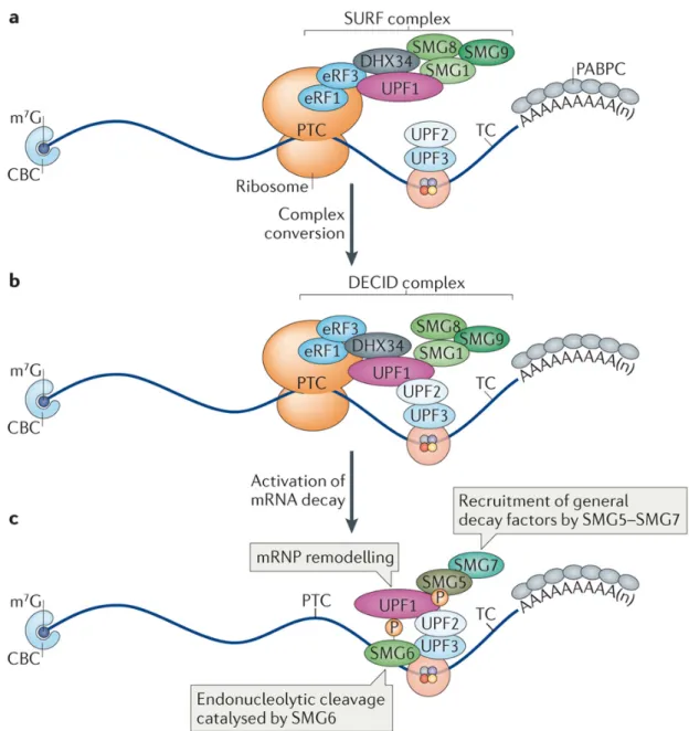
Key factors of NMD are also part of the list of EJC peripheral factors (Figure 3) including UPF3A (up‐frameshift 3A; also known as RENT3A), UPF3B, UPF2, UPF1 and SMG6 (also known as EST1A). The study of EJC composition in both nuclear and cytoplasmic compartments revealed the successive interaction of these factors (Le Hir et al. 2001a). UPF3A and UPF3B are mainly nuclear and bind to nuclear EJCs. Within EJC core, eIF4A3, MAGOH and Y14 together form a composite binding site for the EJC‐binding motif (EBM) of UPF3B (Gehring et al. 2003; Buchwald et al. 2010; Chamieh et al. 2008). UPF2 joins the EJCs in cytoplasm by direct binding to UPF3B while UPF1 joins the complex only in the context of NMD (H Le Hir et al. 2001; Chamieh et al. 2008b; Kashima et al. 2006) . Thus, three UPF proteins form a trimeric complex, in which UPF2 bridges UPF1 and UPF3B (Chamieh et al. 2008). The endonuclease SMG6 interacts with the exon junction complex via two conserved EBMs (Kashima et al. 2010). EBMs may serve as a place of contest for different proteins to hook mRNAs via EJC. Thus EJC occupies a centre stage in events involved in mRNP remodelling.
! "Z! ! ! ! -?N$:"&U>&*?#;&FT&+M.&Q":?QO":9A&T9@;F:#<!3A>51-%>!D8(0!Q%!G.8!%-!52<!VW"^4<! !
D4C4C>&,;O":&+M.&:"A9;"E&T9@;F:#&
&
A158-!D8(0!-$%!=()5D.>%!2.7-!(D!1%8.1$%852!&*/!D5,-(87E!7(0%!(-$%8!18(-%.)7! 58%!527(!8%25-%>!-(!&*/!=+-!-$%.8!D+),-.()52!8(2%!57!5!158-!(D!&*/!.7!)(-!C52.>5-%><! S?0"^W! 3S?:8%25-%>! )+,2%58! 05-8.'! 18(-%.)! (D! "^W! IJ54! .7! 5! ,(5,-.C5-(8! (D! 712.,.)9!-$5-!$57!=%%)!18(1(7%>!-(!%)$5),%!0?@A!Nd!%)>!18(,%77.)9!3Q%!G.8!%-!52<! VWWWe! _2%),(H%! %-! 52<! "]]\4<! G(H%C%8E! .-7! 18%C.(+726! ,()7.>%8%>! 8(2%! 57! 5)! &*/! 18(-%.)! $57! )(-! =%%)! D+226! C52.>5-%><! #$.7! .7! 527(! -8+%! D(8! ShA?! 3S^! I.)57%! "! 3S^h"4!AQO?&L:2.I%e!527(!I)(H)!57!YBQJKYN43P5!%-!52<!VWW\4!H$.,$!.7!5!158-!(D! -$%! 0#B?"rS^h":7.9)522.)9! 15-$H56! -$5-! 1(7.-.C%26! 8%9+25-%7! -85)725-.()! 3L()7%,5!%-!52<!VW"M4<!P5)6!18(-%.)7!(D!S?!D50.26!527(!577(,.5-%!H.-$!&*/!3S.)9$! %-!52<!VW"Ve!S5+2.u8%!%-!52<!VW"V4!=+-!-$%8%!.7!)(!%7-5=2.7$0%)-!(D!-$%.8!(895).;%>! %'.7-%),%! 57! 9%)+.)%! &*/! D5,-(8<! K)-%8%7-.)926E! -$%! 0(7-! 5=+)>5)-! S?! 18(-%.)7!bound to EJCs are SR splicing factor 1 (SRSF1), SRSF3 and SRSF7, which are known to continuously shuttle between the nucleus and the cytoplasm (Long & Caceres 2009). By physically interacting, EJCs and SR proteins could constitute the major driving force for mRNP compaction, which might be necessary for proper mRNP transport and translation (Singh et al. 2015; Singh et al. 2012).
3.0: EJC life cycle
3.1: The localization of EJC core components
Different studies, using diverse strategies, showed that EJCs colocalize with protein components of the spliceosome at nuclear speckles in the nucleoplasm (Custódio et al. 2004; Schmidt et al. 2006). Nuclear speckles, also named “SC35 domains” or “splicing factor compartments,” are nuclear punctuate structures functioning as storage/assembly/modification compartments that supply splicing factors to active transcription sites (Spector & Lamond 2011). In vivo, the four components of the EJC core shuttle between the nucleus and the cytoplasm. Three of the core components MAGOH, Y14, and eIF4A3 are mainly nuclear (Ferraiuolo et al. 2004; Kataoka et al. 2000; Hervé Le Hir et al. 2001; Palacios et al. 2004; Daguenet et al. 2012). In contrast, MLN51 is predominantly cytoplasmic (Degot et al. 2002; Macchi et al. 2003; Daguenet et al. 2012), despite having two nuclear localization domains. The nuclear export signal (NES) in its C‐terminal is responsible for its export from nucleus to cytoplasm and predominant cytoplasmic localization. The export of MLN51 was functionally validated by blocking the crm‐1 nuclear export pathway using inhibitory drugs, which leads to accumulation of MLN51 in nucleoplasm (Daguenet et al. 2012). These four proteins come together to form an EJC core at the periphery of nuclear speckles: a region named “perispeckles”. By using FRET (Fluorescence Resonance Energy Transfer) assays and EJC assembly mutants, Daguenet et al. showed that perispeckles do not store free subunits, but instead are enriched for fully assembled EJC core. These results supported the model that perispeckles are apparently the major assembly sites for EJCs on transcripts. The occurance of perispeckles close to the sites of active RNA Pol‐II also gives strong reasoning
19
that these are the nucleoplasmic locations for co‐transcriptional splicing and hence EJC assembly on mRNAs.
3.2: EJC remodeling and variability
Once mRNAs are properly packed with RBPs in form of mRNPs, they are exported to the cytoplasm. At a moment that is not perfectly defined, mRNP are largely remodeled and see important modification of their protein composition. Many of the nuclear mRNP components are replaced with cytoplasmic ones. One of the important remodeling steps before translation is the replacement of nuclear cap‐binding complex (CBC) with the translation initiation factor eIF4E and exchange of nuclear poly(A)‐ binding protein (PABPN) with its cytoplasmic counterpart poly(A)‐binding protein (PABPC), respectively (Singh et al. 2015). While most mRNPs get engaged into translation, some of them are transported to the specific sites of translation (see section 6.3 – mRNA localization). Following this model, the composition of EJC does not remain fixed during the export and major EJC rearrangements occur. Since ACINUS and PININ are restricted to the nucleus, they must detach from EJCs before export. In the absence of these scaffold proteins (Murachelli et al. 2012), RNPS1 and SAP18 could be stabilized by other peripheral factors, such as UPF3, which can interact with RNPS1 (Gehring et al. 2005; Singh et al. 2007; Lykke‐Andersen et al. 2001), or potentially other SR proteins. Several mutually exclusive interactions in the EJC support the notion of EJC variability even if there is no clear evidence for it. For example, UPF3b and SMG6 compete for the same binding site of the EJC, with UPF3b having a higher affinity for the EJC. This suggests that UPF3b must dissociate from the EJC to make way for SMG6 to bind (Buchwald et al. 2010; Kashima et al. 2010). Similarly ACINUS and PININ cannot be a part of the same complex as they also compete through a similar binding motif to join RNPS1 (Murachelli et al. 2012). However, the coordination of these changes and their functional implications remain unexplored.
3.2.1: EJC disassembly
Initial studies showed that EJC are associated with mRNAs still bound to the nuclear CBC, but not with eIF4E protein suggesting that EJCs are removed during the first round of translation (Lejeune et al. 2002). The study was subsequently complemented with the finding that EJC does not associate with the translating mRNAs and the first ribosome eliminates EJCs contained in the 5'‐UTR region and also in the mRNA ORF (Dostie & Dreyfuss 2002). The observations that translation was necessary and sufficient to dissociate EJCs from mRNA, both in vivo and in vitro, established that EJCs are a mark of untranslated mRNPs. Later, a cytoplasmic RNA‐binding protein was identified and proposed as dissociation factor of the EJC, PYM (Partner of Y14 and MAGOH) (Bono et al. 2004; Diem et al. 2007). Structural and biochemical characterization of the PYM and Y14‐MAGOH ternary complex (Bono et al. 2004) accompanied by other biochemical investigations (Gehring et al. 2009; Bono et al. 2006; Chamieh et al. 2008a) unveiled that PYM disrupts the EJC assembly and/or stability by stably binding to MAGOH‐Y14 at a position that clashes with its binding to eIF4A3. This results in a change in conformation to the open state of eIF4A3 leading to release of mature EJCs from mRNAs. In vitro, PYM dissociates specifically the fully assembled EJCs from spliced mRNA but does not inhibit the EJC assembly (Gehring et al. 2009). By doing so, it also inhibits the NMD. In cells, overexpression of PYM leads to a decreased association of EJCs with spliced mRNAs, while PYM inhibition by RNAi leads to an accumulation of EJC components in cytoplasmic cell fractions. The study points out the important phenomenon that PYM inhibition leads to accumulation of EJCs in the cytoplasmic fraction of cells, indicating the importance of PYM in availing free EJCs in the nucleus. Interestingly, PYM function is regulated by its association with ribosomes. Biochemical analysis revealed that the C‐terminal of PYM interacts with the 40S subunit of ribosome in 48S pre‐initiation complex (Diem et al. 2007). The ribosome mediated disassembly mechanism makes sure that EJCs are not disrupted from the mRNAs until they have fulfilled their functions (e.g., as marker for NMD). However, in D. melanogaster, PYM maintains the
21
homeostasis of not only endogenous mRNA, but also has effects on non‐ translatable reporter, suggesting a ribosome‐independent mode of action (Ghosh et al. 2014). Also in human cells free PYM can disassemble EJCs located outside of the ORFs (Gehring et al. 2009). This confirms a general function of PYM as a self‐sufficient EJC disassembly factor. To date, a direct and active role of PYM in EJC dissociation during translation remains to be proved. We can suppose that PYM prevents the reassembly of the EJC core by binding MAGOH‐Y14 early after EJC dissociation. In contrast to MAGOH and Y14, we cannot exclude that eIF4A3 and MLN51 that can bind together outside the EJC core, re‐bind mRNAs after the first round of translation to further enhance next rounds of translation (Chazal et al. 2013).
3.2.2: Recycling of EJCs to the nucleus
EJC proteins are expressed in a quantity that is not sufficient to be present onto all exons in the cell. The quantification of endogenous proteins by Western blotting in cell extracts shows a large difference between expression level of the EJC proteins and the number of exon junctions: 12000 eIF4A3 and 40000 MAGOH‐Y14 against over 400,000 exon‐exon junctions (Gehring et. al 2009). Therefore, the recycling of EJC from cytoplasm to nucleus must be an effective phenomenon. The mechanism of recycling of Magoh‐Y14 is well understood. In this process, the Importin 13 (IMP13) plays an essential role. This nuclear receptor of karyopherin family imports specifically the MAGOH‐Y14 dimer into the nucleus in a RanGTP dependent manner (Mingot et al. 2001). Binding of Importin 13 to MAGOH‐Y14 dimer is at the interaction site of the PYM protein showing a mutually exclusive binding (Bono et al. 2010). Similarly, MAGOH‐Y14 is sterically inaccessible to IMP13 when in the EJC (Bono et al., 2010). Thus, IMP13 binds to MAGOH‐Y14 after the disassembly of the EJC core by PYM and transports it to the nucleus. Once in the nucleus, binding of one molecule of GTP to the Ran GTPase‐linked leads to the dissociation of Importin 13 of its substrate, allowing the release of MAGOH‐Y14 in the nucleus and its incorporation in new EJCs (Figure 4 (Cook & Conti 2010)). However, the mechanisms of recycling and import of eIF4A3 and MLN51 to the nucleus are still unknown to this day.
!
! !
-?N$:"& C>& & IFE"A& FT& +M.& :"@S@A?GN>! AD-%8! -$%! %'1(8-! (D! 0?@A! .)-(! ,6-(12570E! YOP! 3(85)9%4! .7! .)C(2C%>! .)! -$%! >.77(,.5-.()! (D! &*/7! .)! -$%! ,6-(12570! =6! 3.4! .)-%85,-.)9! H.-$! -$%! 70522! 8.=(7(052! 7+=+).-! C.5! .-7! /:-%80.)+7! 5)>! 3..4! .)-%85,-.)9!H.-$!PAUBG:O"M!C.5!.-7!@:!-%80.)52<!#$%!&*/!,(8%!.7!-$+7!>.77(,.5-%>! .)-(! 5! YOP:PAUBG:O"M! -8.0%8! 5)>! %KLMANE! PQ@R"! 18(-%.)7! 52()%! (8! .)! ,(0=.)5-.()<! PAUBG:O"M! .7! -$%)! j(.)%>! =6! K01(8-.)! "N! .)! -$%! ,6-(12570! 5)>! .01(8-%>! =5,I! -(! -$%! )+,2%+7! -$8(+9$! -$%! )+,2%58! 1(8%7<! #$%! ,589(! .7! -$%)! >.7,$589%>! =6! -$%! 588.C52! (D! -$%! ?5)! U#Y57%! 3A>51-%>! D8(0! _()(! 5)>! U%$8.)9! VW""4<! ! !
C4L>&+M.&T$G@;?FG#&
&
#$%! &*/! .7! 5! >6)50.,! ,(012%'! H$(7%! ,(01(7.-.()! ,$5)9%7! (C%8! 0?@A! -85C%2! .)! -$%! ,%22<! #$%! ,515,.-6! (D! -$%! &*/! -(! 8%,8+.-! >.C%87%! 1%8.1$%852! D5,-(87! 985)-7! .-! -$%! 5=.2.-6! -(! ,((8>.)5-%! 0+2-.12%! 18(,%77%7! 8%25-%>! -(! 05-+85-.()! 5)>! %'18%77.()! (D! -85)7,8.1-7<! _6! C58.(+7! 0%,$5).707E! -$%! &*/! ,5)! 5DD%,-! 18%:0?@A! 712.,.)9!57!H%22!57!0?@A!-85)71(8-E!-85)725-.()!5)>!0?@A!7+8C%.225),%!=6!@PJ! 3-?N$:"& 54<! #$%! .01(8-5),%! (D! &*/:D+),-.()7! .)! 8%9+25-.()! (D! ,%22! 1$67.(2(96! .7!23
highlighted by recent discoveries about EJC‐related developmental defects and diseases. We will discuss here particular examples related to different roles of the EJC, which make this ‘RBP’ a central element of the mRNA fate.
Figure 5: The function of the exon junction complex (EJC) in splicing and translation. a| EJCs deposited after the splicing of flanking introns can facilitate (+) the splicing of a neighbouring weak intron. The activation may be promoted by the EJC peripheral factors ACINUS and RNPS1. b| EJCs deposited during co‐ transcriptional splicing can slow down Pol II elongation rate allowing more time for correct splicing. In the absence of the EJC, the increased Pol II speed will cause more exon skipping. c| Role of EJC in translation activation in mTOR signaling. EJC core protein MLN51 binds to eIF3 directly. The recruitment by MLN51 of eIF3 as part of the 43S PIC enhances translation initiation. (Adapted from Le Hir et al. 2016)
4.1: EJC modulates splicing
The EJC core is assembled on transcripts by the splicing machinery. Most molecular processes that depend on the EJC are post‐splicing events. However, recent studies revealed that EJC can affect pre‐mRNA splicing itself. A genetic screen for mutations affecting photoreceptor in fly showed that mutation and knock‐down of EJC core components (eIF4A3, MAGOH and Y14) causes skipping of several exons of mapk pre‐mRNA containing long introns (Ashton‐Beaucage et al. 2010; Roignant & Treisman 2010). The specific skipping of exons in long intron containing pre‐mRNAs illustrates the downstream application of EJCloading on neighboring exon junctions (Ashton‐Beaucage & Therrien 2011). Similarly, the nuclear EJC is required for splicing of intron4 of piwi transcript (Hayashi et al. 2014a; Malone et al. 2014). Knockdown of any of Y14, RNPS1 or ACINUS caused intron retention of weak intron 4 of piwi transcripts in 100% cases. In this case, the splicing of flanking exons plays role in enhancing the splicing of this weak intron. EJC core and the accessory factors RNPS1 and ACINUS deposited in close proximity aid in definition and efficient splicing of neighboring weak introns (Figure 5a). However, whether the fully assembled EJC or only EJC components were necessary is unknown. The enhancer function of EJC could also be through effectively recruiting SR proteins to enhance exon definition, or through interaction with snRNPs to efficiently recognize neighboring weaker intron. Eventhough intron‐retention and exon‐skipping are both EJC dependent, EJC‐dependent retained introns are generally not very long compared with those found in the previous studies (Ashton‐Beaucage et al. 2010; Roignant & Treisman 2010).This indicates that exon skipping of long intron‐ containing transcripts and intron retention involve different mechanisms that have not yet been characterized (Hayashi et al. 2014a).
The EJC also affects pre‐mRNA splicing in other organisms. In Xenopus laevis, eIF4A3 is required for proper splicing of ryanodine receptor (ryr1) pre‐ mRNA (Haremaki & Weinstein 2012). In the absence of expression of eIF4A3, the expression of the RYR1 protein is drastically reduced due to a defect in splicing of its transcript. These data suggest that eIF4A3, potentially through the EJC, binds to ryr‐1 pre‐messenger and causes the retention of one or more introns.
4.1.1: EJC dependent regulation of splicing in human cells
In human cells, EJC components (eIF4A3, Y14, Acinus, SAP18 and RNPS1), rather than the assembled complex function as regulators of splicing of apoptosis regulator BCL‐X gene (also known as BCL21L1) pre‐mRNA (Michelle et al. 2012). Recently, transcriptome‐wide analysis from the lab revealed that the impact of EJC on alternative splicing is broader than previously expected (Wang et al. 2014). The reduction of EJC causes large numbers of splicing changes for variety of transcripts and for several of them the fully assembled EJC core is required for25
this effect. Interestingly, several constitutive exons were excluded in EJC depleted cells suggesting that EJCs contribute to the recognition of normal splicing events. The mechanism of splicing regulation in human cells can be different than that of Drosophila cells as most of the EJC dependent splicing changes are not dependent on ACINUS or SR proteins. Interestingly, RNA polymerase II (Pol II) transcription accelerates when the EJC proteins amount is reduced suggesting that some splicing changes can be attributed to the faster transcription rate. Variation of transcription elongation rates can change the time available for the recognition of competitive splice sites and thus can regulate alternative splicing (Kornblihtt et al. 2013). By slowing down the transcription elongation rate, EJC may allow more time for splicing factors to recognize alternative exons. How EJCs communicate with the transcription machinery is still a matter of investigation. However, we cannot exclude that EJCs in human also serve in some cases as direct splicing regulators as observed in drosophila. The mechanisms underlying the effect of EJC on specific splicing events may also rely on other direct or indirect factors, which are still unknown.
4.2: EJC enhances translation
The influence of splicing on protein synthesis is a phenomenon that has been observed in most organisms. Following the discovery of the EJC, which marks the spliced exon junctions, it was a matter of curiosity whether the increased expression of transcripts was related to splicing per se or to the presence of an EJC. Early studies showed that splicing can increase the expression of intron‐containing genes compared to their intron‐less counter parts, both at the mRNA and protein levels (Nott et al. 2003; Callis et al. 1987). Then, studies of different reporters producing spliced mRNAs associated or not to EJCs allowed to attribute to EJCs a positive effect on translation independently of effects onto expression level (Wiegand et al. 2003; Nott et al. 2004). One study notably showed that EJCs increase the proportion of mRNAs associated to polysomes (Nott et al. 2004). Although EJCs are not part of translation machinery and are removed in the very first round of translation, it is interesting that EJCs offer a selective advantage to mRNAs that have never experienced translation. This effect is most likely important to reduce the time window between geneactivation at the transcriptional level and mRNA translation producing the final product (Nott et al. 2004). The molecular mechanisms linking the EJC and translation machinery are not clearly elucidated. However, few studies have attempted to clarify the role of the complex in the translation.
4.2.1: EJC enhances translation in mTOR pathway
The first mechanistic insights into EJC role in translation activation is linked to the mTOR pathway (Ma et al. 2008). mTOR is a stress‐sensing signaling pathway which enhances translation via kinase S6K1 to promote cell growth. eIF4A3‐bound SKAR on mRNAs serves to recruit activated S6K1 to the CBC‐ mRNP, leading to phosphorylation of ribosomal proteins and translation initiation factors, thus activating translation initiation in mTOR signaling events (Figure 6). As EJC is loaded only on newly formed mRNAs but not on older templates, associated SKAR communicates with translation machinery as a signal of newly turned‐on gene. Thus mTOR/6K1 promptly targets the translation of freshly formed transcripts in order to reduce the time lag between transcription and translation in stress conditions. However, most EJC‐interactome studies could not identify SKAR as an EJC peripheral factor (Tange et al. 2005; Merz et al. 2007; Singh et al. 2012). This suggests that interaction between SKAR and EJC can be indirect or specific to stress conditions. Alternatively, the EJC‐SKAR synergy might be limited to specific transcripts only, making it difficult to detect in general.