S Y M P O S I U M : L E G G - C A L V E´ -PERTHES DISEASE: WHERE DO WE STAND AFTER 100 YEARS?
Joint-preserving Surgery Improves Pain, Range of Motion,
and Abductor Strength After Legg-Calve´-Perthes Disease
Christoph Emanuel Albers MD,Simon Damian Steppacher MD, Reinhold Ganz MD, Klaus Arno Siebenrock MD, Moritz Tannast MD
Published online: 13 April 2012
Ó The Association of Bone and Joint Surgeons1 2012
Abstract
Background Patients after Legg-Calve´-Perthes disease (LCPD) often develop pain, impaired ROM, abductor weakness, and progression of osteoarthritis (OA) in early adulthood. Based on intraoperative observations during surgical hip dislocation, we established an algorithm for more detailed characterization of the underlying patho-morphologies with a proposed joint-preserving surgical treatment.
Questions/purposes We asked if patients after LCPD treated with our algorithm experienced (1) reduced pain; (2) improved hip function; and/or (3) prevention of OA progression; we then determined (4) the intraoperative damage patterns; (5) the survival of the hip; and (6) factors predicting the need for a conversion to THA; radiographic progression of OA; a Merle d’Aubigne´-Postel score below 15 at last followup; and/or the need for revision surgery. Methods We retrospectively reviewed 53 patients after LCPD who underwent joint-preserving surgery (40 surgical
hip dislocations, eight acetabular osteotomies, four com-bined procedures, and one intertrochanteric osteotomy). We obtained Merle d’Aubigne´-Postel scores to assess pain; OA was assessed using To¨nnis grades. Survival and pre-dictive factors were calculated with the univariate Cox regression. Fifty of the 53 patients were evaluated at a minimum of 5.1 years (mean, 8.2 years; range, 5.1– 12.8 years).
Results Pain and hip function improved at followup from a median of 4 points to 5 points. The mean increase in To¨nnis grades at last followup was 0.3 to 0.8. The survival of surgery at 5 years was 86%; 13 factors related to survival.
Conclusion Patients with symptoms resulting from path-omorphologic deformities after LCPD benefit from joint-preserving surgery with specific treatment of individual structural abnormalities.
Level of Evidence Level IV, therapeutic study. See the Guidelines for Authors for a complete description of levels of evidence.
Introduction
Reasonable clinical and radiographic long-term results can be expected in 40% to 77% of all patients after Legg-Calve´-Perthes disease (LCPD) without the need for THA [8,12,30, 47]. However, in the remaining cases, the patients often develop symptoms in early adulthood including pain, impaired ROM, problems with ambulation, and progressive osteoarthritis (OA) [7,22,48]. Several pathomorphologies seemingly cause these problems: intraarticular femoroace-tabular impingement (FAI) resulting from the aspherical femoral head [7, 35], extraarticular impingement of the greater and the lesser trochanter [17, 31], functional Each author certifies that he or she, or a member of their immediate
family, has no commercial associations (eg, consultancies, stock ownership, equity interest, patent/licensing arrangements, etc) that might pose a conflict of interest in connection with the submitted article.
All ICMJE Conflict of Interest Forms for authors and Clinical Orthopaedics and Related Research editors and board members are on file with the publication and can be viewed on request. Each author certifies that his or her institution approved the human protocol for this investigation, that all investigations were conducted in conformity with ethical principles of research, and that informed consent for participation in the study was obtained.
C. E. Albers, S. D. Steppacher, R. Ganz,
K. A. Siebenrock, M. Tannast (&)
Department of Orthopedic Surgery, Inselspital, University of Bern, Freiburgstrasse, 3010 Bern, Switzerland e-mail: moritz.tannast@insel.ch
Clin Orthop Relat Res (2012) 470:2450–2461
DOI 10.1007/s11999-012-2345-0
and Related Research
®
retrotorsion of the femoral head [18], acetabular retroversion [9,19], associated acetabular dysplasia [14,15,37], and/or joint incongruency [37].
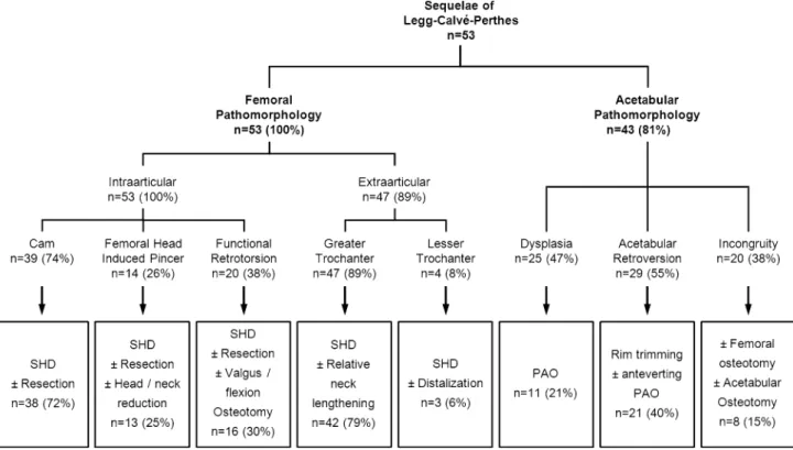
This variety of structural abnormalities can result in different pathomechanical problems that can be present in the same hip. It is crucial to address each problem appro-priately [7, 11] to improve pain, ROM, and abductor weakness. Isolated treatments of a single mechanical problem do not improve pain or limp [13, 29]. Based on descriptions in the literature [7,9,14,15, 17–19, 31,35, 37] and previous preliminary reports on the evaluation of hips after LCPD from our institution [7,11], we developed a treatment algorithm for these complex hips (Fig.1). This algorithm offers a structured way to identify the underlying pathomechanical problem and offers a surgical treatment strategy to correct these structural abnormalities. This algorithm arose based on our intraoperative observations during surgical hip dislocation (SHD) [7] of these hips. This technique has opened the field for novel surgical treatment options for the sequelae of LCPD [11]. However, it is unclear whether treatment according to this algorithm has the potential to relieve pain, restore hip function, or stop progression of OA in hips with previous LCPD.
We therefore asked (1) if patients with LCPD after joint-preserving surgery had relief of pain; (2) improved hip function; and/or (3) and prevention of progressive OA; we then determined (4) the survival of the surgery; (5) the
intraoperative damage patterns; and (6) factors predicting the need for a conversion to THA; the presence of radio-graphic progression of OA; a Merle d’Aubigne´-Postel [24] score below 15 at last followup; and/or the need for revi-sion surgery.
Patients and Methods
We retrospectively reviewed all 73 patients (73 hips) with LCPD in their history presenting with hip pain at our outpatient clinic between October 1997 and August 2005. All patients were evaluated for possible joint-preserving surgery. We excluded 20 patients (20 hips [27%]) with advanced OA (Grade 2 or greater according to To¨nnis [43]) who underwent primary THA. This left 53 patients (53 hips) with no or minor radiographic hip OA (Grade 1 or less) [43] who underwent subsequent joint-preserving sur-gery (Table1). The indications for surgery were: (1) pain; (2) impaired hip function (limp, positive anterior and posterior impingement test, impaired abductor strength); (3) decreased ROM; (4) abductor weakness; and (5) early degenerative changes. The contraindication was advanced OA [ Grade 1 according to To¨nnis. During the study time we treated all patients with joint preservation surgery if they had no advanced OA and met these indications. Using the classification of Stulberg et al. [37], there were three
Fig. 1 The morphologic analysis with the corresponding surgical treatment algorithm of hips with pathomorphologic sequelae of
hips (5%) with Class I, two hips (3%) with Class II, 32 hips (61%) with Class III, and 16 hips (31%) with Class IV. There were no hips with Class V. In 30 hips (57%), pre-vious surgery was performed including 12 hips (23%) with femoral osteotomies, 12 hips (23%) with pelvic osteoto-mies, and in six hips (11%) with other surgical procedures (Table1). All patients were invited to return for a mini-mum followup of 5 years. Three patients (three hips [6%]) were not available for followup. Of those, one patient (one hip [2%]) died from a cause unrelated to surgery without revision. The other two patients (two hips [4%]) were lost to followup. The remaining 50 patients (50 hips [94%]) were evaluated at the outpatient clinic with a minimum followup of 5.1 years (mean, 8.2 ± 2.1 years; range, 5.1–12.8 years). The study was approved by the local Institutional Review Board.
The preoperative clinical evaluation included the patient history, assessment of ambulation, abductor strength (M0-M5) [23], anterior and posterior impingement test [42], and full goniometric ROM. As a clinical scoring system, the Merle d’Aubigne´ and Postel score was used [25]. Routine radiographic evaluations consisted of pre- and postopera-tive AP pelvic radiographs and a lateral crosstable radiograph of the proximal femur. Additional functional views have been used to predict coverage, containment, and congruency after periacetabular osteotomy (PAO) or proximal femoral osteotomies [10]. Abduction views were performed in cases of an acetabular deficiency before PAO if a clinical abduction of more than 20° was possible pre-operatively [10]. Adduction views were performed in cases in which a valgus intertrochanteric osteotomy was planned
[10]. A gadolinium-enhanced MR arthrography was per-formed in all patients to quantify and localize cartilage and labral lesions [20, 21]. In nine patients we obtained an additional CT scan to assess the three-dimensional bone morphology. We analyzed the individual hip morphology using a systematic algorithm for analysis of the patho-morphologic sequelae of LCPD (Fig.1). This analysis comprises both the description of femoral and acetabular pathomorphologies and their surgical treatment with respect to previously introduced surgical approaches and concepts [10,11] for joint-preserving surgery. The aims of the surgery were to decrease pain, improve hip function, and delay secondary OA by eliminating intra- and extra-articular FAI, optimizing the abductor lever arm, and optimizing acetabular coverage and joint congruency.
The intraarticular features comprise the typical aspher-icities related with LCPD subdivided into cam-type deformities (74%) and femoral head induced pincer-type deformities (26%) (Fig.1). In cam-type deformities, the femoral head is still able to enter the acetabular cavity but creates a cam-type FAI. In femoral head-induced pincer-type deformities, the aspherical portion of the femoral head is too large to enter the joint creating a pincer-like type of FAI. The typical surgical treatment consisted of a SHD with trimming of the aspherical portion of the femoral head-neck junction. Rarely, a femoral head reduction osteoplasty (8%) to reduce the femoral head diameter is necessary. The intraarticular pathology can be aggravated by a functional retrotorsion (38%) in which the center of the articulating portion of the femoral head is posterior to the femoral neck axis. In those cases, an additional flexion and/or valgus osteotomy was necessary when femoral head-neck resection did not result in sufficient impinge-ment-free ROM (30%).
The extraarticular features typically refer to abnormali-ties of the greater (89%) and the lesser trochanter (8%) (Fig.1). The typically high-riding greater trochanter can lead to extraarticular impingement with the acetabulum and to decreased abductor muscle force. The high-riding greater trochanter can be corrected by advancing the tro-chanter (relative femoral neck lengthening) during SHD. The lesser trochanter can impinge extraarticularly with the ischium or the posterior acetabulum. The correction com-prises distalization of the lesser trochanter.
Along with alterations of the proximal femur, secondary acetabular pathomorphologies occurred in 81% (Fig.1). These alterations included acetabular deficiency (hip dys-plasia, 47%) and acetabular malorientation (acetabular retroversion, 55%). The surgical treatment consisted of acetabular osteotomy or trimming of the acetabular rim, respectively. In 38%, an additional incongruity of the joint was corrected by either an acetabular and/or femoral osteotomy.
Table 1. Demographic data of patient series
Parameter Value
Number of patients (hips) 53 (53)
Age at surgery (years) 21 ± 10 (7–47)
Sex (percent male of all hips) 55
Side (percent right of all hips) 38
Height (cm) 162 ± 17 (123–188)
Weight (cm) 62 ± 21 (25–107)
Body mass index (kg/m2) 23 ± 5 (16–33)
Previous surgery (percent) 57
Intertrochanteric varus osteotomy 15
Other intertrochanteric osteotomy 8
Pelvic osteotomy 23
Open reduction 6
Distalization of greater trochanter 2
Head trimming 2
Anterior capsulotomy 2
Values of continuous parameters are expressed as mean ± SD with range in parentheses.
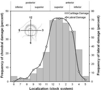
The allocation to one of the pathomorphologies of the algorithm was based on preoperative clinical and radio-graphic information and on a stepwise intraoperative evaluation. Typically, the first step to correct femoral pathomorphologies included the SHD with an osteotomy of the greater trochanter and a reduction of the stable portion of the former greater trochanter (relative neck lengthen-ing). Next, ROM was reevaluated for previously inapparent intraarticular sources of FAI such as a femoral head-neck asphericity or acetabular rim prominence. An additional head-neck trimming was performed to resect the aspherical portion of the femoral head first. In case of a persistent intraarticular FAI, the acetabular rim was trimmed in a second step. We then evaluated the extraarticular impingement of the lesser trochanter, which could be treated by distalization of the lesser trochanter. In case of joint incongruency, functional radiographs were repeated intraoperatively to determine whether we would perform a proximal femoral osteotomy or to check the amount of correction for a PAO. Indications for a PAO were an associated secondary acetabular dysplasia [10], which was defined as a lateral center-edge angle of less than 25° [46]. Indications for a proximal femoral valgus osteotomy were a nonspherical femoral head with good congruence in an adduction view [10]. Often, the acquisition of these radiographs is not possible preoperatively as a result of the lack of hip motion resulting from the femoral patho-morphologies. Forty of the 53 hips (75%) were treated with a SHD, eight (15%) hips with an acetabular osteotomy, four (8%) hips with a combined SHD and acetabular osteotomy, and one hip (2%) with an isolated intertro-chanteric osteotomy (Fig.1). The intraoperative damage pattern was evaluated in the 44 hips (83%) that underwent SHD. This included the status of the articular cartilage and labrum, documented intraoperatively, and graded accord-ing to a previously described gradaccord-ing system [4]. Cartilage damage was objectified as malacia, debonding, cleavage, or defect. Labral damage was objectified as degeneration, full-thickness tear, detachment, or ossification. To describe the exact location of the chondrolabral damage on the acetabulum, we used the clock system. Six o’clock was located at the acetabular notch. All findings were converted to the right side with 3 o’clock consistently representing the most anterior portion of the acetabulum. To describe the exact location of the chondral damage on the femoral head, the head was divided into eight sectors of a sphere (Fig.2) [40].
After SHD the hip was placed in a neutral position in a soft splint. The suction drains were removed after 48 hours. The patient was mobilized with crutches and partial weightbearing with 15 kg and restricted active and passive abduction and adduction to protect the trochanteric oste-otomy. Active flexion was restricted if the lesser trochanter
was advanced. The joint was mobilized on the second postoperative day on a continuous passive motion machine with a maximal flexion of 90° to avoid capsular adhesions. We used low-molecular-weight heparin for 8 weeks for prophylaxis of deep venous thrombosis. Eight weeks postoperatively, the patients were evaluated clinically and radiographically. By then, the trochanteric osteotomy usually was radiographically healed and gradual full weightbearing and muscular strengthening were started. The postoperative treatment regime did not change if an additional PAO or intertrochanteric osteotomy had been performed.
The next clinical and radiographic followup was gen-erally set at 3 months, 1 year, and 2 years postoperatively. Relief of pain was assessed using the definition according to the Merle d’Aubigne´-Postel pain subscore [24]. Improvement of hip function included the complete Merle d’Aubigne´-Postel score [24], incidence of limp, percentage of maximal abductor strength force (M5) according to the British Medical Research Council grading [23], incidence of the anterior and posterior impingement tests [42], and full ROM.
One of us (CEA; not a treating surgeon) assessed 11 descriptive (six acetabular and five femoral) radiographic parameters pre- and postoperatively. The six acetabular parameters were: lateral center-edge angle [46], the ace-tabular index [44], the extrusion index [25], the crossover and the posterior wall sign [28], and the intactness of Shenton’s line. The femoral parameters consisted of the centrum-collum-diaphyseal (CCD) angle, the alpha angle on the AP and crosstable lateral views [26], the sagging rope sign [2], and the trochanteric height. We assessed the trochanteric height by relating the height of the greater trochanter to the femoral head [37]. The head was divided
Fig. 2 The results for intraoperative femoral head damage are shown.
To describe the exact location of the chondral damage on the femoral head, the head was divided into eight sectors of a sphere. The numbers represent the frequency of chondral damage in each of the eight sectors.
into four quadrants with the first quadrant at the inferior and the fourth quadrant at the superior border of the fem-oral head. A fifth quadrant was situated above the femfem-oral head. In the literature, the interclass correlation coefficient or kappa values for the intra- (inter)observer reliability were 0.98 (0.92) for the lateral center-edge angle [41], 0.89 (0.92) for the acetabular index [41], 0.97 (0.91) for the extrusion index [41], 0.77 (0.6) for the crossover sign [41], 0.70 (0.62) for the posterior wall sign [41], and 0.86 (0.81) for the alpha angle [36]. No data exist for intactness of Shenton’s line, CCD angle, sagging rope sign, and assessment of trochanteric height. From the 11 radio-graphic parameters describing the hip morphology, three (27%) changed postoperatively: the alpha angle in AP and crosstable radiograph both decreased, the incidence of a sagging rope sign decreased, and the mean grades for tro-chanteric height decreased (Table2). Progression of OA was graded according to To¨nnis [43].
We tested normal distribution of all continuous param-eters with the Kolmogorov-Smirnov test. We determined differences in pain score between preoperative and fol-lowup using the Wilcoxon rank sum test. We determined differences in hip function using the paired Student’s t-test for normally distributed data (ROM), the Wilcoxon rank sum test for data without normal distribution (Merle d’Aubigne´-Postel score), the Fisher’s exact test for binominal data (incidence of limp, anterior, and posterior impingement test), and the Kruskal-Wallis test for cate-gorical data (abductor strength). We determined
differences in OA using the Kruskal-Wallis test. Survival of surgery was calculated with the following four end points: conversion to THA; radiographic progression of OA; a Merle d’Aubigne´-Postel score below 15 at followup; and revision surgery for any reason (except hardware removal) using the method of Kaplan and Meier [16]. The following factors were evaluated as predictive factors using the univariable Cox proportional hazards model [6]: age, sex, previous surgery, preoperative OA, subluxation of the joint, Stulberg classes [37], preoperative pathomorpholog-ical features (Fig.1), and all radiographic and clinical parameters (Tables2, 3). Hazard ratios were calculated with 95% confidence intervals (CIs).
Results
At the most recent followup, the median pain status repre-sented by the pain subgroup of the Merle d’Aubigne´ score increased (p\0.001) from preoperatively 4 (range, 2–6) to 5 (range, 3–6) for the 45 hips that were not converted to THA. Fourteen patients (31%) presented pain-free and an addi-tional 15 patients (33%) showed a reduction of pain compared with the preoperative status. In three patients (9%), pain was increased at last followup.
All parameters describing hip function except flexion improved at the most recent followup (Table3). The median Merle d’Aubigne´-Postel score increased (p \ 0.001), the incidence of limp decreased (p \ 0.001), the
Table 2. Preoperative and postoperative radiographic evaluation of all hips (n = 53)
Category Parameter Preoperative Postoperative* p value
Acetabular Lateral center-edge angle (degrees) 27 ± 13.7 (2–62) 27 ± 13.4 (3–66) 0.510
Acetabular index [44] (degrees) 11 ± 86.6 ( 5–29) 11 ± 11.1 ( 24–38) 0.682
Extrusion index [25] (percent) 23 ± 14.1 ( 17–48) 23 ± 14 ( 14–57) 0.376
Crossover sign [28] (percent positive) 65 48 0.087
Posterior wall sign [28] (percent positive) 97 94 0.453
Shenton’s line (percent intact) 80 85 0.806
Femoral CCD angle (degrees) 127 ± 8.8 (106–140) 129 ± 6.1 (116–140) 0.090
AP alpha angle AP radiograph (degrees) 79 ± 20.5 (45–114) 52 ± 13 (39–91) \ 0.001
Alpha angle crosstable radiograph [26] (degrees) 79 ± 27.2 (45–120) 44 ± 14.4 (27–96) \ 0.001
Sagging rope sign (percent positive) [2] 78 29 \ 0.001
Trochanteric height [37] (%) Quadrant 1 – 13 \ 0.001 Quadrant 2 6 57 Quadrant 3 17 17 Quadrant 4 44 11 Quadrant 5 33 2
Values of continuous parameters are expressed as mean ± standard deviation with range in parentheses; * osteoarthritis values were obtained at the most recent followup; CCD = centrum-collum-diaphyseal.
percentage of hips with a M5 abductor strength increased (p = 0.002), the incidence of positive anterior and posterior impingement tests both decreased (p \ 0.001), and all amplitudes for ROM except flexion increased (Table3).
The mean OA score increased (p = 0.004) at most recent followup compared with the preoperative status. Preoper-atively, 35 hips (66%) presented with Grade 0 and 18 hips (34%) with Grade 1 according to the classification of To¨nnis [43]. At last followup, 19 hips (42%) of the remaining 45 hips without conversion to THA showed no signs of OA, 17 hips (38%) had osteoarthritic changes Grade 1, seven hips (15%) Grade 2, and two hips (5%) Grade 3 according to To¨nnis [43].
The cumulative survival of surgery was 86% (95% CI, 76%–96%) at 5 years and 61% (46%–75%) at 8 years postoperatively (Fig.3). Twenty-six hips (49%) reached an end point: five hips (9%) converted to THA, 13 hips (25%) showed progression of OA, one hip (2%) had a Merle d’Aubigne´-Postel score of 13, and two hips (4%) had revision surgery. None of the patients had avascular necrosis of the femoral head secondary to surgery. In addition, there were three hips (6%) with combined pro-gression of OA, a Merle d’Aubigne´-Postel score ranging from 13 to 14, and two hips (4%) with progression of OA and revision surgery. The four hips with revision surgery were two acetabular redirection osteotomies, one intertro-chanteric varus osteotomy as a result of loss of femoral head containment, and one patient with excision of heterotopic ossifications.
On the acetabular side, 38 hips (86%) had chondrolabral damage. There were 16 hips (36%) with cartilage malacia, eight hips (18%) with a debonding phenomenon, and two hips (5%) with a cleavage lesion of the cartilage. There were no hips with a full-thickness cartilage defect. The
labrum was degenerated in 14 hips (32%) with a full-thickness tear in 21 (48%), a detachment in two (5%), and ossification in one hip (2%). The chondral damage was located more anteriorly (1.3 ± 2.0 o’clock) in comparison to the labral damage (12.8 ± 1.9 o’clock, p = 0.48; Fig.4). On the femoral side, 25 hips (57%) had evidence of chondral damage. There were 15 hips (34%) with chondral malacia, three hips (7%) with a debonding phenomenon,
Table 3. Clinical results preoperative and at followup of the hips with a preserved joint (n = 45 hips)
Parameter Preoperative value Followup value p value
Merle d’Aubigne´ [24] 14 ± 1.4 (5–17) 16 ± 1.9 (13–18) \ 0.001
Limp (percent of all hips) 71 18 \ 0.001
Abductor strength (percent of all hips with M5) [23] 37 76 0.002
Anterior impingement test [42] (percent of all hips) 91 59 \ 0.001
Posterior impingement test [42] (percent of all hips) 71 21 \ 0.001
Range of motion Flexion 93 ± 17.3 (50–130) 92 ± 15.7 (45–120) 0.741 Extension 3 ± 5.3 (0–20) 7 ± 5.9 (0–20) 0.017 Internal rotation 15 ± 10.7 (0–85) 21 ± 14.9 (0–60) 0.001 External rotation 22 ± 12.6 (0–45) 29 ± 15.6 (0–80) 0.010 Abduction 24 ± 11.17 (0–50) 29 ± 12.9 (0–50) 0.026 Adduction 19 ± 11.0 (0–45) 21.3 ± 8.7 (5–35) 0.029
Values of continuous parameters are expressed as mean ± standard deviation, with range in parentheses; values for the Merle d’Aubigne´ score are expressed as median ± SEM with range in parentheses.
Fig. 3 The Kaplan-Meier survival analysis is shown. End points
were defined as conversion to a THA, reoperation for correction of acetabular coverage or femoral offset, progression of osteoarthritis, or an insufficient clinical result defined as end points. Values are expressed as cumulative survival of surgery with 95% confidence intervals in parentheses.
one hip (2%) with a cleavage lesion, and six hips (14%) with a full-thickness cartilage defect. The chondral lesions were predominantly found in the medial half of the femoral head (Fig.2).
We identified 11 predictors for failure (Table4): one demographic, seven preoperative clinical or radiographic features, and three factors related to surgical treatment (Table4).
Discussion
Pathologic residual hip deformities after LCPD predispose for symptomatic malfunction of the joint and put the hip at high risk for degenerative OA [47]. It is important to address any possible source of extra- and intraarticular impingement, an associated acetabular dysplasia and/or joint incongruence to obtain pain relief, an improved ROM, and abductor strength. The ability of safely dislocating the hip with the recognition of FAI has revolutionized our surgical treatment protocol for surgical management of these conditions. We established a treatment algorithm to characterize the possible pathomechanical problems and proposed the appropriate surgical treatment for each problem. We therefore asked if patients undergoing joint-preserving surgery for symptomatic sequelae after LCPD according to this algorithm had (1) relief of pain; (2) improved hip function; and/or (3) no progression of OA; we determined (4) the survival of the surgery; (5) the intraoperative damage patterns; and (6) factors predicting an end point related to an unsatisfactory outcome. This was defined as the need for a conversion to THA; the presence of radiographic progression of OA; a Merle d’Aubigne´-Postel [24] score below 15 at last followup; or the need for revision surgery.
Our study is subject to a number of limitations. First is the heterogeneity of the patient series, which includes an age range spanning four decades, a high percentage of hips with previous surgeries, and a relatively high number of patients undergoing different surgical procedures. This limits the power of the study. However, we included all patients who had surgery during the study period and in all, the healing stage of LCPD was completed. Nonetheless, we
Fig. 4 The results for intraoperative chondrolabral damage of the
acetabulum are shown. A clock system was used to describe the exact location of the chondrolabral damage on the acetabulum. Six o’clock was located at the acetabular notch. All findings were converted to the right side with 3 o’clock consistently representing the most anterior portion of the acetabulum.
Table 4. Predictive factors for poor outcome with corresponding hazard ratios
Category Parameter Hazard ratio
(95% confidence interval)
p values
Demographic factors Age [ 40 years 6.7 (5.6–7.9) \ 0.01
Preoperative factors Preoperative osteoarthritis (To¨nnis C 1) [43] 4.0 (2.9–5.1) 0.01
Acetabular index [ 14° [44] 2.7 (1.9–3.6) 0.02
Severin classification [ 3 5.7 (3.9–7.5) \ 0.05
Merle d’Aubigne´ score \ 14 points 2.8 (2.0–3.6) 0.01
Range of motion
Internal rotation B 10° 2.6 (1.7–3.4) 0.03
External rotation \ 20° 2.6 (1.7–3.4) 0.04
Abduction \ 20° 2.6 (1.6–3.5) \ 0.05
Alpha angle [ 56° 4.7 (3.2–6.2) 0.04
Stulberg class [ III 6.4 (4.7–8.2) 0.03
Postoperative factors Broken Shenton’s line 2.9 (1.9–4.0) 0.04
Trochanteric height [ Grade 3 [37] 2.7 (1.7–3.7) \ 0.05
Table 5. Selected publications for the outcome after joint-preserving hip surgery for the sequelae of LCPD or Perthes’-like hip deformities Author Number of hips Mean age at surgery (years) Type of surgery Minimum (mean) followup (years) Results Anderson et al. [ 1 ] 14 20 SHD; proximal femoral osteotomy; trochanteric advancement 0.5 (3.8) Improved Harris hip score, reduction of limp, improved center trochanteric distance, no hips with conversion to THA, description of chondrolabral damage pattern Baksi [ 3 ] 3 1 N/A Adductor tenotomy; cheilectomy of the femoral head 3.2–10 Relief of pain, increased range of motion Clohisy et al. [ 5 ] 2 4 2 2 PAO; proximal femoral osteotomy 2.0 (4.5) Improved Harris hip score, improved LCE angle, improved ACE angle, improved acetabular index, medialization of the hip center, slight increase in osteoarthritis Eijer et al. [ 7 ] 1 2 2 1 SHD; various proximal femoral and acetabular osteotomies 1.1 (2.8) Relief of pain, improved ROM, no new femoral head necrosis Joo et al. [ 13 ] 1 5 1 7 Advancement of greater trochanter 2.3 (3.5) No pain relief, no improvement of limp Pecasse et al. [ 27 ] 1 5 3 0 Intertrochanteric osteotomy; advancement of greater trochanter; acetabular roof plasty 8.0 (11.3) Relief of pain, fair Harris hip score, no improvement of ROM, mean survival time of 15.3 years Shinoda et al. [ 32 ] 1 7 3 7 PAO 3.0 (6.6) Improved Harris hip score, no increase in ROM, no decrease in leg length discrepancy, increased acetabular index, increased acetabular head index, progression of osteoarthritis Shore et al. [ 33 ] 2 9 1 7 SHD; various proximal femoral and acetabular osteotomies 1.0 (3.0) Improved WOMAC score, pain reduction, improved ROM and function, low failure rate Vukasinovic et al. [ 45 ] 41 10–20 Chiari osteotomy 4.0 (7.2) Improved Harris hip score, improvement of acetabular angle of Sharp, increase of LCE angle, improved femoral head coverage, improved acetabular depth ratio, increased Shenton’s line continuity Current study 53 21 SHD; PAO; various proximal femoral and acetabular osteotomies 5.0 (8.2) Relief of pain, improvement of range of motion, improved hip abductor strength, slight progression of osteoarthritis LCPD = Legg-Calve ´-Perthes disease; SHD = surgical hip dislocation; PAO = periacetabular osteotomy; LCE = lateral center-edge; ACE = anterior center-edge.
believe the heterogeneity facilitates the identification of predictors for our end points. Second, the surgical tech-nique has evolved over time with growing experience and more precise assessment of the structural problem. Some of the classified pathomorphologies were not consistently addressed according to this proposed treatment algorithm and in some patients we judged an abnormality to be irrelevant. For example, we did not believe acetabular retroversion had to be corrected if impingement-free motion was present after reshaping of the femoral head, particularly with a borderline-type lateral acetabular cov-erage. Furthermore, some structural abnormalities were underestimated and not addressed. For example, in two patients with an additional borderline acetabular dysplasia, the femoral pathology was addressed only, which subse-quently led to a loss of containment needing acetabular redirection osteotomy. Third, the retrospective nature of this study allowed us to only use the Merle d’Aubigne´-Postel score, one that has not been validated. However, the three subgroups of the score (pain, walking ability, ROM) address the problems of patients with residual deformities after healed LCPD. Fourth, the description of the intraar-ticular damage only refers to hips that underwent a SHD, which covers 83% of our cases. Fifth, our univariate
analysis could be misleading because we have inadequate power to perform a multivariate analysis.
Our approach to treat symptomatic hips after LCPD decreases hip pain. This is consistent with other reports describing similar treatment strategies mainly based on a SHD [1,7,33]. This surgical approach offers the option to address any possible intra- and extraarticular source of FAI and can be combined with any form of concomitant femoral and acetabular osteotomies. The authors believe this com-prehensive approach is the key for successful reduction of hip pain in these patients. Surgical treatment of an isolated structural deformity does not improve hip pain as shown with an isolated cheilectomy [29] of the aspherical femoral head portion or a simple trochanteric advancement [13].
We observed improved mean hip function at last fol-lowup. This included an increase in abductor strength together with a decreased prevalence of the anterior and posterior impingement signs. In addition, all amplitudes of hip ROM except flexion improved after surgery (Table3). Although a similar effect on ROM has been described in some articles [3,7], this is not the case with other surgical techniques for treatment of the sequelae of LCPD in the literature [27,32] (Table5). There are two main reasons why hip flexion did not change after surgery. First, hip
Fig. 5A–D The radiographs of a
16-year-old female patient are shown in the (A) AP projection and (B) crosstable projection. Next to the deformity of the femoral head (Grade III according to Stul-berg), a high-riding trochanter, a positive sagging rope sign, and acetabular retroversion were pres-ent. The acetabular index was 4°. (C) A surgical hip dislocation with relative lengthening of the femoral head, distalization of the greater trochanter, and (D) osteochon-droplasty of the femoral head neck junction was performed. The acetabular retroversion was not addressed to avoid joint instability. Eleven years postoperatively, the patient presented with a Merle
d’Aubigne´-Postel score of 18
points without signs of radio-graphic progression of osteoar-thritis and full abductor strength.
flexion is the least restricted preoperative motion with LCPD, particularly in hips with low Stulberg classes [7,39, 48]. Second, an additional acetabular osteotomy can adversely reduce the flexion amplitude even when the femoral pathomorphology was optimized.
Our observations suggest we are unable to stop the progression of OA in these hips. This can be attributed to several factors. A main factor is certainly the preexisting articular damage in these hips. In addition, the restoration of a normal hip anatomy even with extensive surgical corrections of the LCPD pathomorphology is rarely pos-sible. As a result of the lack of comparable followup, the other studies evaluating the use of a SHD have not quan-tified the progression of OA in detail [1,7,33]. However, a similar trend of a slight progression of OA was observed with other joint-preserving techniques [5, 32]. It remains unclear if the progression of OA can be decelerated because of the lack of natural history controls.
We found a survival rate of surgery of 86% at the minimum followup of 5 years and a 61% survival of sur-gery at the mean followup of 8 years. There are only limited comparable studies with a similar approach, none of them performing a survival analysis. Anderson et al. [1] report in their study with 14 hips at a mean followup of 3.8 years a progression of OA in four cases, a poor clinical outcome in three cases, and no conversions to THA. In a recent study by Shore et al. [33], 29 individuals were treated with SHD and various concomitant proximal fem-oral and acetabular osteotomies. In their study, the authors report four failures at a mean followup of 3 years. Of those, there was one patient presenting with a poor clinical result and three patients requiring THA [33]. In addition, seven patients underwent revision surgery. However, the improvement of the clinical scores and the ROM is a
consistent finding in all articles and matches with our results.
The chondrolabral damage on the acetabular side occurs typically in the anterosuperior quadrant, similarly to other hips with FAI [34, 38]. Our findings are consistent with other descriptions of smaller series in the literature [1,7]. On the femoral side, the cartilage lesions are often found in the medial hemisphere, which is typically the location of the necrosis and osteochondral defects [1]. The large lateral aspherical portion is often less affected because it may not even contribute to the articulating process. It can therefore be used as a potential autologous osteochondral allograft for the medial femoral head portion.
We identified no literature reporting predictive factors of outcome in patients with LCPD undergoing joint-preserv-ing surgery. We found 11 univariable predictors for poor outcome. As mentioned, this univariate analysis should be interpreted carefully because we have limited power to perform a multivariate analysis. The predictors can be divided into three main categories: preexisting joint dam-age, extent of the femoral and acetabular pathomorphology, and surgical correction. The preexisting joint damage is reflected by the degree of OA, the age, and the initial clinical score. The extent of the femoral and acetabular pathomorphology is reflected by a worse prognosis or hips with Stulberg [ 3, hips with an accelerated offset problem (alpha angle [ 50°), or a dysplastic morphology with sub-luxation (Severin grade [ 3, acetabular index [14°, and broken Shenton’s line). The surgical correction has to include a sufficient offset creation (alpha angle \ 50°), the restoration of an intact Shenton’s line, and correct tro-chanteric height (Table4).
Our data suggest the pathomorphologic features related to the sequelae of LCPD can be treated using our algorithm
Fig. 6A–C The radiographs of a 43-year-old patient with
Legg-Calve´-Perthes disease are shown. (A) Preoperatively, the patient presented with pain, a severe Trendelenburg limp, and decreased abduction, internal, and external rotation. (B) A surgical hip dislocation with relative lengthening of the femoral head, distalization
of the greater trochanter and osteochondroplasty, and acetabular rim trimming with refixation of the labrum was performed. (C) Five years after surgery, the patient presented with severe progression of osteoarthritis and migration of the femoral head.
(Fig.5). Patients with sufficient pain and disability in whom a primary THA often seems to be the only surgical option can benefit from a SHD together with the specific treatment of individual structural features. The severity of the deformation according to Stulberg et al. [37], the pre-operative grade of OA, the age of the patient, the accuracy of the correction, and the presence of a broken Shenton’s line determine the midterm results of our treatment pro-tocol (Fig.6).
References
1. Anderson LA, Erickson JA, Severson EP, Peters CL. Sequelae of Perthes disease: treatment with surgical hip dislocation and rel-ative femoral neck lengthening. J Pediatr Orthop. 2010;30: 758–766.
2. Apley AG, Wientroub S. The sagging rope sign in Perthes’ dis-ease and allied disorders. J Bone Joint Surg Br. 1981;63:43–47. 3. Baksi DP. Palliative operations for painful old Perthes’ disease.
Int Orthop. 1995;19:46–50.
4. Beck M, Kalhor M, Leunig M, Ganz R. Hip morphology influ-ences the pattern of damage to the acetabular cartilage: femoroacetabular impingement as a cause of early osteoarthritis of the hip. J Bone Joint Surg Br. 2005;87:1012–1018.
5. Clohisy JC, Nunley RM, Curry MC, Schoenecker PL. Periace-tabular osteotomy for the treatment of acePeriace-tabular dysplasia associated with major aspherical femoral head deformities. J Bone Joint Surg Am. 2007;89:1417–1423.
6. Cox DR. Regression models and life-tables. J R Stat Soc Ser B Stat Methodol. 1972;34:187.
7. Eijer H, Podeszwa DA, Ganz R, Leunig M. Evaluation and treatment of young adults with femoro-acetabular impingement secondary to Perthes’ disease. Hip Int. 2006;16:273–280. 8. Engelhardt P. Late prognosis of Perthes’ disease: which factors
determine arthritis risk? Z Orthop Ihre Grenzgeb. 1985;123: 168–181.
9. Ezoe M, Naito M, Inoue T. The prevalence of acetabular retro-version among various disorders of the hip. J Bone Joint Surg Am. 2006;88:372–379.
10. Ganz R, Horowitz K, Leunig M. Algorithm for femoral and periacetabular osteotomies in complex hip deformities. Clin Orthop Relat Res. 2010;468:3168–3180.
11. Ganz R, Huff TW, Leunig M. Extended retinacular soft-tissue flap for intra-articular hip surgery: surgical technique, indications, and results of application. Instr Course Lect. 2009;58:241–255. 12. Ippolito E, Tudisco C, Farsetti P. The long-term prognosis of
unilateral Perthes’ disease. J Bone Joint Surg Br. 1987;69:243– 250.
13. Joo SY, Lee KS, Koh IH, Park HW, Kim HW. Trochanteric advancement in patients with Legg-Calve-Perthes disease does not improve pain or limp. Clin Orthop Relat Res. 2008;466: 927–934.
14. Joseph B. Morphological changes in the acetabulum in Perthes’ disease. J Bone Joint Surg Br. 1989;71:756–763.
15. Kamegaya M, Shinada Y, Moriya H, Tsuchiya K, Akita T, Someya M. Acetabular remodelling in Perthes’ disease after primary healing. J Pediatr Orthop. 1992;12:308–314.
16. Kaplan EL, Meier P. Nonparametric estimation from incomplete observations. J Am Stat Assoc. 1958;53:457–481.
17. Kelikian AS, Tachdjian MO, Askew MJ, Jasty M. Greater tro-chanteric advancement of the proximal femur: a clinical and biomechanical study. Hip. 1983:77–105.
18. Kim HT, Wenger DR. ‘Functional retroversion’ of the femoral head in Legg-Calve-Perthes disease and epiphyseal dysplasia: analysis of head-neck deformity and its effect on limb position using three-dimensional computed tomography. J Pediatr Orthop. 1997;17:240–246.
19. Larson AN, Stans AA, Sierra RJ. Ischial spine sign reveals ace-tabular retroversion in Legg-Calve-Perthes disease. Clin Orthop Relat Res. 2011;469:2012–2018.
20. Leunig M, Podeszwa D, Beck M, Werlen S, Ganz R. Magnetic resonance arthrography of labral disorders in hips with dysplasia and impingement. Clin Orthop Relat Res. 2004;418:74–80. 21. Leunig M, Werlen S, Ungersbock A, Ito K, Ganz R. Evaluation
of the acetabular labrum by MR arthrography. J Bone Joint Surg Br. 1997;79:230–234.
22. McAndrew MP, Weinstein SL. A long-term follow-up of Legg-Calve-Perthes disease. J Bone Joint Surg Am. 1984;66:860–869. 23. Medical Resarch Council. Aids to the Investigation of Peripheral Nerve Injuries. London, UK: HMSO: War memorandum No. 7 (revised 2nd edition); 1943.
24. Merle d’Aubigne´ R, Postel M. Functional results of hip arthroplasty with acrylic prostesis. J Bone Joint Surg Am. 1954; 36: 451–475.
25. Murphy SB, Ganz R, Muller ME. The prognosis in untreated dysplasia of the hip. A study of radiographic factors that predict the outcome. J Bone Joint Surg Am. 1995;77:985–989. 26. Notzli HP, Wyss TF, Stoecklin CH, Schmid MR, Treiber K,
Hodler J. The contour of the femoral head-neck junction as a predictor for the risk of anterior impingement. J Bone Joint Surg Br. 2002;84:556–560.
27. Pecasse GA, Eijer H, Haverkamp D, Marti RK. Intertrochan-teric osteotomy in young adults for sequelae of Legg-Calve-Perthes’ disease—a long term follow-up. Int Orthop. 2004;28: 44–47.
28. Reynolds D, Lucas J, Klaue K. Retroversion of the acetabulum. A cause of hip pain. J Bone Joint Surg Br. 1999;81:281–288. 29. Rowe SM, Jung ST, Cheon SY, Choi J, Kang KD, Kim KH.
Outcome of cheilectomy in Legg-Calve-Perthes disease: mini-mum 25-year follow-up of five patients. J Pediatr Orthop. 2006; 26:204–210.
30. Saito S, Takaoka K, Ono K, Minobe Y, Inoue A. Residual deformities related to arthrotic change after Perthes’ disease. A long-term follow-up of fifty-one cases. Arch Orthop Trauma Surg. 1985;104:7–14.
31. Schneidmueller D, Carstens C, Thomsen M. Surgical treatment of overgrowth of the greater trochanter in children and adolescents. J Pediatr Orthop. 2006;26:486–490.
32. Shinoda T, Naito M, Nakamura Y, Kiyama T. Periacetabular osteotomy for the treatment of dysplastic hip with Perthes-like deformities. Int Orthop. 2009;33:71–75.
33. Shore BJ, Novais EN, Millis MB, Kim YJ. Low early failure rates using a surgical dislocation approach in healed Legg-Calve-Perthes disease. Clin Orthop Relat Res. 2011 Nov 29 [Epub ahead
of print]. DOI:10.1007/s11999-011-2187-1.
34. Siebenrock KA, Schoeniger R, Ganz R. Anterior femoro-acetabular impingement due to femoro-acetabular retroversion. Treatment with periacetabular osteotomy. J Bone Joint Surg Am. 2003;85: 278–286.
35. Snow SW, Keret D, Scarangella S, Bowen JR. Anterior impingement of the femoral head: a late phenomenon of Legg-Calve-Perthes’ disease. J Pediatr Orthop. 1993;13:286–289. 36. Steppacher SD, Tannast M, Werlen S, Siebenrock KA. Femoral
morphology differs between deficient and excessive acetabular coverage. Clin Orthop Relat Res. 2008;466:782–790.
37. Stulberg SD, Cooperman DR, Wallensten R. The natural history of Legg-Calve-Perthes disease. J Bone Joint Surg Am. 1981;63: 1095–1108.
38. Tannast M, Goricki D, Beck M, Murphy SB, Siebenrock KA. Hip damage occurs at the zone of femoroacetabular impingement. Clin Orthop Relat Res. 2008;466:273–280.
39. Tannast M, Hanke M, Ecker TM, Murphy SB, Albers CE, Puls M. LCPD: reduced range of motion resulting from extra-and intraarticular impingement. Clin Orthop Relat Res. 2012.
DOI:10.1007/s11999-012-2344-1.
40. Tannast M, Kruger A, Mack PW, Powell JN, Hosalkar HS, Siebenrock KA. Surgical dislocation of the hip for the fixation of acetabular fractures. J Bone Joint Surg Br. 2010;92:842–852. 41. Tannast M, Mistry S, Steppacher SD, Reichenbach S, Langlotz F,
Siebenrock KA, Zheng G. Radiographic analysis of femoroace-tabular impingement with Hip2Norm-reliable and validated. J Orthop Res. 2008;26:1199–1205.
42. Tannast M, Siebenrock KA, Anderson SE. Femoroacetabular impingement: radiographic diagnosis—what the radiologist should know. AJR Am J Roentgenol. 2007;188:1540–1552.
43. To¨nnis D. Gerneral radiography of the hip joint. In: To¨nnis D, ed. Congenital Dysplasia, Dislocation of the Hip. New York, NY, USA: Springer; 1987:100–142.
44. To¨nnis D, Heinecke A. Acetabular and femoral anteversion: relationship with osteoarthritis of the hip. J Bone Joint Surg Am. 1999;81:1747–1770.
45. Vukasinovic Z, Spasovski D, Slavkovic N, Bascarevic Z, Zivkovic Z, Starcevic B. Chiari pelvic osteotomy in the treatment of adolescent hip disorders: possibilities, limitations and com-plications. Int Orthop. 2011;35:1203–1208.
46. Wiberg G. The anatomy and roentgenographic appearance of a normal hip joint. Acta Chir Scand. 1939;83:7–38.
47. Yrjonen T. Long-term prognosis of Legg-Calve-Perthes disease: a meta-analysis. J Pediatr Orthop B. 1999;8:169–172.
48. Zilkens C, Bittersohl B, Jager M, Haamberg T, Westhoff B, Krauspe R. Clinical presentation of young adults after Legg-Calve-Perthes disease. Acta Orthop Belg. 2009;75:754–760.