ORIGINAL ARTICLE
Malignant minor salivary gland tumors: a retrospective
study of 27 cases
Astrid L. D. Kruse&Klaus W. Grätz&
Joachim A. Obwegeser&Heinz-Theo Lübbers
Published online: 6 April 2010 # Springer-Verlag 2010 Abstract
Purpose Malignant tumors of the intra-oral minor salivary glands are uncommon. The aim of this study was to give information concerning the clinical features of these tumors, the distribution of location, treatment opportunities, and outcome.
Methods Twenty-seven patients with malignant salivary gland tumors that were treated between January 1999 and December 2008 were evaluated retrospectively.
Results Of the 27 minor salivary gland carcinomas, 48.1% were adenoid cystic carcinomas (ACC), 29.7% mucoepi-dermoid carcinomas (MEC), 22.2% adenocarcinomas (ADCA). The most common first symptom was a painless swelling in 60% of the cases, with the second most common symptom being ulcers (28%). Four recurrences and two metastases were found. No recurrence was observed in ADCA. All four patients experiencing a recurrence developed it in the first 3 years after treatment. Conclusion Wide excision with a clinical margin of 1 cm and in large tumors, positive surgical margins or perineural infiltration and postoperative radiotherapy (RT) can be recommended; but in order to give exact information concerning the possible benefit from postoperative RT, it needs large prospective multicenter studies. Long-term follow-up controls and in particularly longer than 5 years in ACC including yearly chest X-rays should be offered to these patients because of late metastasis and recurrences. Keywords Malignant tumor . Neoplasm . Retrospective study . Outcome . Treatment . Salivary gland . Radiotherapy . Oncology
Introduction
Between 450 and 750 minor glands can be found scattered throughout the head and neck and are present in many sites, such as the lips, cheek, gingival, palate, tongue, oropharynx, parapharyngeal space, and paranasal sinuses [1]. Malignant tumors of the intra-oral minor salivary glands constitute 2–3% of all malignant neoplasms of the upper aerodigestive tract and less than 25% of all salivary gland tumors [2]. Tumors arising in major salivary glands due to their well-defined anatomic borders and their higher incidence cannot be directly compared to minor salivary gland tumors. Most tumors arising at minor salivary glands are malignant. The pathogenesis is still not completely solved, but factors like smoking, poor mouth hygiene or alcohol abuse seems to play not the same rule like in oral squamous cell carcinoma. The most common sites are the hard palate, nasal cavity, and paranasal sinuses [3]. Squamous cell carcinomas of the oral cavity are well studied, but malignant tumors of the minor salivary glands are rare. However, controversy still sur-rounds several issues including length of follow-up time and indications for radiotherapy (RT). Most of the studies that are available are retrospective studies.
The purpose of this study was to determine the histological types, sites and prognostic factors of patients with malignant minor salivary gland carcinomas and to compare the results with data from different reports.
Patients
The records of all patients with malignant minor salivary gland tumors from January 1999 and December 2008, treated in the Department of Craniomaxillofacial and Oral Surgery, University Hospital Zurich were systematically reviewed for tumor site, histological type, involvement A. L. D. Kruse
:
K. W. Grätz:
J. A. Obwegeser:
H.-T. Lübbers (*)
Clinic for Cranio-Maxillofacial Surgery,
University Hospital of Zurich, Zurich, Switzerland e-mail: t.luebbers@gmail.com
of lymph nodes, metastases, treatment, and recurrence. Exclusion criteria were recurrent disease plus lack of information, therefore two patients were excluded. In all patients, an incisional biopsy and computed tomography (CT), positron-emissions tomography (PET) or magnetic resonance imaging (MRI) was performed before the definitive treatment.
Results
Age, sex, primary sites, and kinds of carcinoma
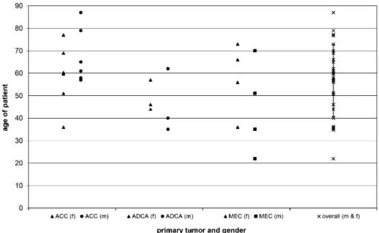
Together, 27 patients with intra-oral minor salivary gland tumors were treated and evaluated retrospectively. Out of these 27 patients, 48.1% had an adenoid cystic carcinoma Fig. 1 Distribution of age
(ACC adenoid cystic carcinoma, ADCA adenocarcinoma, MEC mucoepidermoid carcinoma)
ACC ADCA MEC
Number of patients 13 6 8 Age Mean 64.4 47.3 51.1 Minimum 36 35 22 Maximum 87 62 73 Sex Male 6 3 4 Female 7 3 4
Tumor site Upper jaw alveolar ridge 4 2 1
Hard palate 5 2 5 Soft palate 2 2 1 Lower jaw 2 0 1 Tumor status T1 3 2 4 T2 5 3 3 T3 2 0 0 T4 3 1 1 Node status N1 1 0 0 N2a 2 0 0 N2b 0 0 0 N2c 0 0 0 N3 0 0 0 Metastases status M0 0 0 0 M1 1 0 0
Table 1 Tumor location and stage at initial clinical presentation
ACC adenoid cystic carcinoma, ADCA adenocarcinoma, MEC mucoepidermoid carcinoma
(ACC), 29.7% a mucoepidermoid carcinoma (MEC), and 22.2% an adenocarcinoma (ADCA). The female to male ratio was 14:13, and the average age for MEC was 47.9 years (28– 83 years); for ADCA, 47.3 years (35–62 years); and for ACC, 64.4 years (36–87 years). The most common age period for developing a malignant tumor of the intra-oral minor salivary gland was between 55 and 70 years (Fig.1).
The distribution of location is presented in Table1: in the region of the alveolar ridge, the most common minor salivary cancer is ACC; and in the hard palate, it is the MEC. Forty-four percent of these tumors are located in the hard palate. Regarding the tumors located in the lower jaw, one ACC was located on the ascending ramus and one on the alveolar crest in an edentulous region. One MEC was located on the alveolar crest in an edentulous region. Figure2does give an overview about the primary location of all included cases. Initial symptoms and therapy
Most patients were referred from private dentists. The most common first symptom, found in 60% of patients, was a painless swelling, followed by an ulcer (28%).
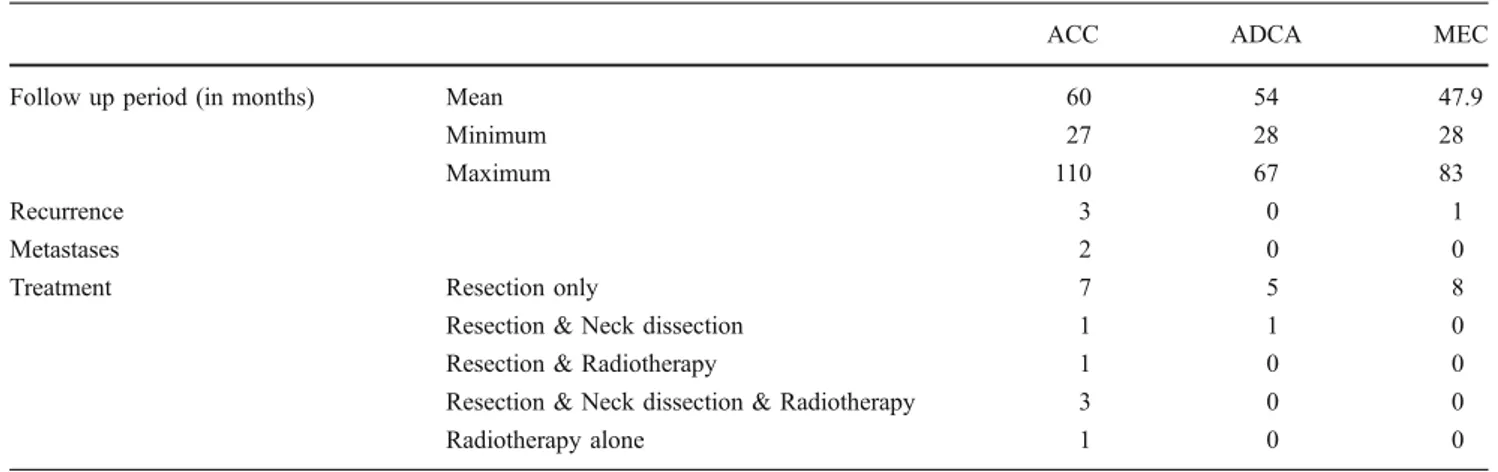
The tumors in the present study were resected clinically with 1-cm margins. Out of 27 patients, five patients (four with ACC and one with MEC) had a supraomohyoidal neck dissection because of clinically positive lymph nodes. Recurrence, positive lymph nodes, large tumors, or positive surgical margins were an indication for radio- and/or chemotherapy (Table2).
Treatment results and follow-up strategy
In four out of 27 patients, a recurrence (three in ACC and one in MEC) was observed. No recurrence was observed in ADCA (Table2).
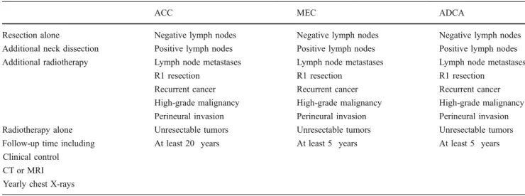
All four patients experiencing a recurrence developed it in the first 3 years after treatment. Two patients were treated first in a different hospital, so that the original pathohistology was not available. Five patients (all with ACC) had perineural infiltration in their first resection, but in only one, recurrence and metastases developed until now (Table 3). Neck dissections were only performed in cases of clinically positive lymph nodes. The median follow-up time for ACC was 60 months, for ADCA 54 months and for MEC 47.9 months (Table 2). The patients were seen in the first year on a monthly basis, in the second year every 2 months, in the third year every 3 months, in the fourth year every 6 months followed by a yearly control (Table4).
Discussion
In several studies, adenoid cystic carcinoma has been shown as being most common [4–6], with several other studies showing, on the other hand, that mucoepidermoid carcinoma is the most common (Table 5) [2,7–10]. Some authors describe the tumor distribution pattern as varying by country.
Most reports describe a female predominance in minor salivary gland tumors [4,5] similar to the proportion in the present study, which shows female to male as 7:6. The main age group was found between 60–79 years; that is also supported in other studies [11,12]. Also, the present study shows the palate as the predominant location (62.9% in the hard and soft palate), a finding comparable to those of other current studies [4, 5, 10]. Concerning recurrence, only a very few studies are available because of late metastasis, older age of patients, and lower incidence of these tumors. A lower rate of cervical lymph node metastases in comparison to oral squamous cell carcinomas has been reported by several authors [1, 13] and this was also supported by the present study; in only three out of 27 patients were positive lymph nodes found. Therefore, there is probably little benefit from elective neck dissection. In this study, neck dissection was performed in patients with Fig. 2 Tumor locations
clinical or radiological suspect of lymph node metastasis or in patients with T4 tumor stage.
Regarding the initial therapy, there was no difference between the patients developing recurrence (Table3) on the one hand and the patients not experiencing recurrence on the other hand.
Further therapy strategies recommend on a type-2 level of evidence [14] in cases of unresectable/inoperable locore-gional disease neutron, heavy ions or proton radiotherapy. But there are still controversies concerning radiotherapy, on the one hand surgical resection and postoperative RT is recommended [15]. But on the other hand, Spiro et al.
could not show a survival benefit for patients receiving postoperative RT, after matching for stage, site, and histology [12].
Concerning neutron beam, one problem seems to be the limited number of institutions providing this treatment besides the reported complication rate like osteroradionec-rosis, optic neuritis/retinitis, and oral/pharyngeal-cutaneous fistulas [14,16].
Due to overlapping clinicopathological features of ACC and polymorphous low-grade adenocarcinoma (PLGA), the differentiation can be difficult. Up until now, only vimentin presents a clear difference, which is negative in ACC and
Table 3 Patients with recurrence and/or metastases Patient Tumor
type
TNM Perineural infiltration
Recurrence Metastases Outcome Follow-up time
Initial Therapy
1 ACC T2N2bM0 Yes 32 months 44 months DOD 48 months Tumor resection with free margins, unilateral neck dissection due to known lymph node metastasis, no extracapsular tumor cells 2 ACC T2N0M0 No None 68 months Palliative 74 months Tumor resection with free margins,
no neck dissection due to no sign of lymph node metastasis in clinical and radiological examination 3 ACC T4N0M0 No 34 months None Tumor free 110 months Tumor resection with R1 margins,
postoperative radiotherapy, neck dissection due to T4 state despite no clinical or radiological signs of lymph node metastasis, neck dissection revealed no lymph node infiltration
4 MEC T4N0M0 No 14 months None Tumor free 42 months Tumor resection with free margins, neck dissection due to T4 state despite no clinical or radiological signs of lymph node metastasis, neck dissection revealed no lymph node infiltration
ACC adenoid cystic carcinoma, DOD death of disease, MEC mucoepidermoid carcinoma Table 2 Treatment strategies and outcome
ACC ADCA MEC
Follow up period (in months) Mean 60 54 47.9
Minimum 27 28 28
Maximum 110 67 83
Recurrence 3 0 1
Metastases 2 0 0
Treatment Resection only 7 5 8
Resection & Neck dissection 1 1 0
Resection & Radiotherapy 1 0 0
Resection & Neck dissection & Radiotherapy 3 0 0
Radiotherapy alone 1 0 0
positive in PLGA. CEA, EMA, and S100 protein can be positive in both tumors [17]. Concerning salivary gland tumors, Ki-67 has been discussed as a prognostic factor because of being significantly higher in cases of treatment failure and large tumors [18], but in clinical routine, the application of Ki-67 is still missing.
Adenoid cystic carcinoma
The peak incidence of these tumors was between 60 and 79 years, although the literature describes a wide age distribution [18]. Also, the predominance of females, as in our study, is confirmed (7:4). Nearly half of all intra-oral ACC occur in the palate [17].
The growth pattern of ACC is described as slow but aggressive, with a predominantly perineural spread and less lymphatic spread, but also with more frequent distant metastasis (particularly in the lungs) after long-term survival and even in the absence of local or regional recurrence [19]. Therefore, it is difficult to distinguish between recurrence and a second tumor in adenoid cystic carcinomas. Distant metastases, when affecting the lungs,
are usually slow growing, sometimes apparently isolated, and frequently surgically resectable [20]. The longest time elapsing before a distant metastasis in the present study was 68 months.
Compared to other head and neck malignancies, ACC are more difficult to clear surgically, often resulting in positive margins [17]. In the present study, only in one case was a second resection performed immediately after histopatholog-ical diagnosis of involved margin (not detected in frozen section), this patient showed no recurrence or metastases.
Predictors for distant metastasis seem to be large tumor size and lymph node involvement [21]. Therefore, initial aggressive surgery, combined with radiation for high-stage tumors or involved surgical margins, is suggested [18,21]. These principles were also used in the present study. Mucoepidermoid carcinoma
MEC was indicated in 32% of the cases as the second most common type of tumor. The female to male ratio was 1:1.
In contrast to ACC, MEC show their malignant behavior within the first 5 years after surgery [18,22]. In only one
Author Year N ACC ADCA MEC others
Vander Poorten et al. [11] 2000 55 22 5 9 16
Jansisyanont et al. [29] 2002 61 7 9 33 9 Strick et al. [1] 2004 21 7 6 6 2 Toida et al. [5] 2005 27 10 0 8 6 Yih et al. [10] 2005 94 22 18 45 8 Pires et al. [32] 2007 241 35 28 125 32 Buchner et al. [7] 2007 156 24 27 83 16 Copelli et al. [33] 2008 43 26 1 12 2 Mücke et al. [34] 2009 95 33 14 20 28
Table 5 Studies emphasized on malignant minor salivary gland tumors
ACC adenoid cystic carcinoma, ADCA adenocarcinoma, MEC mucoepidermoid carcinoma
Table 4 Treatment and follow-up strategy
ACC MEC ADCA
Resection alone Negative lymph nodes Negative lymph nodes Negative lymph nodes
Additional neck dissection Positive lymph nodes Positive lymph nodes Positive lymph nodes Additional radiotherapy Lymph node metastases Lymph node metastases Lymph node metastases
R1 resection R1 resection R1 resection
Recurrent cancer Recurrent cancer Recurrent cancer
High-grade malignancy High-grade malignancy High-grade malignancy Perineural invasion Perineural invasion Perineural invasion
Radiotherapy alone Unresectable tumors Unresectable tumors Unresectable tumors
Follow-up time including At least 20 years At least 5 years At least 5 years
Clinical control CT or MRI Yearly chest X-rays
case out of eight was a recurrence seen after 14 months and resected again. An increase of Ki-67 has been discussed in order to differentiate between malignant and benign gland tissue [18, 23] but has not been established in clinical routine to date. Although radio-resistance is discussed, radiation is recommended in patients with positive surgical margins and high-grade tumors [15,24]. Tran et al. reported an improvement of local control by RT in patients with positive surgical margins from 50% to 71% [25]. Recurrence of MEC appears mostly in the first 5 years after surgery [18]. Adenocarcinoma
A predominance in female patients was also described by Pogodzinski et al. [26]. No recurrence was found in the patients in the present study but delayed local recurrences and regional nodal metastases have been mentioned in the literature [26–28]. In contrast to adenoid cystic carcinoma, adenocarcinoma has an excellent cure rate after complete excision infrequent recurrences and rare regional metastasis [29]. In the present study, no recurrence or metastasis was observed in this patient group.
Limitations
The main weakness of this study was the small number of patients. Within the limitations of this investigation, the results show the need of long follow-up times in patients with ACC. Also, there were considerable differences in the surgical and postoperative follow-up protocols, as well as in the length of follow-up time. Most of those protocols suffer from a lack of long-term results: most studies cover less than 5 years of postoperative controls; and, due to the higher age of this patient group, long-term controls are not always possible.
Therapy
Postoperative RT should be recommended for patients with high-grade malignancies, lymph node metastases, perineural invasion, and recurrent cancer. It is mentioned that RT alone can cure patients with minor salivary gland malignancy, but the overall survival rates are lower compared to RT in combination with surgery [3].
Prognosis
Concerning prognosis, one must differ between oral and paranasal sinus malignancies, the latter seems to be associated with advanced-stage lesions, bone invasion, nerve involvement, adenoid cystic carcinoma, and positive margins [30]. Probably, in these cases, one should consider
proton beam RT in order to reduce complications associated with central nervous system or visual apparatus.
Vander et al. and Spiro et al. recommend that the oral squamous cell carcinoma TNM classification as one of the major prognostic factor for all oncologic outcomes after diagnosis of minor salivary gland carcinoma [11, 12]. Our small study indicated that perineural infiltration is not directly associated with distant metastases, but for exact evaluation it needs larger and longer studies.
Seventy percent of local recurrences are observed within 3 years. But there is a clear exception in cases of high-grade and adenoid cystic histology [31]. Therefore, along with Guzzo et al., we recommend a follow-up period of 20 years in particular in adenoid cystic carcinomas or high-grade tumors including yearly chest X-rays.
Conclusion
Wide excision with a clinical margin of 1 cm and in large tumors, positive surgical margins or perineural infiltration additional postoperative radiotherapy can be recommended. But in order to give exact information concerning the possible benefit from postoperative radiotherapy, it needs large prospective multicenter studies. Long-term follow-up controls and in particularly longer than 5 years in adenoid cystic carcinomas including yearly chest X-rays should be offered to these patients because of late metastasis and recurrences.
References
1. Strick MJ et al (2004) Malignant tumours of the minor salivary glands—a 20 year review. Br J Plast Surg 57(7):624–631 2. Rivera-Bastidas H, Ocanto RA, Acevedo AM (1996) Intraoral
minor salivary gland tumors: a retrospective study of 62 cases in a Venezuelan population. J Oral Pathol Med 25(1):1–4
3. Cianchetti M et al (2009) Radiation therapy for minor salivary gland carcinoma. Laryngoscope 119(7):1334–1338
4. Takahashi H et al (1990) Intraoral minor salivary gland tumors: a demographic and histologic study of 200 cases. Tohoku J Exp Med 161(2):111–128
5. Toida M et al (2005) Intraoral minor salivary gland tumors: a clinicopathological study of 82 cases. Int J Oral Maxillofac Surg 34(5):528–532
6. Wang D et al (2007) Intraoral minor salivary gland tumors in a Chinese population: a retrospective study on 737 cases. Oral Surg Oral Med Oral Pathol Oral Radiol Endod 104(1):94–100 7. Buchner A, Merrell PW, Carpenter WM (2007) Relative frequency
of intra-oral minor salivary gland tumors: a study of 380 cases from northern California and comparison to reports from other parts of the world. J Oral Pathol Med 36(4):207–214
8. Jaber MA (2006) Intraoral minor salivary gland tumors: a review of 75 cases in a Libyan population. Int J Oral Maxillofac Surg 35 (2):150–154
9. Lopes MA et al (1999) A clinicopathologic study of 196 intraoral minor salivary gland tumours. J Oral Pathol Med 28(6):264–267 10. Yih WY, Kratochvil FJ, Stewart JC (2005) Intraoral minor
salivary gland neoplasms: review of 213 cases. J Oral Maxillofac Surg 63(6):805–810
11. Vander Poorten VL et al (2000) Stage as major long term outcome predictor in minor salivary gland carcinoma. Cancer 89(6):1195–204 12. Spiro RH et al (1991) The importance of clinical staging of minor
salivary gland carcinoma. Am J Surg 162(4):330–336
13. Terhaard CH et al (2004) Salivary gland carcinoma: independent prognostic factors for locoregional control, distant metastases, and overall survival: results of the Dutch head and neck oncology cooperative group. Head Neck 26(8):681–92, discussion 692–3 14. Laramore GE et al (1993) Neutron versus photon irradiation for
unresectable salivary gland tumors: final report of an RTOG-MRC randomized clinical trial. Radiation Therapy Oncology Group. Medical Research Council. Int J Radiat Oncol Biol Phys 27 (2):235–240
15. Le QT et al (1999) Postoperative irradiation of minor salivary gland malignancies of the head and neck. Radiother Oncol 52 (2):165–171
16. Krull A et al (1998) Neutron therapy in malignant salivary gland tumors: results at European centers. Recent Results Cancer Res 150:88–99
17. Darling MR, Schneider JW, Phillips VM (2002) Polymorphous low-grade adenocarcinoma and adenoid cystic carcinoma: a review and comparison of immunohistochemical markers. Oral Oncol 38(7):641–645
18. Triantafillidou K et al (2006) Management of adenoid cystic carcinoma of minor salivary glands. J Oral Maxillofac Surg 64 (7):1114–1120
19. van der Wal JE et al (2002) Distant metastases of adenoid cystic carcinoma of the salivary glands and the value of diagnostic examinations during follow-up. Head Neck 24(8):779–783 20. Fordice J et al (1999) Adenoid cystic carcinoma of the head and
neck: predictors of morbidity and mortality. Arch Otolaryngol Head Neck Surg 125(2):149–152
21. Spiro RH (1997) Distant metastasis in adenoid cystic carcinoma of salivary origin. Am J Surg 174(5):495–498
22. Brandwein MS et al (2001) Mucoepidermoid carcinoma: a clinicopathologic study of 80 patients with special reference to histological grading. Am J Surg Pathol 25(7):835–845
23. Hicks J, Flaitz C (2000) Mucoepidermoid carcinoma of salivary glands in children and adolescents: assessment of proliferation markers. Oral Oncol 36(5):454–460
24. Hosokawa Y et al (1999) Role of radiotherapy for mucoepidermoid carcinoma of salivary gland. Oral Oncol 35(1):105–111
25. Tran L et al (1986) Major salivary gland tumors: treatment results and prognostic factors. Laryngoscope 96(10):1139–1144 26. Pogodzinski MS et al (2006) Retrospective study and review of
polymorphous low-grade adenocarcinoma. Laryngoscope 116 (12):2145–2149
27. Evans HL, Luna MA (2000) Polymorphous low-grade adenocarci-noma: a study of 40 cases with long-term follow up and an evaluation of the importance of papillary areas. Am J Surg Pathol 24(10):1319– 1328
28. Castle JT et al (1999) Polymorphous low grade adenocarcinoma: a clinicopathologic study of 164 cases. Cancer 86(2):207–219 29. Jansisyanont P, Blanchaert RH Jr, Ord RA (2002) Intraoral minor
salivary gland neoplasm: a single institution experience of 80 cases. Int J Oral Maxillofac Surg 31(3):257–261
30. Garden AS et al (1994) Postoperative radiation therapy for malignant tumors of minor salivary glands. Outcome and patterns of failure. Cancer 73(10):2563–2569
31. Guzzo M et al (2009) Major and minor salivary gland tumors. Crit Rev Oncol Hematol. doi:10.1016/j.critrevonc.2009.10.004
32. Pires FR et al (2007) Intra-oral minor salivary gland tumors: a clinicopathological study of 546 cases. Oral Oncol 43(5):463– 470
33. Copelli C et al (2008) Malignant tumors of intraoral minor salivary glands. Oral Oncol 44(7):658–663
34. Mucke T et al (2009) Advanced malignant minor salivary glands tumors of the oral cavity. Oral Surg Oral Med Oral Pathol Oral Radiol Endod 108(1):81–89